Embed Size (px)
Citation preview
RESEARCH ARTICLE
Notch signalling patterns retinal composition by regulating atoh7during post-embryonic growthAlicia Perez Saturnino1,2, Katharina Lust1,* and Joachim Wittbrodt1,‡
ABSTRACTPatterning of a continuously growing naive field in the context of a life-long growing organ such as the teleost eye is of high functionalrelevance. Intrinsic and extrinsic signals have been proposed toregulate lineage specification in progenitors that exit the stem cell nichein the ciliary marginal zone (CMZ). The proper cell-type compositionarising from those progenitors is a prerequisite for retinal function. Ourfindings in the teleost medaka (Oryzias latipes) uncover that the Notch-Atoh7 axis continuously patterns the CMZ. The complement of celltypes originating from the two juxtaposed progenitors marked byNotch or Atoh7 activity contains all constituents of a retinal column.Modulation of Notch signalling specifically in Atoh7-expressing cellsdemonstrates the crucial role of this axis in generating the correct cell-type proportions. After transiently blocking Notch signalling, retinalpatterning and differentiation is re-initiated de novo. Taken together, ourdata show that Notch activity in the CMZ continuously structures thegrowing retina by juxtaposingNotchandAtoh7progenitors that give riseto distinct complementary lineages, revealing coupling of de novopatterning and cell-type specification in the respective lineages.
KEY WORDS: Notch, Atoh7, Retinal progenitors, Post-embryonicgrowth, Cell specification, Medaka
INTRODUCTIONThe central nervous system (CNS) presents an extraordinarydiversity of neuronal cell types. Even now, the exact number ofdistinct neuronal cell types is unclear. Moreover, their lineagespecification from a common progenitor pool is also very complex(Edlund and Jessell, 1999; Pearson and Doe, 2004). The retina, eventhough it is part of the CNS, has a relatively simple cellularcomposition, which has been extensively studied (Bassett andWallace, 2012). It consists of six neuronal cell types and one glialcell type, which are distributed into three nuclear layers: the outernuclear layer (ONL) containing the rod and cone photoreceptors(PRCs); the inner nuclear layer (INL) where bipolar cells (BCs),amacrine cells (ACs), horizontal cells (HCs) and Müller glia (MG)cells are located; and the ganglion cell layer (GCL) where the retinalganglion cells (RGCs) as well as some ACs reside. Its well-
characterized structure, together with its accessibility and easymanipulation, make the retina a particularly suitable tissue in whichto study the principles of lineage specification.
During lineage specification in retinal development, intrinsic aswell as extrinsic factors influence the progression of progenitorsthrough different competence states to achieve the production of thedifferent cell types (Hufnagel and Brown, 2013; Livesey and Cepko,2001). This process continues throughout life in constantly growingorganisms, such as fish and amphibians, and is supported by stemcells, which reside in the most peripheral domain of the retina, theciliary marginal zone (CMZ) (Johns and Easter, 1977; Hollyfield,1968). Similarly to embryonic development, this pool ofmultipotent stem cells gives rise to the whole spectrum of retinalcell types during post-embryonic growth (Centanin et al., 2011,2014; Wan et al., 2016). However, how lineage specification andpatterning are coordinated in a continuously growing organ remainselusive.
Post-embryonic growth shows some marked differences fromembryonic development. Whereas new structures and organs needto be formed during embryonic development, already existingfunctional structures need to be expanded during post-embryonicgrowth. The Notch signalling pathway has been previously identifiedas extrinsic factor influencing cell-fate decisions during retinaldevelopment (Andreazzoli, 2009; Livesey and Cepko, 2001). Despiteextensive studies in retinal development, little is known about cellspecification of post-embryonic retinal stem and progenitor cells. Thetransmembrane receptor Notch as well as other components ofthe Notch signalling pathway have been reported to be expressed inthe CMZ in frogs and fish, suggesting a role in retinal post-embryonicgrowth (Coffman et al., 1990; Dorsky et al., 1995; Ohnuma et al.,2002; Perron et al., 1998; Raymond et al., 2006). However, the natureof this role has not yet been addressed.
The potential implication of Notch signalling in cell-fatespecification during retinal post-embryonic growth is stronglysupported by the classical role of Notch signalling in neuraldevelopment: patterning regulation and cell-fate determination(Louvi and Artavanis-Tsakonas, 2006). Notch signalling is knownto regulate tissue diversification by generating a mosaic pattern:Notch signalling propagates in an equipotent tissue, generating abinary pattern, where adjacent cells differ from each other withrespect to the activation of the pathway (lateral inhibition). Theactivation of the pathway occurs when the transmembrane receptorNotch binds its ligand Delta, a protein located in the membrane ofthe neighbouring cell. This binding triggers proteolytic activity onthe receptor releasing its intracellular domain, which translocatesinto the nucleus and regulates gene expression (Fiúza and Arias,2007). Notch signalling downstream target genes, Hairy/E(spl)-related factors (Her factors in fish, Hes in mouse) function astranscriptional repressors (Borggrefe and Oswald, 2009). Amongtheir targets, Her factors have been shown to act on basic helix-loop-helix (bHLH) proneural factors (Kageyama et al., 2007).Received 5 July 2018; Accepted 9 October 2018
1Centre for Organismal Studies, Heidelberg University, Heidelberg 69120,Germany. 2Heidelberg Biosciences International Graduate School (HBIGS),Heidelberg 69120, Germany.*Present address: Research Institute of Molecular Pathology (IMP), ViennaBiocenter (VBC), Vienna, Austria.
‡Author for correspondence ( [email protected])
K.L., 0000-0002-2580-5492; J.W., 0000-0001-8550-7377
This is an Open Access article distributed under the terms of the Creative Commons AttributionLicense (https://creativecommons.org/licenses/by/4.0), which permits unrestricted use,distribution and reproduction in any medium provided that the original work is properly attributed.
1
© 2018. Published by The Company of Biologists Ltd | Development (2018) 145, dev169698. doi:10.1242/dev.169698
DEVELO
PM
ENT
Eventually, this results in a salt-and-pepper pattern of proneuralgene expression (Lai, 2004; Neves et al., 2013). Among otherbHLH factors, atoh7 expression has been previously shown to benegatively regulated by Her factors during development (Maureret al., 2014; Schneider et al., 2001). The role of Atoh7 in retinaldevelopment has been previously described in teleost fish. It hasbeen shown to be necessary and sufficient for the development ofRGCs (Kanekar et al., 1997; Kay et al., 2001). Atoh7-positiveprogenitors also give rise to ACs, HCs and PRCs during retinaldevelopment (Poggi et al., 2005). Interestingly, atoh7 has been alsoshown to be expressed in the progenitor area of the post-embryonicteleost retina (Lust et al., 2016). However, the role for Notchsignalling as well as its crosstalk with atonal genes in retinal post-embryonic growth is still unknown.Here, we show that Notch signalling is active in a subset of
progenitors in the transit-amplifying zone of the CMZ in the Japaneserice fish medaka (Oryzias latipes). This progenitor population is fate-restricted to BCs, ACs and MG cells. Moreover, Notch signallingactivation shows a mutually exclusive pattern with the expression ofthe bHLH transcription factor Atoh7. Manipulation of Notchsignalling by targeted activation as well as its chemical inhibitiondemonstrate its crucial role in generating the correct cell-typeproportions within the Atoh7 lineage. All this occurs continuouslyde novo in the CMZ where, after transient Notch inhibition, theNotch-Atoh7 axis is re-initiated from scratch and maintainedthereafter. Our data provide mechanistic insight into how a growingorgan is patterned continuously de novo and how this two-dimensional patterning, the juxtaposition of Notch and Atoh7 cellsin the CMZ, impacts on the third dimension of cell-type compositionby distinct lineage specification.
RESULTSNotch signalling is active in a subset of retinal progenitorsin the post-embryonic retina in medakaNotch signalling is known to be active in MG cells and the transit-amplifying zone of the CMZ in the zebrafish post-embryonic retina(Link and Darland, 2001; Raymond et al., 2006). Its role in MGcells, which are the retinal stem cells responsible for retinalregeneration in zebrafish, has been extensively studied (Wan andGoldman, 2017; Wan et al., 2012). However, the function of Notchsignalling in lineage specification in the transit-amplifying zone ofthe CMZ is still unknown. We addressed this in the medaka retina.Here, retinal stem cells residing in the CMZ have been recentlycharacterized: they have been shown to be multipotent and thetranscriptional network regulating their stemness has also beenidentified (Centanin et al., 2011, 2014; Reinhardt et al., 2015).To visualize active Notch signalling in the post-embryonic retina
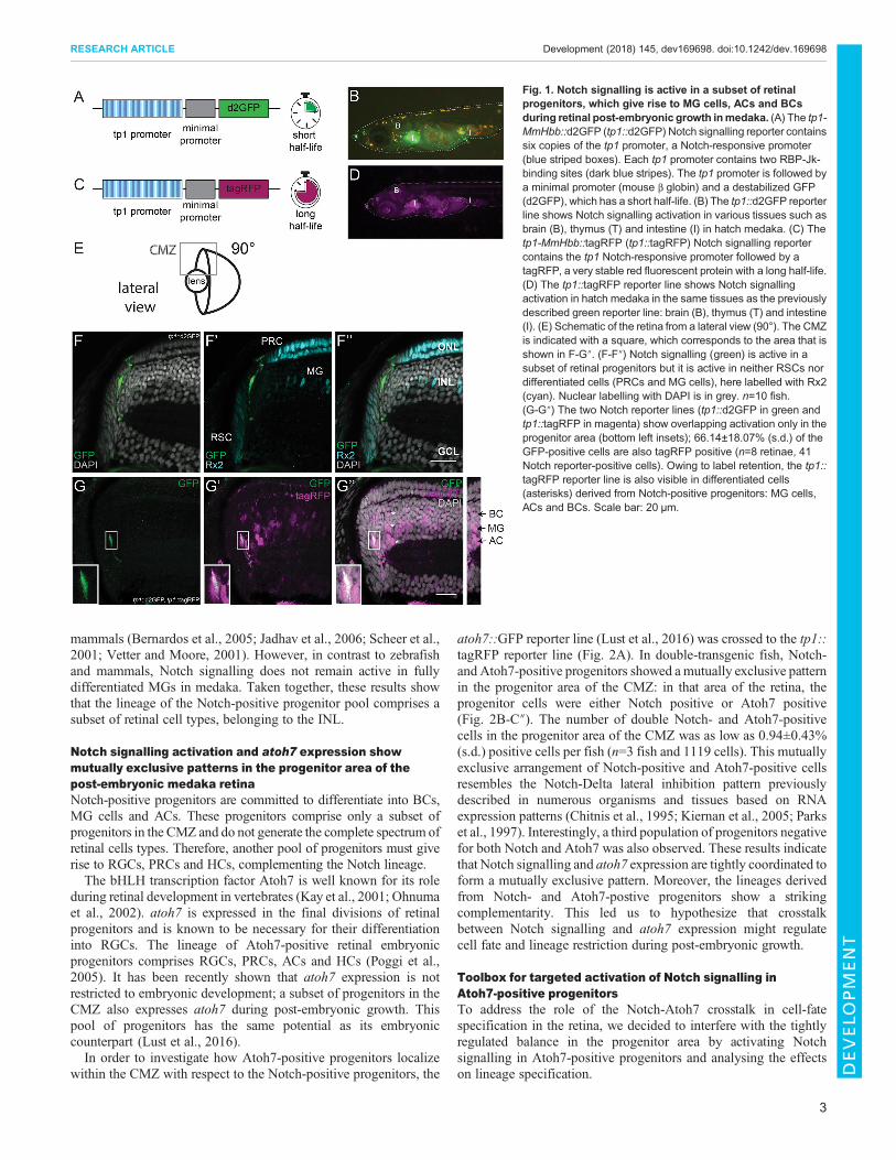
in medaka, the previously characterized tp1-MmHbb::d2GFP (tp1::d2GFP) Notch signalling reporter (Clark et al., 2012; Lust et al.,2016) was used. The reporter construct carries six copies of the tp1promoter, a Notch-responsive promoter containing 2 RBP-Jk-binding sites, followed by a minimal promoter (mouse beta globin)and a destabilized GFP (d2GFP) (Fig. 1A). The tp1::d2GFPreporter line showed Notch signalling activation in various tissues,such as thymus, brain and intestine, in hatch medaka (8 days postfertilization at 28°C) (Fig. 1B). The Notch signalling activationpattern is highly conserved and activation in these tissues has beenpreviously reported in other organisms from flies to mouse(Bajoghli et al., 2009; Bigas and Espinosa, 2012; Bray, 2016;Siebel and Lendahl, 2017). In addition, we generated a secondNotch reporter line for short-term lineage analysis: tp1-MmHbb::tagRFP (tp1::tagRFP). This line carries the same construct as the
tp1::d2GFP line but the short-lived d2GFP is replaced by a tagRFP(Fig. 1C). TagRFP is not targeted for degradation and therefore hasa long half-life allowing short-term lineage tracing due to labelretention (Merzlyak et al., 2007). The tp1::tagRFP reporter lineshowed activation in the same tissues as the tp1::d2GFP line,including the brain, the thymus and the intestine in a medakahatchling (Fig. 1D).
In the retina, we detected Notch signalling activation in a subset ofprogenitors (Fig. 1E-F″). Retinal progenitors are located in the transit-amplifying zone of the CMZ between the retinal stem cells (RSCs)and the central differentiated retina. RSCs are labelled by antibodystaining against the transcription factor retinal homeobox gene two(Rx2) (Reinhardt et al., 2015). They are located in the most peripheralpart of the retina. The differentiated retina occupies the layered andmost central part of the tissue. Among the other retinal cell types, itcontains PRCs and MG cells, which also express Rx2. Importantly,Notch signalling was not detected in RSCs. MG cells and PRCs didnot show Notch signalling either (n=10 fish). This is in contrast tozebrafish, in which Notch signalling is active in MG cells (Wan andGoldman, 2017). The lack of overlap of Notch signalling with RSCs,MG cells or PRCs can be clearly appreciated in the tp1::d2GFPretina 3D reconstruction from a frontal (Fig. S1A,A′) and lateral(Fig. S1B,B′) view. Our data show that Notch signalling is confinedto a subset of progenitors in the CMZ of the medaka retina.
Notch-positiveprogenitors give rise toMGcells, ACs andBCsNotch signalling has been previously shown to be involved in cell-fate choices in the embryonic vertebrate retina (Andreazzoli, 2009;Livesey and Cepko, 2001). During post-embryonic retinal growth,we hypothesize lineage specification to occur in the transit-amplifying zone of the CMZ, between RSCs and the morecentrally located terminally differentiated retina. Factors involvedin lineage specification during retinal development, such as atoh7and neuroD, have been shown to be expressed in that zone (Lustet al., 2016; Poggi et al., 2005; Raymond et al., 2006; Taylor et al.,2015). Consistently, the activation of Notch signalling in the transit-amplifying zone of the CMZ points towards a role for Notchsignalling in cell-fate specification during post-embryonic growth.Thus, we next addressed the differentiation potential of the Notch-positive progenitor pool.
To assess this, we crossed the tp1::tagRFP reporter line to thetp1::d2GFP reporter. In the retina of these double-transgenic fish,we distinguished two cell populations. One population, locatedtowards the periphery of the CMZ, was double positive reflectingactual Notch signalling activation (Fig. 1G-G″, insets). The secondpopulation, located more centrally in the differentiated retina, wasonly positive for tagRFP and corresponds to cells derived from aNotch-positive progenitor pool (Fig. 1G-G″). Interestingly, thesecells were only located in the INL and were identified as BCs, MGcells and ACs based onmorphology and position: BCs are located inthe apical part of the INL; the nucleus of MG cells is elongated andlocated in the centre of the INL; ACs have a round nucleus and arelocated in the basal part of the INL (Fig. 1G″, asterisks and inset onthe right). No tagRFP-positive nuclei were observed in the ONL orGCL. The tagRFP signal that can be observed in those layerscorresponds to cellular projections of BCs and MG cells. Inaddition, we validated the identity of the descendants of Notch-positive progenitor cells by co-staining with cell type-specificmarkers (Fig. S2). Interestingly, we observed a longer activation ofNotch signalling in cells that will become MG cells (Fig. S3). Thispoints towards a conserved role for Notch signalling in specifyingMG cell fate, as has been previously reported in zebrafish and
2
RESEARCH ARTICLE Development (2018) 145, dev169698. doi:10.1242/dev.169698
DEVELO
PM
ENT
mammals (Bernardos et al., 2005; Jadhav et al., 2006; Scheer et al.,2001; Vetter and Moore, 2001). However, in contrast to zebrafishand mammals, Notch signalling does not remain active in fullydifferentiated MGs in medaka. Taken together, these results showthat the lineage of the Notch-positive progenitor pool comprises asubset of retinal cell types, belonging to the INL.
Notch signalling activation and atoh7 expression showmutually exclusive patterns in the progenitor area of thepost-embryonic medaka retinaNotch-positive progenitors are committed to differentiate into BCs,MG cells and ACs. These progenitors comprise only a subset ofprogenitors in the CMZ and do not generate the complete spectrum ofretinal cells types. Therefore, another pool of progenitors must giverise to RGCs, PRCs and HCs, complementing the Notch lineage.The bHLH transcription factor Atoh7 is well known for its role
during retinal development in vertebrates (Kay et al., 2001; Ohnumaet al., 2002). atoh7 is expressed in the final divisions of retinalprogenitors and is known to be necessary for their differentiationinto RGCs. The lineage of Atoh7-positive retinal embryonicprogenitors comprises RGCs, PRCs, ACs and HCs (Poggi et al.,2005). It has been recently shown that atoh7 expression is notrestricted to embryonic development; a subset of progenitors in theCMZ also expresses atoh7 during post-embryonic growth. Thispool of progenitors has the same potential as its embryoniccounterpart (Lust et al., 2016).In order to investigate how Atoh7-positive progenitors localize
within the CMZ with respect to the Notch-positive progenitors, the
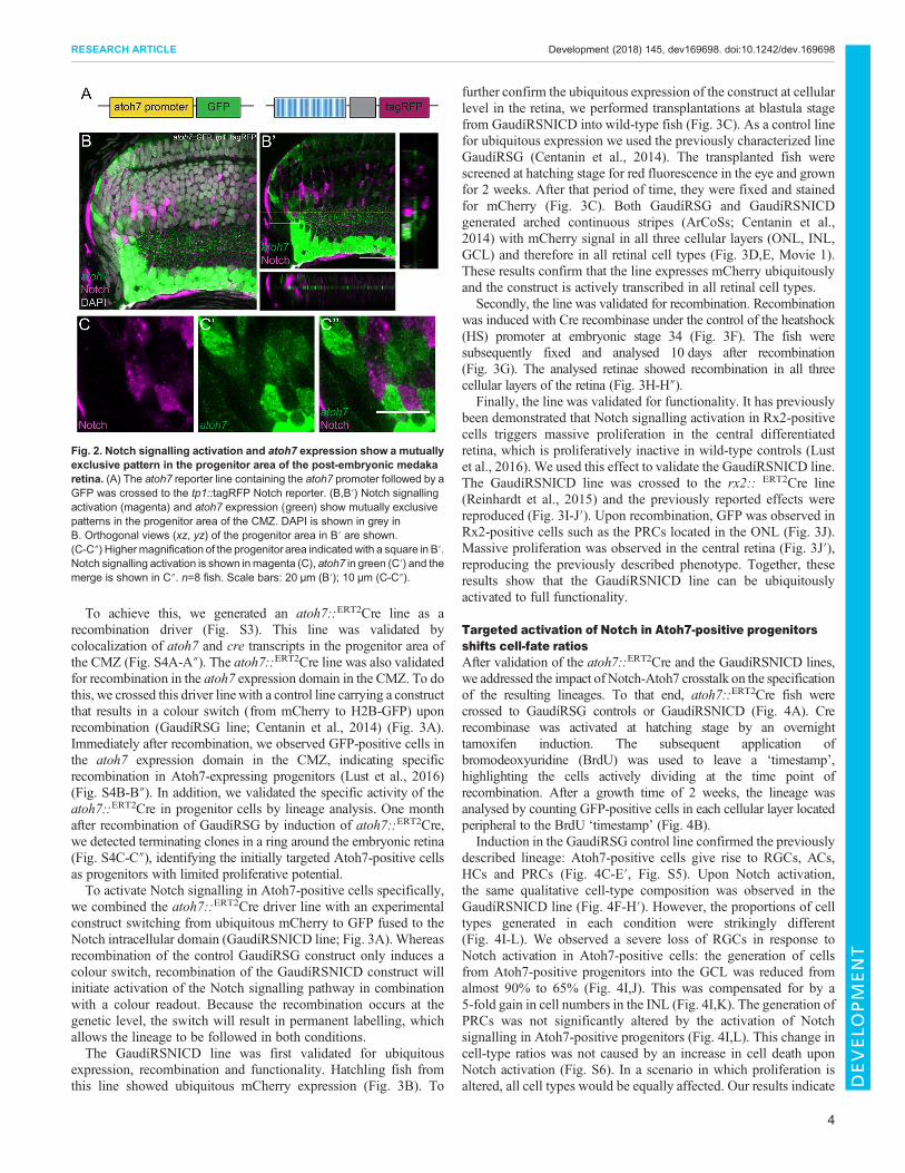
atoh7::GFP reporter line (Lust et al., 2016) was crossed to the tp1::tagRFP reporter line (Fig. 2A). In double-transgenic fish, Notch-and Atoh7-positive progenitors showed amutually exclusive patternin the progenitor area of the CMZ: in that area of the retina, theprogenitor cells were either Notch positive or Atoh7 positive(Fig. 2B-C″). The number of double Notch- and Atoh7-positivecells in the progenitor area of the CMZ was as low as 0.94±0.43%(s.d.) positive cells per fish (n=3 fish and 1119 cells). This mutuallyexclusive arrangement of Notch-positive and Atoh7-positive cellsresembles the Notch-Delta lateral inhibition pattern previouslydescribed in numerous organisms and tissues based on RNAexpression patterns (Chitnis et al., 1995; Kiernan et al., 2005; Parkset al., 1997). Interestingly, a third population of progenitors negativefor both Notch and Atoh7 was also observed. These results indicatethat Notch signalling and atoh7 expression are tightly coordinated toform a mutually exclusive pattern. Moreover, the lineages derivedfrom Notch- and Atoh7-postive progenitors show a strikingcomplementarity. This led us to hypothesize that crosstalkbetween Notch signalling and atoh7 expression might regulatecell fate and lineage restriction during post-embryonic growth.
Toolbox for targeted activation of Notch signalling inAtoh7-positive progenitorsTo address the role of the Notch-Atoh7 crosstalk in cell-fatespecification in the retina, we decided to interfere with the tightlyregulated balance in the progenitor area by activating Notchsignalling in Atoh7-positive progenitors and analysing the effectson lineage specification.
Fig. 1. Notch signalling is active in a subset of retinalprogenitors, which give rise to MG cells, ACs and BCsduring retinal post-embryonic growth inmedaka. (A) The tp1-MmHbb::d2GFP (tp1::d2GFP) Notch signalling reporter containssix copies of the tp1 promoter, a Notch-responsive promoter(blue striped boxes). Each tp1 promoter contains two RBP-Jk-binding sites (dark blue stripes). The tp1 promoter is followed bya minimal promoter (mouse β globin) and a destabilized GFP(d2GFP), which has a short half-life. (B) The tp1::d2GFP reporterline shows Notch signalling activation in various tissues such asbrain (B), thymus (T) and intestine (I) in hatch medaka. (C) Thetp1-MmHbb::tagRFP (tp1::tagRFP) Notch signalling reportercontains the tp1 Notch-responsive promoter followed by atagRFP, a very stable red fluorescent protein with a long half-life.(D) The tp1::tagRFP reporter line shows Notch signallingactivation in hatch medaka in the same tissues as the previouslydescribed green reporter line: brain (B), thymus (T) and intestine(I). (E) Schematic of the retina from a lateral view (90°). The CMZis indicated with a square, which corresponds to the area that isshown in F-G″. (F-F″) Notch signalling (green) is active in asubset of retinal progenitors but it is active in neither RSCs nordifferentiated cells (PRCs and MG cells), here labelled with Rx2(cyan). Nuclear labelling with DAPI is in grey. n=10 fish.(G-G″) The two Notch reporter lines (tp1::d2GFP in green andtp1::tagRFP in magenta) show overlapping activation only in theprogenitor area (bottom left insets); 66.14±18.07% (s.d.) of theGFP-positive cells are also tagRFP positive (n=8 retinae, 41Notch reporter-positive cells). Owing to label retention, the tp1::tagRFP reporter line is also visible in differentiated cells(asterisks) derived from Notch-positive progenitors: MG cells,ACs and BCs. Scale bar: 20 μm.
3
RESEARCH ARTICLE Development (2018) 145, dev169698. doi:10.1242/dev.169698
DEVELO
PM
ENT
To achieve this, we generated an atoh7::ERT2Cre line as arecombination driver (Fig. S3). This line was validated bycolocalization of atoh7 and cre transcripts in the progenitor area ofthe CMZ (Fig. S4A-A″). The atoh7::ERT2Cre line was also validatedfor recombination in the atoh7 expression domain in the CMZ. To dothis, we crossed this driver linewith a control line carrying a constructthat results in a colour switch (from mCherry to H2B-GFP) uponrecombination (GaudíRSG line; Centanin et al., 2014) (Fig. 3A).Immediately after recombination, we observed GFP-positive cells inthe atoh7 expression domain in the CMZ, indicating specificrecombination in Atoh7-expressing progenitors (Lust et al., 2016)(Fig. S4B-B″). In addition, we validated the specific activity of theatoh7::ERT2Cre in progenitor cells by lineage analysis. One monthafter recombination of GaudíRSG by induction of atoh7::ERT2Cre,we detected terminating clones in a ring around the embryonic retina(Fig. S4C-C″), identifying the initially targeted Atoh7-positive cellsas progenitors with limited proliferative potential.To activate Notch signalling in Atoh7-positive cells specifically,
we combined the atoh7::ERT2Cre driver line with an experimentalconstruct switching from ubiquitous mCherry to GFP fused to theNotch intracellular domain (GaudíRSNICD line; Fig. 3A). Whereasrecombination of the control GaudíRSG construct only induces acolour switch, recombination of the GaudíRSNICD construct willinitiate activation of the Notch signalling pathway in combinationwith a colour readout. Because the recombination occurs at thegenetic level, the switch will result in permanent labelling, whichallows the lineage to be followed in both conditions.The GaudíRSNICD line was first validated for ubiquitous
expression, recombination and functionality. Hatchling fish fromthis line showed ubiquitous mCherry expression (Fig. 3B). To
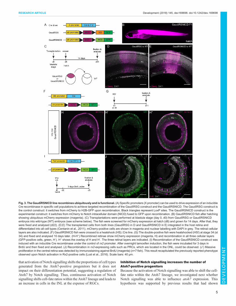
further confirm the ubiquitous expression of the construct at cellularlevel in the retina, we performed transplantations at blastula stagefrom GaudíRSNICD into wild-type fish (Fig. 3C). As a control linefor ubiquitous expression we used the previously characterized lineGaudíRSG (Centanin et al., 2014). The transplanted fish werescreened at hatching stage for red fluorescence in the eye and grownfor 2 weeks. After that period of time, they were fixed and stainedfor mCherry (Fig. 3C). Both GaudíRSG and GaudíRSNICDgenerated arched continuous stripes (ArCoSs; Centanin et al.,2014) with mCherry signal in all three cellular layers (ONL, INL,GCL) and therefore in all retinal cell types (Fig. 3D,E, Movie 1).These results confirm that the line expresses mCherry ubiquitouslyand the construct is actively transcribed in all retinal cell types.
Secondly, the line was validated for recombination. Recombinationwas induced with Cre recombinase under the control of the heatshock(HS) promoter at embryonic stage 34 (Fig. 3F). The fish weresubsequently fixed and analysed 10 days after recombination(Fig. 3G). The analysed retinae showed recombination in all threecellular layers of the retina (Fig. 3H-H″).
Finally, the line was validated for functionality. It has previouslybeen demonstrated that Notch signalling activation in Rx2-positivecells triggers massive proliferation in the central differentiatedretina, which is proliferatively inactive in wild-type controls (Lustet al., 2016). We used this effect to validate the GaudíRSNICD line.The GaudíRSNICD line was crossed to the rx2:: ERT2Cre line(Reinhardt et al., 2015) and the previously reported effects werereproduced (Fig. 3I-J′). Upon recombination, GFP was observed inRx2-positive cells such as the PRCs located in the ONL (Fig. 3J).Massive proliferation was observed in the central retina (Fig. 3J′),reproducing the previously described phenotype. Together, theseresults show that the GaudíRSNICD line can be ubiquitouslyactivated to full functionality.
Targeted activation of Notch in Atoh7-positive progenitorsshifts cell-fate ratiosAfter validation of the atoh7::ERT2Cre and the GaudíRSNICD lines,we addressed the impact of Notch-Atoh7 crosstalk on the specificationof the resulting lineages. To that end, atoh7::ERT2Cre fish werecrossed to GaudíRSG controls or GaudíRSNICD (Fig. 4A). Crerecombinase was activated at hatching stage by an overnighttamoxifen induction. The subsequent application ofbromodeoxyuridine (BrdU) was used to leave a ‘timestamp’,highlighting the cells actively dividing at the time point ofrecombination. After a growth time of 2 weeks, the lineage wasanalysed by counting GFP-positive cells in each cellular layer locatedperipheral to the BrdU ‘timestamp’ (Fig. 4B).
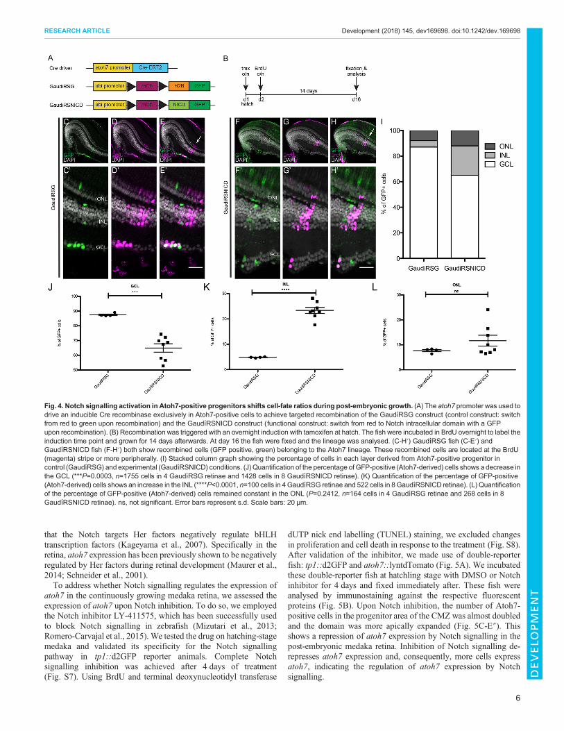
Induction in the GaudíRSG control line confirmed the previouslydescribed lineage: Atoh7-positive cells give rise to RGCs, ACs,HCs and PRCs (Fig. 4C-E′, Fig. S5). Upon Notch activation,the same qualitative cell-type composition was observed in theGaudíRSNICD line (Fig. 4F-H′). However, the proportions of celltypes generated in each condition were strikingly different(Fig. 4I-L). We observed a severe loss of RGCs in response toNotch activation in Atoh7-positive cells: the generation of cellsfrom Atoh7-positive progenitors into the GCL was reduced fromalmost 90% to 65% (Fig. 4I,J). This was compensated for by a5-fold gain in cell numbers in the INL (Fig. 4I,K). The generation ofPRCs was not significantly altered by the activation of Notchsignalling in Atoh7-positive progenitors (Fig. 4I,L). This change incell-type ratios was not caused by an increase in cell death uponNotch activation (Fig. S6). In a scenario in which proliferation isaltered, all cell types would be equally affected. Our results indicate
Fig. 2. Notch signalling activation and atoh7 expression show a mutuallyexclusive pattern in the progenitor area of the post-embryonic medakaretina. (A) The atoh7 reporter line containing the atoh7 promoter followed by aGFP was crossed to the tp1::tagRFP Notch reporter. (B,B′) Notch signallingactivation (magenta) and atoh7 expression (green) show mutually exclusivepatterns in the progenitor area of the CMZ. DAPI is shown in grey inB. Orthogonal views (xz, yz) of the progenitor area in B′ are shown.(C-C″) Highermagnification of the progenitor area indicated with a square in B′.Notch signalling activation is shown inmagenta (C), atoh7 in green (C′) and themerge is shown in C″. n=8 fish. Scale bars: 20 μm (B′); 10 μm (C-C″).
4
RESEARCH ARTICLE Development (2018) 145, dev169698. doi:10.1242/dev.169698
DEVELO
PM
ENT
that activation of Notch signalling shifts the proportions of cell typesgenerated from the Atoh7-positive progenitors but it does notimpact on their differentiation potential, suggesting a regulation ofAtoh7 by Notch signalling. Thus, continuous activation of Notchsignalling shifts cell-fate ratios within the Atoh7 lineage and leads toan increase in cells in the INL at the expense of RGCs.
Inhibition of Notch signalling increases the number ofAtoh7-positive progenitorsBecause the activation of Notch signalling was able to shift the cell-fate ratio within the Atoh7 lineage, we investigated next whetherNotch signalling was able to influence atoh7 expression. Thishypothesis was supported by previous results that had shown
Fig. 3. TheGaudıRSNICD line recombines ubiquitously and is functional. (A) Specific promoters (X promoter) can be used to drive expression of an inducibleCre recombinase in specific cell populations to achieve targeted recombination of the GaudıRSG construct and the GaudıRSNICD. The GaudıRSG construct isthe control construct: it switches from mCherry to H2B-GFP upon recombination. Black triangles represent LoxP sites. The GaudıRSNICD construct is theexperimental construct: it switches from mCherry to Notch intracellular domain (NICD) fused to GFP upon recombination. (B) GaudıRSNICD fish after hatchingshowing ubiquitous mCherry expression (magenta). (C) Transplantations were performed at blastula stage (day 0, d0) from GaudıRSG or GaudıRSNICDembryos into wild-type (WT) embryos (see scheme below). The fish were screened for mCherry expression at hatch (d8) and grown for 14 days. After that, theywere fixed and analysed (d22). (D,E) The transplanted cells from both lines (GaudıRSG in D and GaudıRSNICD in E) integrated in the host retina anddifferentiated into all cell types (Centanin et al., 2011). mCherry-positive cells are shown in magenta and nuclear labelling with DAPI in grey. The retinal cellularlayers are also indicated. (F) GaudıRSNICD fish were crossed to a heatshock (HS)::Cre line. (G) The double-positive fish were heatshocked (HS!) at stage 34 (st34) and fixed and analysed 10 days later. (H-H″) Recombined retinae show mCherry expression (magenta, H) and recombination in all three cellular layers(GFP-positive cells, green, H′). H″ shows the overlay of H and H′. The three retinal layers are indicated. (I) Recombination of the GaudıRSNICD construct wasinduced with an inducible Cre recombinase under the control of rx2 promoter. After overnight tamoxifen induction, the fish were incubated for 3 days inBrdU and then fixed and analysed. (J) Recombination in rx2-expressing cells such as PRCs, which are located in the ONL, could be observed. (J′) Massiveproliferation in the central retina was detected by immunostaining against BrdU (magenta) (n=7 fish). This result recapitulated the previously reported phenotypeobserved upon Notch activation in Rx2-positive cells (Lust et al., 2016). Scale bars: 40 µm.
5
RESEARCH ARTICLE Development (2018) 145, dev169698. doi:10.1242/dev.169698
DEVELO
PM
ENT
that the Notch targets Her factors negatively regulate bHLHtranscription factors (Kageyama et al., 2007). Specifically in theretina, atoh7 expression has been previously shown to be negativelyregulated by Her factors during retinal development (Maurer et al.,2014; Schneider et al., 2001).To address whether Notch signalling regulates the expression of
atoh7 in the continuously growing medaka retina, we assessed theexpression of atoh7 upon Notch inhibition. To do so, we employedthe Notch inhibitor LY-411575, which has been successfully usedto block Notch signalling in zebrafish (Mizutari et al., 2013;Romero-Carvajal et al., 2015). We tested the drug on hatching-stagemedaka and validated its specificity for the Notch signallingpathway in tp1::d2GFP reporter animals. Complete Notchsignalling inhibition was achieved after 4 days of treatment(Fig. S7). Using BrdU and terminal deoxynucleotidyl transferase
dUTP nick end labelling (TUNEL) staining, we excluded changesin proliferation and cell death in response to the treatment (Fig. S8).After validation of the inhibitor, we made use of double-reporterfish: tp1::d2GFP and atoh7::lyntdTomato (Fig. 5A). We incubatedthese double-reporter fish at hatchling stage with DMSO or Notchinhibitor for 4 days and fixed immediately after. These fish wereanalysed by immunostaining against the respective fluorescentproteins (Fig. 5B). Upon Notch inhibition, the number of Atoh7-positive cells in the progenitor area of the CMZ was almost doubledand the domain was more apically expanded (Fig. 5C-E″). Thisshows a repression of atoh7 expression by Notch signalling in thepost-embryonic medaka retina. Inhibition of Notch signalling de-represses atoh7 expression and, consequently, more cells expressatoh7, indicating the regulation of atoh7 expression by Notchsignalling.
Fig. 4. Notch signalling activation in Atoh7-positive progenitors shifts cell-fate ratios during post-embryonic growth. (A) The atoh7 promoter was used todrive an inducible Cre recombinase exclusively in Atoh7-positive cells to achieve targeted recombination of the GaudıRSG construct (control construct: switchfrom red to green upon recombination) and the GaudıRSNICD construct (functional construct: switch from red to Notch intracellular domain with a GFPupon recombination). (B) Recombination was triggered with an overnight induction with tamoxifen at hatch. The fish were incubated in BrdU overnight to label theinduction time point and grown for 14 days afterwards. At day 16 the fish were fixed and the lineage was analysed. (C-H′) GaudıRSG fish (C-E′) andGaudıRSNICD fish (F-H′) both show recombined cells (GFP positive, green) belonging to the Atoh7 lineage. These recombined cells are located at the BrdU(magenta) stripe or more peripherally. (I) Stacked column graph showing the percentage of cells in each layer derived from Atoh7-positive progenitor incontrol (GaudıRSG) and experimental (GaudıRSNICD) conditions. (J) Quantification of the percentage of GFP-positive (Atoh7-derived) cells shows a decrease inthe GCL (***P=0.0003, n=1755 cells in 4 GaudıRSG retinae and 1428 cells in 8 GaudıRSNICD retinae). (K) Quantification of the percentage of GFP-positive(Atoh7-derived) cells shows an increase in the INL (****P<0.0001, n=100 cells in 4GaudıRSG retinae and 522 cells in 8GaudıRSNICD retinae). (L) Quantificationof the percentage of GFP-positive (Atoh7-derived) cells remained constant in the ONL (P=0.2412, n=164 cells in 4 GaudıRSG retinae and 268 cells in 8GaudıRSNICD retinae). ns, not significant. Error bars represent s.d. Scale bars: 20 μm.
6
RESEARCH ARTICLE Development (2018) 145, dev169698. doi:10.1242/dev.169698
DEVELO
PM
ENT
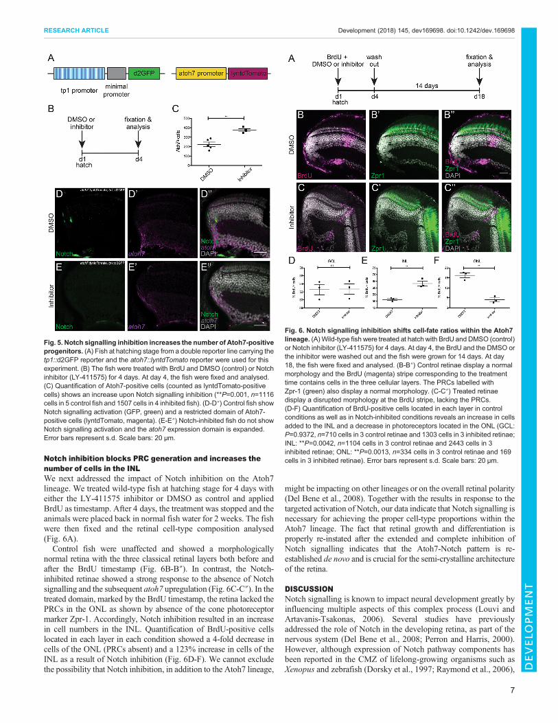
Notch inhibition blocks PRC generation and increases thenumber of cells in the INLWe next addressed the impact of Notch inhibition on the Atoh7lineage. We treated wild-type fish at hatching stage for 4 days witheither the LY-411575 inhibitor or DMSO as control and appliedBrdU as timestamp. After 4 days, the treatment was stopped and theanimals were placed back in normal fish water for 2 weeks. The fishwere then fixed and the retinal cell-type composition analysed(Fig. 6A).Control fish were unaffected and showed a morphologically
normal retina with the three classical retinal layers both before andafter the BrdU timestamp (Fig. 6B-B″). In contrast, the Notch-inhibited retinae showed a strong response to the absence of Notchsignalling and the subsequent atoh7 upregulation (Fig. 6C-C″). In thetreated domain, marked by the BrdU timestamp, the retina lacked thePRCs in the ONL as shown by absence of the cone photoreceptormarker Zpr-1. Accordingly, Notch inhibition resulted in an increasein cell numbers in the INL. Quantification of BrdU-positive cellslocated in each layer in each condition showed a 4-fold decrease incells of the ONL (PRCs absent) and a 123% increase in cells of theINL as a result of Notch inhibition (Fig. 6D-F). We cannot excludethe possibility that Notch inhibition, in addition to the Atoh7 lineage,
might be impacting on other lineages or on the overall retinal polarity(Del Bene et al., 2008). Together with the results in response to thetargeted activation of Notch, our data indicate that Notch signalling isnecessary for achieving the proper cell-type proportions within theAtoh7 lineage. The fact that retinal growth and differentiation isproperly re-instated after the extended and complete inhibition ofNotch signalling indicates that the Atoh7-Notch pattern is re-established de novo and is crucial for the semi-crystalline architectureof the retina.
DISCUSSIONNotch signalling is known to impact neural development greatly byinfluencing multiple aspects of this complex process (Louvi andArtavanis-Tsakonas, 2006). Several studies have previouslyaddressed the role of Notch in the developing retina, as part of thenervous system (Del Bene et al., 2008; Perron and Harris, 2000).However, although expression of Notch pathway components hasbeen reported in the CMZ of lifelong-growing organisms such asXenopus and zebrafish (Dorsky et al., 1997; Raymond et al., 2006),
Fig. 5. Notch signalling inhibition increases the number of Atoh7-positiveprogenitors. (A) Fish at hatching stage from a double reporter line carrying thetp1::d2GFP reporter and the atoh7::lyntdTomato reporter were used for thisexperiment. (B) The fish were treated with BrdU and DMSO (control) or Notchinhibitor (LY-411575) for 4 days. At day 4, the fish were fixed and analysed.(C) Quantification of Atoh7-positive cells (counted as lyntdTomato-positivecells) shows an increase upon Notch signalling inhibition (**P=0.001, n=1116cells in 5 control fish and 1507 cells in 4 inhibited fish). (D-D″) Control fish showNotch signalling activation (GFP, green) and a restricted domain of Atoh7-positive cells (lyntdTomato, magenta). (E-E″) Notch-inhibited fish do not showNotch signalling activation and the atoh7 expression domain is expanded.Error bars represent s.d. Scale bars: 20 μm.
Fig. 6. Notch signalling inhibition shifts cell-fate ratios within the Atoh7lineage. (A)Wild-type fish were treated at hatch with BrdU and DMSO (control)or Notch inhibitor (LY-411575) for 4 days. At day 4, the BrdU and the DMSO orthe inhibitor were washed out and the fish were grown for 14 days. At day18, the fish were fixed and analysed. (B-B″) Control retinae display a normalmorphology and the BrdU (magenta) stripe corresponding to the treatmenttime contains cells in the three cellular layers. The PRCs labelled withZpr-1 (green) also display a normal morphology. (C-C″) Treated retinaedisplay a disrupted morphology at the BrdU stripe, lacking the PRCs.(D-F) Quantification of BrdU-positive cells located in each layer in controlconditions as well as in Notch-inhibited conditions reveals an increase in cellsadded to the INL and a decrease in photoreceptors located in the ONL (GCL:P=0.9372, n=710 cells in 3 control retinae and 1303 cells in 3 inhibited retinae;INL: **P=0.0042, n=1104 cells in 3 control retinae and 2443 cells in 3inhibited retinae; ONL: **P=0.0013, n=334 cells in 3 control retinae and 169cells in 3 inhibited retinae). Error bars represent s.d. Scale bars: 20 μm.
7
RESEARCH ARTICLE Development (2018) 145, dev169698. doi:10.1242/dev.169698
DEVELO
PM
ENT
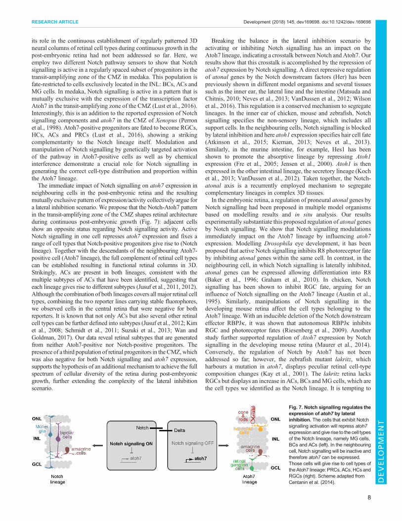
its role in the continuous establishment of regularly patterned 3Dneural columns of retinal cell types during continuous growth in thepost-embryonic retina had not been addressed so far. Here, weemploy two different Notch pathway sensors to show that Notchsignalling is active in a regularly spaced subset of progenitors in thetransit-amplifying zone of the CMZ in medaka. This population isfate-restricted to cells exclusively located in the INL: BCs, ACs andMG cells. In medaka, Notch signalling is active in a pattern that ismutually exclusive with the expression of the transcription factorAtoh7 in the transit-amplifying zone of the CMZ (Lust et al., 2016).Interestingly, this is an addition to the reported expression of Notchsignalling components and atoh7 in the CMZ of Xenopus (Perronet al., 1998). Atoh7-positive progenitors are fated to become RGCs,HCs, ACs and PRCs (Lust et al., 2016), showing a strikingcomplementarity to the Notch lineage itself. Modulation andmanipulation of Notch signalling by genetically targeted activationof the pathway in Atoh7-positive cells as well as by chemicalinterference demonstrate a crucial role for Notch signalling ingenerating the correct cell-type distribution and proportion withinthe Atoh7 lineage.The immediate impact of Notch signalling on atoh7 expression in
neighbouring cells in the post-embryonic retina and the resultingmutually exclusive pattern of expression/activity collectively argue fora lateral inhibition scenario. We propose that the Notch-Atoh7 patternin the transit-amplifying zone of the CMZ shapes retinal architectureduring continuous post-embryonic growth (Fig. 7): adjacent cellsshow an opposite status regarding Notch signalling activity. ActiveNotch signalling in one cell represses atoh7 expression and fixes arange of cell types that Notch-positive progenitors give rise to (Notchlineage). Together with the descendants of the neighbouring Atoh7-positive cell (Atoh7 lineage), the full complement of retinal cell typescan be established resulting in functional retinal columns in 3D.Strikingly, ACs are present in both lineages, consistent with themultiple subtypes of ACs that have been identified, suggesting thateach lineage gives rise to different subtypes (Jusuf et al., 2011, 2012).Although the combination of both lineages covers all major retinal celltypes, combining the two reporter lines carrying stable fluorophores,we observed cells in the central retina that were negative for bothreporters. It is known that not only ACs but also several other retinalcell types can be further defined into subtypes (Jusuf et al., 2012; Kimet al., 2008; Schmidt et al., 2011; Suzuki et al., 2013; Wan andGoldman, 2017). Our data reveal retinal subtypes that are generatedfrom neither Atoh7-positive nor Notch-positive progenitors. Thepresence of a third population of retinal progenitors in the CMZ,whichwas also negative for both Notch signalling and atoh7 expression,supports the hypothesis of an additional mechanism to achieve the fullspectrum of cellular diversity of the retina during post-embryonicgrowth, further extending the complexity of the lateral inhibitionscenario.
Breaking the balance in the lateral inhibition scenario byactivating or inhibiting Notch signalling has an impact on theAtoh7 lineage, indicating a crosstalk between Notch and Atoh7. Ourresults show that this crosstalk is accomplished by the repression ofatoh7 expression by Notch signalling. A direct repressive regulationof atonal genes by the Notch downstream factors (Her) has beenpreviously shown in different model organisms and several tissuessuch as the inner ear, the lateral line and the intestine (Matsuda andChitnis, 2010; Neves et al., 2013; VanDussen et al., 2012; Wilsonet al., 2016). This regulation is a conserved mechanism to segregatelineages. In the inner ear of chicken, mouse and zebrafish, Notchsignalling specifies the non-sensory lineage, which includes allsupport cells. In the neighbouring cells, Notch signalling is blockedby lateral inhibition and here atoh1 expression specifies hair cell fate(Atkinson et al., 2015; Kiernan, 2013; Neves et al., 2013).Similarly, in the murine intestine, for example, Hes1 has beenshown to promote the absorptive lineage by repressing Atoh1expression (Fre et al., 2005; Jensen et al., 2000). Atoh1 is thenexpressed in the other intestinal lineage, the secretory lineage (Kochet al., 2013; VanDussen et al., 2012). Taken together, the Notch-atonal axis is a recurrently employed mechanism to segregatecomplementary lineages in complex 3D tissues.
In the embryonic retina, a regulation of proneural atonal genes byNotch signalling had been proposed in multiple model organismsbased on modelling results and in situ analysis. Our resultsexperimentally substantiate this proposed regulation of atonal genesby Notch signalling. We show that Notch signalling modulationsimmediately impact on the Atoh7 lineage by influencing atoh7expression. Modelling Drosophila eye development, it has beenproposed that active Notch signalling inhibits R8 photoreceptor fateby inhibiting atonal genes within the same cell. In contrast, in theneighbouring cell, in which Notch signalling is laterally inhibited,atonal genes can be expressed allowing differentiation into R8(Baker et al., 1996; Graham et al., 2010). In chicken, Notchsignalling has been shown to inhibit RGC fate, arguing for aninfluence of Notch signalling on the Atoh7 lineage (Austin et al.,1995). Similarly, manipulations of Notch signalling in thedeveloping mouse retina affect the cell types belonging to theAtoh7 lineage. With an inducible deletion of the Notch downstreameffector RBPJκ, it was shown that autonomous RBPJκ inhibitsRGC and photoreceptor fates (Riesenberg et al., 2009). Anotherstudy further supported regulation of Atoh7 expression by Notchsignalling in the developing mouse retina (Maurer et al., 2014).Conversely, the regulation of Notch by Atoh7 has not beenaddressed so far; however, the zebrafish mutant lakritz, whichharbours a mutation in atoh7, displays peculiar retinal cell-typecomposition changes (Kay et al., 2001). The lakritz retina lacksRGCs but displays an increase in ACs, BCs andMG cells, which arethe cell types we identified as the Notch lineage. It is tempting to
Fig. 7. Notch signalling regulates theexpression of atoh7 by lateralinhibition. The cells that exhibit Notchsignalling activation will repress atoh7expression and give rise to the cell typesof the Notch lineage, namely MG cells,BCs and ACs (left). In the neighbouringcell, Notch signalling will be inactive andtherefore atoh7 can be expressed.Those cells will give rise to cell types oftheAtoh7 lineage: PRCs, ACs, HCs andRGCs (right). Scheme adapted fromCentanin et al. (2014).
8
RESEARCH ARTICLE Development (2018) 145, dev169698. doi:10.1242/dev.169698
DEVELO
PM
ENT
speculate that Atoh7 has a repressing effect on Notch signalling,which is released in in lakritz mutants, leading to an increase in celltypes of the Notch lineage. Our direct modulation of Notchsignalling and its impact on atoh7 in conjunction with those datademonstrate that the Notch-atonal axis is fundamental for thesegregation of retinal lineages to ultimately achieve the correct cell-type composition and pseudo-crystalline architecture of the retina.To address the link between growth and continuous patterning of
the differentiating retina, it was crucial to address whether the patternestablished by the Notch-atonal axis is perpetuated from the alreadydifferentiated central retina and thus impacts on the newly formingretinal columns or whether it is de novo established in a continuousfashion in the retinal progenitor cells. Our experiments completelyblocking Notch signalling allowed the existing, highly patternedcentral retina to be disconnected from the newly forming tissueoriginating from the distal CMZ. Strikingly, even though cells aredisturbed in their fate, impacting on retinal lamination in the domainexperiencing the Notch signalling block, the retina continues to growand re-instates proper lamination and differentiation de novo. Thisuncovers an effective self-organization capacity that establishesretinal patterning in the growth zone of the fish retina. It remains to beaddressed whether this is deterministically initiated via anasymmetric division generating a Notch-positive and Notch-negative cell or whether, alternatively, Notch-positive or -negativecells are stochastically initiated upon exit from the niche to eventuallypropagate a mutually exclusive pattern with Atoh7 to the progenitorpopulation by lateral inhibition. These scenarios show strikingparallels to the proposed self-organization driven by Notch-Deltalateral inhibition interactions in the sensory organ in Drosophila(Corson et al., 2017). In these diverse contexts, the Notch-atonal axisacts a fundamental and highly evolutionarily conserved mechanismcombining pattern establishment and cell-lineage specification toshift the temporal specification axis into the third dimension of celltypes arranged in the retinal column.
MATERIALS AND METHODSAnimals and transgenic linesMedaka (Oryzias latipes) used in this study were kept as closed stocks inaccordance with Tierschutzgesetz 111, Abs. 1, Nr. 1 and with EuropeanUnion animal welfare guidelines. Fish were maintained in a constantrecirculating system at 28°C on a 14 h light/10 h dark cycle(Tierschutzgesetz 111, Abs. 1, Nr. 1, Haltungserlaubnis AZ35–9185.64and AZ35–9185.64/BH KIT). The following stocks and transgenic lineswere used: wild-type Cabs, GaudíRSG (Reinhardt et al., 2015),GaudíRSNICD, rx2::LoxPN3ICD (Lust et al., 2016), atoh7::iCre, rx2::iCre (Reinhardt et al., 2015),HS::Cre (Centanin et al., 2014), tp1-MmHbb::d2GFP (Lust et al., 2016), tp1-MmHbb::tagRFP, atoh7::GFP (Lust et al.,2016), atoh7::lyntdTomato. All transgenic lines were created bymicroinjection with Meganuclease (I-SceI) in medaka embryos at theone-cell stage, as previously described (Thermes et al., 2002), except fortp1-MmHbb::tagRFP, which was created by microinjection with Tol2.
BrdU incorporationFor BrdU incorporation, embryos were incubated in 2.5 mM BrdU (Sigma-Aldrich) diluted in 1× embryo rearing medium [ERM; 17 mM sodiumchloride, 0.4 mM potassium chloride, 0.27 mM calcium chloride dihydrate,0.66 mM magnesium sulfate heptahydrate (pH 7)] for the amount of timeindicated in the respective experiment.
Induction of Cre/lox systemFor ERT2Cre induction, embryos were treated with a 5 µM tamoxifen solution(Sigma-Aldrich) in 1× ERM overnight. For HS::Cre induction, stage 34embryos were moved to room temperature where the medium was removedcompletely from the plastic dish and replaced with 42°C ERM. Immediately
after, the embryos were placed in a 37°C incubator for 2 h. Finally, they werereturned to 28°C.
Inhibitor treatmentLY-411575 (Sigma-Aldrich) was dissolved in DMSO to a 50 mM stockconcentration. The stock solution was diluted in 2.5 mM BrdU to reach thefinal working concentration of 5 μM. Hatching-stage fish were treated with5 μM LY-411575 in BrdU for 4 days at 28°C in the dark. The solution wasexchanged after 2 days.
Immunohistochemistry on cryosectionsFish were euthanized using 1×Tricaine (Sigma-Aldrich) and fixed overnight in4% paraformaldehyde (PFA) in 1× PTW [1× PBS (pH 7.3), 0.1% Tween] at4°C. After fixation, samples were washed with 1× PTW and cryoprotected in30% sucrose in 1× PTW. To improve section quality, the samples wereincubated in a 1:1mixture of 30% sucrose and Tissue FreezingMedium (Leica)for at least 3 days. Serial sections (16-µm thick) were obtained on a LeicaCM3050 S cryostat. Sections were rehydrated in 1× PTW for 30 min at roomtemperature. Blocking was performed for 1-2 h with 10% normal goat serum(NGS; Sigma-Aldrich) in 1× PTW at room temperature. Primary antibodieswere applied at 1:500 in 1%NGS overnight at 4°C. Secondary antibodies wereused at 1:750 in 1%NGS together withDAPI (Roth; 1:500 in 1× PTWof 5 mg/ml stock) and applied for 2 h at 37°C. Slides were mounted with 60% glyceroland kept at 4°C until imaging.
BrdU immunohistochemistry on cryosectionsBrdU antibody staining was performed with an antigen retrieval step. Afterall antibody and DAPI staining, except for BrdU, were complete, a 30 minfixation was performed with 4% PFA. Slides were incubated for 1 h at 37°Cin 2 N HCl solution, and pH was recovered by washing with a 40% boraxsolution before incubation with the primary BrdU antibody.
Immunohistochemistry on whole-mount retinaeFish were euthanized using 20× Tricaine and fixed overnight in 4% PFA in1× PTW at 4°C. After fixation, samples were washed with 1× PTW. Fishwere bleached with 3% H2O2, 0.5% KOH in H2O for 2-3 h in the dark.Retinae were enucleated and permeabilized with acetone for 15 min at−20°C. Blocking was performed in 1% bovine serum albumin (Sigma-Aldrich), 1% DMSO (Roth/Merck), 4% sheep serum (Sigma-Aldrich) in 1×PTW for 2 h. Samples were incubated with primary antibody in blockingbuffer overnight at 4°C. The secondary antibody was applied together withDAPI in blocking buffer overnight at 4°C. Primary antibodies were used at1:200, secondary antibodies 1:250 and DAPI 1:500. The stained retinaewere sectioned (40-μm thick) with the Vibratome Leica VT 1000S. Prior tosectioning, the retinae were embedded into 4% agarose (Sigma).
AntibodiesThe following primary antibodies were used: anti-EGFP (chicken; LifeTechnologies, A10262; 1:500), rabbit anti-Rx2 (Reinhardt et al., 2015; 1:500),anti-tagRFP (rabbit; Evrogen, AB233; 1:500), anti-Pax6 (rabbit; HissDiagnostics, PRB-278P; 1:200), PKCα (rabbit; Santa Cruz, sc-208; 1:200),anti-GS (mouse; Chemicon, MAB302; 1:500), anti-Sox2 (rabbit; Genetex,GTX101506; 1:500), anti-HuC/D (mouse; Thermo Fisher, A21271; 1:500),anti-recoverin (rabbit;Millipore,AB5585; 1:500), anti-Zpr-1 (mouse;ZebrafishInternational Resource Center; 1:500), anti-DsRed (rabbit; Clontech, 632496;1:500), anti-BrdU (rat; AbDSerotec, BU1/75; 1:200). The following secondaryantibodies were used: anti-mouse Cy5 (Jackson ImmunoResearch, 715-175-151), anti-chicken 488 (Jackson ImmunoResearch, 703-485-155), anti-ratDyLight549 (Jackson ImmunoResearch, 112-505-143), anti-rabbitDyLight549 (Jackson ImmunoResearch), anti-mouse Alexa546 (LifeTechnologies, A-11030) and anti-rat Alexa633 (Life Technologies, A21094).DAPI (Sigma-Aldrich, D9564) nuclear counterstaining was performed asdescribed by Inoue and Wittbrodt (2011).
Whole-mount double-fluorescence in situWhole-mount double-fluorescence in situ was performed with the TSA-Plus Cyanine 5 system from PerkinElmer as previously described (Reinhardtet al., 2015).
9
RESEARCH ARTICLE Development (2018) 145, dev169698. doi:10.1242/dev.169698
DEVELO
PM
ENT
TUNELTUNEL staining on cryosections was performed after all other antibodystainings were completed using the In Situ Cell Death Detection Kit TMRRed from Roche. Staining was performed according to the manufacturer’sprotocol with the following modification: washes were performed with 1×PTW instead of PBS.
Immunohistochemistry and fluorescence in situ imagingAll immunohistochemistry and fluorescence in situ images were acquired byconfocal microscopy using a Leica TCS SPEwith either a 20×water objectiveor a 40× oil objective or a Leica TCS SP8 with 20× or 63× oil objective.
Image processing and statistical analysisImages were processed using Fiji image processing software. Statisticalanalyses and graphical representation of the data were performed using thePrism software package (GraphPad). Unpaired t-tests were performed todetermine the statistical significances. P<0.05 was considered significantand P-values are given in the figure legends. Sample size (n) is mentioned inevery figure legend. No statistical methods were used to predeterminesample sizes, but our sample sizes are similar to those generally used in thefield. The experimental groups were allocated randomly, with no blindingduring allocation.
AcknowledgementsWe thank C. Helker and D. Stainier for tp1::tagRFP reporter construct; T. Tavhelidseand B. Wittbrodt for designing and establishing the atoh7::ERT2Cre construct; andR. Sinn for the atoh7::lyntdTomato medaka line. We are grateful to A. Saraceno,E. Leist and M. Majewski for fish husbandry. We thank L. Centanin and all themembers of the Wittbrodt group for constructive feedback on the project and criticalreading of the manuscript.
Competing interestsThe authors declare no competing or financial interests.
Author contributionsConceptualization: A.P.S., K.L., J.W.; Methodology: A.P.S., K.L.; Validation: A.P.S.,K.L.; Formal analysis: A.P.S., K.L., J.W.; Investigation: A.P.S., K.L.; Resources:A.P.S., K.L.; Data curation: A.P.S., K.L.; Writing - original draft: A.P.S., K.L., J.W.;Writing - review & editing: A.P.S., K.L., J.W.; Visualization: A.P.S., K.L.; Supervision:J.W.; Project administration: J.W.; Funding acquisition: J.W.
FundingA.P.S. and K.L. were members of the Heidelberg Biosciences InternationalGraduate School and were both supported by a LGFG Fellowship funded by theMinisterium fur Wissenschaft, Forschung und Kunst Baden-Wurttemberg. This workwas supported by the European Research Council (GA 294354-ManISteC to J.W.)and the Deutsche Forschungsgemeinschaft (SFB 873 TP A3 to J.W.). Deposited inPMC for immediate release.
Supplementary informationSupplementary information available online athttp://dev.biologists.org/lookup/doi/10.1242/dev.169698.supplemental
ReferencesAndreazzoli, M. (2009). Molecular regulation of vertebrate retina cell fate. BirthDefects Res. Part C - Embryo Today 87, 284-295.
Atkinson, P. J., Huarcaya Najarro, E., Sayyid, Z. N. and Cheng, A. G. (2015).Sensory hair cell development and regeneration: similarities and differences.Development 142, 1561-1571.
Austin, C. P., Feldman, D. E., Ida, J. A. and Cepko, C. L. (1995). Vertebrate retinalganglion cells are selected from competent progenitors by the action of Notch.Development 121, 3637-3650.
Bajoghli, B., Aghaallaei, N., Hess, I., Rode, I., Netuschil, N., Tay, B.-H.,Venkatesh, B., Yu, J.-K., Kaltenbach, S. L., Holland, N. D. et al. (2009).Evolution of genetic networks underlying the emergence of thymopoiesis invertebrates. Cell 138, 186-197.
Baker, N. E., Yu, S. and Han, D. (1996). Evolution of proneural atonal expressionduring distinct regulatory phases in the developing Drosophila eye. Curr. Biol. 6,1290-1302.
Bassett, E. A. and Wallace, V. A. (2012). Cell fate determination in the vertebrateretina. Trends Neurosci. 35, 565-573.
Bernardos, R. L., Lentz, S. I., Wolfe, M. S. and Raymond, P. A. (2005). Notch-Delta signaling is required for spatial patterning and Muller glia differentiation inthe zebrafish retina. Dev. Biol. 278, 381-395.
Bigas, A. and Espinosa, L. (2012). Hematopoietic stem cells: to be or Notch to be.Blood 119, 3226-3235.
Borggrefe, T. and Oswald, F. (2009). The Notch signaling pathway: transcriptionalregulation at Notch target genes. Cell. Mol. Life Sci. 66, 1631-1646.
Bray, S. J. (2016). Notch signalling in context. Nat. Rev. Mol. Cell Biol. 17, 722-735.Centanin, L., Hoeckendorf, B. and Wittbrodt, J. (2011). Fate restriction and
multipotency in retinal stem cells. Cell Stem Cell 9, 553-562.Centanin, L., Ander, J.-J., Hoeckendorf, B., Lust, K., Kellner, T., Kraemer, I.,
Urbany, C., Hasel, E., Harris, W. A., Simons, B. D. et al. (2014). Exclusivemultipotency and preferential asymmetric divisions in post-embryonic neural stemcells of the fish retina. Development 141, 3472-3482.
Chitnis, A., Henrique, D., Lewis, J., Ish-Horowicz, D. and Kintner, C. (1995).Primary neurogenesis in Xenopus embryos regulated by a homologue of theDrosophila neurogenic gene Delta. Nature 375, 761-766.
Clark, B. S., Cui, S., Miesfeld, J. B., Klezovitch, O., Vasioukhin, V. and Link,B. A. (2012). Loss of Llgl1 in retinal neuroepithelia reveals links between apicaldomain size, Notch activity and neurogenesis. Development 139, 1599-1610.
Coffman, C., Harris, W. and Kintner, C. (1990). Xotch, the Xenopus homolog ofDrosophila Notch. Science (80-.) 249, 1438-1441.
Corson, F., Couturier, L., Rouault, H., Mazouni, K. and Schweisguth, F. (2017).Self-organized Notch dynamics generate stereotyped sensory organ patterns inDrosophila. Science (80-.). 356, eaai7407.
Del Bene, F., Wehman, A. M., Link, B. A. and Baier, H. (2008). Regulation ofneurogenesis by interkinetic nuclear migration through an apical-basal notchgradient. Cell 134, 1055-1065.
Dorsky, R. I., Rapaport, D. H. and Harris, W. A. (1995). Xotch inhibits celldifferentiation in the Xenopus retina. Neuron 14, 487-496.
Dorsky, R. I., Chang, W. S., Rapaport, D. H. and Harris, W. A. (1997). Regulationof neuronal diversity in the Xenopus retina by Delta signalling. Nature 385, 67-70.
Edlund, T. and Jessell, T. M. (1999). Progression from extrinsic to intrinsic signalingin cell fate specification: a view from the nervous system. Cell 96, 211-224.
Fiuza, U.-M. and Arias, A. M. (2007). Cell and molecular biology of Notch.J. Endocrinol. 194, 459-474.
Fre, S., Huyghe, M., Mourikis, P., Robine, S., Louvard, D. and Artavanis-Tsakonas, S. (2005). Notch signals control the fate of immature progenitor cells inthe intestine. Nature 435, 964-968.
Graham, T. G. W., Tabei, S. M. A., Dinner, A. R. and Rebay, I. (2010). Modelingbistable cell-fate choices in the Drosophila eye: qualitative and quantitativeperspectives. Development 137, 2265-2278.
Hollyfield, J. G. (1968). Differential addition of cells to the retina in rana pipienstadpoles. Dev. Biol. 18, 163-179.
Hufnagel, R. B. and Brown, N. L. (2013). Specification of retinal cell types. InPatterning and Cell Type Specification in the Developing CNS and PNS:Comprehensive Developmental Neuroscience (ed. J. Rubenstein and P. Rakic),pp. 519-536. Academic Press.
Inoue, D. and Wittbrodt, J. (2011). One for all-a highly efficient and versatilemethod for fluorescent immunostaining in fish embryos. PLoS One 6, e19713.
Jadhav, A. P., Cho, S.-H. and Cepko, C. L. (2006). Notch activity permits retinalcells to progress through multiple progenitor states and acquire a stem cellproperty. Proc. Natl. Acad. Sci. USA 103, 18998-19003.
Jensen, J., Pedersen, E. E., Galante, P., Hald, J., Heller, R. S., Ishibashi, M.,Kageyama, R., Guillemot, F., Serup, P. and Madsen, O. D. (2000). Control ofendodermal endocrine development by Hes-1. Nat. Genet. 24, 36-44.
Johns, P. R. and Easter, S. S.Jr (1977). Growth of the adult goldfish eye, II:Increase in retinal cell number. J. Comp. Neurol. 176, 331-341.
Jusuf, P. R., Almeida, A. D., Randlett, O., Joubin, K., Poggi, L. and Harris, W. A.(2011). Origin and determination of inhibitory cell lineages in the vertebrate retina.J. Neurosci. 31, 2549-2562.
Jusuf, P. R., Albadri, S., Paolini, A., Currie, P. D., Argenton, F., Higashijima, S.,Harris, W. A. and Poggi, L. (2012). Biasing amacrine subtypes in the Atoh7lineage through expression of Barhl2. J. Neurosci. 32, 13929-13944.
Kageyama, R., Ohtsuka, T. and Kobayashi, T. (2007). The Hes gene family:repressors and oscillators that orchestrate embryogenesis. Development 134,1243-1251.
Kanekar, S., Perron, M., Dorsky, R., Harris, W. A., Jan, L. Y., Jan, Y. N. andVetter, M. L. (1997). Xath5 participates in a network of bHLH genes in thedeveloping xenopus retina. Neuron 19, 981-994.
Kay, J. N., Finger-Baier, K. C., Roeser, T., Staub,W. and Baier, H. (2001). Retinalganglion cell genesis requires lakritz, a Zebrafish atonal Homolog. Neuron 30,725-736.
Kiernan, A. (2013). Notch signaling during cell fate determination in the inner ear.Semin. Cell Dev. Biol. 24, 470-479.
Kiernan, A. E., Cordes, R., Kopan, R., Gossler, A. and Gridley, T. (2005). TheNotch ligands DLL1 and JAG2 act synergistically to regulate hair cell developmentin the mammalian inner ear. Development 132, 4353-4362.
10
RESEARCH ARTICLE Development (2018) 145, dev169698. doi:10.1242/dev.169698
DEVELO
PM
ENT
Kim, D. S., Ross, S. E., Trimarchi, J. M., Aach, J., Greenberg, M. E. and Cepko,C. L. (2008). Identification of molecular markers of bipolar cells in the murineretina. J. Comp. Neurol. 507, 1795-1810.
Koch, U., Lehal, R. and Radtke, F. (2013). Stem cells living with a Notch.Development 140, 689-704.
Lai, E. C. (2004). Notch signaling: control of cell communication and cell fate.Development 131, 965-973.
Link, B. A. and Darland, T. (2001). Genetic analysis of initial and ongoingretinogenesis in the zebrafish: comparing the central neuroepithelium andmarginal zone. Prog. Brain Res. 131, 565-577.
Livesey, F. J. and Cepko, C. L. (2001). Vertebrate neural cell-fate determination:lessons from the retina. Nat. Rev. Neurosci. 2, 109-118.
Louvi, A. and Artavanis-Tsakonas, S. (2006). Notch signalling in vertebrate neuraldevelopment. Nat. Rev. Neurosci. 7, 93-102.
Lust, K., Sinn, R., Perez Saturnino, A., Centanin, L. and Wittbrodt, J. (2016). Denovo neurogenesis by targeted expression of Atoh7 to Muller glia cells.Development 143, 1874-1883.
Matsuda, M. and Chitnis, A. B. (2010). Atoh1a expression must be restricted byNotch signaling for effective morphogenesis of the posterior lateral lineprimordium in zebrafish. Development 137, 3477-3487.
Maurer, K. A., Riesenberg, A. N. and Brown, N. L. (2014). Notch signalingdifferentially regulates Atoh7 and Neurog2 in the distal mouse retina.Development 141, 3243-3254.
Merzlyak, E. M., Goedhart, J., Shcherbo, D., Bulina, M. E., Shcheglov, A. S.,Fradkov, A. F., Gaintzeva, A., Lukyanov, K. A., Lukyanov, S., Gadella, T. W. J.et al. (2007). Bright monomeric red fluorescent protein with an extendedfluorescence lifetime. Nat. Methods 4, 555-557.
Mizutari, K., Fujioka, M., Hosoya, M., Bramhall, N., Okano, H. J., Okano, H. andEdge, A. S. B. (2013). Notch inhibition induces cochlear hair cell regeneration andrecovery of hearing after acoustic trauma. Neuron 77, 58-69.
Neves, J., Abello, G., Petrovic, J. and Giraldez, F. (2013). Patterning and cell fatein the inner ear: a case for Notch in the chicken embryo. Dev. Growth Differ. 55,96-112.
Ohnuma, S., Hopper, S., Wang, K. C., Philpott, A. and Harris, W. A. (2002). Co-ordinating retinal histogenesis: early cell cycle exit enhances early cell fatedetermination in the Xenopus retina. Development 129, 2435-2446.
Parks, A. L., Huppert, S. S. and Muskavitch, M. A. T. (1997). The dynamics ofneurogenic signalling underlying bristle development in Drosophila melanogaster.Mech. Dev. 63, 61-74.
Pearson, B. J. and Doe, C. Q. (2004). Specification of temporal identity in thedeveloping nervous system. Annu. Rev. Cell Dev. Biol. 20, 619-647.
Perron, M. and Harris, W. A. (2000). Determination of vertebrate retinal progenitorcell fate by the Notch pathway and basic helix-loop-helix transcription factors.Cell.Mol. Life Sci. 57, 215-223.
Perron, M., Kanekar, S., Vetter, M. L. and Harris, W. A. (1998). The geneticsequence of retinal development in the ciliary margin of the xenopus eye. Dev.Biol. 199, 185-200.
Poggi, L., Vitorino, M., Masai, I. and Harris, W. A. (2005). Influences on neurallineage and mode of division in the zebrafish retina in vivo. J. Cell Biol. 171,991-999.
Raymond, P. A., Barthel, L. K., Bernardos, R. L. and Perkowski, J. J. (2006).Molecular characterization of retinal stem cells and their niches in adult zebrafish.BMC Dev. Biol. 6, 36.
Reinhardt, R., Centanin, L., Tavhelidse, T., Inoue, D., Wittbrodt, B., Concordet,J.-P., Martinez-morales, J. R. andWittbrodt, J. (2015). Sox 2, Tlx, Gli 3, and Her9 converge on Rx 2 to define retinal stem cells in vivo. EMBO J. 34, 1572-1588.
Riesenberg, A. N., Liu, Z., Kopan, R. and Brown, N. L. (2009). Rbpj cellautonomous regulation of retinal ganglion cell and cone photoreceptor fates in themouse retina. J. Neurosci. 29, 12865-12877.
Romero-Carvajal, A., Acedo Navajas, J., Jiang, L., Kozlovskaja-Gumbrien, A.,Alexander, R., Li, H. and Piotrowski, T. (2015). Regeneration of sensory haircells requires localized interactions between the Notch and Wnt pathways. Dev.Cell 34, 267-282.
Scheer, N., Groth, A., Hans, S. and Campos-Ortega, J. A. (2001). An instructivefunction for Notch in promoting gliogenesis in the zebrafish retina. Development128, 1099-1107.
Schmidt, T. M., Chen, S.-K. and Hattar, S. (2011). Intrinsically photosensitiveretinal ganglion cells: many subtypes, diverse functions. Trends Neurosci. 34,572-580.
Schneider, M. L., Turner, D. L. and Vetter, M. L. (2001). Notch signaling can inhibitXath5 function in the neural plate and developing retina. Mol. Cell. Neurosci. 18,458-472.
Siebel, C. and Lendahl, U. (2017). Notch signaling in development, tissuehomeostasis, and disease. Physiol. Rev. 97, 1235-1294.
Suzuki, S. C., Bleckert, A., Williams, P. R., Takechi, M., Kawamura, S. andWong, R. O. L. (2013). Cone photoreceptor types in zebrafish are generated bysymmetric terminal divisions of dedicated precursors. Proc. Natl. Acad. Sci. 110,15109-15114.
Taylor, S. M., Alvarez-Delfin, K., Saade, C. J., Thomas, J. L., Thummel, R.,Fadool, J. M. and Hitchcock, P. F. (2015). The bHLH transcription factor neuroDgoverns photoreceptor genesis and regeneration through delta-notch signaling.Investig. Ophthalmol. Vis. Sci. 56, 7496-7515.
Thermes, V., Grabher, C., Ristoratore, F., Bourrat, F., Choulika, A., Wittbrodt, J.and Joly, J.-S. (2002). I-SceI meganuclease mediates highly efficienttransgenesis in fish. Mech. Dev. 118, 91-98.
VanDussen, K. L., Carulli, A. J., Keeley, T. M., Patel, S. R., Puthoff, B. J.,Magness, S. T., Tran, I. T., Maillard, I., Siebel, C., Kolterud, A. et al. (2012).Notch signaling modulates proliferation and differentiation of intestinal crypt basecolumnar stem cells. Development 139, 488-497.
Vetter, M. L. andMoore, K. B. (2001). Becoming glial in the neural retina.Dev. Dyn.221, 146-153.
Wan, J. and Goldman, D. (2017). Opposing actions of Fgf8a on notch signalingdistinguish two muller glial cell populations that contribute to retina growth andregeneration. Cell Rep. 19, 849-862.
Wan, J., Ramachandran, R. and Goldman, D. (2012). HB-EGF is necessary andsufficient for Muller glia dedifferentiation and retina regeneration. Dev. Cell 22,334-347.
Wan, Y., Almeida, A. D., Rulands, S., Chalour, N., Muresan, L., Wu, Y., Simons,B. D., He, J. and Harris, W. A. (2016). The ciliary marginal zone of the zebrafishretina: clonal and time-lapse analysis of a continuously growing tissue.Development 143, 1099-1107.
Wilson, S. G., Wen, W., Pillai-kastoori, L. and Morris, A. C. (2016). Tracking thefate of her4 expressing cells in the regenerating retina using her4 : Kaede zebra fish. Exp. Eye Res. 145, 75-87.
11
RESEARCH ARTICLE Development (2018) 145, dev169698. doi:10.1242/dev.169698
DEVELO
PM
ENT