Embed Size (px)
Citation preview
This is the author’s version of a work that was submitted/accepted for pub-lication in the following source:
Jeon, J.E., Schrobback, K., Hutmacher, D.W., & Klein, T.J. (2012) Dynamiccompression improves biosynthesis of human zonal chondrocytes from os-teoarthritis patients. Osteoarthritis and Cartilage, 20(8), pp. 906-915.
This file was downloaded from: http://eprints.qut.edu.au/51569/
c© Copyright 2012 Elsevier
This is the author’s version of a work that was accepted for publicationin <Osteoarthritis and Cartilage>. Changes resulting from the publish-ing process, such as peer review, editing, corrections, structural format-ting, and other quality control mechanisms may not be reflected in thisdocument. Changes may have been made to this work since it wassubmitted for publication. A definitive version was subsequently pub-lished in Osteoarthritis and Cartilage, [VOL: 20, ISSUE: 8, (2012)] DOI:10.1016/j.joca.2012.04.019
Notice: Changes introduced as a result of publishing processes such ascopy-editing and formatting may not be reflected in this document. For adefinitive version of this work, please refer to the published source:
http://dx.doi.org/10.1016/j.joca.2012.04.019

Dynamic compression of human zonal osteoarthritic chondrocytes
1
Title: Dynamic compression improves biosynthesis of human zonal chondrocytes from 1 osteoarthritis patients 2 3 Authors: 4 June E. Jeon, B.S. 5 Karsten Schrobback, Ph.D. 6 Dietmar W. Hutmacher, Ph.D. 7 Travis J. Klein, Ph.D. 8 9 Corresponding author: 10 Travis J. Klein 11 Address: 12 Institute of Health and Biomedical Innovation, Queensland University of Technology 13 60 Musk Ave., Kelvin Grove, QLD, 4059, Australia 14 Email: [email protected] 15 Phone: +61 7 3138 6142 16 Fax: +61 7 3138 6030 17 18 19

Dynamic compression of human zonal osteoarthritic chondrocytes
2
Abstract 20 Objective: We hypothesize that chondrocytes from distinct zones of articular cartilage 21 respond differently to compressive loading, and that zonal osteoarthritic (OA) chondrocytes 22 can benefit from optimized compressive stimulation. Therefore, we aimed to determine the 23 transcriptional response of superficial (S) and middle/deep (MD) zone chondrocytes to 24 varying dynamic compressive strain and loading duration. To confirm effects of compressive 25 stimulation on overall matrix production, we subjected zonal chondrocytes to compression 26 for 2 weeks. 27 Design: Human S and MD chondrocytes from OA joints were encapsulated in 2% alginate, 28 pre-cultured, and subjected to compression with varying dynamic strain (5, 15, 50% at 1 Hz) 29 and loading duration (1, 3, 12 hr). Temporal changes in cartilage-specific, zonal, and 30 dedifferentiation genes following compression were evaluated using qRT-PCR. The benefits 31 of long-term compression (50% strain, 3 hr/day, for 2 wks) were assessed by measuring 32 construct glycosaminoglycan (GAG) content and compressive moduli, as well as 33 immunostaining. 34 Results: Compressive stimulation significantly induced ACAN, COL2A1, COL1A1, PRG4, 35 and COL10A1 gene expression after 2 hours of unloading, in a zone-dependent manner (p < 36 0.05). ACAN and PRG4 mRNA levels depended on strain and load duration, with 50% and 3 37 hour loading resulting in highest levels (p < 0.05). Long-term compression increased collagen 38 type II and aggrecan immunostaining and total GAG (p < 0.05), but only S constructs showed 39 more PRG4 stain, retained more GAG (p < 0.01), and developed higher compressive moduli 40 than non-loaded controls. 41 Conclusions: The biosynthetic activity of zonal chondrocytes from OA joints can be 42 enhanced with selected compression regimes, indicating the potential for cartilage tissue 43 engineering applications. 44 45 46 Keywords: cartilage; osteoarthritis; chondrocyte subpopulation; mechanical loading; 47 alginate. 48 49 50 Running Headline: Dynamic compression of human zonal osteoarthritic chondrocytes 51
52
Dynamic compression of human zonal osteoarthritic chondrocytes
3
Introduction53 Osteoarthritis (OA) is a highly prevalent disease affecting more than 27 million adults in
the USA [1]. Approximately 50% of people over the age of 65 were diagnosed with some form of arthritis between years 2007-2009 [2], and it is well known that occurrence of osteoarthritis increases with age. Current clinical interventions available such as microfracture, osteochondral autograft, and autologous chondrocyte implantation (ACI) can help promote cartilage repair for focal defects of younger patients [3, 4]. However, since OA affects larger areas of the joint in an unconfined manner, these methods are usually not feasible. One alternative strategy to overcome the limitations of the current clinical treatment methods is to make a functional cartilage construct in vitro using autologous chondrocytes and implant it in place of the damaged cartilage [5]. Even for OA patients in need of total joint replacement surgeries there are typically regions of macroscopically normal cartilage, and biopsies can be taken to obtain chondrocytes needed for lab-generated cartilaginous tissue. However, without developing ways to enhance biosynthetic capabilities of the aging chondrocytes, it will be difficult to produce cartilage replacement tissues in vitro. Recently, studies have shown that chondrocytes from OA joints are sensitive to growth factor stimuli and their matrix protein synthesis can be improved depending on the culture conditions [6, 7]. These reports are encouraging, and with further improvements to in vitro culture conditions, it may be possible to use chondrocytes from OA patients to engineer a functional cartilage construct.
Advancement in our understanding of the benefits of in vitro mechanical stimulation of cartilage and cartilaginous constructs suggests that such stimulation may promote biosynthesis of chondrocytes from OA patients. Prior investigations using cartilage explants and chondrocyte-seeded hydrogels have shown that enhanced matrix protein biosynthesis and improved mechanical properties through in vitro compressive stimulation depends on the frequency [8, 9], magnitude (compressive strains) [10], and duration of the load applied [11]. Mechanical loading not only influences matrix-protein biosynthesis and matrix remodelling [12], but zonal protein secretion as well, such as proteoglycan 4 (PRG4), a superficial zonal marker that helps lubricate the joint [13]. While there has been much research effort focused on the compression-induced biosynthesis of full-thickness immature bovine chondrocytes, research has been limited regarding mechanically stimulating human chondrocytes from elderly patients, which represents the largest age group affected by OA. Furthermore, the different strain levels found in each zone of cartilage indicate that chondrocytes from different zones may require different loading regimes during in vitro compression. Superficial (surface), middle (transitional), and deep (radial) zones have physiological compressive strains of >50%, 10-20%, and 0-5%, respectively [14]. Developing ways to stimulate zonal OA chondrocytes in a compression bioreactor to enhance their matrix- and zone-specific protein synthesis will be an important step towards generating an autologous replacement tissue using chondrocytes from OA patients.
We hypothesize that compressive stimulation can enhance the biosynthesis of zonal OA chondrocytes depending on the loading protocols applied. We further postulate that monitoring the temporal gene expression following loading will aid in the development of appropriate loading protocols. Therefore, our first aim was to observe the time-course of cartilage- and zone-specific mRNA levels in superficial (S) and middle/deep (MD) zone chondrocytes from OA patients following dynamic compression. In our experiments, we encapsulated the chondrocytes in 2% alginate hydrogels, which have been shown to support chondrocyte differentiation and matrix synthesis [15, 16]. The second aim was to understand how various dynamic loading conditions (strain levels and loading durations) influence the short-term transcriptional response. After identifying a loading protocol that enhanced
Dynamic compression of human zonal osteoarthritic chondrocytes
4
mRNA expressions, we applied compressive stimulation over 2 weeks to confirm its long-term effect on matrix synthesis and construct mechanical properties. Method Chondrocyte isolation and culture
Chondrocytes were isolated from macroscopically normal cartilage of the femoral condyles from 4 total knee replacement surgery patients (Age: 49-78; Mean ± SD: 67.1 ± 10.2) with ethical approval from Queensland University of Technology (QUT) and Prince Charles Hospital. In order to harvest the zonal chondrocytes, a scalpel was used to gently slice the top 100-300 m (S) from the articulating surface as well as the remaining middle/deep (MD) zone cartilage which were then diced into ~1 mm2 pieces. This technique has been extensively tested in our lab as described previously [17]. Cartilage pieces were digested overnight in low glucose Dulbecco’s modified Eagle’s medium (LG-DMEM) containing 0.15% collagenase type 2 (Worthington, NJ, USA). Freshly isolated chondrocytes (Mean ± 95% confidence interval, S: 5.27 ± 2.73 million; MD: 8.19 ± 3.86 million) were plated at 3000 cells/cm2 in T175 flasks (Nunc, Australia), and expanded for 2 passages (mean population doublings: ~5). Media used for expansion consisted of LG-DMEM with 10% foetal bovine serum (FBS) (Hyclone, UT, USA) and additives (2 mM GlutaMAX-1, 110 mg/L sodium pyruvate, 10 mM HEPES, 0.1 mM nonessential amino acids, 50 U/mL penicillin, 50 g/mL streptomycin, 0.5 g/mL fungizone (all Invitrogen, CA, USA), 0.4 mM L-proline (Sigma, MO, USA), 0.1 mM L-ascorbic acid (WAKO Chemical, Japan). Media was changed twice a week and the volume of media used was 19 ml per media change for T175 flasks. Total duration of time for cells to reach confluency was about 2 weeks for P0 cells and about 1 week for P1 cells. When ~95% confluent, cells were passaged by incubation in 0.125% trypsin-EDTA solution for 5-10 min. Encapsulation in alginate disks
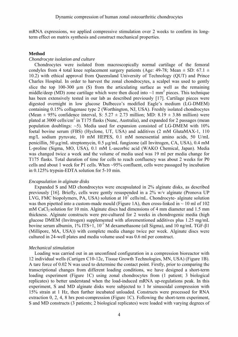
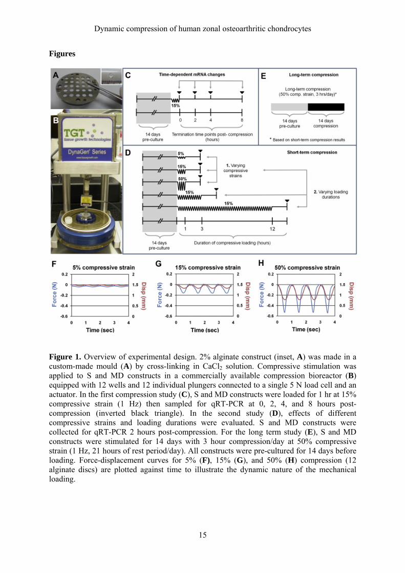
Expanded S and MD chondrocytes were encapsulated in 2% alginate disks, as described previously [16]. Briefly, cells were gently resuspended in a 2% w/v alginate (Pronova UP LVG, FMC biopolymers, PA, USA) solution at 107 cells/mL. Chondrocyte- alginate solution was then pipetted into a custom-made mould (Figure 1A), then cross-linked in ~ 10 ml of 102 mM CaCl2 solution for 10 min. Alginate discs had dimensions of 4 mm diameter and 1.5 mm thickness. Alginate constructs were pre-cultured for 2 weeks in chondrogenic media (high glucose DMEM (Invitrogen) supplemented with aforementioned additives plus 1.25 mg/mL bovine serum albumin, 1% ITS+1, 10-7 M dexamethasone (all Sigma), and 10 ng/mL TGF-β1 (Millipore, MA, USA)) with complete media change twice per week. Alginate discs were cultured in 24-well plates and media volume used was 0.6 ml per construct. Mechanical stimulation Loading was carried out in an unconfined configuration in a compression bioreactor with 12 individual wells (Cartigen C10-12c, Tissue Growth Technologies, MN, USA) (Figure 1B). A tare force of 0.02 N was used to determine the contact point. Firstly, prior to comparing the transcriptional changes from different loading conditions, we have designed a short-term loading experiment (Figure 1C) using zonal chondrocytes from (1 patient; 3 biological replicates) to better understand when the load-induced mRNA up-regulations peak. In this experiment, S and MD alginate disks were subjected to 1 hr sinusoidal compression with 15% strain at 1 Hz, then further incubated unloaded. Constructs were processed for RNA extraction 0, 2, 4, 8 hrs post-compression (Figure 1C). Following the short-term experiment, S and MD constructs (3 patients; 2 biological replicates) were loaded with varying degrees of

Dynamic compression of human zonal osteoarthritic chondrocytes
5
compressive strain and loading durations were tested by applying dynamic compression for 1 hr with 5%, 15%, or 50% strain at 1 Hz. Under 1 Hz sinusoidal loading, calculated loading rate for each strain levels were 0.15 mm/s (5% strain), 0.45 mm/s (15% strain), and 1.5 mm/s (50% strain). The effect of loading duration was tested by dynamically compressing the constructs for 1 hr, 3 hr, or 12 hr with 15% strain at 1 Hz. Based on our result from the first experiment, samples were terminated after 2 hours of unloading for RNA extraction in all subsequent experiments. For long-term evaluation of the constructs, S and MD alginate constructs (3 patients; 2-3 biological replicates) were subjected to 2 week pre-culture, followed by 2 week dynamic compression at 50% compressive strain for 3 hrs per day (Figure 1E). During the intermittent resting periods, loading plungers were raised at least 3 mm above the constructs. Bioreactor was kept within the incubator, and free-swelling controls were also kept within the same incubator in a 24-well plate with equal media volumes. Loaded constructs as well as the controls were cultured in chondrogenic media, and the media volume and media change schedules were the same as during pre-culture times. A 2 week pre-culture period was chosen as it has been shown that delayed loading may enhance chondrocytes response to long-term compression [18]. qRT-PCR
qRT-PCR was performed to quantify the mRNA levels of aggrecan (ACAN), collagen type II (COL2A1), collagen type I (COL1A1), collagen type X (COL10A1), and PRG4 with previously described primers [16]. RNA was extracted from the alginate disks using TRIzol (Invitrogen) reagent according to the manufacturer’s protocol. Distinct 18S and 28S rRNA bands were confirmed through electrophoresis. After DNase treatment (DNase I, Invitrogen), cDNA was synthesized using SuperScriptTM III first-strand synthesis supermix for qRT-PCR (Invitrogen) according to the manufacturer’s protocol. Express SYBR GreenERTM qPCR supermix universal kit (Invitrogen) and a 7900HT fast real-time PCR system (Applied Biosystems, CA, USA) was used to carry out the PCR reaction. The cycle threshold (Ct) value of each gene was normalized to the housekeeping gene, 18S rRNA, using the comparative Ct method (2–ΔΔCt). To quantitate the fold-changes in each of the genes due to compression, compressed samples were normalized to unloaded controls at each time point. Immunohistochemistry S and MD constructs were exposed to 0.1 M monensin (Sigma) in media overnight in order to enhance intracellular detection of secreted proteins such as PRG4. Constructs were then fixed in 4% (w/v) paraformaldehyde containing 100 mM sodium cacodylate trihydrate (Sigma) and 10 mM CaCl2. After fixation, constructs were incubated at 4°C in 50 mM BaCl2 solution containing 100 mM sodium cacodylate trihydrate prior to dehydration and paraffin-embedding to stabilize the alginate [19]. Paraffin-embedded constructs were sectioned at 5 µm. For antigen retrieval, 0.1% (w/v) pronase and 0.1% (w/v) hyaluronidase (Sigma) were used. Sections blocked with 2% FBS solution prior to exposure to primary antibodies (Table 1). Following incubation in fluorescence-labelled goat anti-mouse secondary antibody (Alexa Fluor® 488, Invitrogen) and DAPI (Invitrogen), sections were visualized using a fluorescence microscope (Axio Imager A1, Zeiss). Integrated intensities of immunofluorescence signals around each cell were measured in ImageJ software (NIH) from 10 cells chosen randomly from 6 images for each of the conditions. (2 different microscopy images from 3 different sections; total number of cells analysed = 60). These values were represented in a box plot (Figure 6B). To reduce bleaching, we have mounted our sections with antifade mounting medium (Prolong Gold, Invitrogen) and minimized the delay in obtaining the fluorescence images as well as the time they were exposed to the light source.

Dynamic compression of human zonal osteoarthritic chondrocytes
6
Live/dead assay In order to confirm viability of the chondrocytes following compression, S and MD construct that were pre-cultured for 2 weeks in chondrogenic media were subjected to a 3 hour dynamic compression with 50% compressive strain. Immediately after compression, alginate gels were stained with fluorescein diacetate (FDA) and propidium iodide (PI) in PBS to stain for live and dead cells, respectively. Following 20 minutes of staining, alginate disks that were either labelled as FS (free-swelling control) or Comp (compressed samples) were imaged under fluorescence microscope (Eclipse, Nikon). Biochemical Analysis Glycosaminoglycan (GAG) content was determined to assess compression-induced matrix synthesis. S and MD alginate constructs were digested in 0.5 mg/ml proteinase K solution (Invitrogen) overnight at 60°C. Digested samples (3 patients; 2 biological replicates) were analysed for GAG and DNA. GAG levels were assessed using the modified 1,9-dimethylmethylene blue (DMMB, Sigma) assay at pH 1.5 [20]. DNA content was quantified using the Quant-iT PicoGreen dsDNA assay kit (Invitrogen) according to the manufacturer’s instructions. GAG content was normalized to the DNA content. Conditioned media collected during the entire culture period (3 patients; 3 biological replicates) were also assessed for GAG content. Mechanical testing
The compressive moduli of the S and MD constructs were measured to evaluate the effects of long-term dynamic compression (3 patients; 2-3 biological replicates). Alginate disks without cells cultured under the same conditions were also included in the mechanical testing. Constructs were compressed on an Instron 5848 microtester fitted with a 5 N load cell (Instron, Australia). The compression testing protocol comprised of slow ramp down (10 µm/sec) to 50% with 10 min hold time for collection of stress relaxation data. The stress relaxation protocol was developed from extensive validation tests involving different duration of hold times on alginate disks. Dynamic properties were determined from tests with a sinusoidal strain of 15% amplitude at 1 Hz. Samples were kept in media until the time of mechanical test, which occurred within 12 hours following the last cycle of compression. Using equations 1.1-1.5 [21] and the Solver function in Microsoft Excel, stress-relaxation data were used to determine the equilibrium modulus, and dynamic stress-strain data were used to calculate the compressive storage (E’) and loss (E”) moduli. (Equation 1.1) ))1(1(*)(
)(
t
seqEt
(Equation 1.2) 11 ))(cos(*)( DTtDtD o
(Equation 1.3) 12 ))(cos(*)( FTtFtF o
(Equation 1.4) ))(cos(*' 12 TT
LD
AF
Eo
o
(Equation 1.5) ))(sin(*" 12 TT
LD
AF
Eo
o
Variable definition: σ = stress
Eeq = equilibrium modulus τs = creep time constant τε = stress relaxation time constant
Dynamic compression of human zonal osteoarthritic chondrocytes
7
t = time ε = strain
D = displacement ω = frequency
F = force E’ = storage modulus E” = loss modulus T1, T2 = time lag constants D1, F1 = offset constants A = construct area L = construct thickness
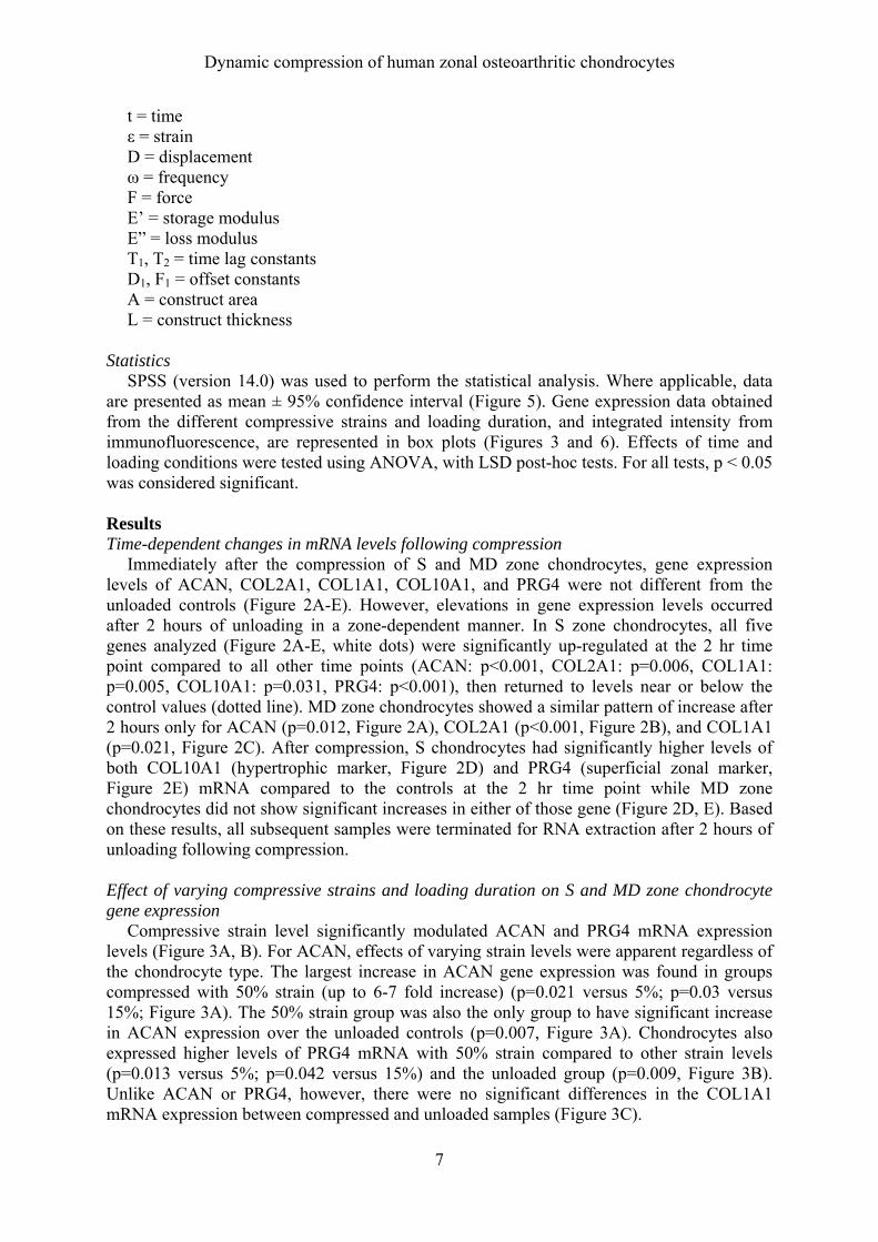
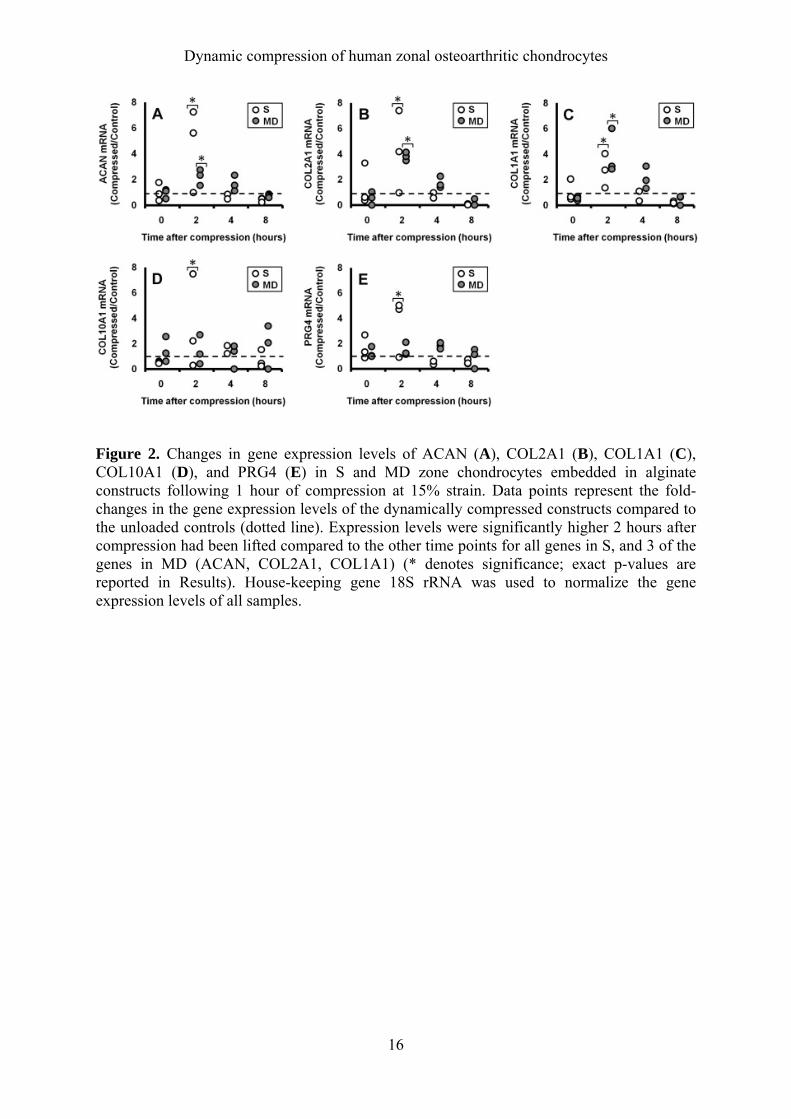
Statistics SPSS (version 14.0) was used to perform the statistical analysis. Where applicable, data are presented as mean ± 95% confidence interval (Figure 5). Gene expression data obtained from the different compressive strains and loading duration, and integrated intensity from immunofluorescence, are represented in box plots (Figures 3 and 6). Effects of time and loading conditions were tested using ANOVA, with LSD post-hoc tests. For all tests, p < 0.05 was considered significant. Results Time-dependent changes in mRNA levels following compression Immediately after the compression of S and MD zone chondrocytes, gene expression levels of ACAN, COL2A1, COL1A1, COL10A1, and PRG4 were not different from the unloaded controls (Figure 2A-E). However, elevations in gene expression levels occurred after 2 hours of unloading in a zone-dependent manner. In S zone chondrocytes, all five genes analyzed (Figure 2A-E, white dots) were significantly up-regulated at the 2 hr time point compared to all other time points (ACAN: p<0.001, COL2A1: p=0.006, COL1A1: p=0.005, COL10A1: p=0.031, PRG4: p<0.001), then returned to levels near or below the control values (dotted line). MD zone chondrocytes showed a similar pattern of increase after 2 hours only for ACAN (p=0.012, Figure 2A), COL2A1 (p<0.001, Figure 2B), and COL1A1 (p=0.021, Figure 2C). After compression, S chondrocytes had significantly higher levels of both COL10A1 (hypertrophic marker, Figure 2D) and PRG4 (superficial zonal marker, Figure 2E) mRNA compared to the controls at the 2 hr time point while MD zone chondrocytes did not show significant increases in either of those gene (Figure 2D, E). Based on these results, all subsequent samples were terminated for RNA extraction after 2 hours of unloading following compression. Effect of varying compressive strains and loading duration on S and MD zone chondrocyte gene expression
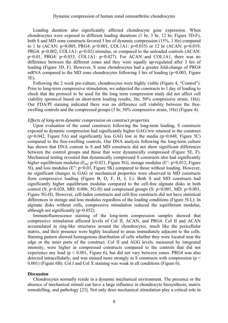
Compressive strain level significantly modulated ACAN and PRG4 mRNA expression levels (Figure 3A, B). For ACAN, effects of varying strain levels were apparent regardless of the chondrocyte type. The largest increase in ACAN gene expression was found in groups compressed with 50% strain (up to 6-7 fold increase) (p=0.021 versus 5%; p=0.03 versus 15%; Figure 3A). The 50% strain group was also the only group to have significant increase in ACAN expression over the unloaded controls (p=0.007, Figure 3A). Chondrocytes also expressed higher levels of PRG4 mRNA with 50% strain compared to other strain levels (p=0.013 versus 5%; p=0.042 versus 15%) and the unloaded group (p=0.009, Figure 3B). Unlike ACAN or PRG4, however, there were no significant differences in the COL1A1 mRNA expression between compressed and unloaded samples (Figure 3C).
Dynamic compression of human zonal osteoarthritic chondrocytes
8
Loading duration also significantly affected chondrocyte gene expression. When chondrocytes were exposed to different loading durations (1 hr, 3 hr, 12 hr; Figure 3D-F), both S and MD zone constructs favored 3 hrs of dynamic compression (15%, 1 Hz) compared to 1 hr (ACAN: p=0.005, PRG4: p<0.001, COL1A1: p=0.035) or 12 hr (ACAN: p=0.019, PRG4: p=0.002, COL1A1: p=0.02) stimulus, or compared to the unloaded controls (ACAN: p=0.01, PRG4: p=0.033, COL1A1: p=0.027). For ACAN and COL1A1, there was no difference between the different zones and they were equally up-regulated after 3 hrs of loading (Figure 3D, F). However, S zone chondrocytes had a greater fold-change of PRG4 mRNA compared to the MD zone chondrocytes following 3 hrs of loading (p=0.003, Figure 3E).
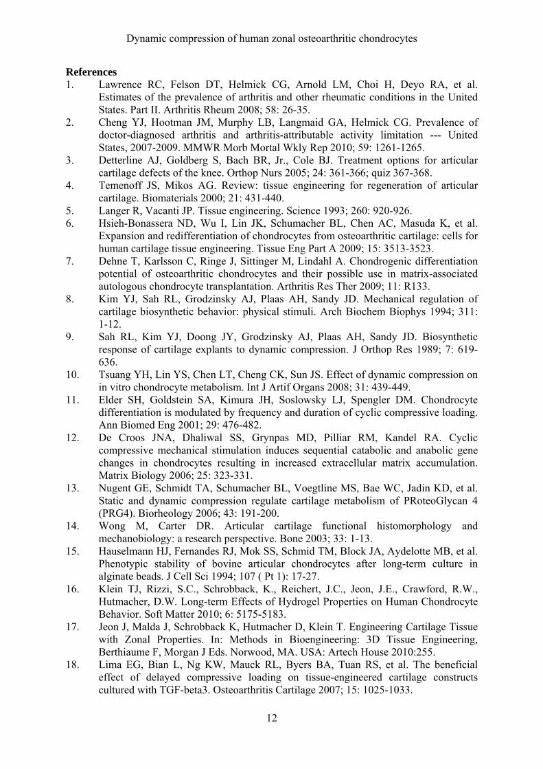
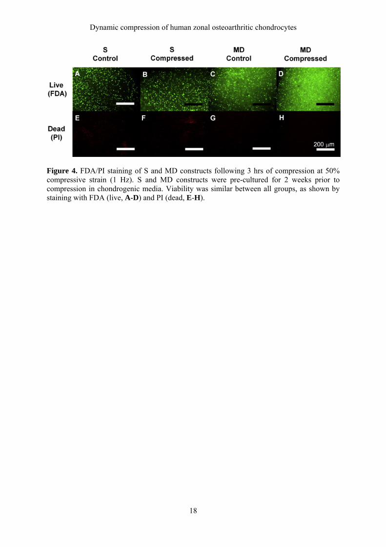
Following the 2 week pre-culture, chondrocytes were highly viable (Figure 4, “Control”). Prior to long-term compressive stimulation, we subjected the constructs to 1 day of loading to check that the protocol to be used for the long term compression study did not affect cell viability (protocol based on short-term loading results; 3hr, 50% compressive strain, 1Hz). Our FDA/PI staining indicated there was no difference cell viability between the free-swelling controls and the compressed groups (3 hr, 50% compressive strain, 1Hz) (Figure 4).
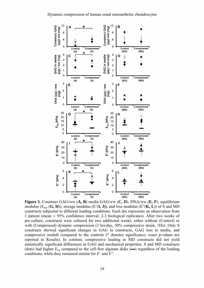
Effects of long-term dynamic compression on construct properties Upon evaluation of the zonal constructs following the long-term loading, S constructs exposed to dynamic compression had significantly higher GAG/ww retained in the construct (p=0.042, Figure 5A) and significantly less GAG lost in the media (p=0.048, Figure 5C) compared to the free-swelling controls. Our DNA analysis following the long-term culture has shown that DNA content in S and MD constructs did not show significant differences between the control groups and those that were dynamically compressed (Figure 5E, F). Mechanical testing revealed that dynamically compressed S constructs also had significantly higher equilibrium modulus (Eeq: p=0.021, Figure 5G), storage modulus (E’: p=0.012, Figure 5I), and loss modulus (E”: p=0.03, Figure 5K) compared to those without loading. However, no significant changes in GAG or mechanical properties were observed in MD constructs from compressive loading (Figure B, D, F, H, J, L). Both S and MD constructs had significantly higher equilibrium modulus compared to the cell-free alginate disks in both control (S: p=0.028, MD: 0.006, 5G-H) and compressed groups (S: p<0.001, MD: p<0.001, Figure 5G-H). However, cell-laden constructs and cell-free constructs did not have statistical differences in storage and loss modulus regardless of the loading conditions (Figure 5I-L). In alginate disks without cells, compressive stimulation reduced the equilibrium modulus, although not significantly (p=0.052).
Immunofluorescence staining of the long-term compression samples showed that compressive stimulation affected levels of Col II, ACAN, and PRG4. Col II and ACAN accumulated in ring-like structures around the chondrocytes, much like the pericellular matrix, and their presence were highly localized to areas immediately adjacent to the cells. Staining pattern showed homogenous distribution of cells whether they were located near the edge or the inner parts of the construct. Col II and AGG levels, measured by integrated intensity, were higher in compressed constructs compared to the controls that did not experience any load (p < 0.001, Figure 6), but did not vary between zones. PRG4 was also detected intracellularly, and was stained more strongly in S constructs with compression (p < 0.001) (Figure 6B). Col I and Col X staining was weak in all conditions (Figure 6). Discussion
Chondrocytes normally reside in a dynamic mechanical environment. The presence or the absence of mechanical stimuli can have a large influence in chondrocyte biosynthesis, matrix remodelling, and pathology [23]. Not only does mechanical stimulation play a critical role in

Dynamic compression of human zonal osteoarthritic chondrocytes
9
chondrocyte homeostasis in vivo, its ability to change chondrocyte biosynthetic behavior has been reported through in vitro studies using explants [9] and engineered tissues [24-26]. Our results show that S and MD zone chondrocytes from OA patients are significantly influenced by loading conditions. Following compression, we observed a transient elevation in matrix and zone-specific mRNA levels peaking at 2 hours after loading. We also observed that compression-induced increase in mRNA levels varied depending on the compressive strain and loading duration. Long-term compression data indicate that compression enhances Col II and ACAN expression irrespective of the zonal origin of the chondrocytes. However, GAG/DNA and compressive moduli suggest that S constructs benefit more from this loading regime than MD constructs. Furthermore, S constructs increased levels of PRG4 with compressive loading compared to MD constructs, showing that compression can differentially modulate protein secretion in a zone-dependent fashion.
Since mRNA expression is likely to change over time, our first objective was to define the
time-dependent gene expression changes after compression has been lifted (Figure 2). Immediately after the samples were released from the compression, (time point 0, Figure 2) gene expression levels of the compressed samples were similar to the unloaded controls (dotted line). However, mRNA expression was transiently up-regulated, peaking 2 hours after compression finished, with S and MD chondrocytes responding distinctly (Figure 2). Milward-Sadler, et al., also found a peak in ACAN mRNA expression by normal human chondrocytes in monolayer after 1-3 hours of cyclic pressurization [27]. The transient nature of ACAN, COL2A1, COL1A1, COL10A1, and PRG4 gene expression highlights the importance of sampling time, and suggests that terminating compressed samples immediately after unloading may not reflect the resultant changes in gene expression. Therefore, observations of little to no increase in gene expression reported in other compression studies [24, 28] could be due to terminating samples immediately after loading. When we examined the effects of 1, 3, and 12 hour compression on S and MD zone chondrocytes, we found that 3 hour compression produced the highest gene expression levels of ACAN, PRG4, and COL1A1 (Figure 3D-F). This also suggests chondrocytes are only positively responsive to load for a limited time, and that sustained compression (e.g., 12 hour compression) does not necessarily prolong increase in matrix gene expression levels. According to Stoddart, et al., and Valhmu, et al., [29, 30], samples terminated immediately after unloading showed an ACAN mRNA peak 1-3 hours from the start of the compression, suggesting that mRNA levels can peak while samples are being compressed. Thus, it is possible that gene expression level of chondrocytes dynamically compressed for 12 hours might have reached a peak within the 12-hour loading period, which would result in lower mRNA levels when terminated after compression. Further studies are needed to determine whether the time-dependent changes in mRNAs are altered by varying loading conditions.
The different levels of compressive strains experienced by each of the cartilage zones in
vivo [14] led to our hypothesis that chondrocytes from the different zones respond differently to varying compressive strains. Unexpectedly, for both S and MD zone chondrocytes, 50% compressive strain induced the highest increase of ACAN and PRG4 mRNA, whereas COL1A1, a chondrocyte dedifferentiation marker [31], did not vary significantly between compressive strain levels (Figure 3C). This suggests that compression does not simply up-regulate all genes, but can induce cartilage-specific genes, depending on the strain levels applied. Initially, we expected 50% compressive strain to inhibit matrix gene expression of MD zone chondrocytes, since these cells normally do not experience such large compressive strains [32]. Yet, cartilage and 2% alginate gels vary significantly in mechanical properties. The equilibrium compressive modulus of human cartilage ranges from about 1.16±0.20 MPa

Dynamic compression of human zonal osteoarthritic chondrocytes
10
in the superficial zone to 7.75±1.45 MPa in the deep zone [33]. Previous experiments from our lab showed that without cells, 2% alginate gels have compressive modulus of about 10 kPa (day 0) to 30 kPa (~3 months following long-term culture in media) [16]. While isolated human chondrocytes are softer (Young’s Modulus: ~4 kPa [34]) than the alginate gel, it is likely that 2 weeks of culture prior to compression resulted in the accumulation of much stiffer pericellular matrix [35]). This may impart a stress-shielding effect, reducing the actual compressive strain of the chondrocytes. This hypothesis is supported by data from Knight, et al., which showed that after 6 days of culture in 3% agarose gels, 20% compression yielded little chondrocyte deformation because they were surrounded by the pericellular matrix [36]. Recent findings also indicate that chondrocytes in alginate have sensitive Ca2+ signalling response to fluid flow when they have cell-matrix interaction [37]. Since 50% compressive strain would result in more fluid flow around chondrocytes, the extent of Ca2+ signalling, which plays an important part in mechanotransduction [38], may have been greater in the 50% strain groups compared to the 5% or 15% strain groups. On a similar note, studies also show that fast strain rates induce greater Ca2+ transients during compressive loading [39]. The different strain levels in our experiment also resulted in different strain rates at 1 Hz (5%: 0.15mm/s; 15%: 0.45mm/s; 50%: 1.5mm/s), and it is possible that 50% strain group, with the fastest strain rate, may have had more load-induced Ca2+ signalling than the other strain groups, indicating that the results that we see may not only be due to the strain levels, but also strain rates as well. Apart from the Ca2+ signalling effects, the benefits of 50% compressive strain may simply be due to improved nutrient transport due to increased fluid flow during high-strain dynamic compression [40]. Long-term compression studies indicate mixed results in regards to zonal differences. While compression significantly enhanced GAG retention in S constructs (Figure 5A), we did not observe such increase in the MD constructs (Figure 5B). Interestingly, however, both S and MD constructs had significantly brighter Col II and ACAN staining with compression (Figure 6). It is possible that our immunofluorescence results may be showing only a part of the effect that compressive stimulation had in S and MD constructs. One explanation could be that there may be proteoglycan molecules other than ACAN that may be contributing to the increased GAG levels observed in S constructs. In case the synthesis or composition of various proteoglycan molecules are differentially modulated by compression in a zone-dependent manner, there is a possibility that it could have contributed to the differences in GAG and mechanical properties. While our immunofluorescence images show that compressive stimulation benefits both S and MD constructs, high amplitude (50%) dynamic loading is perhaps more important for S constructs and influences GAG biosynthesis in S chondrocytes to a great extent compared to the MD constructs. It is important to note, however, that with or without loading, MD chondrocytes produced GAG at a level comparable or higher than those of the dynamically stimulated S constructs, confirming the zone-dependent differences in GAG production in vivo [42] and in vitro [43]. Our findings which indicate that dynamic compression selectively benefits S chondrocytes in terms of GAG biosynthesis is in alignment with other studies. For example, dynamic compression of bovine cartilage explants showed that proteoglycan synthesis in S chondrocytes was stimulated to a greater degree following 7 day compression in comparison to the deep (D) zone chondrocytes [44]. Judging from the varying degree of benefits long-term compression has on expanded S and MD zone chondrocytes and its effect in zonal marker expression (PRG4, Figure 6), it appears that zonal chondrocytes retain a certain degree of innate programming even after monolayer culture.

Dynamic compression of human zonal osteoarthritic chondrocytes
11
While chondrocytes from OA joints are often disregarded as possible cell sources for cartilage tissue engineering due to less robust biosynthesis compared to those from young, healthy donors [46], we have shown that biosynthetic capacity of the chondrocytes from OA joints can be enhanced with selected compression regimes. Under loading conditions of 50% strain or 3 hrs of loading, chondrocytes from 4 different patients showed consistent elevation of ACAN and PRG4 mRNAs. We also observed zonal differences using these cells, and postulate that similar or greater differences in response to compression will be seen in the young healthy chondrocytes. In future cartilage engineering endeavours, appreciating and taking note of the different characteristics of the articular cartilage zones will be helpful in producing cartilage constructs that better mimic the native tissue.
Acknowledgments We would like to thank Prof. Ross Crawford for supplying the samples from joint replacement surgeries he performed at the Prince Charles Hospital. The antibodies against collagen type II (II-II6B3), type I (M-38), and type X (X-AC9), were obtained from the DSHB maintained by the University of Iowa, Department of Biology, Iowa City, IA 52242. Contributions All listed authors have made substantial contribution to the following aspects of the manuscript: (1) The conception and design of the study, analysis, and interpretation of data (2) Drafting the article or revising it critically for important intellectual content (3) Final approval of the version to be submitted The integrity of this work is guaranteed by June E. Jeon ([email protected]) and Dr. Travis Klein ([email protected]). Role of the funding source The authors would like to acknowledge the Australian Research Council for funding. Competing interest statement Authors have no conflict of interest with regard to the work.
Dynamic compression of human zonal osteoarthritic chondrocytes
12
References 1. Lawrence RC, Felson DT, Helmick CG, Arnold LM, Choi H, Deyo RA, et al.
Estimates of the prevalence of arthritis and other rheumatic conditions in the United States. Part II. Arthritis Rheum 2008; 58: 26-35.
2. Cheng YJ, Hootman JM, Murphy LB, Langmaid GA, Helmick CG. Prevalence of doctor-diagnosed arthritis and arthritis-attributable activity limitation --- United States, 2007-2009. MMWR Morb Mortal Wkly Rep 2010; 59: 1261-1265.
3. Detterline AJ, Goldberg S, Bach BR, Jr., Cole BJ. Treatment options for articular cartilage defects of the knee. Orthop Nurs 2005; 24: 361-366; quiz 367-368.
4. Temenoff JS, Mikos AG. Review: tissue engineering for regeneration of articular cartilage. Biomaterials 2000; 21: 431-440.
5. Langer R, Vacanti JP. Tissue engineering. Science 1993; 260: 920-926. 6. Hsieh-Bonassera ND, Wu I, Lin JK, Schumacher BL, Chen AC, Masuda K, et al.
Expansion and redifferentiation of chondrocytes from osteoarthritic cartilage: cells for human cartilage tissue engineering. Tissue Eng Part A 2009; 15: 3513-3523.
7. Dehne T, Karlsson C, Ringe J, Sittinger M, Lindahl A. Chondrogenic differentiation potential of osteoarthritic chondrocytes and their possible use in matrix-associated autologous chondrocyte transplantation. Arthritis Res Ther 2009; 11: R133.
8. Kim YJ, Sah RL, Grodzinsky AJ, Plaas AH, Sandy JD. Mechanical regulation of cartilage biosynthetic behavior: physical stimuli. Arch Biochem Biophys 1994; 311: 1-12.
9. Sah RL, Kim YJ, Doong JY, Grodzinsky AJ, Plaas AH, Sandy JD. Biosynthetic response of cartilage explants to dynamic compression. J Orthop Res 1989; 7: 619-636.
10. Tsuang YH, Lin YS, Chen LT, Cheng CK, Sun JS. Effect of dynamic compression on in vitro chondrocyte metabolism. Int J Artif Organs 2008; 31: 439-449.
11. Elder SH, Goldstein SA, Kimura JH, Soslowsky LJ, Spengler DM. Chondrocyte differentiation is modulated by frequency and duration of cyclic compressive loading. Ann Biomed Eng 2001; 29: 476-482.
12. De Croos JNA, Dhaliwal SS, Grynpas MD, Pilliar RM, Kandel RA. Cyclic compressive mechanical stimulation induces sequential catabolic and anabolic gene changes in chondrocytes resulting in increased extracellular matrix accumulation. Matrix Biology 2006; 25: 323-331.
13. Nugent GE, Schmidt TA, Schumacher BL, Voegtline MS, Bae WC, Jadin KD, et al. Static and dynamic compression regulate cartilage metabolism of PRoteoGlycan 4 (PRG4). Biorheology 2006; 43: 191-200.
14. Wong M, Carter DR. Articular cartilage functional histomorphology and mechanobiology: a research perspective. Bone 2003; 33: 1-13.
15. Hauselmann HJ, Fernandes RJ, Mok SS, Schmid TM, Block JA, Aydelotte MB, et al. Phenotypic stability of bovine articular chondrocytes after long-term culture in alginate beads. J Cell Sci 1994; 107 ( Pt 1): 17-27.
16. Klein TJ, Rizzi, S.C., Schrobback, K., Reichert, J.C., Jeon, J.E., Crawford, R.W., Hutmacher, D.W. Long-term Effects of Hydrogel Properties on Human Chondrocyte Behavior. Soft Matter 2010; 6: 5175-5183.
17. Jeon J, Malda J, Schrobback K, Hutmacher D, Klein T. Engineering Cartilage Tissue with Zonal Properties. In: Methods in Bioengineering: 3D Tissue Engineering, Berthiaume F, Morgan J Eds. Norwood, MA. USA: Artech House 2010:255.
18. Lima EG, Bian L, Ng KW, Mauck RL, Byers BA, Tuan RS, et al. The beneficial effect of delayed compressive loading on tissue-engineered cartilage constructs cultured with TGF-beta3. Osteoarthritis Cartilage 2007; 15: 1025-1033.

Dynamic compression of human zonal osteoarthritic chondrocytes
13
19. Heywood HK, Sembi PK, Lee DA, Bader DL. Cellular utilization determines viability and matrix distribution profiles in chondrocyte-seeded alginate constructs. Tissue Eng 2004; 10: 1467-1479.
20. Enobakhare BO, Bader DL, Lee DA. Quantification of sulfated glycosaminoglycans in chondrocyte/alginate cultures, by use of 1,9-dimethylmethylene blue. Anal Biochem 1996; 243: 189-191.
21. Fung YC. Biomechanics: mechanical properties of living tissues. New York, Springer-Verlag 1993.
22. Sharma B, Williams CG, Kim TK, Sun D, Malik A, Khan M, et al. Designing zonal organization into tissue-engineered cartilage. Tissue Eng 2007; 13: 405-414.
23. Grodzinsky AJ, Levenston ME, Jin M, Frank EH. Cartilage tissue remodeling in response to mechanical forces. Annu Rev Biomed Eng 2000; 2: 691-713.
24. Demarteau O, Wendt D, Braccini A, Jakob M, Schafer D, Heberer M, et al. Dynamic compression of cartilage constructs engineered from expanded human articular chondrocytes. Biochem Biophys Res Commun 2003; 310: 580-588.
25. Mauck RL, Soltz MA, Wang CC, Wong DD, Chao PH, Valhmu WB, et al. Functional tissue engineering of articular cartilage through dynamic loading of chondrocyte-seeded agarose gels. J Biomech Eng 2000; 122: 252-260.
26. Ragan PM, Chin VI, Hung HH, Masuda K, Thonar EJ, Arner EC, et al. Chondrocyte extracellular matrix synthesis and turnover are influenced by static compression in a new alginate disk culture system. Arch Biochem Biophys 2000; 383: 256-264.
27. Millward-Sadler SJ, Wright MO, Davies LW, Nuki G, Salter DM. Mechanotransduction via integrins and interleukin-4 results in altered aggrecan and matrix metalloproteinase 3 gene expression in normal, but not osteoarthritic, human articular chondrocytes. Arthritis Rheum 2000; 43: 2091-2099.
28. Hunter CJ, Imler SM, Malaviya P, Nerem RM, Levenston ME. Mechanical compression alters gene expression and extracellular matrix synthesis by chondrocytes cultured in collagen I gels. Biomaterials 2002; 23: 1249-1259.
29. Stoddart MJ, Ettinger L, Häuselmann HJ. Enhanced matrix synthesis in de novo, scaffold free cartilage-like tissue subjected to compression and shear. Biotechnology and Bioengineering 2006; 95: 1043-1051.
30. Valhmu WB, Stazzone EJ, Bachrach NM, Saed-Nejad F, Fischer SG, Mow VC, et al. Load-Controlled Compression of Articular Cartilage Induces a Transient Stimulation of Aggrecan Gene Expression. Arch Biochem Biophys 1998; 353: 29-36.
31. Benya PD, Shaffer JD. Dedifferentiated chondrocytes reexpress the differentiated collagen phenotype when cultured in agarose gels. Cell 1982; 30: 215-224.
32. Schinagl RM, Gurskis D, Chen AC, Sah RL. Depth-dependent confined compression modulus of full-thickness bovine articular cartilage. J Orthop Res 1997; 15: 499-506.
33. Chen SS, Falcovitz YH, Schneiderman R, Maroudas A, Sah RL. Depth-dependent compressive properties of normal aged human femoral head articular cartilage: relationship to fixed charge density. Osteoarthritis Cartilage 2001; 9: 561-569.
34. Guilak F, Alexopoulos LG, Upton ML, Youn I, Choi JB, Cao L, et al. The pericellular matrix as a transducer of biomechanical and biochemical signals in articular cartilage. Ann N Y Acad Sci 2006; 1068: 498-512.
35. Alexopoulos LG, Haider MA, Vail TP, Guilak F. Alterations in the mechanical properties of the human chondrocyte pericellular matrix with osteoarthritis. J Biomech Eng 2003; 125: 323-333.
36. Knight MM, Lee DA, Bader DL. The influence of elaborated pericellular matrix on the deformation of isolated articular chondrocytes cultured in agarose. Biochim Biophys Acta 1998; 1405: 67-77.

Dynamic compression of human zonal osteoarthritic chondrocytes
14
37. Degala S, Zipfel WR, Bonassar LJ. Chondrocyte calcium signaling in response to fluid flow is regulated by matrix adhesion in 3-D alginate scaffolds. Arch Biochem Biophys; 505: 112-117.
38. Han SK, Wouters W, Clark A, Herzog W. Mechanically induced calcium signaling in chondrocytes in situ. J Orthop Res; 30: 475-481.
39. Pingguan-Murphy B, El-Azzeh M, Bader DL, Knight MM. Cyclic compression of chondrocytes modulates a purinergic calcium signalling pathway in a strain rate- and frequency-dependent manner. J Cell Physiol 2006; 209: 389-397.
40. Zhang L, Gardiner BS, Smith DW, Pivonka P, Grodzinsky A. The effect of cyclic deformation and solute binding on solute transport in cartilage. Arch Biochem Biophys 2007; 457: 47-56.
41. Schumacher BL, Block JA, Schmid TM, Aydelotte MB, Kuettner KE. A novel proteoglycan synthesized and secreted by chondrocytes of the superficial zone of articular cartilage. Arch Biochem Biophys 1994; 311: 144-152.
42. Buckwalter JA, Mankin HJ. Articular cartilage: tissue design and chondrocyte-matrix interactions. Instr Course Lect 1998; 47: 477-486.
43. Coates EE, Fisher JP. Phenotypic variations in chondrocyte subpopulations and their response to in vitro culture and external stimuli. Ann Biomed Eng 2010; 38: 3371-3388.
44. Korver TH, van de Stadt RJ, Kiljan E, van Kampen GP, van der Korst JK. Effects of loading on the synthesis of proteoglycans in different layers of anatomically intact articular cartilage in vitro. J Rheumatol 1992; 19: 905-912.
45. Nugent GE, Aneloski NM, Schmidt TA, Schumacher BL, Voegtline MS, Sah RL. Dynamic shear stimulation of bovine cartilage biosynthesis of proteoglycan 4. Arthritis Rheum 2006; 54: 1888-1896.
46. Buckwalter JA, Mankin HJ. Articular cartilage: degeneration and osteoarthritis, repair, regeneration, and transplantation. Instr Course Lect 1998; 47: 487-504.
Dynamic compression of human zonal osteoarthritic chondrocytes
15
Figures
Figure 1. Overview of experimental design. 2% alginate construct (inset, A) was made in a custom-made mould (A) by cross-linking in CaCl2 solution. Compressive stimulation was applied to S and MD constructs in a commercially available compression bioreactor (B) equipped with 12 wells and 12 individual plungers connected to a single 5 N load cell and an actuator. In the first compression study (C), S and MD constructs were loaded for 1 hr at 15% compressive strain (1 Hz) then sampled for qRT-PCR at 0, 2, 4, and 8 hours post-compression (inverted black triangle). In the second study (D), effects of different compressive strains and loading durations were evaluated. S and MD constructs were collected for qRT-PCR 2 hours post-compression. For the long term study (E), S and MD constructs were stimulated for 14 days with 3 hour compression/day at 50% compressive strain (1 Hz, 21 hours of rest period/day). All constructs were pre-cultured for 14 days before loading. Force-displacement curves for 5% (F), 15% (G), and 50% (H) compression (12 alginate discs) are plotted against time to illustrate the dynamic nature of the mechanical loading.
Dynamic compression of human zonal osteoarthritic chondrocytes
16
Figure 2. Changes in gene expression levels of ACAN (A), COL2A1 (B), COL1A1 (C), COL10A1 (D), and PRG4 (E) in S and MD zone chondrocytes embedded in alginate constructs following 1 hour of compression at 15% strain. Data points represent the fold-changes in the gene expression levels of the dynamically compressed constructs compared to the unloaded controls (dotted line). Expression levels were significantly higher 2 hours after compression had been lifted compared to the other time points for all genes in S, and 3 of the genes in MD (ACAN, COL2A1, COL1A1) (* denotes significance; exact p-values are reported in Results). House-keeping gene 18S rRNA was used to normalize the gene expression levels of all samples.

Dynamic compression of human zonal osteoarthritic chondrocytes
17
Figure 3: Gene expression levels of ACAN (A, D), PRG4 (B, E), and COL1A1 (C, F) in S and MD zone chondrocytes after compression either with varying compressive strains (5%, 15%, 50%) for 1 h (A-C) or with 15% compressive strain for various durations (1 hr, 3 hr, 12 hr) (D-F) normalized to unloaded controls. Loading groups with higher fold-changes in the gene expression compared to other loading groups were indicated with (*) (exact p-values are reported in Results). Dotted line denotes free-swelling control levels. S and MD groups were combined for statistical analysis. House-keeping gene 18S rRNA was used to normalize the gene expression levels of all samples. The distribution represents the variation between 3 patients (n=3, 2 technical repeats/patient). Comparing the different compressive strain levels, cartilage-associated genes ACAN and PRG4 were significantly up regulated with 50% compressive strain (A,B). The evaluation of the different loading durations showed that 3 hr compression significantly up-regulated all three genes (D-F) compared to 1hr and 12hr.
Dynamic compression of human zonal osteoarthritic chondrocytes
18
Figure 4. FDA/PI staining of S and MD constructs following 3 hrs of compression at 50% compressive strain (1 Hz). S and MD constructs were pre-cultured for 2 weeks prior to compression in chondrogenic media. Viability was similar between all groups, as shown by staining with FDA (live, A-D) and PI (dead, E-H).
Dynamic compression of human zonal osteoarthritic chondrocytes
19
Figure 5. Construct GAG/ww (A, B) media GAG/ww (C, D), DNA/ww (E, F), equilibrium modulus (Eeq, (G, H)), storage modulus (E’(I, J)), and loss modulus (E”(K, L)) of S and MD constructs subjected to different loading conditions. Each dot represents an observation from 1 patient (mean ± 95% confidence interval; 2-3 biological replicates). After two weeks of pre-culture, constructs were cultured for two additional weeks, either without (Control) or with (Compressed) dynamic compression (3 hrs/day, 50% compressive strain, 1Hz). Only S constructs showed significant changes in GAG in constructs, GAG loss to media, and compressive moduli compared to the controls (* denotes significance; exact p-values are reported in Results). In contrast, compressive loading in MD constructs did not yield statistically significant differences in GAG and mechanical properties. S and MD constructs (dots) had higher Eeq compared to the cell-free alginate disks (▬) regardless of the loading conditions, while they remained similar for E’ and E”.

Dynamic compression of human zonal osteoarthritic chondrocytes
20
Figure 6. Immunofluorescence images of S and MD constructs showing Col II, ACAN, PRG4, Col I, and Col X (green) with nucleus (blue) (A) and box plots (B) showing integrated intensity values (total number of cells evaluated/condition = 60). S and MD constructs subjected to 2 weeks of compressive stimulation had significantly enhanced Col II and ACAN staining compared to free-swelling (FS) controls. PRG4 stains were more intense in compressed S constructs compared to S controls and MD compressed groups. No differences were found in Col I and Col X stained sections with regards to loading conditions and zones. Statistical significance was denoted with § (p-values are reported in Results). (scale bar = 100 µm, inset scale bar = 20 µm)

Dynamic compression of human zonal osteoarthritic chondrocytes
21
Table 1. List of target antigens, dilution, and sources for primary antibodies used for immunofluorescence analysis of the long-term compression constructs. Target Antigen Abbreviation Dilution Reference No. Source Collagen type II Col II 1:200 II-II6B3 DSHB
Aggrecan ACAN Undiluted supernatant
MA75A95 Abcam
Lubricin PRG4 1:200 Ab28484 Abcam Collagen type I Col I 1:20 M-38 DSHB Collagen type X Col X 1:100 X-AC9 DSHB