Embed Size (px)
Citation preview
ORIGINAL ARTICLE
NTPDase3 and ecto-5′-nucleotidase/CD73 are differentiallyexpressed during mouse bladder cancer progression
Liliana Rockenbach & Elizandra Braganhol & Fabrícia Dietrich & Fabrício Figueiró &
Manoella Pugliese & Maria Isabel Albano Edelweiss & Fernanda Bueno Morrone &
Jean Sévigny & Ana Maria Oliveira Battastini
Received: 24 October 2013 /Accepted: 6 January 2014# Springer Science+Business Media Dordrecht 2014
Abstract According to the World Health Organization, blad-der cancer is the seventh most common cancer among men inthe world. The current treatments for this malignancy are notefficient to prevent the recurrence and progression of tumors.Then, researches continue looking for better therapeutic tar-gets which can end up in new and more efficient treatments.One of the recent findings was the identification that thepurinergic system was involved in bladder tumorigenesis.The ectonucleotidases, mainly ecto-5′-nucleotidase/CD73have been revealed as new players in cancer progression andmalignity. In this work, we investigated the NTPDase3 andecto-5′-nucleotidase/CD73 expression in cancer progressionin vivo. Bladder tumor was induced in mice by the addition of0.05 % of N-butyl-N-(hydroxybutyl)-nitrosamine (BBN) inthe drinking water for 4, 8, 12, 18, and 24 weeks. After thisperiod, mice bladders were removed for histopathology anal-ysis and immunofluorescence assays. The bladder of animals
which has received BBN had alterations, mainly inflamma-tion, in initial times of tumor induction. After 18 weeks,mice’s bladder has developed histological alterations similarto human transitional cell carcinoma. The cancerousurothelium, frommice that receivedBBN for 18 and 24weeks,presented a weak immunostaining to NTPDase3, in contrast toan increased expression of ecto-5′-nucleotidase/CD73. Thealtered expression of NTPDase3 and ecto-5′-nucleotidase/CD73 presented herein adds further evidence to support theidea that alterations in ectonucleotidases are involved in blad-der tumorigenesis and reinforce the ecto-5′-nucleotidase/CD73 as a future biomarker and/or a target for pharmacolog-ical therapy of bladder cancer.
Keywords Bladder cancer . BBN . Purinergic signaling .
NTPDase3 . Ecto-5′-nucleotidase/CD73
M. Pugliese :A. M. O. BattastiniDepartamento de Bioquímica, Instituto de Ciências Básicas daSaúde, Universidade Federal do Rio Grande do Sul (UFRGS),Porto Alegre, Rio Grande do Sul, Brazil
L. Rockenbach : F. Dietrich : F. Figueiró :A. M. O. Battastini (*)Programa de Pós-Graduação em Ciências Biológicas: Bioquímica,Instituto de Ciências Básicas da Saúde, Universidade Federal do RioGrande do Sul (UFRGS), Porto Alegre, Rio Grande do Sul, Brazile-mail: [email protected]
E. BraganholPrograma de Pós-Graduação em Bioquímica e Bioprospecção,Centro de Ciências Químicas, Farmacêuticas e de Alimentos,Universidade Federal de Pelotas (UFPEL),Pelotas, Rio Grande do Sul, Brazil
M. I. A. EdelweissHospital de Clínicas de Porto Alegre, Universidade Federal do RioGrande do Sul (UFRGS), Porto Alegre, Rio Grande do Sul, Brazil
F. B. MorroneFaculdade de Farmácia, Pontifícia Universidade Católica do RioGrande do Sul (PUCRS), Porto Alegre, Rio Grande do Sul, Brazil
J. SévignyCentre de Recherche en Rhumatologie et Immunologie,Centre de recherche du CHU de Québec, and Département demicrobiologie-infectiologie et d’immunologie,Faculté de Médecine, Université Laval, Québec, QC, Canada
L. Rockenbach (*)Departamento de Bioquímica, Instituto de Ciências Básicas daSaúde, UFRGS, Rua Ramiro Barcelos, 2600-anexo,90035-003 Porto Alegre, Rio Grande do Sul, Brazile-mail: [email protected]
Purinergic SignallingDOI 10.1007/s11302-014-9405-8
Introduction
Bladder cancer is the second most prevalent tumor in thegenitourinary tract [1, 2]. It is the seventh most commoncancer worldwide with about 336,000 new cases per year[3]. The main risk factors are smoking, which increase therisk up to six times, and occupational and environmentalexposure to carcinogens [4, 5]. About 90 % of bladder cancercorresponds to transitional cell carcinoma (TCC) [1, 6] due tothe fact that urothelium is constantly exposed to potentialcarcinogens [7]. The tumor invasiveness of TCC defines thepatient’s prognosis. For example, 70–80 % of patients presentsuperficial non-muscle-invasive TCCs which are generallynot life threatening; while 20–30 % of individuals havemuscle-invasive TCCs with increased risk of metastasis anddeath [8, 9]. Superficial cancers are treated by transurethralresection (TUR), followed by chemo/immunotherapy. How-ever, nearly 70 % of patients present tumor recurrence, where30 % of the recurrent tumors progress to muscle-invasivedisease within 5 years of TUR [8, 10]. Given the high recur-rence rates and the need for frequent monitoring, this diseaseis one of the most expensive cancers to treat on a per patientbasis from diagnosis until death [11], and hence, poses atremendous burden on health systems worldwide [4]. There-fore, new therapeutic targets, which end up in more efficienttreatments, are necessary to prevent bladder cancer recurrenceand progression.
Accordingly, recent researches have focused in the poten-tial involvement of purinergic system in bladder tumors [1,12–14]. Nucleosides and nucleotides mediate a variety ofbiological functions in both short- and long-term signalingfunctions (development, regeneration, differentiation, prolif-eration, and cell death) [1, 12]. These events are mediated bythe activation of P1 (for adenosine) or P2 (for ATP, ADP,UDP, and UTP) receptors and are controlled by the action ofectonucleotidases [15]. The ecto-nucleoside triphosphatediphosphohydrolases refer to a family of cell-surface enzymesthat hydrolyze extracellular ATP and ADP to AMP. Studieshave demonstrated the involvement of these enzymes in can-cer progression [16–18]. The final dephosphorylation of nu-cleotides, conversion of nucleoside monophosphates (e.g.,AMP) to their respective nucleosides (e.g., adenosine), iscatalyzed by ecto-5′-nucleotidase/CD73 (ecto-5′-NT/CD73).This enzyme is highly expressed in a variety of solid tumors[19–22] and has both its enzymatic activity and its adhesionprotein function associated with cancer progression [23, 24].Besides, ecto-5′-NT/CD73 was found to be involved in cancercell growth, maturation, differentiation, adhesion, migration,invasiveness, metastasis, immune escape, and drug resistance[19, 21–27].
A previous study with mouse bladders showed that mousehealthy urothelium expresses only NTPDase3, not expressingthe ecto-5′-NT/CD73 [28]. This is in agreement with a
previous work from our group, where we showed a differen-tial pattern of ectonucleotidases expression in two malignantbladder cancer cells. We showed that a less malignant lineagefrom a TCC grade 1 of malignancy (RT4) expressesNTPDase3 and ecto-5′-NT/CD73, while a more malignantlineage, from a TCC grade 4 of malignancy (T24) only ex-presses ecto-5′-NT/CD73 [14]. Although little is known aboutthe role of NTPDase3 in cancer, these findings prompted us tosuspect that the loss of NTPDase3 expression and the parallelecto-5′-NT/CD73 expression might be involved in the bladdercancer progression.
Therefore, herein we investigate the NTPDase3 and ecto-5′-NT/CD73 expression in a model of mouse bladder cancerinduced by N-butyl-N-(hydroxybutyl)-nitrosamine (BBN).
Materials and methods
Reagents
BBN was purchased from Sigma (Sigma Chemical Company,St. Louis, MO, USA). Rabbit antibodies anti-rat ecto-5′-nu-cleotidase (rNu-9L) and anti-rat NTPDase3 (rN3-1L) were ob-tained from http://ectonucleotidases-ab.com. Alexa fluor 568goat anti-rabbit IgG and Alexa fluor 488 phalloidin werepurchased from Invitrogen (Invitrogen Co., Carlsbad, CA,USA). Optimum cutting temperature (OCT) freezing medium(Tissue-Tek; Sakura Finetek, Torrance, CA, USA). All otherchemicals and solvents used were of analytical grade.
Animals
Male mice from the Balb-c lineage were used at the age of10 weeks. Animals were obtained and maintained in theUnidade de Experimentação Animal do Hospital de Clínicasde Porto Alegre (HCPA) under a standard dark–light cycle(lights on between 7:00 a.m. and 7:00 p.m.) at room-controlled temperature (22±2 °C). The mice had free accessto standard laboratory chow and water. Mice were euthana-tized by isoflurane inhalation. After euthanasia, the bladderswere rapidly excised and processed as described below. Allanimal studies were carried out in strict accordance with therecommendations in the Brazilian national law number11.794, from October 8, 2008, which determines the proce-dures to scientific use of animals. The protocol (protocol 10-0104) was approved by the Research Ethics Committee atgroup of research and graduation of HCPA. All efforts weremade to minimize animal suffering.
Bladder cancer induction
The animal model of bladder cancer induction by BBN havebeen used in many studies [29–33] mainly because BBN
Purinergic Signalling
induced alterations in the bladder of rodents that are corre-spondent to histopathological and molecular features of hu-man transitional cell carcinoma [34–36]. In this work, bladdercancer was induced by the addition of 0.05 % of BBN in thedrinking water for 4, 8, 12, 18, and 24 weeks with a respectivecontrol group for each induction time. The number of mice pergroup was 8 for control groups of 4, 8, and 12 weeks and 9,10, and 10 for BBN groups of 4, 8, and 12 weeks, respective-ly; and for the cancer induction times of 18 and 24 weeks, itwas 3 for control groups and 5 and 4 for groups that receivedBBN, respectively. The animals were weighed in an analyticalbalance, as well as the excised bladders. Then, the bladder wetweight was expressed as milligrams of bladder per 100 g ofanimal as an additional measure of edema [37].
Bladder processing
A Y-shaped cut was made in the excised bladders for furthertissue processing. Thereby, bladders were disposed as a mono-layer and were divided in two parts: apex and base. The tissueswere embedded in OCT freezing medium and snap frozen inisopentane in dry ice and stored at −80 °C until use. In thisstudy, we analyzed only the base of bladders due to its majorand constant contact with urine, which ends up in the majorprobability of cancer. For this, the frozen base of bladders wassliced in cryostat to achieve histological slides (5 μm) thatwere used for his topathological analysis or forimmunofluorescence.
Histopathological analysis
Histological slides were stained with hematoxylin and eosin(HE) for histopathological analysis. The lesions induced byBBN were classified in three groups: degeneration and in-flammation, pre-neoplastic lesion, and cancer in accordancewith the histological characteristics of each one. The analyseswere done by a pathologist, blinded for the experimental data.
Immunofluorescence analysis of NTPDase3and ecto-5′-NT/CD73 expression
Frozen cryostat sections (5 μm) from mouse bladder tissueswere fixed in 10 % phosphate-buffered formalin mixed withcold acetone and washed three times for 5 min in Tris-bufferedsaline (TBS). Tissue sections were then incubated in 5 % fetalbovine serum prepared in TBS containing 0.25 % TritonX-100 for 30 min at room temperature. These sections wereincubated 120 min at room temperature with the followingprimary antibodies: rabbit rN3-1L [38]; rabbit rNu-9L [39, 40];each diluted in 5 % fetal bovine serum prepared in TBScontaining 0.25 % Triton X-100. They were then incubatedwith Alexa 568-conjugated goat anti-rabbit IgG secondaryantibody and Alexa 488 phalloindin (1:40) for 120 min at
room temperature. Sections were counterstained with 4′, 6-diamidino-2-phenylindole, dihydrochloride (DAPI) blue(1:10,000) for 5 min at room temperature. All immunofluo-rescent localization data shown are representative images ofstaining performed on at least three individual bladders.
Scanning laser confocal analysis of fluorescently labeled cells
Imaging was performed on a Olympus FluoView™ 1,000confocal microscope equipped with solid state lasers of 405,473, 559, 635 nm (Centro de Microscopia Eletrônica daUniversidade Federal do Rio Grande do Sul). Images wereacquired by sequential scanning with Olympus UPLSAPO×40 N.A 0.9 objective and the appropriate filter combinations.All images were acquired with the same power of lasers thatresulted in images with the same size (512×512 pixels). Theimages quantification was made in MacBiophotonics ImageJsoftware. Only the red channel of the enzyme (ecto-5′-NT/CD73 or NTPDase3) immunostaining was chosen, and thenbackground subtraction was done. Following, the whole areaof urothelium (with or without cancer) was selected with aregion of interest (ROI) and the mean of fluorescence fromROI was acquired in number. With the mean of fluorescences,statistical analysis comparing the different times of bladdercancer induction with control was performed. The imageswere quantified and saved as TIFF files in MacBiophotonicsImageJ software, and finally imported in CorelDRAW X6software.
Statistical analysis
All results are presented as mean ± SD. Bladder weight datawere analyzed by two-way ANOVA (between-group factor:treatments; within-group factor: weeks of treatment), followedby Bonferroni post test. Quantified immunofluorescence datawere analyzed by one-way ANOVA, followed by Tukey posthoc test. Differences between mean values were consideredsignificant at p<0.05.
Results
Mouse bladder cancer induction by BBN exposition
Over the time of bladder cancer induction, the animals did notexhibit unusual or altered behavior. Importantly, BBN toxicitywas limited to bladder tissue and the visual analysis of otherorgans, including liver, kidney, lung, and heart showed nomorphological alterations between BBN-treated and controlmice. Macroscopically, the bladders of animals that receivedBBN had thicker walls than that of control animals. Figure 1shows the bladder wet weight in different times of bladdercancer induction where animals that received BBN for 4 and
Purinergic Signalling
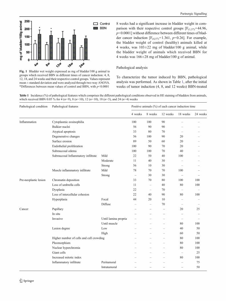
8 weeks had a significant increase in bladder weight in com-parison with their respective control groups [F(1,57)=44.96,p<0.0001] without difference between different times of blad-der cancer induction [F(4,57)=1.361, p=0.26]. For example,the bladder weight of control (healthy) animals killed at4 weeks, was 103±22 mg of bladder/100 g animal, whilethe bladder weight of animals which received BBN for4 weeks was 166±28 mg of bladder/100 g of animal.
Pathological analysis
To characterize the tumor induced by BBN, pathologicalanalysis was performed. As shown in Table 1, after the initialweeks of tumor induction (4, 8, and 12 weeks) BBN-treated
Fig. 1 Bladder wet weight expressed as mg of bladder/100 g animal togroups which received BBN in different times of cancer induction: 4, 8,12, 18, and 24weeks and their respective control groups. Values representmean ± standard deviation and were analyzed through two-way ANOVA.*Differences between mean values of control and BBN, with p<0.0001
Table 1 Incidence (%) of pathological features which comprises the different pathological conditions observed in HE staining of bladders from animals,which received BBN 0.05 % for 4 (n=9), 8 (n=10), 12 (n=10), 18 (n=5), and 24 (n=4) weeks
Pathological condition Pathological features Positive animals (%) of each cancer induction time
4 weeks 8 weeks 12 weeks 18 weeks 24 weeks
Inflammation Cytoplasmic eosinophilia 100 100 90 – –
Bulkier nuclei 56 90 90 – –
Atypical apoptosis 33 80 70 – –
Degenerative changes 56 100 90 20 –
Surface erosion 89 50 60 20 –
Endothelial proliferation 100 90 70 20 –
Submucosal edema 100 100 70 40 –
Submucosal Inflammatory infiltrate Mild 22 50 40 100 –
Moderate 11 40 30 – –
Strong 56 10 30 – –
Muscle inflammatory infiltrate Mild 78 70 70 100 –
Strong – 30 30 – –
Pre-neoplastic lesion Chromatin deposition 33 70 80 100 100
Loss of umbrella cells 11 – 40 80 100
Dysplasia 22 – 70 – –
Loss of intercellular cohesion 22 40 90 80 100
Hyperplasia Focal 44 20 10 – –
Diffuse – – 70 – –
Cancer Papillary – – – 20 25
In situ – – – – –
Invasive Until lamina propria – – – – –
Until muscle – – – 80 100
Lesion degree Low – – – 40 50
High – – – 60 50
Higher number of cells and cell crowding – – – 80 100
Pleomorphism – – – 80 100
Nuclear hyperchromia – – – 80 100
Giant cells – – – – 25
Increased mitotic index – – – 80 100
Inflammatory infiltrate Peritumoral – – – – 75
Intratumoral – – – – 50
Purinergic Signalling
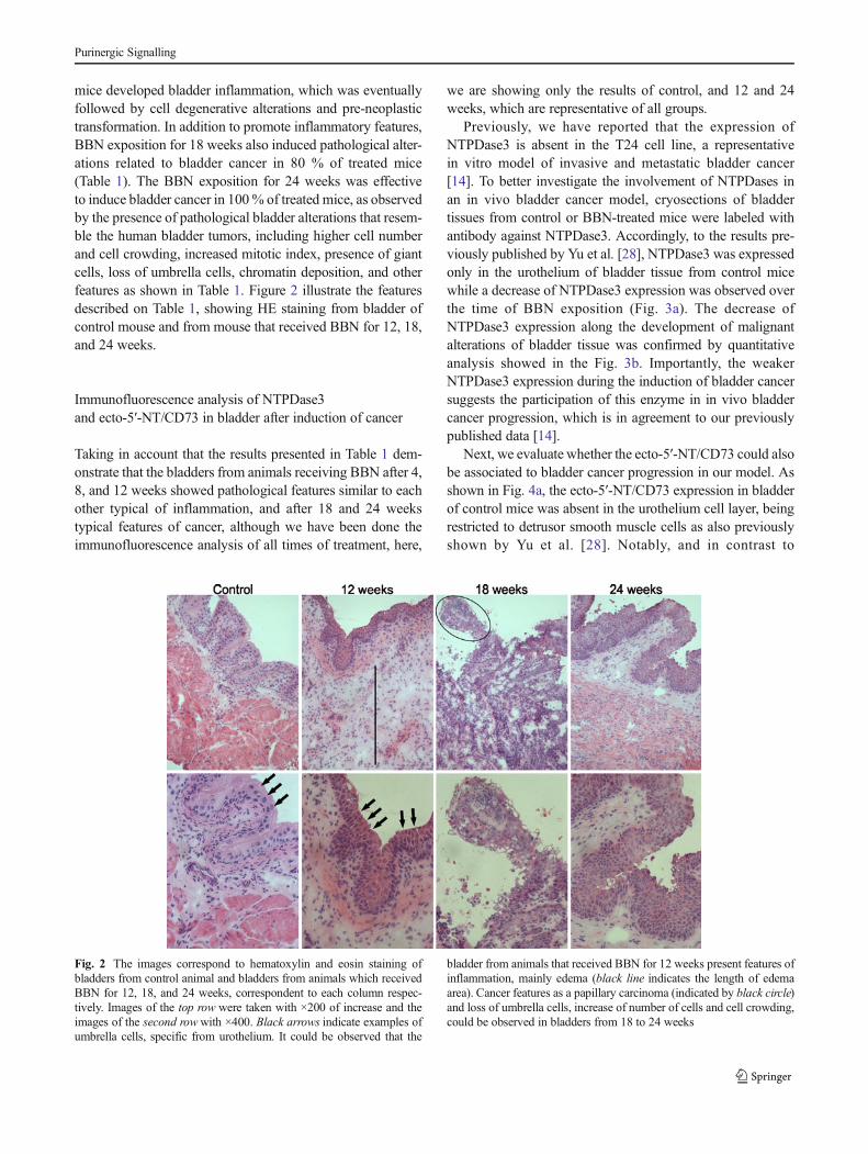
mice developed bladder inflammation, which was eventuallyfollowed by cell degenerative alterations and pre-neoplastictransformation. In addition to promote inflammatory features,BBN exposition for 18 weeks also induced pathological alter-ations related to bladder cancer in 80 % of treated mice(Table 1). The BBN exposition for 24 weeks was effectiveto induce bladder cancer in 100% of treated mice, as observedby the presence of pathological bladder alterations that resem-ble the human bladder tumors, including higher cell numberand cell crowding, increased mitotic index, presence of giantcells, loss of umbrella cells, chromatin deposition, and otherfeatures as shown in Table 1. Figure 2 illustrate the featuresdescribed on Table 1, showing HE staining from bladder ofcontrol mouse and from mouse that received BBN for 12, 18,and 24 weeks.
Immunofluorescence analysis of NTPDase3and ecto-5′-NT/CD73 in bladder after induction of cancer
Taking in account that the results presented in Table 1 dem-onstrate that the bladders from animals receiving BBN after 4,8, and 12 weeks showed pathological features similar to eachother typical of inflammation, and after 18 and 24 weekstypical features of cancer, although we have been done theimmunofluorescence analysis of all times of treatment, here,
we are showing only the results of control, and 12 and 24weeks, which are representative of all groups.
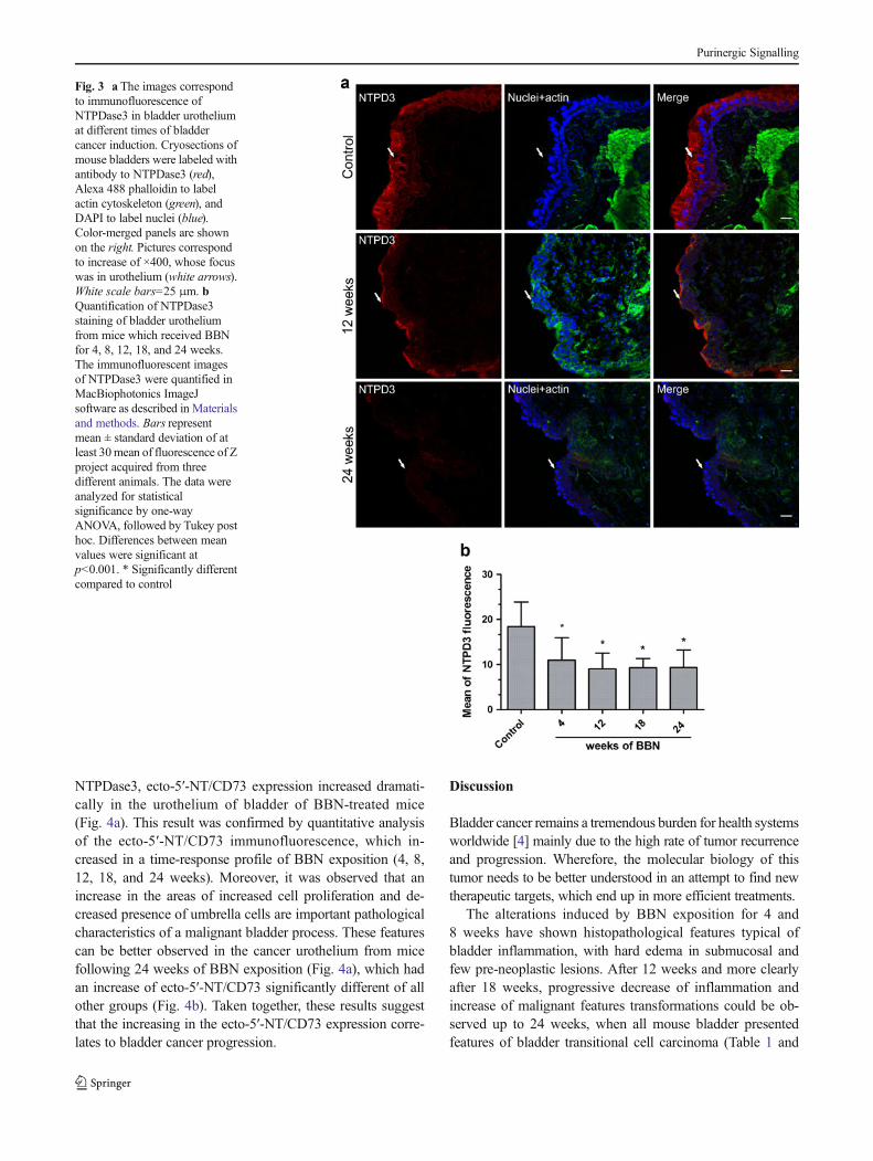
Previously, we have reported that the expression ofNTPDase3 is absent in the T24 cell line, a representativein vitro model of invasive and metastatic bladder cancer[14]. To better investigate the involvement of NTPDases inan in vivo bladder cancer model, cryosections of bladdertissues from control or BBN-treated mice were labeled withantibody against NTPDase3. Accordingly, to the results pre-viously published by Yu et al. [28], NTPDase3 was expressedonly in the urothelium of bladder tissue from control micewhile a decrease of NTPDase3 expression was observed overthe time of BBN exposition (Fig. 3a). The decrease ofNTPDase3 expression along the development of malignantalterations of bladder tissue was confirmed by quantitativeanalysis showed in the Fig. 3b. Importantly, the weakerNTPDase3 expression during the induction of bladder cancersuggests the participation of this enzyme in in vivo bladdercancer progression, which is in agreement to our previouslypublished data [14].
Next, we evaluate whether the ecto-5′-NT/CD73 could alsobe associated to bladder cancer progression in our model. Asshown in Fig. 4a, the ecto-5′-NT/CD73 expression in bladderof control mice was absent in the urothelium cell layer, beingrestricted to detrusor smooth muscle cells as also previouslyshown by Yu et al. [28]. Notably, and in contrast to
Fig. 2 The images correspond to hematoxylin and eosin staining ofbladders from control animal and bladders from animals which receivedBBN for 12, 18, and 24 weeks, correspondent to each column respec-tively. Images of the top row were taken with ×200 of increase and theimages of the second rowwith ×400. Black arrows indicate examples ofumbrella cells, specific from urothelium. It could be observed that the
bladder from animals that received BBN for 12 weeks present features ofinflammation, mainly edema (black line indicates the length of edemaarea). Cancer features as a papillary carcinoma (indicated by black circle)and loss of umbrella cells, increase of number of cells and cell crowding,could be observed in bladders from 18 to 24 weeks
Purinergic Signalling
NTPDase3, ecto-5′-NT/CD73 expression increased dramati-cally in the urothelium of bladder of BBN-treated mice(Fig. 4a). This result was confirmed by quantitative analysisof the ecto-5′-NT/CD73 immunofluorescence, which in-creased in a time-response profile of BBN exposition (4, 8,12, 18, and 24 weeks). Moreover, it was observed that anincrease in the areas of increased cell proliferation and de-creased presence of umbrella cells are important pathologicalcharacteristics of a malignant bladder process. These featurescan be better observed in the cancer urothelium from micefollowing 24 weeks of BBN exposition (Fig. 4a), which hadan increase of ecto-5′-NT/CD73 significantly different of allother groups (Fig. 4b). Taken together, these results suggestthat the increasing in the ecto-5′-NT/CD73 expression corre-lates to bladder cancer progression.
Discussion
Bladder cancer remains a tremendous burden for health systemsworldwide [4] mainly due to the high rate of tumor recurrenceand progression. Wherefore, the molecular biology of thistumor needs to be better understood in an attempt to find newtherapeutic targets, which end up in more efficient treatments.
The alterations induced by BBN exposition for 4 and8 weeks have shown histopathological features typical ofbladder inflammation, with hard edema in submucosal andfew pre-neoplastic lesions. After 12 weeks and more clearlyafter 18 weeks, progressive decrease of inflammation andincrease of malignant features transformations could be ob-served up to 24 weeks, when all mouse bladder presentedfeatures of bladder transitional cell carcinoma (Table 1 and
Fig. 3 a The images correspondto immunofluorescence ofNTPDase3 in bladder urotheliumat different times of bladdercancer induction. Cryosections ofmouse bladders were labeled withantibody to NTPDase3 (red),Alexa 488 phalloidin to labelactin cytoskeleton (green), andDAPI to label nuclei (blue).Color-merged panels are shownon the right. Pictures correspondto increase of ×400, whose focuswas in urothelium (white arrows).White scale bars=25 μm. bQuantification of NTPDase3staining of bladder urotheliumfrom mice which received BBNfor 4, 8, 12, 18, and 24 weeks.The immunofluorescent imagesof NTPDase3 were quantified inMacBiophotonics ImageJsoftware as described in Materialsand methods. Bars representmean ± standard deviation of atleast 30mean of fluorescence of Zproject acquired from threedifferent animals. The data wereanalyzed for statisticalsignificance by one-wayANOVA, followed by Tukey posthoc. Differences between meanvalues were significant atp<0.001. * Significantly differentcompared to control
Purinergic Signalling
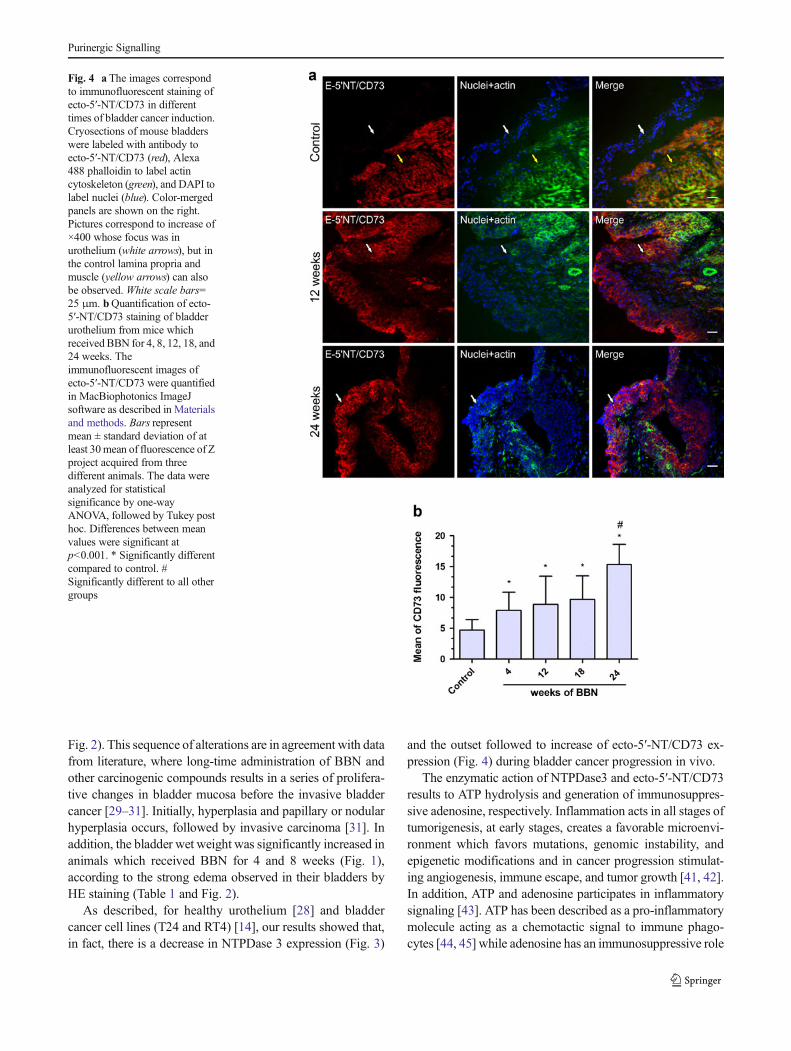
Fig. 2). This sequence of alterations are in agreement with datafrom literature, where long-time administration of BBN andother carcinogenic compounds results in a series of prolifera-tive changes in bladder mucosa before the invasive bladdercancer [29–31]. Initially, hyperplasia and papillary or nodularhyperplasia occurs, followed by invasive carcinoma [31]. Inaddition, the bladder wet weight was significantly increased inanimals which received BBN for 4 and 8 weeks (Fig. 1),according to the strong edema observed in their bladders byHE staining (Table 1 and Fig. 2).
As described, for healthy urothelium [28] and bladdercancer cell lines (T24 and RT4) [14], our results showed that,in fact, there is a decrease in NTPDase 3 expression (Fig. 3)
and the outset followed to increase of ecto-5′-NT/CD73 ex-pression (Fig. 4) during bladder cancer progression in vivo.
The enzymatic action of NTPDase3 and ecto-5′-NT/CD73results to ATP hydrolysis and generation of immunosuppres-sive adenosine, respectively. Inflammation acts in all stages oftumorigenesis, at early stages, creates a favorable microenvi-ronment which favors mutations, genomic instability, andepigenetic modifications and in cancer progression stimulat-ing angiogenesis, immune escape, and tumor growth [41, 42].In addition, ATP and adenosine participates in inflammatorysignaling [43]. ATP has been described as a pro-inflammatorymolecule acting as a chemotactic signal to immune phago-cytes [44, 45] while adenosine has an immunosuppressive role
Fig. 4 a The images correspondto immunofluorescent staining ofecto-5′-NT/CD73 in differenttimes of bladder cancer induction.Cryosections of mouse bladderswere labeled with antibody toecto-5′-NT/CD73 (red), Alexa488 phalloidin to label actincytoskeleton (green), and DAPI tolabel nuclei (blue). Color-mergedpanels are shown on the right.Pictures correspond to increase of×400 whose focus was inurothelium (white arrows), but inthe control lamina propria andmuscle (yellow arrows) can alsobe observed. White scale bars=25 μm. bQuantification of ecto-5′-NT/CD73 staining of bladderurothelium from mice whichreceived BBN for 4, 8, 12, 18, and24 weeks. Theimmunofluorescent images ofecto-5′-NT/CD73 were quantifiedin MacBiophotonics ImageJsoftware as described in Materialsand methods. Bars representmean ± standard deviation of atleast 30mean of fluorescence of Zproject acquired from threedifferent animals. The data wereanalyzed for statisticalsignificance by one-wayANOVA, followed by Tukey posthoc. Differences between meanvalues were significant atp<0.001. * Significantly differentcompared to control. #Significantly different to all othergroups
Purinergic Signalling
suppressing innate and adaptative immune responses [44].The participation of altered ectonucleotidases expression inthe modulation of immune cells to contribute to cancer prog-ress have been described to gliomas [46], that have a similarATP/ADP/AMPase activity pattern of bladder cancer. Thisprofile of nucleotide metabolism may favor extracellularATP and adenosine accumulation within the tumor interstice,so while ATP could induce the dead of healthy cells, tumorproliferation, and recruitment of immune cells, adenosine isresponsible for angiogenesis and immunosuppression [46].Although, the BCG antimutor activity is due to stimulationof the local and acute immune response and the recruitment ofpolymorphonuclear neutrophil granulocytes [33], the pres-ence of ATP and adenosine may be important to create chronicinflammatory conditions observed in tumor microenviron-ment, which suppress the immune response. This hypothesisis reinforced by the increasing density of immune cells in thebladder tissues during the course of BBN treatment. More-over, the massive presence of tumor-associated macrophagesin late clinical staging of patients with bladder cancer [47] andthe association between tumor infiltrating lymphocytes andthe recurrence of non-muscle-invasive bladder cancer [48]further suggest the chronic inflammatory process and cancerprogression association.
Moreover, both, ATP and adenosine have been extensivelydescribed to participate in bladder signaling [28, 49–53].Urothelium is a source of ATP release [28, 50, 51] and it isalso an important site of adenosine biosynthesis. Importantly,both ATP and adenosine have functions in exocytosis ofumbrella cell layer [52, 53], which is the mechanism toincrease the luminal surface area when the bladder fills(cytoplasmatic discoidal/fusiform vesicles fuse with the apicalplasma membrane) [54]. The ATP released by urothelium isresponsible for micturition reflex, through P2X3 fromsubepithelial nerve fibers [55]. Then, although it needs to bebetter elucidated, the changes in NTPDase3 and ecto-5′-NT/CD73 expression with malignant transformation of urothelialcells described in these work, would be expected to perturbnucleotide signaling in the bladder and thus affect somebladder functions.
Although little is known about the role of NTPDase3 incancer, an important finding of the present study was theabsence of ecto-5′-NT/CD73 in healthy urothelium [28]followed by increase in ecto-5′-NT/CD73 expression withthe increase of features of malignancy in mice that receivedBBN (Fig. 4). These findings indicate that this enzyme isinvolved in bladder cancer progression, and makes it a pro-missory therapeutic target to local treatments by instillation ofinhibitors of this enzyme. This result is in agreement with theliterature that shows the increase of ecto-5′-NT/CD73 expres-sion in many other cancers such as breast cancer, glioma, andmelanoma [19, 23, 56]. Furthermore, ecto-5′-NT/CD73 over-expression promotes invasion, migration, adhesion, and
metastasis of human breast cancer cells [20, 57], indicatinghigher invasiveness and metastatic capability to melanomas[56, 58] and poor prognosis in human colorectal cancer [22].Ecto-5′-NT/CD73 expression in cancer cells has been alsolinked with drug resistance [26, 59] and immune escape [44,60]. Moreover, researchers have already targeted ecto-5′-NT/CD73 in an attempt to develop promising therapies, for in-stance, ecto-5′-NT/CD73 inhibitors have shown antiprolifera-tive effects in bladder cancer cell lines and gliomas [61, 62],and decrease in ovarian cancer progression [63]. The treat-ment of mice with anti-CD73 antibody inhibited breast tumorgrowth and metastasis [20], and the use of RNA interferenceof ecto-5′-NT/CD73 inhibited cell growth and invasion inhuman breast cancer cells [25].
In conclusion, the altered expression of NTPDase3 andecto-5′-NT/CD73 presented herein add further evidence tosupport the idea that alterations in the activity and expres-sion of ectonucleotidases are involved in bladder tumori-genesis. Although additional studies are needed to deter-mine whether the alterations in the ectonucleotidases ex-pression are the cause or consequence of malignant trans-formation of urothelium, we bring the ecto-5′-NT/CD73 asa future biomarker and/or a target for pharmacologicaltherapy.
Acknowledgments This work was supported by Fundo de Incentivo aPesquisa do Hospital de Clínicas de Porto Alegre (FIPE-HCPA protocol10-0104), Conselho Nacional de Desenvolvimento Científico eTecnológico (CNPq), Coordenação de Aperfeiçoamento de Pessoal deNível Superior (Capes) e Fundação de Amparo à Pesquisa do Estado doRio Grande do Sul (FAPERGS). L. Rockenbach and F. Dietrich wererecipients of CAPES fellowships and F. Figueiró was a recipient of CNPqfellowship. JS was supported by grants from the Canadian Institutes ofHealth Research (CIHR) and was the recipient of a Senior Scholarshipfrom the “Fonds de la Recherche en Santé du Québec”. We thank L.R.Blazina, M.S. Quevedo, H.B. Biehl, L.A. Martins, and the team ofUnidade de Experimentação Animal of HCPA for their excellent techni-cal assistance.
References
1. Burnstock G (2001) Therapeutic potential of purinergic signaling fordiseases of the urinary tract. BJU Int 107:192–204
2. Shabbir M, Ryten M, Thompson C, Mikhailidis D, Burnstock G(2007) Purinergic receptor-mediated effects of ATP in high-gradebladder cancer. BJU Int 101:106–112
3. Eble JN, Sauter G, Epstein JI, Sesterhenn IA, editors. (2004)Pathology and genetics of tumours of the urinary system and malegenital organs. In: Kleihues P, Sobin LH editors. World HealthOrganization Classification of Tumours. IARCPress. pp. 89–157.Available http://www.iarc.fr/en/publications/pdfs-online/pat-gen/bb7/bb7-chap2.pdf. Accessed 10 January 2013.
4. Bachir BG, Kassouf W (2012) Cause–effect? Understanding the riskfactors associated with bladder cancer. Expert Rev Anticancer Ther12:1499–1502
5. van Roekel EH, Cheng KK, James ND,Wallace DM, Billingham LJ,Murray PG, Bryan RT, Zeegers MP (2013) Smoking is associated
Purinergic Signalling
with lower age, higher grade, higher stage, and larger size of malig-nant bladder tumours at diagnosis. Int J Cancer. doi:10.1002/ijc.28017
6. Rivas A, Burzio V, Landerer E, Borgna V, Gatica S, Ávila R, López C,Villota C, de la Fuente R, Echenique J, Burzio LO, Villegas J (2012)Determination of the differential expression of mitochondrial longnon-coding RNAs as noninvasive diagnosis of bladder cancer. BMCUrol 12:37
7. Wu XR (2005) Urothelial tumorigenesis: a tale of divergent path-ways. Nat Rev Cancer 5:713–725
8. Ariel I, Ayesh S, Gofrit O, Ayesh B, Abdul-ghani R, Pizov G, SmithY, Sidi AA, Birman T, Schneider T, de Groot N, Hochberg A (2004)Gene expression in the bladder carcinoma rat model. Mol Carcinog41:69–76
9. Gui Y, Guo G, Huang Y, Hu X, Tang A, Gao S,Wu R, Chen C, Li X,Zhou L, He M, Li Z, Sun X, Jia W, Chen J, Yang S, Zhou F, Zhao X,Wan S, Ye R, Liang C, Liu Z, Huang P, Liu C, Jiang H, Wang Y,ZhengH, Sun L, Liu X, Jiang Z, FengD, Chen J,Wu S, Zou J, ZhangZ, Yang R, Zhao J, Xu C, Yin W, Guan Z, Ye J, Zhang H, Li J,Kristiansen K, Nickerson ML, Theodorescu D, Li Y, Zhang X, Li S,Wang J, Yang H, Wang J, Cai Z (2011) Frequent mutations ofchromatin remodeling genes in transitional cell carcinoma of thebladder. Nat Genet 43:875–878
10. Schenk-Braat EA, Bangma CH (2005) Immunotherapy for superfi-cial bladder cancer. Cancer Immunol Immunother 54:414–423
11. Avritscher EB, Cooksley CD, Grossman HB, Sabichi AL, HamblinL, Dinney CP, Elting LS (2006) Clinical model of lifetime cost oftreating bladder cancer and associated complications. Urology 68:549–553
12. Burnstock G (2002) Potential therapeutic targets in the rapidlyexpanding field of purinergic signalling. Clin Med 2:45–53
13. Shabbir M, Burnstock G (2009) Purinergic receptor-mediated effectsof adenosine-5′-triphosphate in urological malignant deseases. Int JUrol 16:143–150
14. Stella J, Bavaresco L, Braganhol E, Rockenbach L, Farias PF, WinkMR, Azambuja AA, Barrios CH, Morrone FB, Oliveira BattastiniAM (2010) Differential ectonucleotidase expression in human blad-der cancer cell lines. Urol Oncol 28:260–267
15. Zimmermann H (2001) Ectonucleotidases: some recent develop-ments and a note on nomenclature. Drug Dev Res 52:44–56
16. Feng L, Sun X, Csizmadia E, Han L, Bian S, Murakami T, Wang X,Robson SC, Wu Y (2011) Vascular CD39/ENTPD1 directly pro-motes tumor cell growth by scavenging extracellular adenosinetriphosphate. Neoplasia 13:206–216
17. Jackson SW, Hoshi T, Wu Y, Sun X, Enjyoji K, Cszimadia E,Sundberg C, Robson SC (2007) Disordered purinergic signalinginhibits pathological angiogenesis in cd39/Entpd1-null mice. Am JPathol 171:1395–1404
18. Braganhol E, Morrone FB, Bernardi A, Huppes D, Meurer L,Edelweiss MI, Lenz G, Wink MR, Robson SC, Battastini AM(2009) Selective NTPDase2 expression modulates in vivo rat gliomagrowth. Cancer Sci 100:1434–1442
19. Spychala J (2000) Tumor-promoting functions of adenosine.Pharmacol Ther 87:161–173
20. Stagg J, Divisekera U, McLaughlin N, Sharkey J, Pommey S,Denoyer D, Dwyer KM, Smyth MJ (2010) Anti-CD73 antibodytherapy inhibits breast tumor growth and metastasis. Proc NatlAcad Sci U S A 107:1547–1552
21. Zhang B (2012) CD73 promotes tumor growth and metastasis.Oncoimmunology 1:67–70
22. Wu XR, He XS, Chen YF, Yuan RX, Zeng Y, Lian L, Zou YF, LanN, Wu XJ, Lan P (2012) High expression of CD73 as a poorprognostic biomarker in human colorectal cancer. J Surg Oncol106:130–137
23. Bavaresco L, Bernardi A, Braganhol E, Cappellari AR, Rockenbach L,Farias PF, Wink MR, Delgado-Cañedo A, Battastini AM (2008) The
role of ecto-5′-nucleotidase/CD73 in glioma cell line proliferation.Mol Cell Biochem 319:61–68
24. Cappellari AR, Vasques GJ, Bavaresco L, Braganhol E, BattastiniAM (2012) Involvement of ecto-5′-nucleotidase/CD73 in U138MGglioma cell adhesion. Mol Cell Biochem 359:315–322
25. Zhi X, Chen S, Zhou P, Shao Z, Wang L, Ou Z, Yin L (2007) RNAinterference of ecto-5′-nucleotidase (CD73) inhibits human breastcancer cell growth and invasion. Clin Exp Metastasis 24:439–448
26. Quezada C, Garrido W, Oyarzún C, Fernández K, Segura R, Melo R,Casanello P, Sobrevia L, San Martín R (2012) 5′-ectonucleotidasemediates multiple-drug resistance in glioblastoma multiforme cells. Jcell Physiol 228:602–608
27. Wang L, Zhou X, Zhou T, Ma D, Chen S, Zhi X, Yin L, Shao Z, OuZ, Zhou P (2008) Ecto-5′-nucleotidase promotes invasion, migrationand adhesion of human breast cancer cells. J Cancer Res Clin Oncol134:365–372
28. Yu W, Robson SC, Hill WG (2011) Expression and distribu-tion of ectonucleotidases in mouse urinary bladder. Plos One6:e18704
29. Cohen SM, Jacobs JB, Arai M, Johansson S, Friedell GH (1976)Early lesions in experimental bladder cancer: experimental designand light microscopic findings. Cancer Res 36:2508–2511
30. Ito N (1976) Early changes caused by N-Butyl-N-(4-hydroxybutyl)nitrosamine in the bladder epithelium of differentanimal species. Cancer Res 36:2528–2531
31. Oyasu R, Iwasaki T, Matsumoto M, Hirao Y, Tabuchi Y (1978)Induction of tumors in heterotopic bladder by topical application ofN-methyl-N-nitrosourea and N-butyl-N-(3-carboxypropyl) nitrosa-mine. Cancer Res 38:3019–3025
32. Chihara Y, Fujimoto K, Miyake M, Hiasa Y, Hirao Y (2009) Anti-tumor effect of cimetidine via inhibiting angiogenesis factors in N-butyl-N-(4-hydroxybutyl) nitrosamine-induced mouse and rat blad-der carcinogenesis. Oncol Rep 22:23–28
33. Lee SJ, Cho YH, Park K, Kim EJ, Kang BS, JungKH, Kim CH, KimWJ, Moon SK (2009) Inhibitory effects of the aqueous extract ofMagnolia officinalis on the responses of human urinary bladdercancer 5637cells in vitro and mouse urinary bladder tumors inducedby N-Butyl-N-(4-hydroxybutyl) nitrosamine in vivo. Phytother Res23:20–27
34. Hirose M, Fukushima S, Hananouchi M, Shirai T, Ogiso T (1976)Different susceptibilities of the urinary bladder epithelium of animalspecies to three nitroso compounds. Gann 67:175–189
35. Chen G, Chan FL, Zhang X, Chan PS (2009) Identification ofdifferently expressed genes in chemical carcinogen-induced rat blad-der cancers. Med Sci 29:220–226
36. Williams PD, Lee JK, TheodorescuD (2008)Molecular credentialingof rodent bladder carcinogenesis models. Neoplasia 10:838–846
37. Batista CK, Brito GA, Souza ML, Leitão BT, Cunha FQ, Ribeiro RA(2006) A model of hemorrhagic cystitis induced with acrolein inmice. Braz J Med Biol Res 39:1475–1481
38. Vekaria RM, Shirley DG, Sévigny J, Unwin RJ (2006)Immunolocalization of ectonucleotidases along the rat nephron.Am J Physiol Renal Physiol 290:F550–F560
39. Fausther M, Lecka J, Soliman E, Kauffenstein G, Pelletier J, SheungN, Dranoff JA, Sévigny J (2011) Co-expression of ecto-5′-nucleotid-ase/CD73 with specific NTPDases differentially regulates adenosineformation in the rat liver. Am J Physiol Gastrointest Liver Physiol302:G447–G459
40. Koszalka P, Özüyaman B, Huo Y, Zernecke A, Flögel U, Braun N,Buchheiser A, Decking UK, Smith ML, Sévigny J, Gear A, WeberAA, Molojavyi A, Ding Z, Weber C, Ley K, Zimmermann H,Gödecke A, Schrader J (2004) Targeted disruption of cd73/ecto-5′-nucleotidase alters thromboregulation and augments vascular inflam-matory response. Circ Res 95:814–821
41. Coussens LM, Werb Z (2002) Inflammation and cancer. Nature 420:860–867
Purinergic Signalling
42. Grivennikov SI, Greten FR, Karin M (2010) Immunity, inflamma-tion, and cancer. Cell 140:883–899
43. Bours MJ, Swennen EL, Di Virgilio F, Cronstein BN, Dagnelie PC(2006) Adenosine 5′-triphosphate and adenosine as endogenous sig-naling molecules in immunity and in inflammation. Pharmacol Ther112:358–404
44. Stagg J, Smyth MJ (2010) Extracellular adenosine triphophate andadenosine in cancer. Oncogene 29:5346–5358
45. Säve S, Persson K (2010) Extracellular ATP and P2Y receptoractivation induce a proinflammatory host response in the humanurinary tract. Infect Immun 78:3609–3615
46. Bergamin LS, Braganhol E, Zanin RF, Edelweiss MI, BattastiniAM (2012) Ectonucleotidases in tumor cells and tumor-associated immune cells: an overview. J Biomed Biotechnol 2012:959848
47. Zhang QW, Liu L, Gong CY, Shi HS, Zeng YH, Wang XZ, ZhaoYW, Wei YQ (2012) Prognostic significance of tumor-associatedmacrophages in solid tumor: a meta-analysis of the literature. 7:e50946
48. Krpina K, Babarović E, Dorđević G, Fuckar Z, Jonjić N (2012) Theassociation between the recurrence of solitary non-muscle invasivebladder cancer and tumor infiltrating lymphocytes. Croat Med J 53:598–604
49. Burnstock G (2013) Purinergic signalling in the lower urinary tract.Acta Physiol (Oxf) 207:40–52
50. Kumar V, Chapple CC, Chess-Williams R (2004) Characteristics ofadenosine triphosphate [corrected] release from porcine and humannormal bladder. J Urol 172:744–747
51. Munoz A, Gangitano DA, Smith CP, Boone TB, Somogyi GT(2010) Removal of urothelium affects bladder contractility andrelease of ATP but not release of NO in rat urinary bladder. BMCUrol 10:10
52. Wang EC, Lee JM, Ruiz WG, Balestreire EM, von Bodungen M,Barrick S, Cockayne DA, Birder LA, Apodaca G (2005) ATP andpurinergic receptor-dependent membrane traffic in bladder umbrellacells. J Clin Invest 115:2412–2422
53. Yu W, Zacharia LC, Jackson EK, Apodaca G (2006) Adenosinereceptor expression and function in bladder uroepithelium. Am JPhysiol Cell Physiol 291:C254–C265
54. Truschel ST, Wang E, Ruiz WG, Leung SM, Rojas R, Lavelle J,Zeidel M, Stoffer D, Apodaca G (2002) Stretch-regulated exocytosis/endocytosis in bladder umbrella cells. Mol Biol Cell 13:830–846
55. Ford AP, Cockayne DA (2011) ATP and P2X purinoceptors inurinary tract disorders. Handb Exp Pharmacol 202:485–526
56. Sadej R, Spychala J, Skladanowski AC (2006) Expression of ecto-5′-nucleotidase (eN, CD73) in cell lines from various stages of humanmelanoma. Melanoma Res 16:213–222
57. Zhou P, Zhi X, Zhou T, Chen S, Li X, Wang L, Yin L, Shao Z, Ou Z(2007) Overexpression of ecto-5′-nucleotidase (CD73) promotesT-47D human breast cancer cells invasion and adhesion to extracel-lular matrix. Cancer Biol Ther 6:426–431
58. Stagg J, Divisekera U, Duret H, Sparwasser T, Teng MW, Darcy PK,Smyth MJ (2011) CD73-deficient mice have increased antitumorimmunity and are resistant to experimental metastasis. Cancer Res71:2892–2900
59. Ujházy P, Berleth ES, Pietkiewicz JM, Kitano H, Skaar JR, EhrkeMJ, Mihich E (1996) Evidence for the involvement of ecto-5′-nucle-otidase (CD73) in drug resistance. Int J Cancer 68:493–500
60. Zhang B (2012) Opportunities and challenges for anti-CD73 cancertherapy. Immunotherapy 4:861–865
61. Rockenbach L, Bavaresco L, Fernandes Farias P, Cappellari AR,Barrios CH, Bueno Morrone F, Oliveira Battastini AM (2011)Alterations in the extracellular catabolism of nucleotides are involvedin the antiproliferative effect of quercetin in human bladder cancerT24 cells. Urol Oncol. doi:10.1016/j.urolonc.2011.10.009
62. Braganhol E, Tamajusuku AS, Bernardi A, Wink MR, Battastini AM(2007) Ecto-5′-nucleotidase/CD73 inhibition by quercetin in the humanU138MG glioma cell line. Biochim Biophys Acta 1770:1352–1359
63. Jin D, Fan J,Wang L, Thompson LF, LiuA, Daniel BJ, Shin T, CurielTJ, Zhang B (2010) CD73 on tumor cells impairs antitumor T-cellresponses: a novel mechanism of tumor-induced immune suppres-sion. Cancer Res 70:2245–2255
Purinergic Signalling