Embed Size (px)
Citation preview
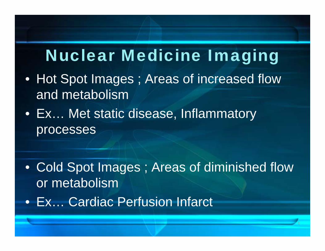
Nuclear Medicine Imaging• Hot Spot Images ; Areas of increased flow• Hot Spot Images ; Areas of increased flow
and metabolismE M t t ti di I fl t• Ex… Met static disease, Inflammatory processes
• Cold Spot Images ; Areas of diminished flow p g ;or metabolism
• Ex Cardiac Perfusion Infarct1
Ex… Cardiac Perfusion Infarct
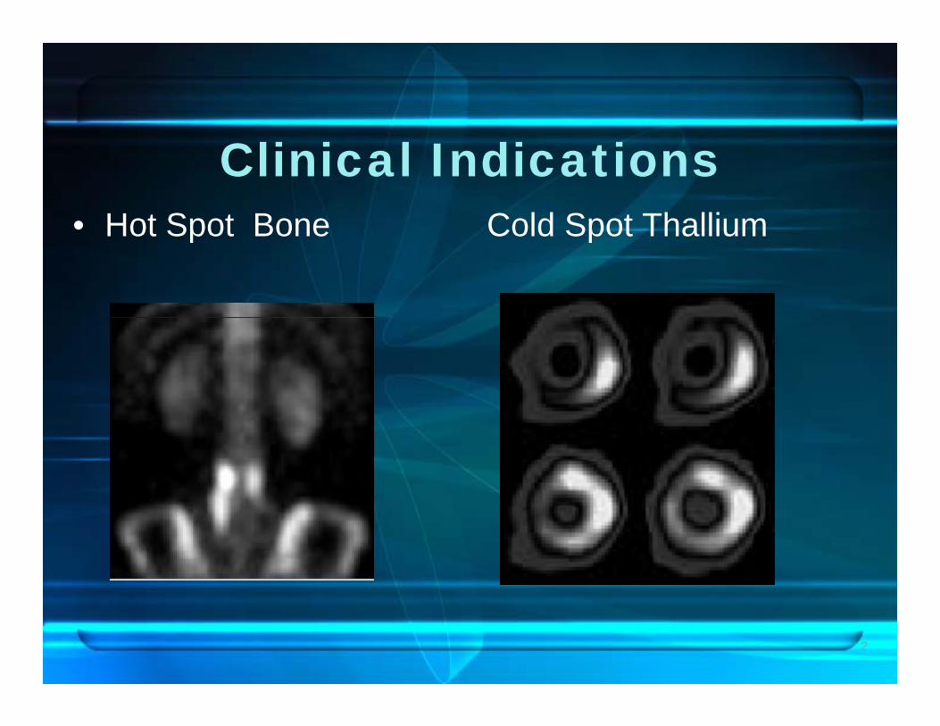
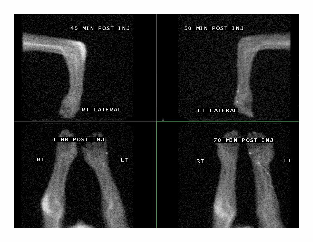
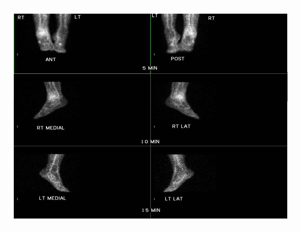
Clinical Indications• Hot Spot Bone Cold Spot Thallium• Hot Spot Bone Cold Spot Thallium
2
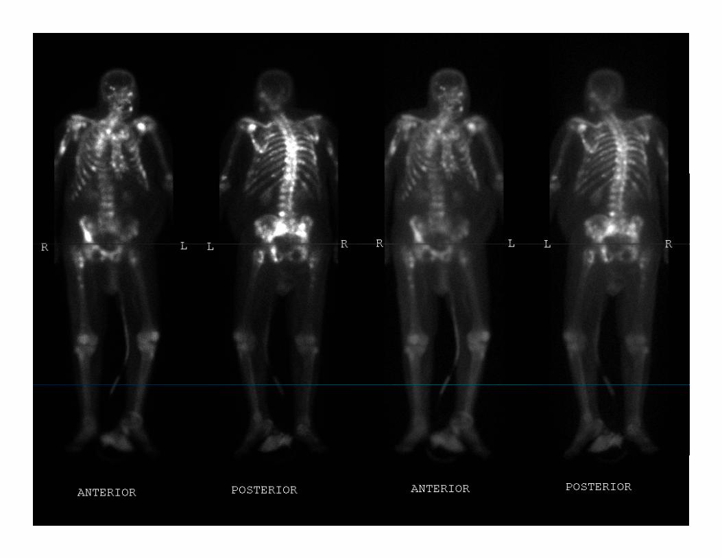
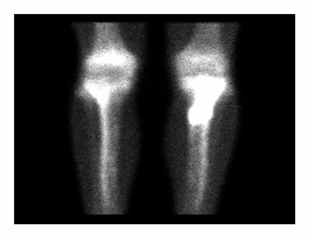
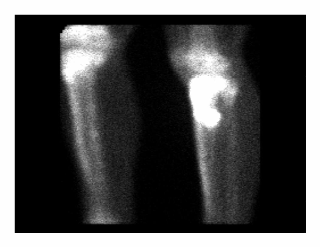
Frequently Performed ExaminationsExaminations
• Bone scans– Reasons to have a bone scan:
• Fractures• Bone metastases• To investigate unexplained bone or back pain• Follow up lesion seen on plain x-ray• A vascular necrosis
3
Frequently Performed ExaminationsExaminations
– Reasons to have a bone scan:• Investigate possible child abuseg• To establish viability of bone graph• Reflex sympathetic dystrophy (RSD)
4
5
6
7
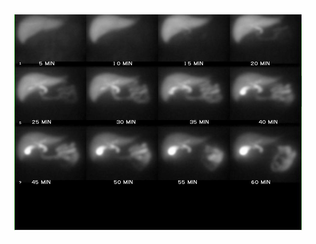
Frequently Performed ExaminationsExaminations
• Hepatobiliary scansp y– Reasons to have hepatobiliary scans:
• To diagnose suspected acute cholecystitisg y• To investigate possible biliary obstruction• To detect biliary leak
8
9
10
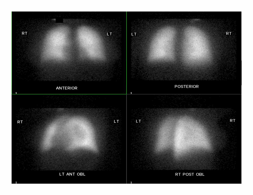
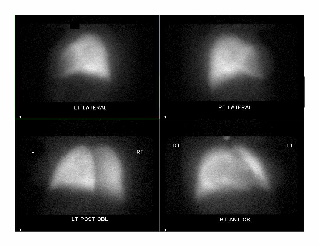
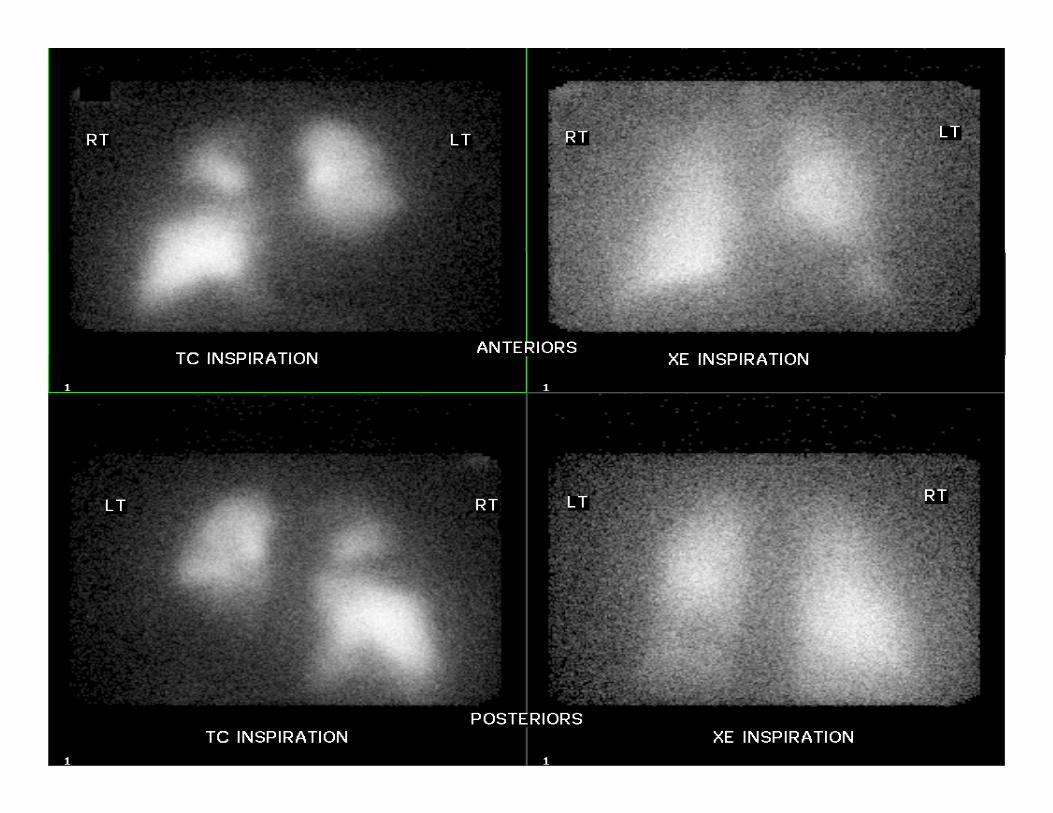
Frequently Performed ExaminationsExaminations
• Lung Imagingg g g– Reasons for lung imaging:
• To diagnose pulmonary embolismg y• To diagnose recurrence of pulmonary embolism• To predict quantitative residual lung function after lung
surgery
11
12
13
14
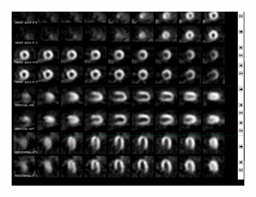
Frequently Performed ExaminationsExaminations
• Myocardial perfusion imagingR f di l f i i i– Reasons for myocardial perfusion imaging:
• To confirm known or suspected coronary artery diseasedisease
• To differentiate between coronary and non-coronary chest pain
• To assess cardiac risk for preoperative patients having surgeryF ll ti t ft h t
15
• Follow up patients after heart surgery
16
Frequently Performed ExaminationsExaminations
• Renal ImagingR f l i i– Reasons for renal imaging :
• To assess function of kidneysTo diagnose or exclude urinary tract obstruction• To diagnose or exclude urinary tract obstruction
17
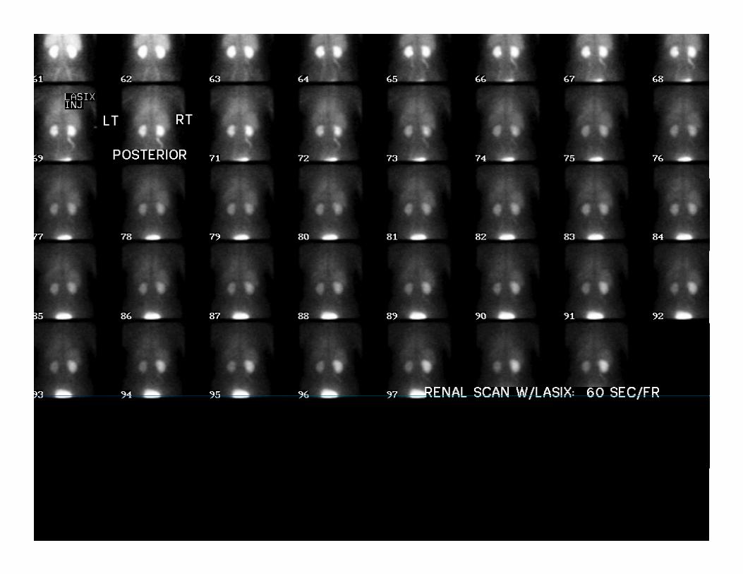
18
Frequently Performed ExaminationsExaminations
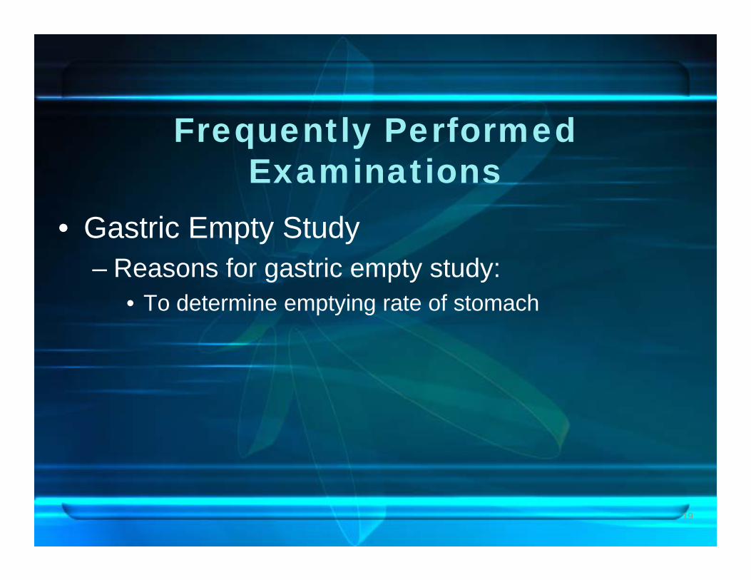
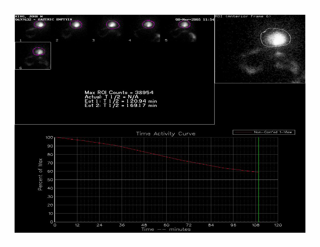
• Gastric Empty Study– Reasons for gastric empty study:
• To determine emptying rate of stomach
19
20
Frequently Performed ExaminationsExaminations
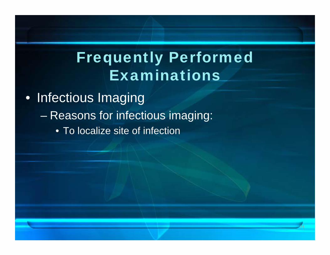
• Infectious ImagingR f i f ti i i– Reasons for infectious imaging:
• To localize site of infection
21
22
23
Frequently Performed ExaminationsExaminations
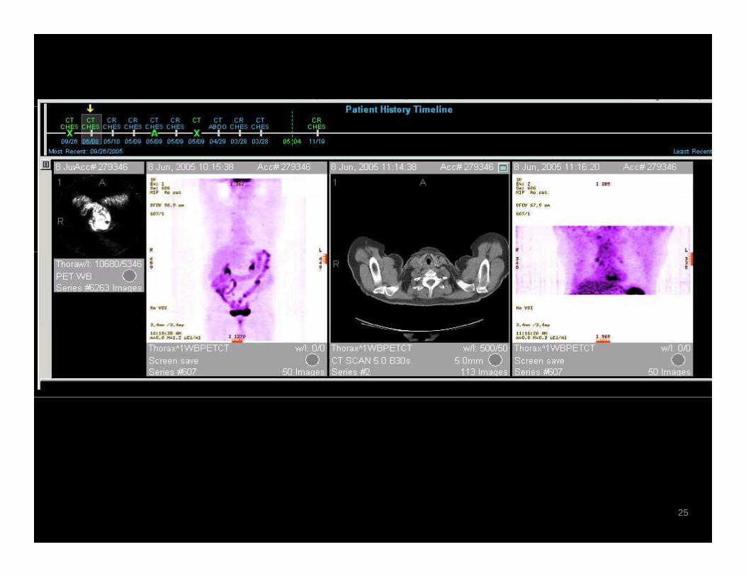
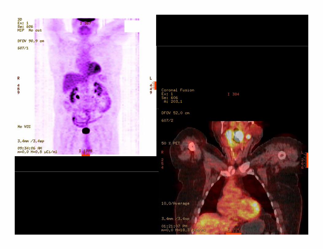
• Positron Emission Tomography PETg p y– Reasons to have pet imaging:
• Detect diagnose, stage, and restage cancerg g g• Monitor cancer therapy• Myocardial viability• Evaluate patients for dementia
24
25
26

27