Embed Size (px)
DESCRIPTION
Nuclear medicine Pet/Spect. Chapters 18 to 22. Activity. Number of radioactive atoms undergoing nuclear transformation per unit time. Change in radioactive atoms N in time dt Number of radioactive atoms decreases with time (- minus sign). Activity. Expressed in Curie - PowerPoint PPT Presentation
Citation preview
Nuclear medicine
Pet/Spect
Chapters 18 to 22
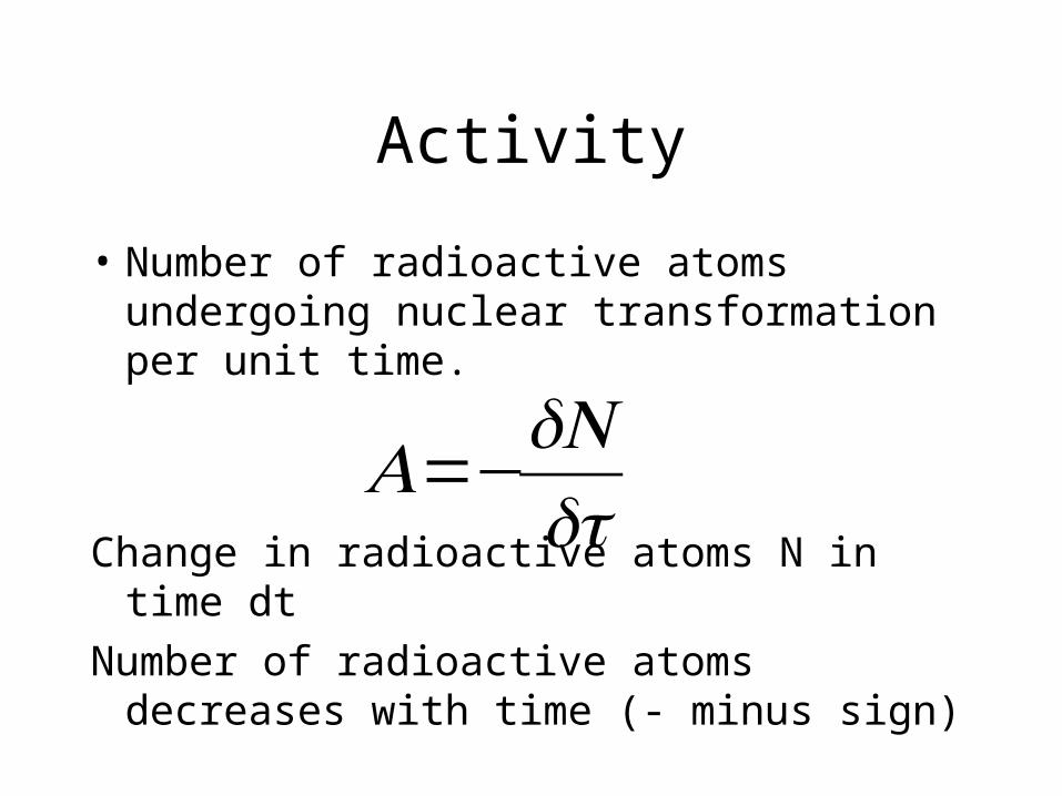
Activity
• Number of radioactive atoms undergoing nuclear transformation per unit time.
Change in radioactive atoms N in time dt
Number of radioactive atoms decreases with time (- minus sign)
€
A=−dNdt
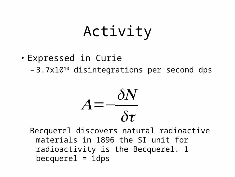
Activity
• Expressed in Curie – 3.7x10¹⁰ disintegrations per second dps
Becquerel discovers natural radioactive materials in 1896 the SI unit for radioactivity is the Becquerel. 1 becquerel = 1dps
€
A=−dNdt
Nuclear medicine
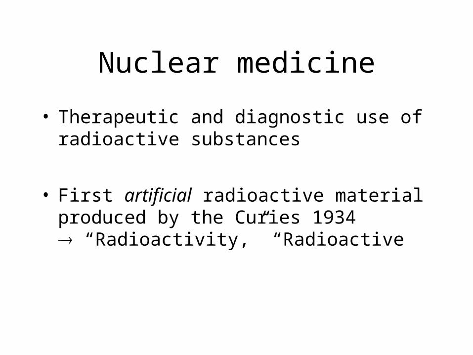
• Therapeutic and diagnostic use of radioactive substances
• First artificial radioactive material produced by the Curies 1934 “Radioactivity,” “Radioactive
Definitions: Nuclide
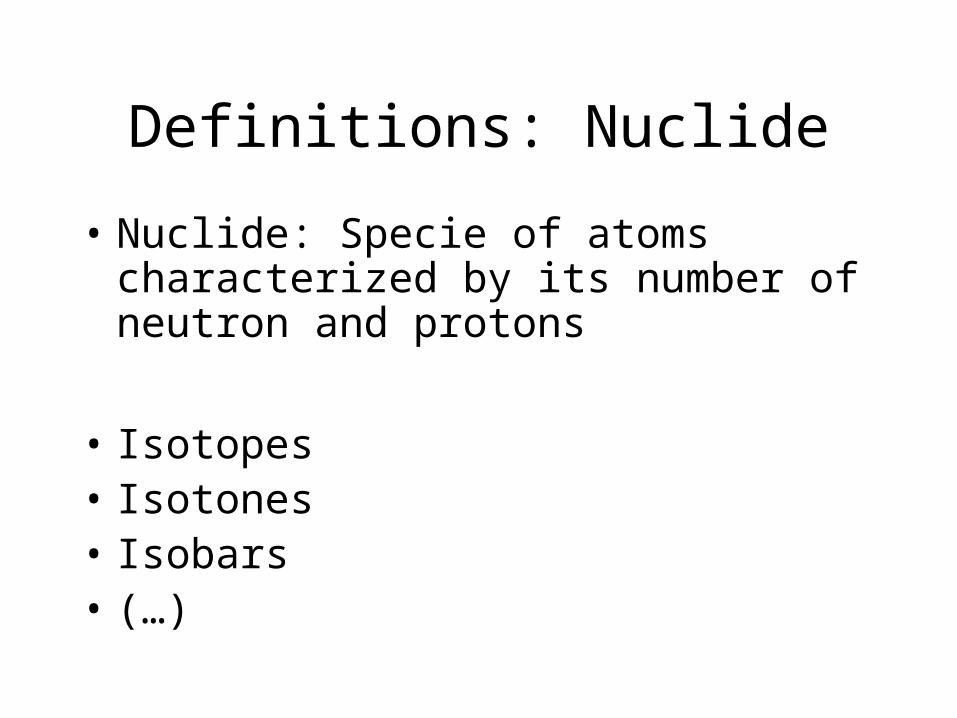
• Nuclide: Specie of atoms characterized by its number of neutron and protons
• Isotopes• Isotones• Isobars• (…)
Definitions: Nuclide
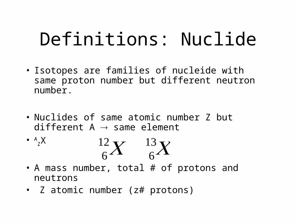
• Isotopes are families of nucleide with same proton number but different neutron number.
• Nuclides of same atomic number Z but different A same element
• AZX
• A mass number, total # of protons and neutrons• Z atomic number (z# protons)
€
612C 6
13C
Definitions: Nuclide
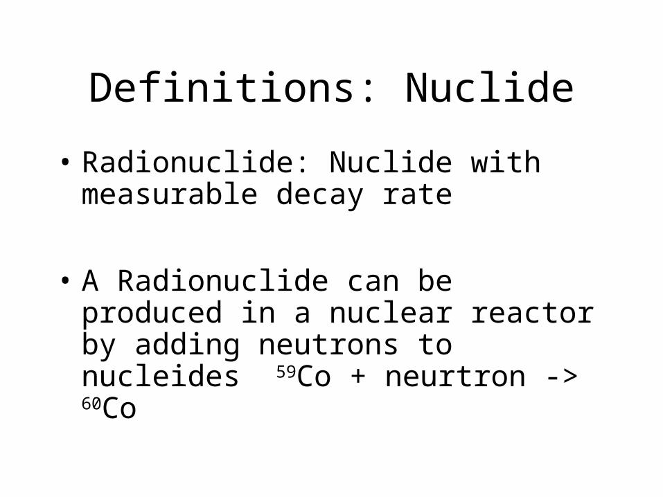
• Radionuclide: Nuclide with measurable decay rate
• A Radionuclide can be produced in a nuclear reactor by adding neutrons to nucleides 59Co + neurtron -> 60Co
Radioactive Decay
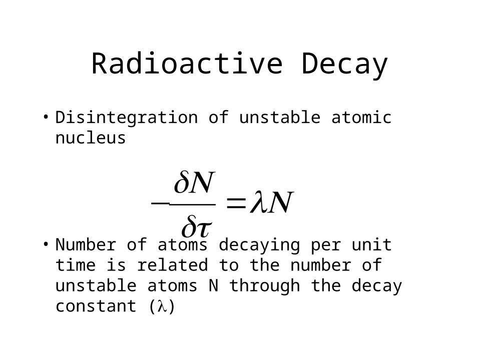
• Disintegration of unstable atomic nucleus
• Number of atoms decaying per unit time is related to the number of unstable atoms N through the decay constant ()
€
−dNdt
=N
Radioactive Decay
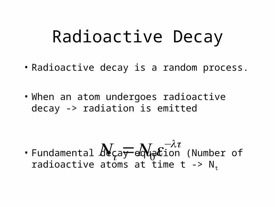
• Radioactive decay is a random process.
• When an atom undergoes radioactive decay -> radiation is emitted
• Fundamental decay equation (Number of radioactive atoms at time t -> Nt
€
Nt =N0e−t
Radioactive Decay
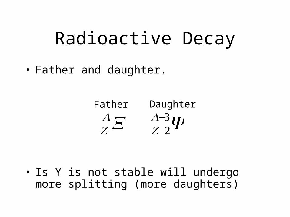
• Father and daughter.
• Is Y is not stable will undergo more splitting (more daughters)
€
ZAX Z−2
A−3YFather Daughter
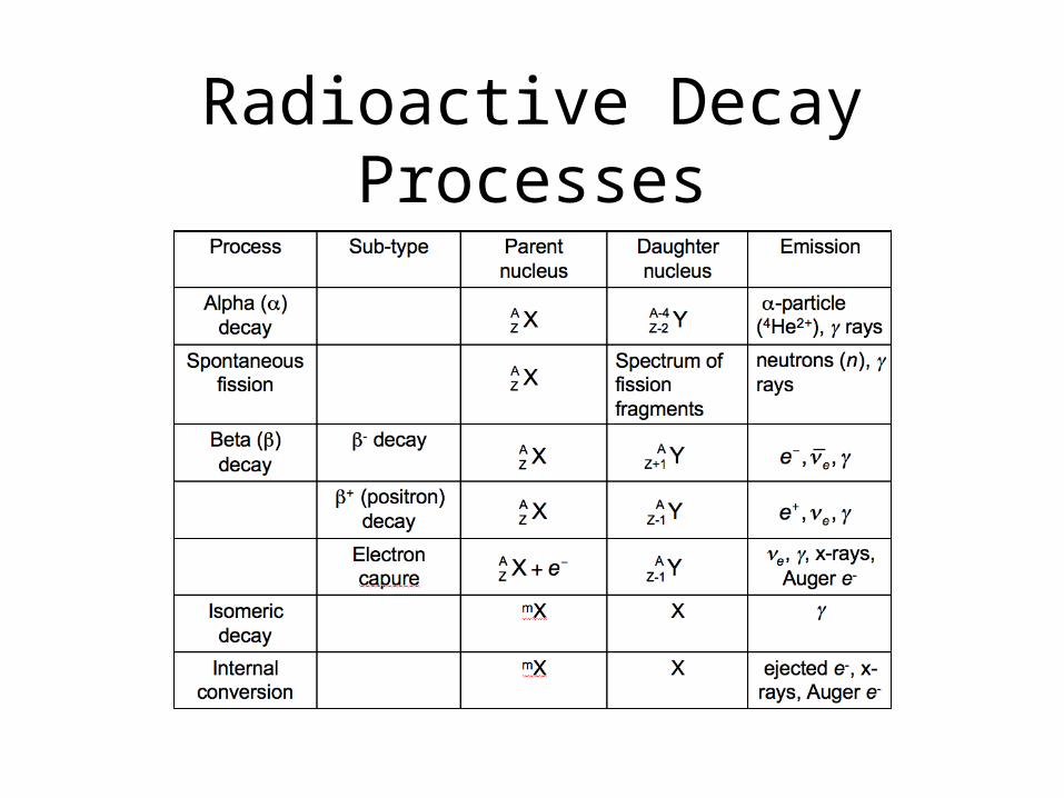
Radioactive Decay Processes
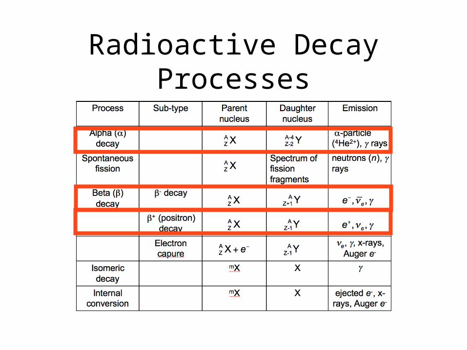
Radioactive Decay Processes
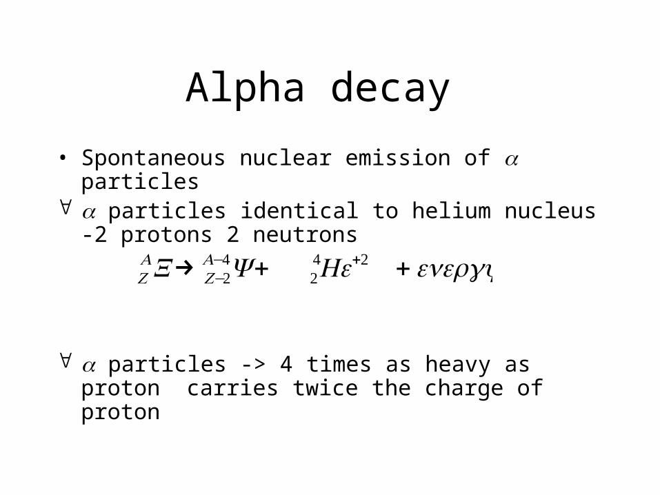
Alpha decay
• Spontaneous nuclear emission of particles particles identical to helium nucleus -2 protons 2
neutrons
particles -> 4 times as heavy as proton carries twice the charge of proton
€
ZAX→ Z−2
A−4Y + 24He+2 +energy
Alpha decay
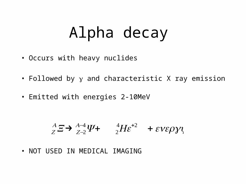
• Occurs with heavy nuclides
• Followed by and characteristic X ray emission • Emitted with energies 2-10MeV
• NOT USED IN MEDICAL IMAGING
€
ZAX→ Z−2
A−4Y + 24He+2 +energy
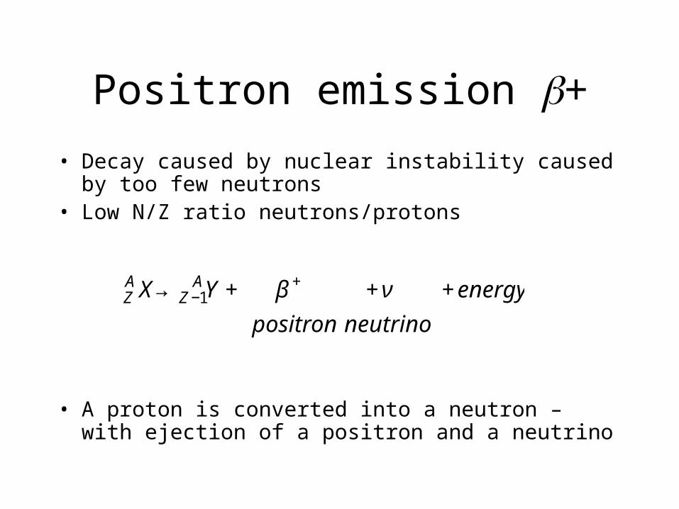
Positron emission +
• Decay caused by nuclear instability caused by too few neutrons
• Low N/Z ratio neutrons/protons
• A proton is converted into a neutron – with ejection of a positron and a neutrino€
ZA X→Z −1
AY + β + + ν + energy
positron neutrino
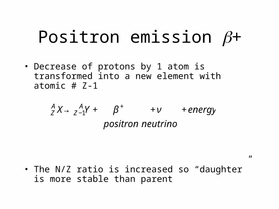
Positron emission +
• Decrease of protons by 1 atom is transformed into a new element with atomic # Z-1
• The N/Z ratio is increased so “daughter” is more stable than parent€
ZA X→Z −1
AY + β + + ν + energy
positron neutrino

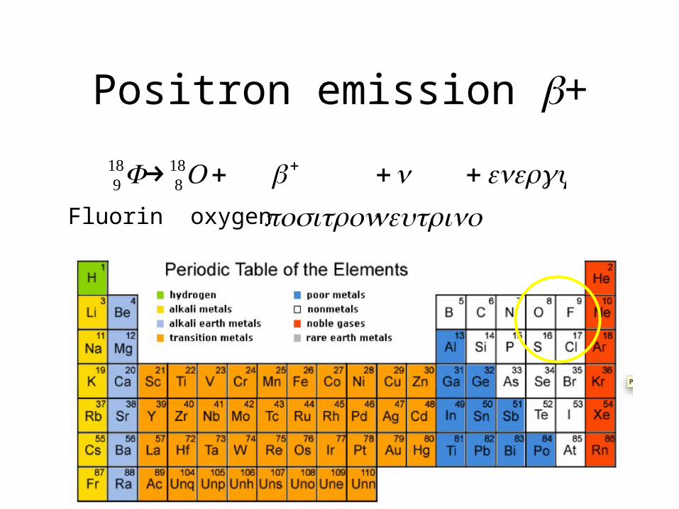
Positron emission +
€
918F→ 8
18O+ + +ν +energy positron neutrinoFluorin oxygen
Positron emission +
€
918F→ 8
18O+ + +ν +energy positron neutrinoFluorin oxygen
Positron emission +
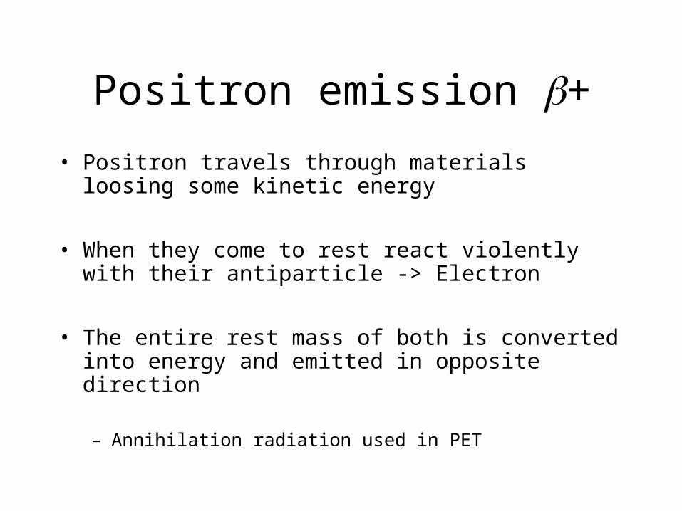
• Positron travels through materials loosing some kinetic energy
• When they come to rest react violently with their antiparticle -> Electron
• The entire rest mass of both is converted into energy and emitted in opposite direction
– Annihilation radiation used in PET
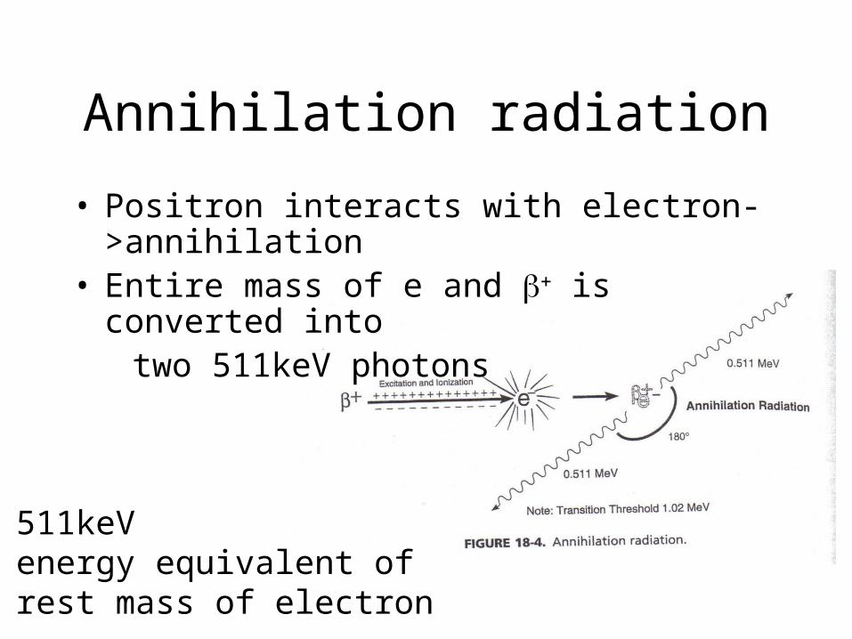
Annihilation radiation
• Positron interacts with electron->annihilation• Entire mass of e and is converted into two 511keV photons
511keVenergy equivalent ofrest mass of electron
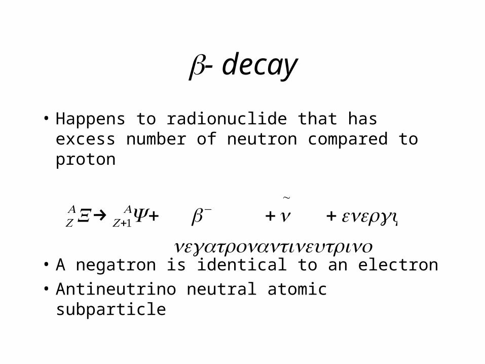
- decay
• Happens to radionuclide that has excess number of neutron compared to proton
• A negatron is identical to an electron• Antineutrino neutral atomic subparticle
€
ZAX→ Z+1
AY + − +ν~
+energy negatron antineutrino
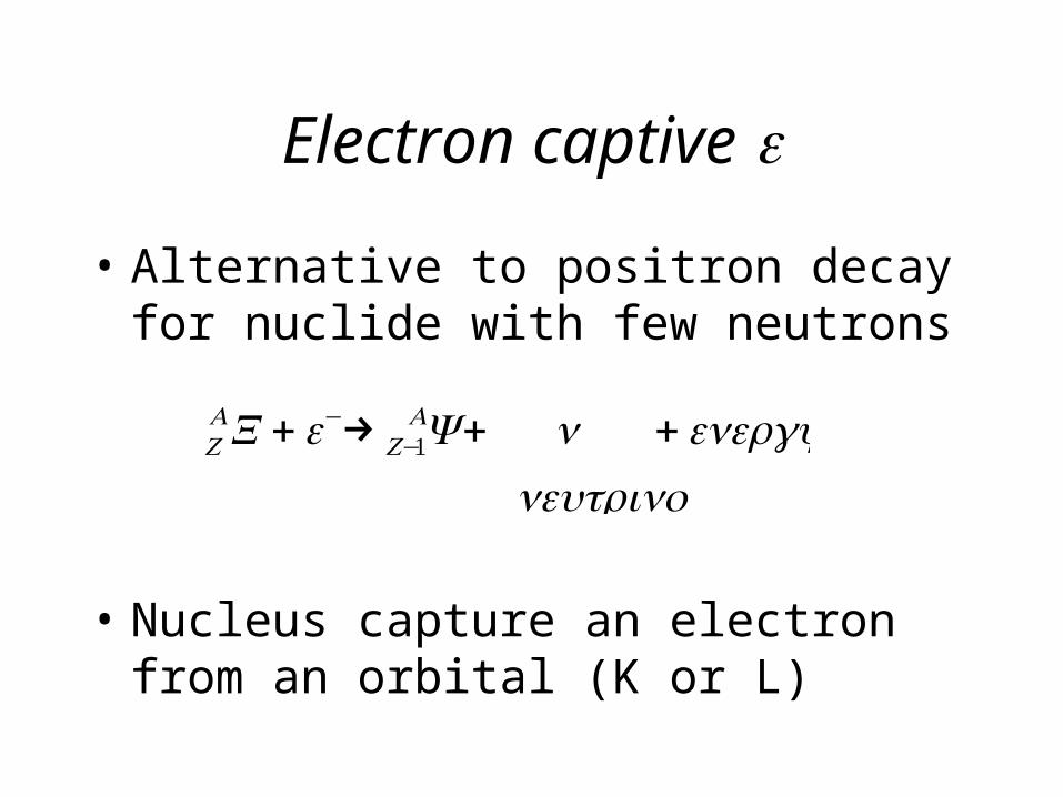
Electron captive
• Alternative to positron decay for nuclide with few neutrons
• Nucleus capture an electron from an orbital (K or L)
€
ZAX +e−→ Z−1
AY + ν +energy neutrino
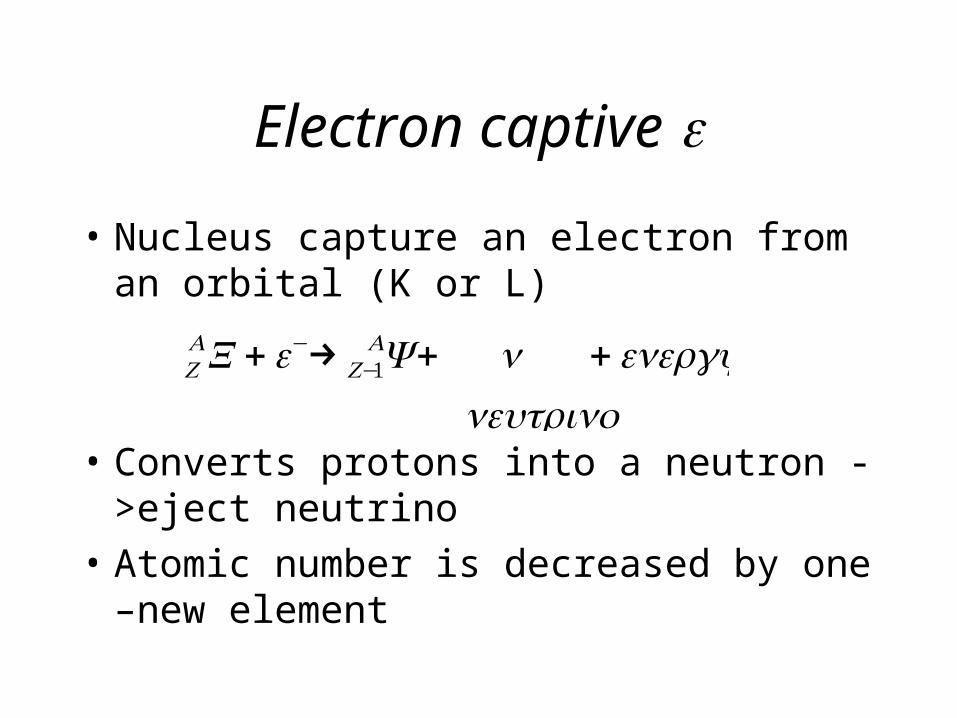
Electron captive
• Nucleus capture an electron from an orbital (K or L)
• Converts protons into a neutron ->eject neutrino
• Atomic number is decreased by one –new element
€
ZAX +e−→ Z−1
AY + ν +energy neutrino
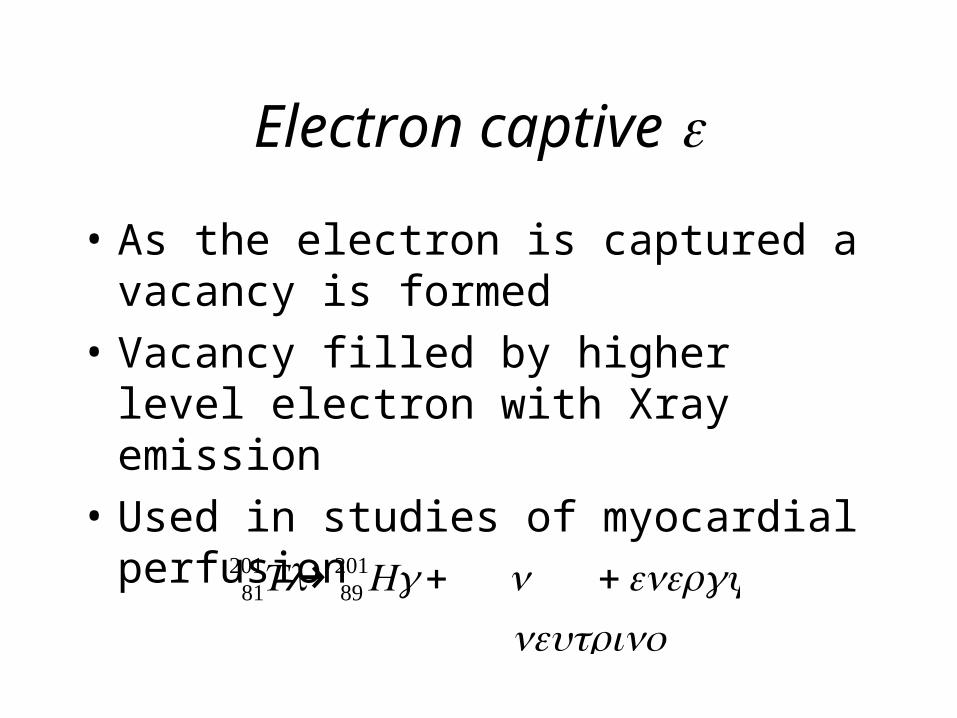
Electron captive
• As the electron is captured a vacancy is formed
• Vacancy filled by higher level electron with Xray emission
• Used in studies of myocardial perfusion
€
81201Tl→ 89
201Hg+ ν +energy neutrino
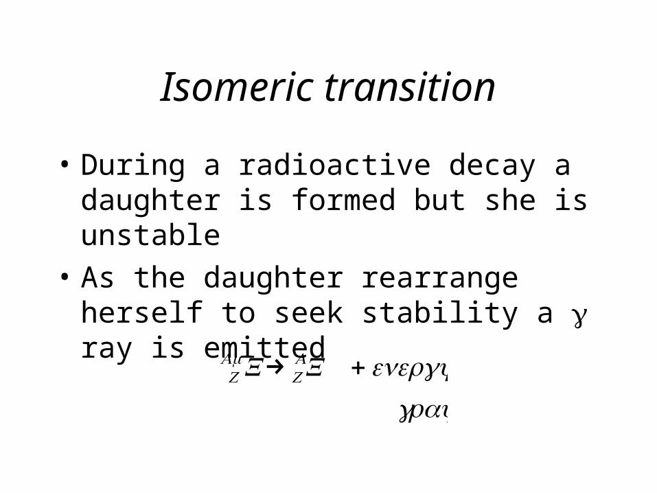
Isomeric transition
• During a radioactive decay a daughter is formed but she is unstable
• As the daughter rearrange herself to seek stability a ray is emitted
€
ZAmX→ Z
AX +energy ray
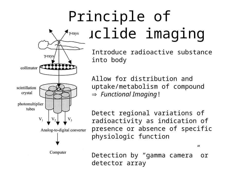
Principle of radionuclide imaging
Principle of radionuclide imaging
Introduce radioactive substance into body
Allow for distribution and uptake/metabolism of compound Functional Imaging!
Detect regional variations of radioactivity as indication of presence or absence of specific physiologic function
Detection by “gamma camera” or detector array
(Image reconstruction)
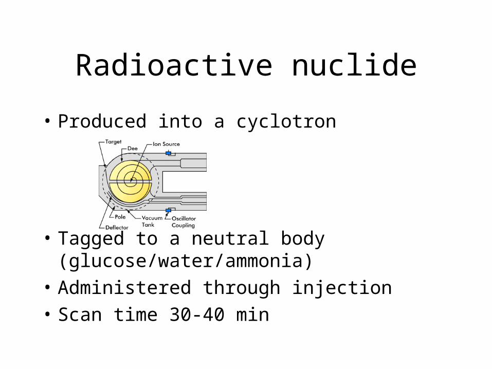
Radioactive nuclide
• Produced into a cyclotron
• Tagged to a neutral body (glucose/water/ammonia)
• Administered through injection• Scan time 30-40 min
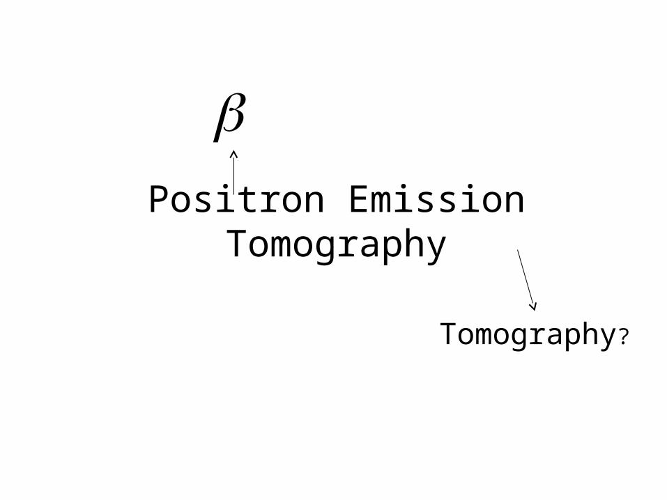
Positron Emission Tomography
Tomography?
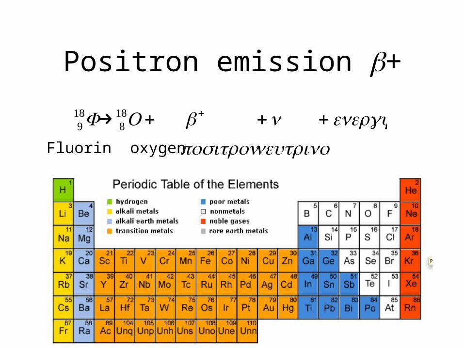
Positron emission +
€
918F→ 8
18O+ + +ν +energy positron neutrinoFluorin oxygen
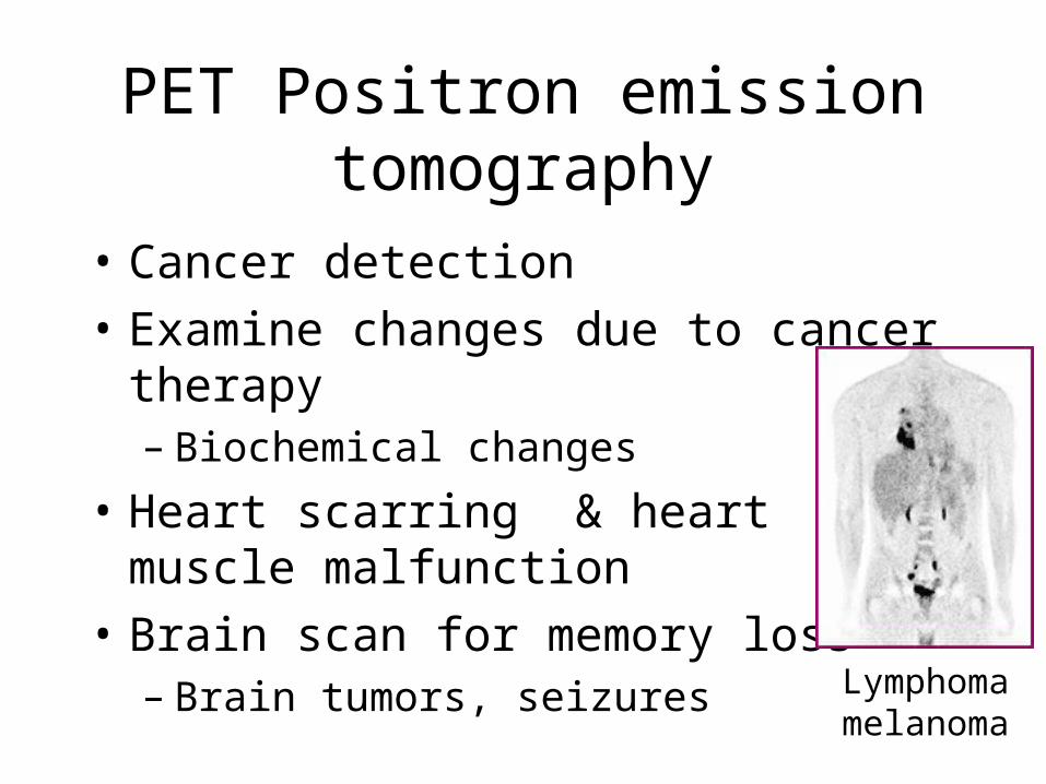
• Cancer detection
• Examine changes due to cancer therapy– Biochemical changes
• Heart scarring & heart muscle malfunction
• Brain scan for memory loss– Brain tumors, seizures Lymphoma
melanoma
PET Positron emission tomography
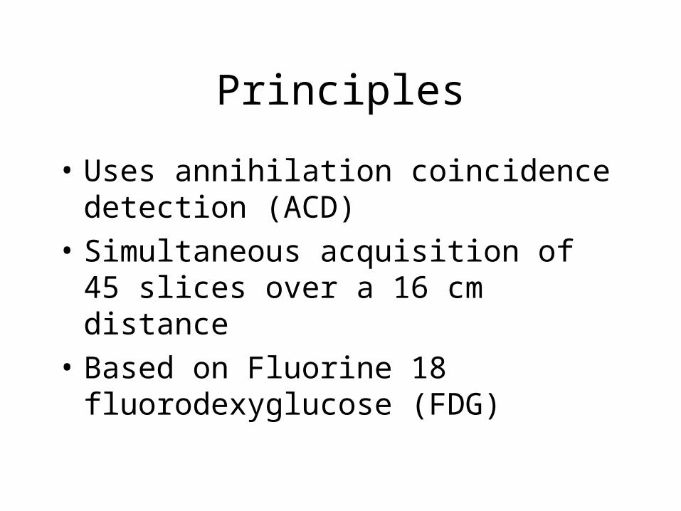
Principles
• Uses annihilation coincidence detection (ACD)
• Simultaneous acquisition of 45 slices over a 16 cm distance
• Based on Fluorine 18 fluorodexyglucose (FDG)
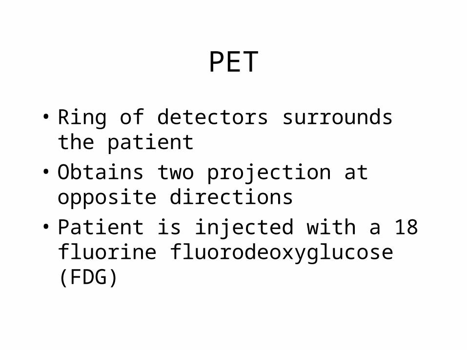
PET
• Ring of detectors surrounds the patient
• Obtains two projection at opposite directions
• Patient is injected with a 18 fluorine fluorodeoxyglucose (FDG)
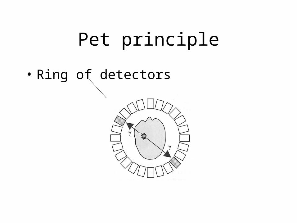
Pet principle
• Ring of detectors
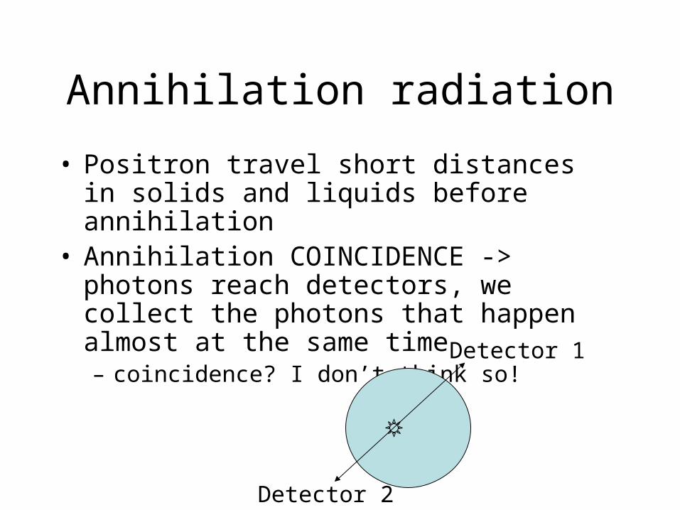
Annihilation radiation
• Positron travel short distances in solids and liquids before annihilation
• Annihilation COINCIDENCE -> photons reach detectors, we collect the photons that happen almost at the same time – coincidence? I don’t think so!
Detector 1
Detector 2
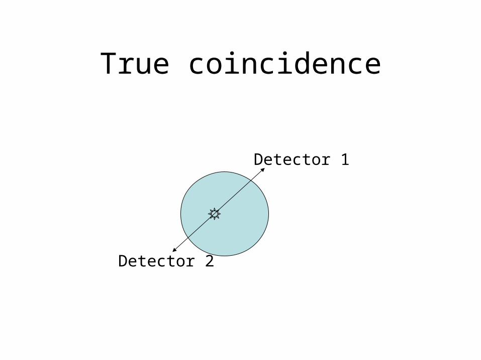
True coincidence
Detector 1
Detector 2
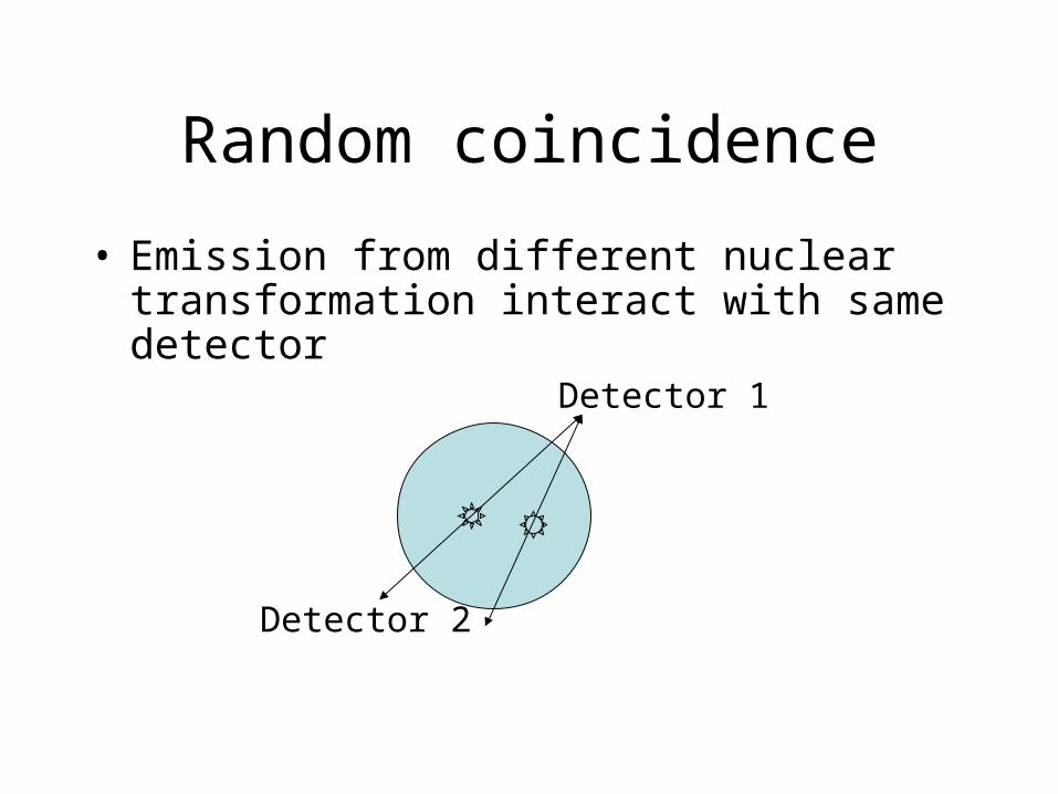
Random coincidence
• Emission from different nuclear transformation interact with same detector
Detector 1
Detector 2
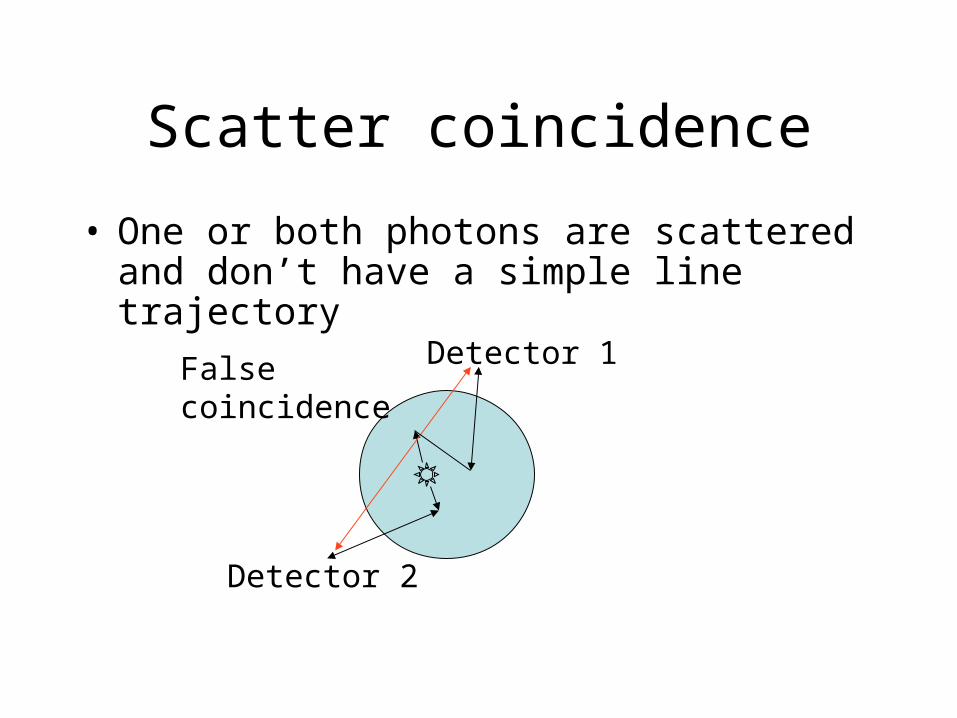
Scatter coincidence
• One or both photons are scattered and don’t have a simple line trajectory
Detector 1
Detector 2
Falsecoincidence
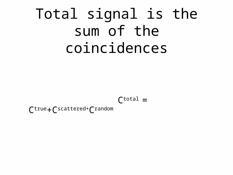
Total signal is the sum of the coincidences
Ctotal = Ctrue+Cscattered+Crandom
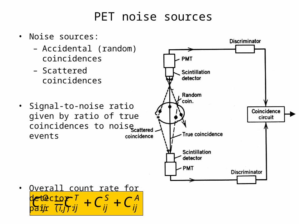
PET noise sourcesPET noise sources
O T S Aij ij ij ijC C C C= + +
• Noise sources:– Accidental (random)
coincidences– Scattered coincidences
• Signal-to-noise ratio given by ratio of true coincidences to noise events
• Overall count rate for detector pair (i,j):
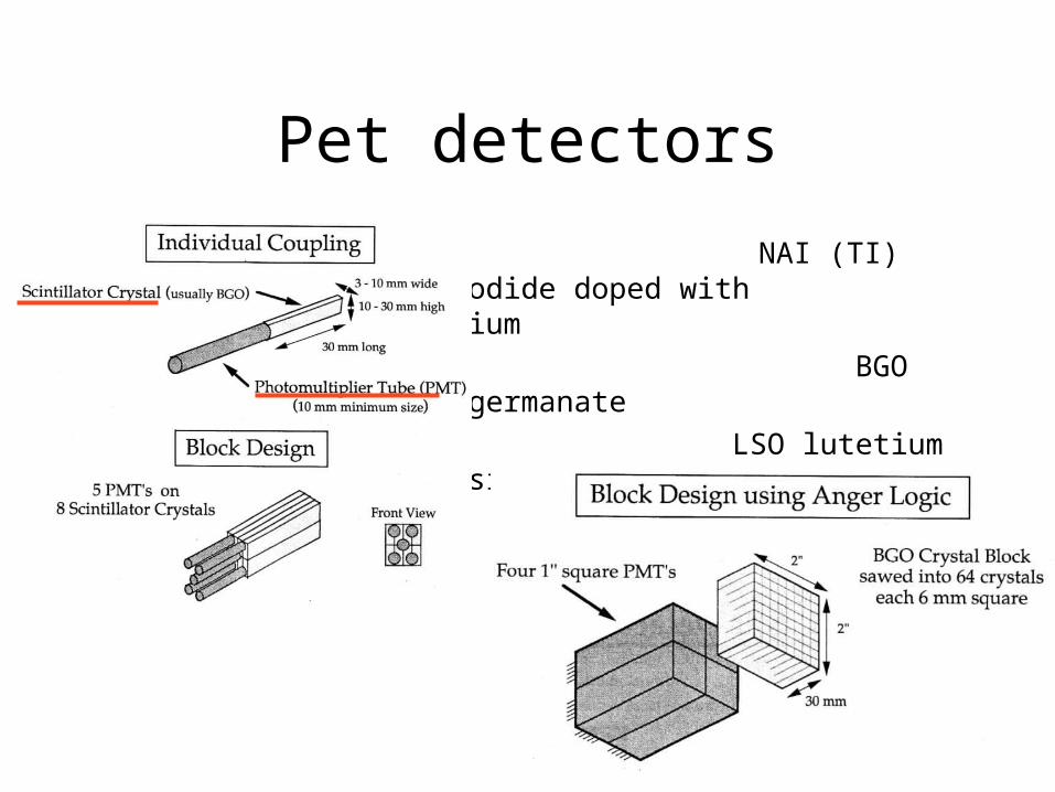
Pet detectors
NAI (TI) Sodium iodide doped with thallium
BGO bismuth germanate
LSO lutetium oxyorthosilicate
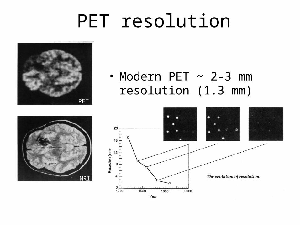
PET resolutionPET resolution
• Modern PET ~ 2-3 mm resolution (1.3 mm)
MRI
PET
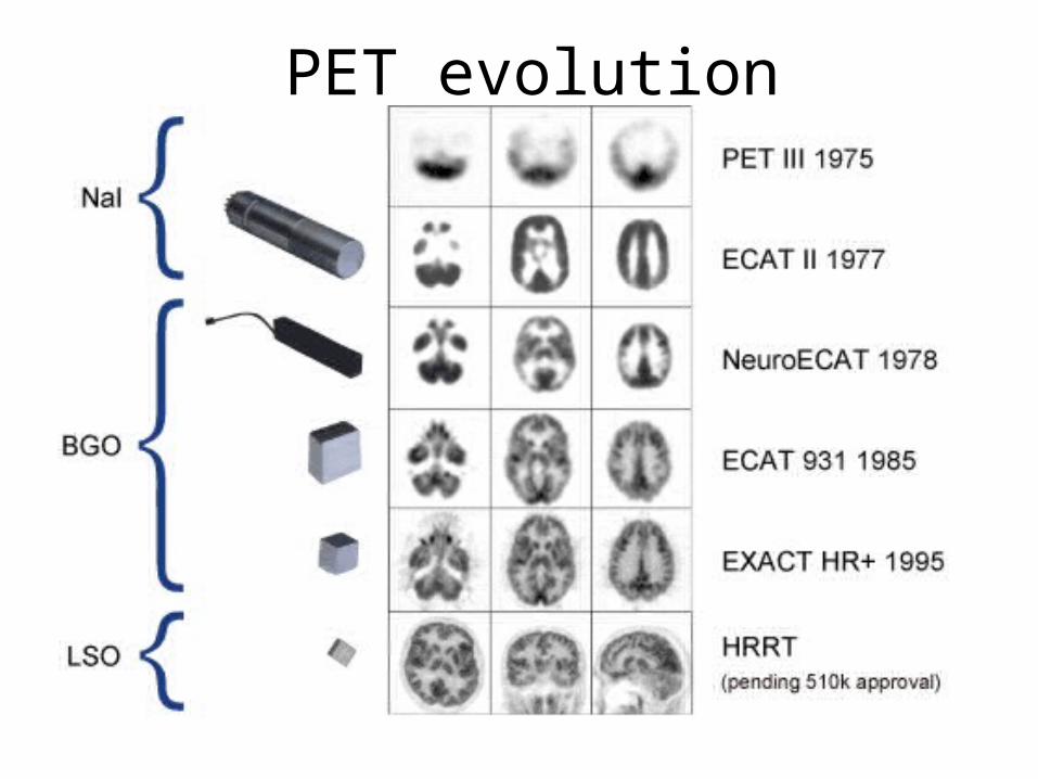
PET evolutionPET evolution
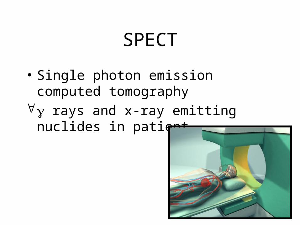
SPECT
• Single photon emission computed tomography
rays and x-ray emitting nuclides in patient
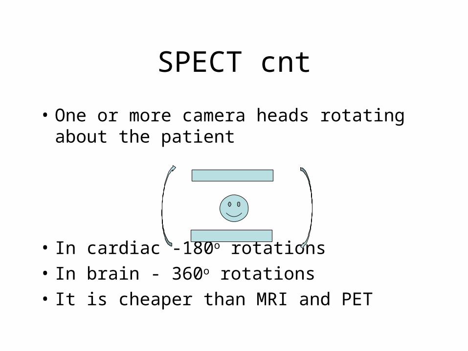
SPECT cnt
• One or more camera heads rotating about the patient
• In cardiac -180o rotations• In brain - 360o rotations• It is cheaper than MRI and PET
SPECT cnt
• 60-130 projections
• Technetium is the isothope
• Decays with ray emission
• Filtered back projection to reconstruct an image of a solid
Typical studies
• Bone scan
• Myocardial perfusion
• Brain
• Tumor
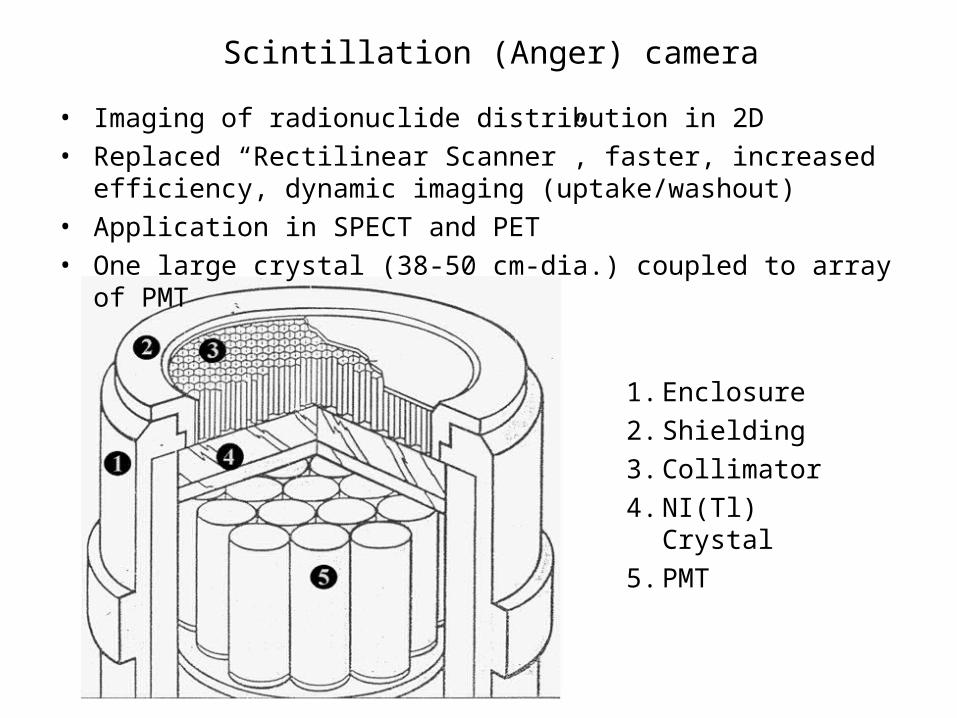
Scintillation (Anger) cameraScintillation (Anger) camera
1. Enclosure
2. Shielding
3. Collimator
4. NI(Tl) Crystal
5. PMT
• Imaging of radionuclide distribution in 2D• Replaced “Rectilinear Scanner”, faster, increased efficiency,
dynamic imaging (uptake/washout)• Application in SPECT and PET• One large crystal (38-50 cm-dia.) coupled to array of PMT
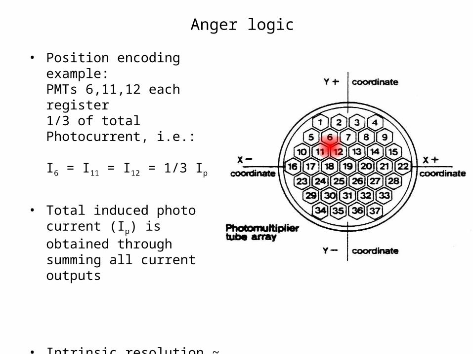
Anger logicAnger logic
• Position encoding example: PMTs 6,11,12 each register 1/3 of total Photocurrent, i.e.:
I6 = I11 = I12 = 1/3 Ip
• Total induced photo current (Ip) is obtained through summing all current outputs
• Intrinsic resolution ~ 4 mm
Ld
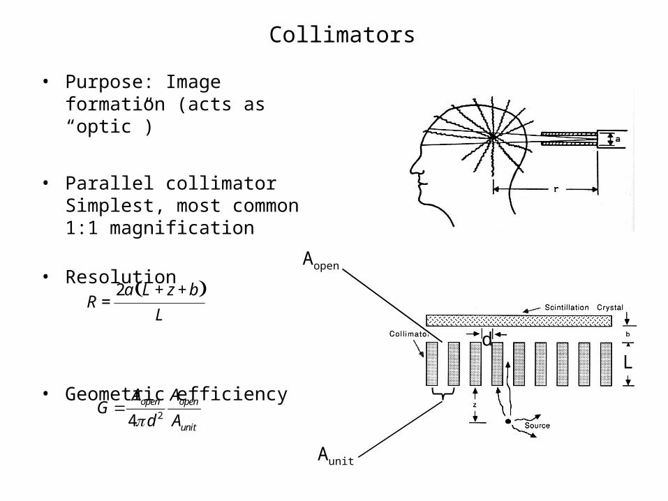
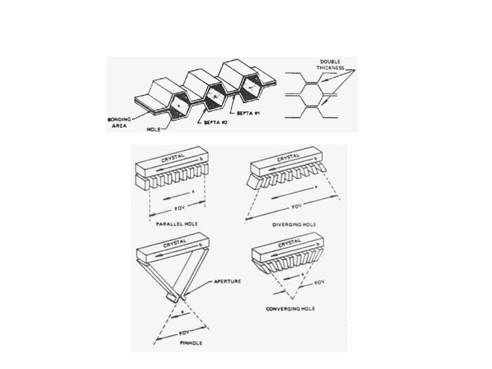
CollimatorsCollimators
• Purpose: Image formation (acts as “optic”)
• Parallel collimatorSimplest, most common 1:1 magnification
• Resolution
• Geometric efficiency
• Tradeoff: Resolution Efficiency
( )2a L z bR
L
+ +=
24open open
unit
A AG
d Aπ=
Aopen
Aunit
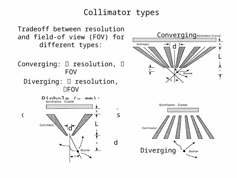
Collimator typesCollimator types
Tradeoff between resolution and field-of view (FOV) for different types:
Converging: resolution, FOV
Diverging: resolution, FOV
Pinhole (~ mm):High resolution of small organs at close
distances
Diverging
L
d
d
Converging
L
d
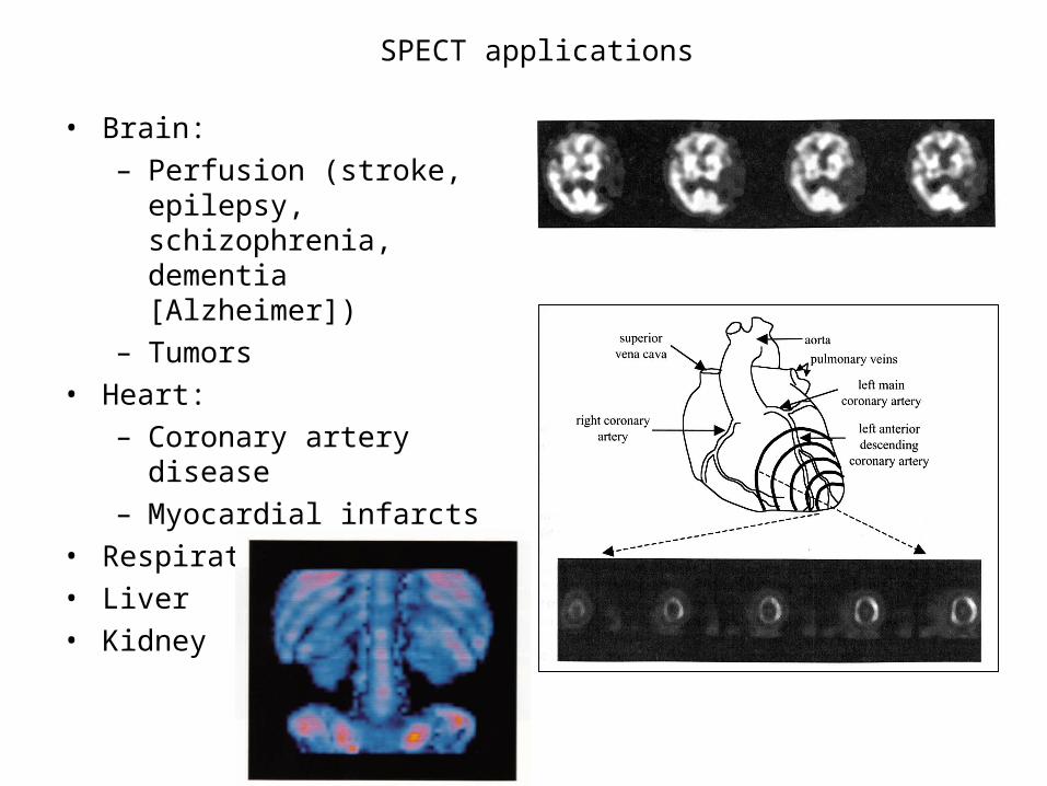
SPECT applicationsSPECT applications
• Brain: – Perfusion (stroke, epilepsy,
schizophrenia, dementia [Alzheimer])
– Tumors• Heart:
– Coronary artery disease– Myocardial infarcts
• Respiratory• Liver• Kidney