Embed Size (px)
Citation preview
Nursing patients with
Fractures
Tania Yelland VN
Fractures
A fracture occurs when there is a break in the continuity of the bone.
It can occur either as:
A result of trauma
Or
A pathological fracture
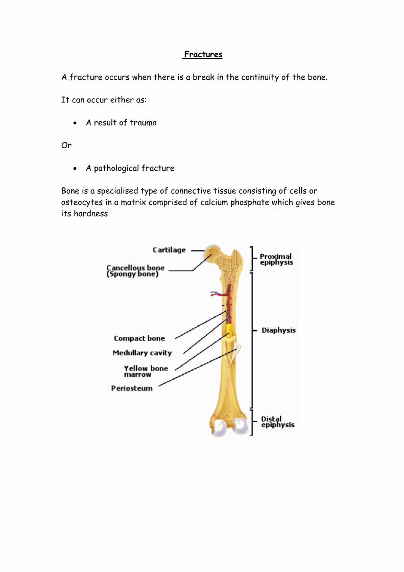
Bone is a specialised type of connective tissue consisting of cells or
osteocytes in a matrix comprised of calcium phosphate which gives bone
its hardness
Classification of fractures
Fractures can either be open or closed
Closed fracture = A fracture with no break in the skin
Open fracture = a woind that has penetrated the skin and the fracture
ends are open to the outside environment
Fractures can also be described in many other ways
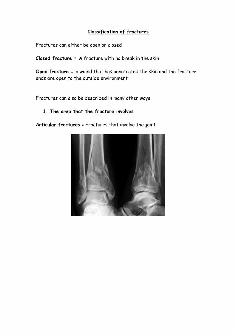
1. The area that the fracture involves
Articular fractures = Fractures that involve the joint
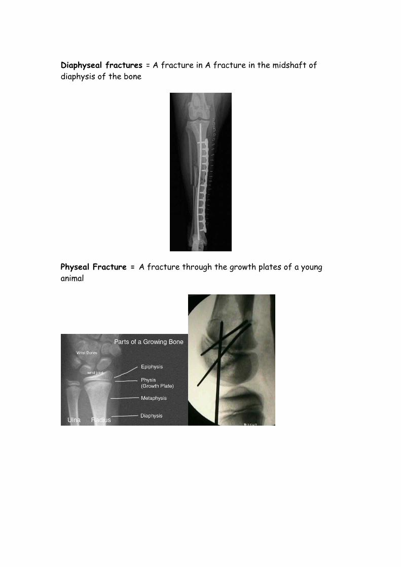
Diaphyseal fractures = A fracture in A fracture in the midshaft of
diaphysis of the bone
Physeal Fracture = A fracture through the growth plates of a young
animal
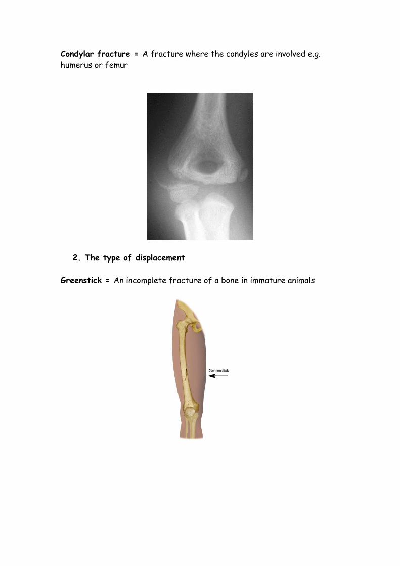
Condylar fracture = A fracture where the condyles are involved e.g.
humerus or femur
2. The type of displacement
Greenstick = An incomplete fracture of a bone in immature animals
Fissure = A fine crack which may displace during surgery or if the site is
put under any type of stress
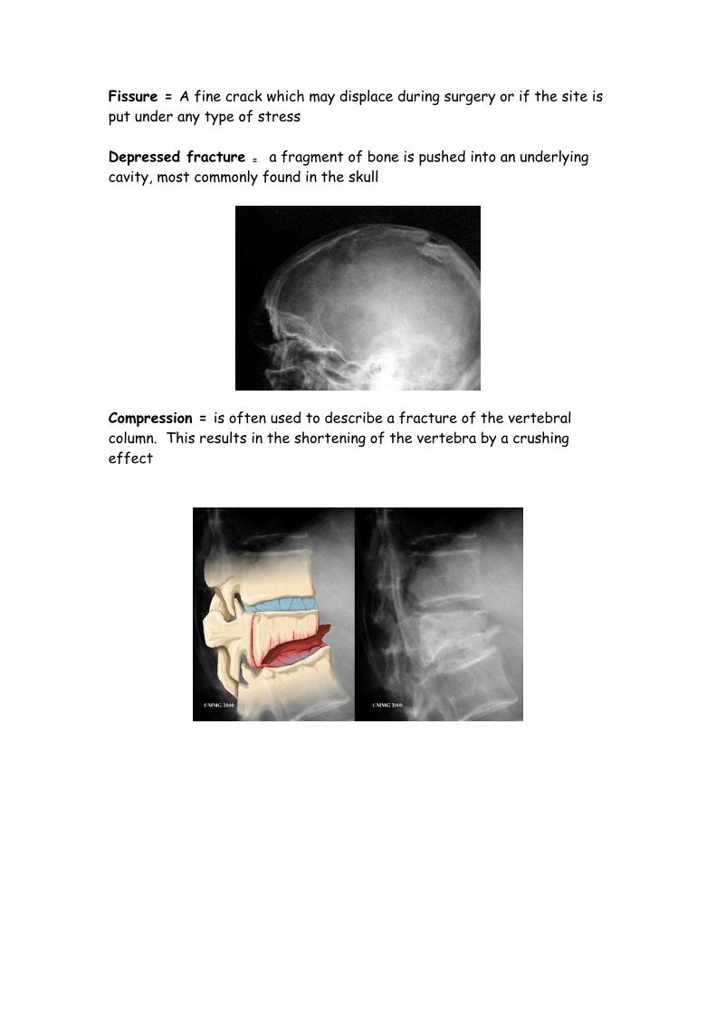
Depressed fracture = a fragment of bone is pushed into an underlying
cavity, most commonly found in the skull
Compression = is often used to describe a fracture of the vertebral
column. This results in the shortening of the vertebra by a crushing
effect
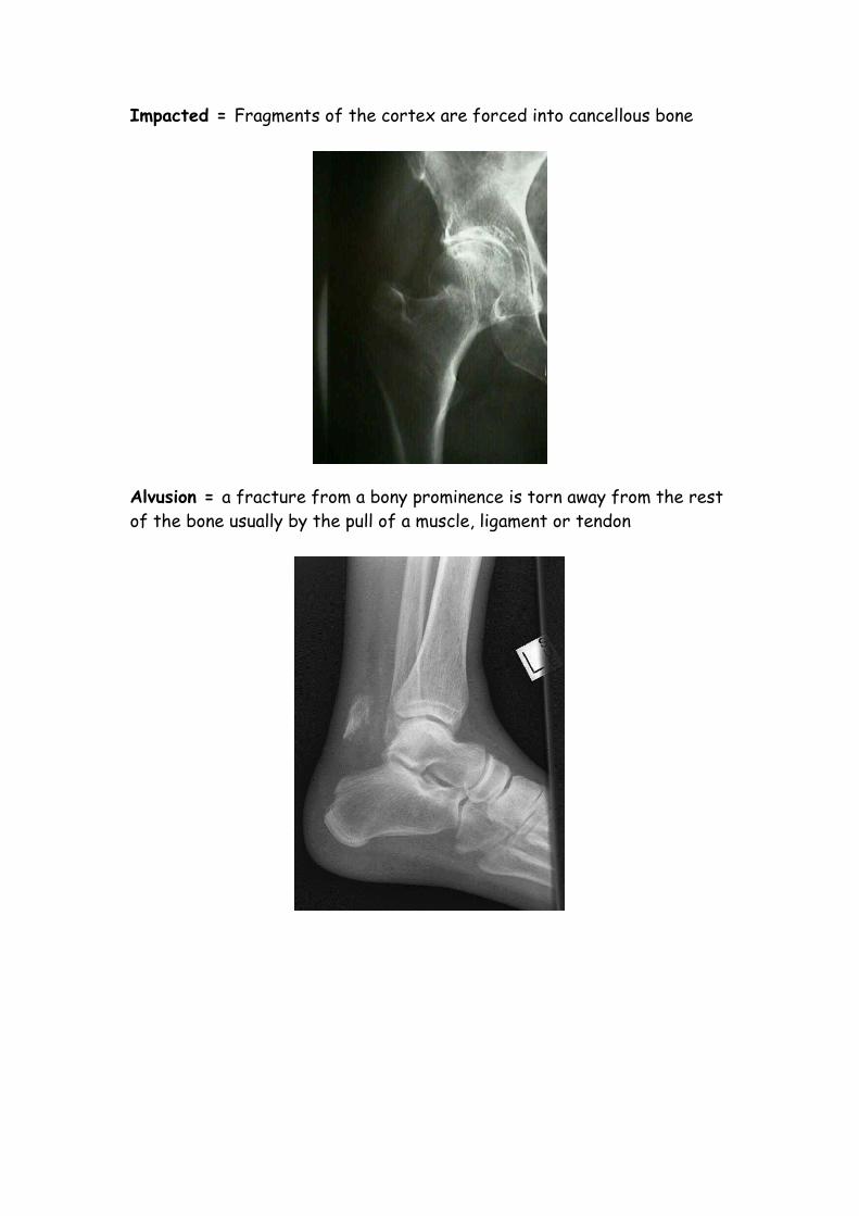
Impacted = Fragments of the cortex are forced into cancellous bone
Alvusion = a fracture from a bony prominence is torn away from the rest
of the bone usually by the pull of a muscle, ligament or tendon
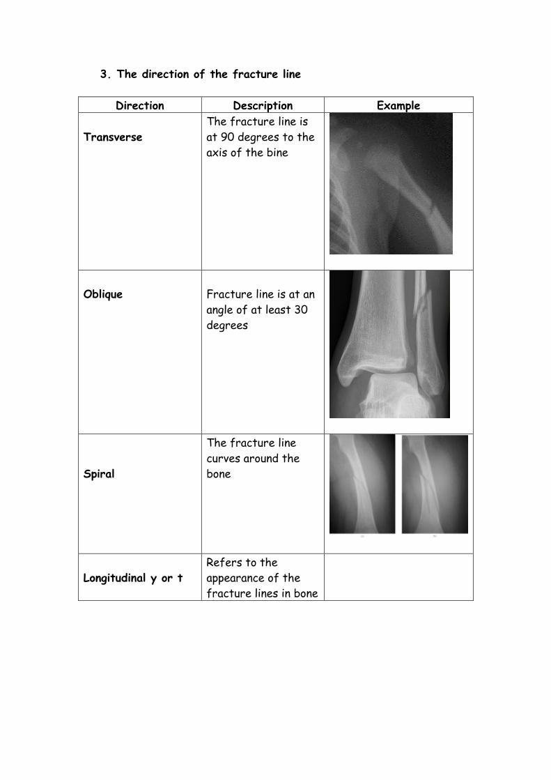
3. The direction of the fracture line
Direction Description Example
Transverse
The fracture line is
at 90 degrees to the
axis of the bine
Oblique
Fracture line is at an
angle of at least 30
degrees
Spiral
The fracture line
curves around the
bone
Longitudinal y or t
Refers to the
appearance of the
fracture lines in bone
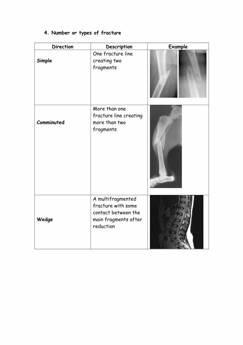
4. Number or types of fracture
Direction Description Example
Simple
One fracture line
creating two
fragments
Comminuted
More than one
fracture line creating
more than two
fragments
Wedge
A multifragmented
fracture with some
contact between the
main fragments after
reduction
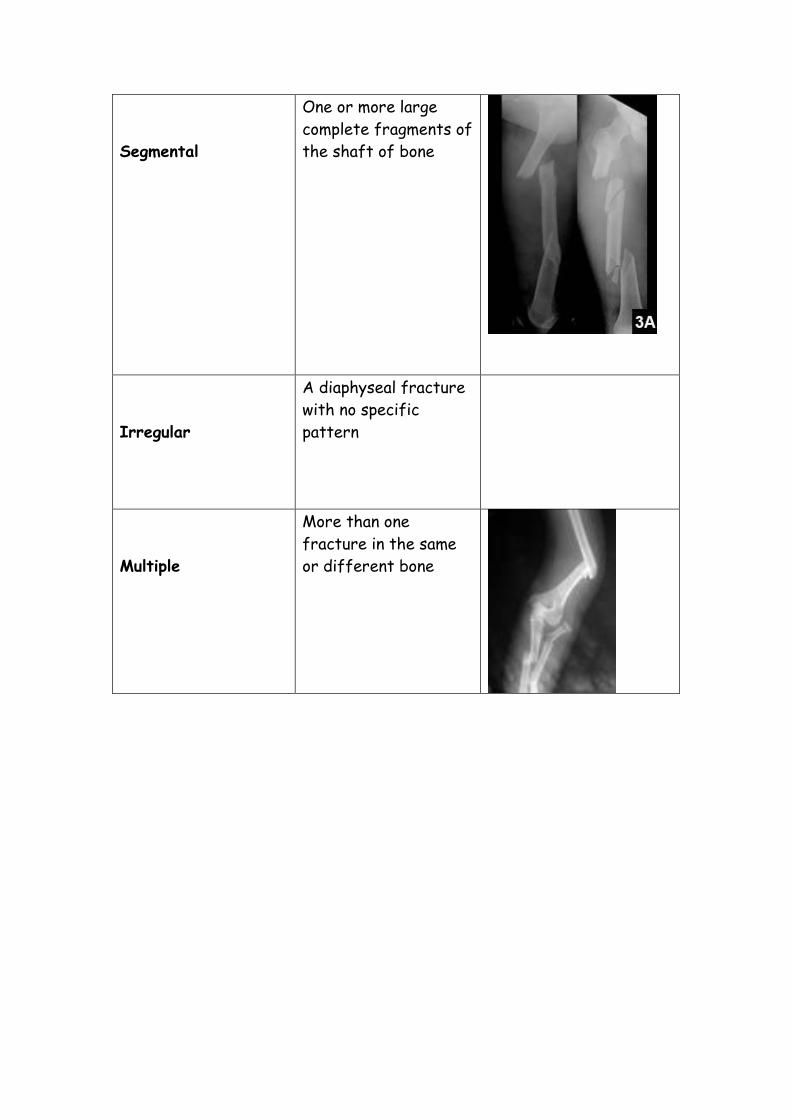
Segmental
One or more large
complete fragments of
the shaft of bone
Irregular
A diaphyseal fracture
with no specific
pattern
Multiple
More than one
fracture in the same
or different bone
Diagnosis of a fracture
Clinical signs
History from the owner – has the animal fallen or had some sort of
trauma
Clinical examination
Signs seen on clinical examination will include:
Inflammation
Pain localised to the affected bone
Local swelling and heat
Bruising at the fracture site leading to discolouration
Marked loss of function (lameness/non weight bearing)
Visible or palpable deformity of the affected bone
Abnormal mobility at the fracture site
Crepitus when the injured part is moved
Radiographs should be taken – 2 views are essential to enable the
Veterinary Surgeon to make a proper diagnosis and plan for a repair
Even if it is obviously fractured a good quality radiograph will confirm
details such as a hairline fracture etc that may affect a treatment and
surgical plan
Fracture Repair
The aim of fracture fixation is to restore the functional anatomy of the
fractured bone
We do this by:
Restoring the continuity of the bone
Restore the length
Restoring the shape of the bone
Maintaining soft tissue function
All essential tissue (blood vessels, muscles and nerves) need to removed
from the fracture site especially when fixation is involved, or repaired
Stabilisation of the fractures
Reduction – the fragments should be brought together in the correct
alignment
Fixation – The fragments should be immobilised in the correct alignment
until union occurs
It is important that the fracture is properly looked after before surgery.
This will prevent displacement of the fracture, pain and non union
Fracture fixation techniques are classified into 3 areas
1. External - Using casts or spints
2. Internal – Using pins, screws, plates etc
3. external – Internal – Using external fixators
There are several factors that need to be taken into account before
repairing a fracture. These include:
Identifying the classification of the fracture
The age of the patient
The size of the patient
The temperament of the patient
Presence of bacteria
Cost
Expectations of the owner (working vs. pet)
External
Aim = to limit movement at the site of the fracture
This type of fixation is not suitable if the joints above and below the
fracture cannot be immobilised
Advantages
Simple
Economical
Non Invasive
Disadvantages
Limited applications,
They don’t provide sufficient stability for many fractures
Risk of ulcers
Restrict activity of joints
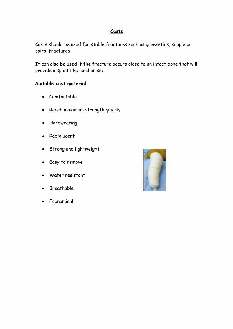
Casts
Casts should be used for stable fractures such as greenstick, simple or
spiral fractures
It can also be used if the fracture occurs close to an intact bone that will
provide a splint like mechanism
Suitable cast material
Comfortable
Reach maximum strength quickly
Hardwearing
Radiolucent
Strong and lightweight
Easy to remove
Water resistant
Breathable
Economical
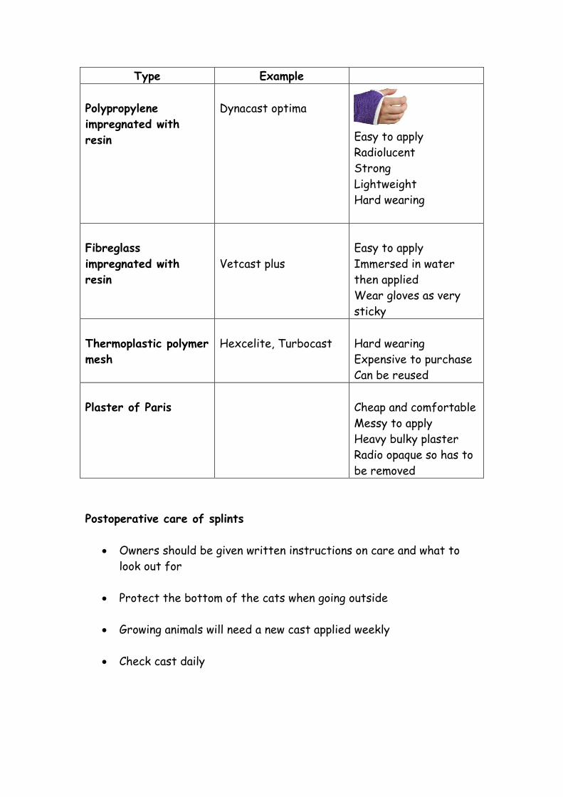
Type Example
Polypropylene
impregnated with
resin
Dynacast optima
Easy to apply
Radiolucent
Strong
Lightweight
Hard wearing
Fibreglass
impregnated with
resin
Vetcast plus
Easy to apply
Immersed in water
then applied
Wear gloves as very
sticky
Thermoplastic polymer
mesh
Hexcelite, Turbocast
Hard wearing
Expensive to purchase
Can be reused
Plaster of Paris
Cheap and comfortable
Messy to apply
Heavy bulky plaster
Radio opaque so has to
be removed
Postoperative care of splints
Owners should be given written instructions on care and what to
look out for
Protect the bottom of the cats when going outside
Growing animals will need a new cast applied weekly
Check cast daily
Complication of a cast
Limb swelling
Decubital ulcers
Cast loosening
Prolonged immobilisation of the limb causing limb stiffness, muscle
atrophy
Joint laxity
Delayed union, Malunion, non union
Refracture on removal of cast
Limbs should remain in the cast from 4-6 weeks. Radiographs should be
taken at intervals to assess healing.
Internal fixation
This involves the use of pins, plate’s screws and wire
Advantages
Suitable for fractures in any bone
Versatile
Allows accurate reduction
Allows limb function to return early on
Disadvantages
Expensive and time consuming
Technically demanding
Cost of buying equipment in
Risk of surgery and wound healing
Open fractures with soft tissue involvement may not be suitable
Implants and techniques
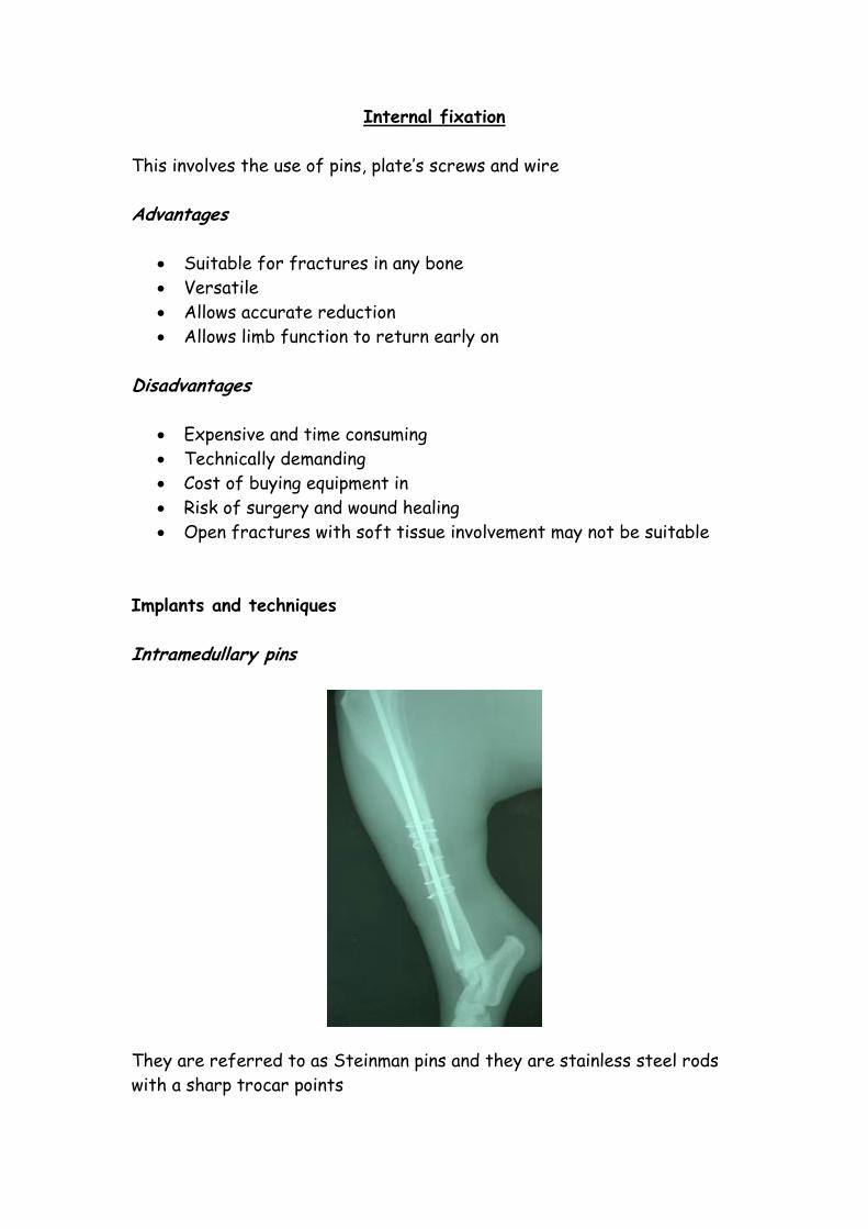
Intramedullary pins
They are referred to as Steinman pins and they are stainless steel rods
with a sharp trocar points
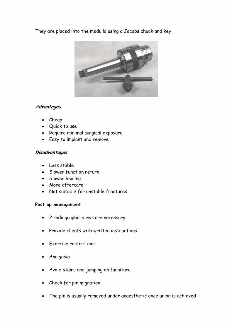
They are placed into the medulla using a Jacobs chuck and key
Advantages
Cheap
Quick to use
Require minimal surgical exposure
Easy to implant and remove
Disadvantages
Less stable
Slower function return
Slower healing
More aftercare
Not suitable for unstable fractures
Post op management
2 radiographic views are necessary
Provide clients with written instructions
Exercise restrictions
Analgesia
Avoid stairs and jumping on furniture
Check for pin migration
The pin is usually removed under anaesthetic once union is achieved
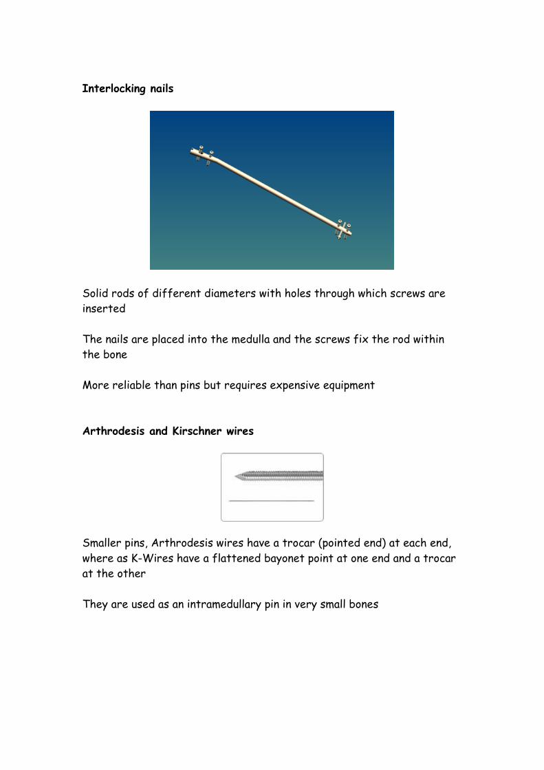
Interlocking nails
Solid rods of different diameters with holes through which screws are
inserted
The nails are placed into the medulla and the screws fix the rod within
the bone
More reliable than pins but requires expensive equipment
Arthrodesis and Kirschner wires
Smaller pins, Arthrodesis wires have a trocar (pointed end) at each end,
where as K-Wires have a flattened bayonet point at one end and a trocar
at the other
They are used as an intramedullary pin in very small bones
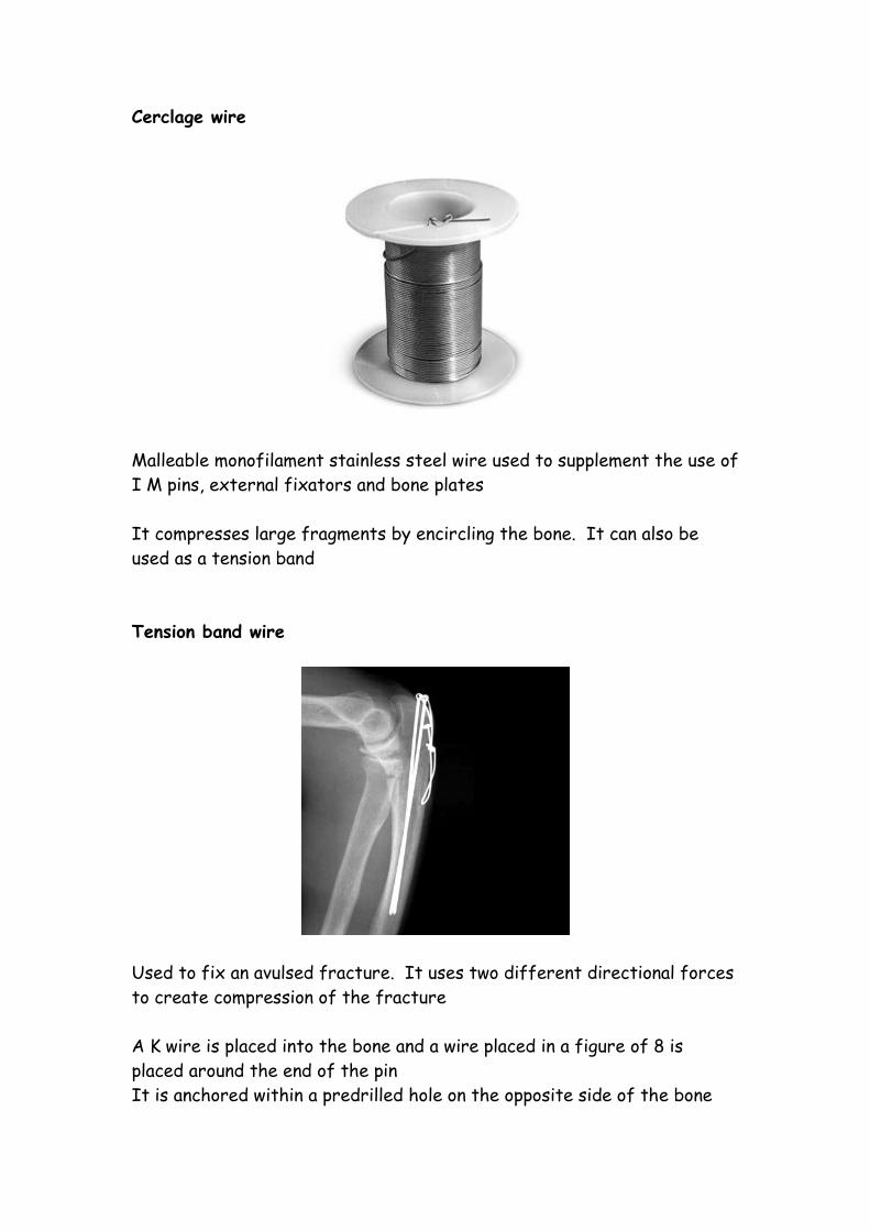
Cerclage wire
Malleable monofilament stainless steel wire used to supplement the use of
I M pins, external fixators and bone plates
It compresses large fragments by encircling the bone. It can also be
used as a tension band
Tension band wire
Used to fix an avulsed fracture. It uses two different directional forces
to create compression of the fracture
A K wire is placed into the bone and a wire placed in a figure of 8 is
placed around the end of the pin
It is anchored within a predrilled hole on the opposite side of the bone
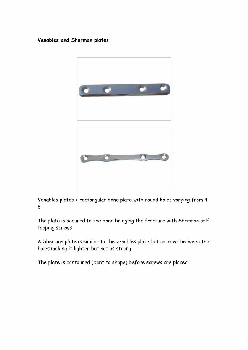
Venables and Sherman plates
Venables plates = rectangular bone plate with round holes varying from 4-
8
The plate is secured to the bone bridging the fracture with Sherman self
tapping screws
A Sherman plate is similar to the venables plate but narrows between the
holes making it lighter but not as strong
The plate is contoured (bent to shape) before screws are placed
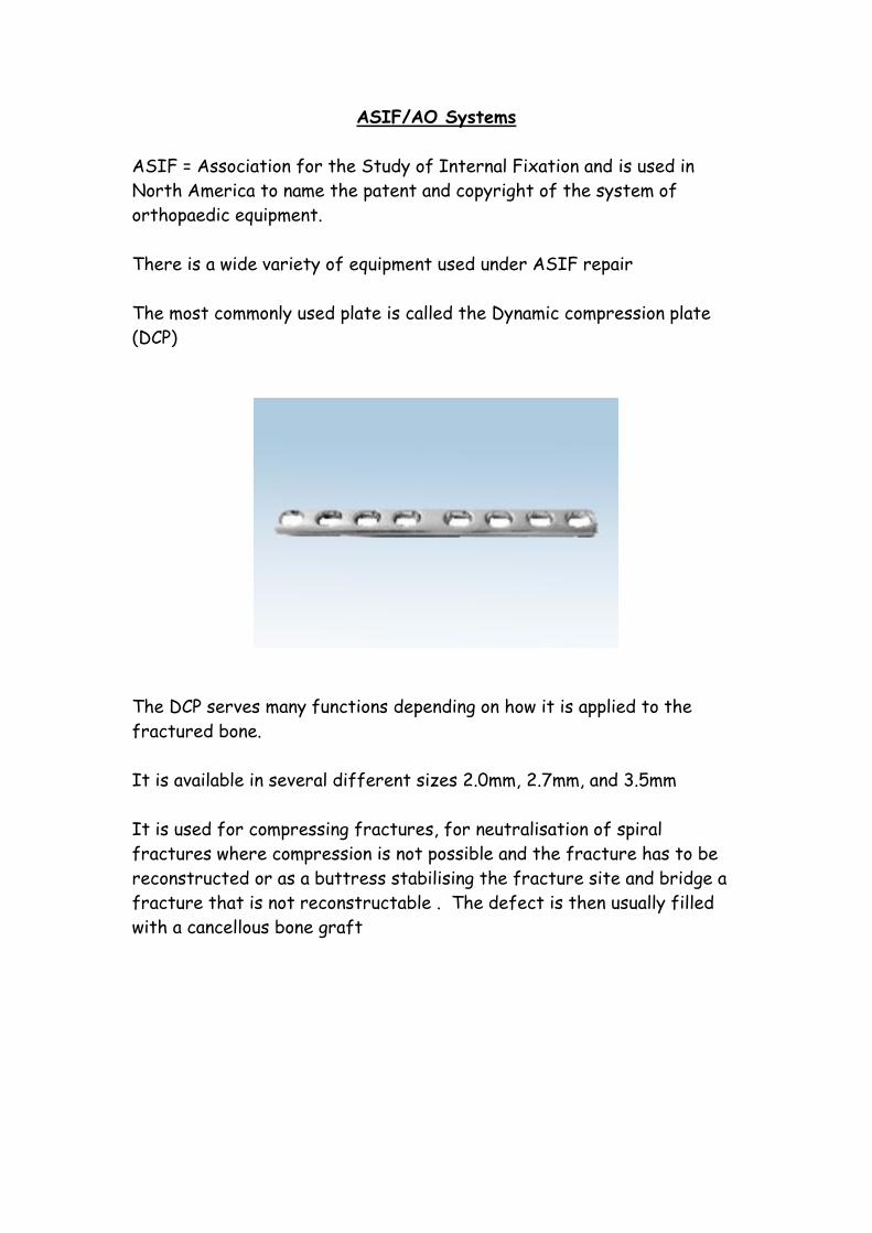
ASIF/AO Systems
ASIF = Association for the Study of Internal Fixation and is used in
North America to name the patent and copyright of the system of
orthopaedic equipment.
There is a wide variety of equipment used under ASIF repair
The most commonly used plate is called the Dynamic compression plate
(DCP)
The DCP serves many functions depending on how it is applied to the
fractured bone.
It is available in several different sizes 2.0mm, 2.7mm, and 3.5mm
It is used for compressing fractures, for neutralisation of spiral
fractures where compression is not possible and the fracture has to be
reconstructed or as a buttress stabilising the fracture site and bridge a
fracture that is not reconstructable . The defect is then usually filled
with a cancellous bone graft
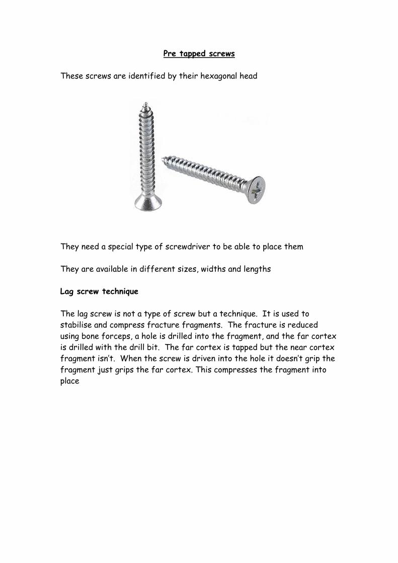
Pre tapped screws
These screws are identified by their hexagonal head
They need a special type of screwdriver to be able to place them
They are available in different sizes, widths and lengths
Lag screw technique
The lag screw is not a type of screw but a technique. It is used to
stabilise and compress fracture fragments. The fracture is reduced
using bone forceps, a hole is drilled into the fragment, and the far cortex
is drilled with the drill bit. The far cortex is tapped but the near cortex
fragment isn’t. When the screw is driven into the hole it doesn’t grip the
fragment just grips the far cortex. This compresses the fragment into
place
Postoperative care following internal fixation
2 radiographic views should be taken to assess repair
Analgesia throughout recovery
Adequate nutrition
IVFT
Assisted walking
Daily monitoring of TPR
Sutures should be removed 10 days post op
Clients should be given written instructions on care
Exercise restrictions with short bouts of lead exercise in the first
3-4 weeks. Hydrotherapy can be used once the wounds have healed
Complications
Infection
This if often due to poor technique or choice of implants. In some cases
postop care in the home environment is not good enough to protect the
implants from failure
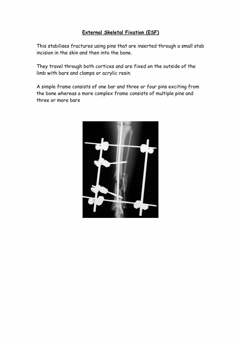
External Skeletal Fixation (ESF)
This stabilises fractures using pins that are inserted through a small stab
incision in the skin and then into the bone.
They travel through both cortices and are fixed on the outside of the
limb with bars and clamps or acrylic resin.
A simple frame consists of one bar and three or four pins exciting from
the bone whereas a more complex frame consists of multiple pins and
three or more bars
Advantages
Minimal instrumentation required
Clamps and bars are reusable
Minimal disruption of soft tissue
Minimal foreign body at fracture site
Open wound management easy
Easy to combine with other implants
Alignment is adjustable
Assessment of fracture healing easy
Easy to remove
Disadvantages
Soft tissue problems possible
Application technique requires practice
Premature pin loosening common
Difficult to apply to proximal limb
Can be difficult to obtain good x-rays
Types of fractures that are suitable for external fixation
Long one fractures
Comminuted fractures
Open and infected fractures
Delayed union and non union
Mandibular fractures
APEF System
APEF = Acrylic pin external fixator
It uses corrugated tubing which is filled with polymethylmethacrylate
which is a type of bone cement
The pins are placed into bones with the corrugated tubing fixed to the
ends of the pins
The tubes are then filled with cement and held in place until hardened.
This is particularly useful in mandibular fractures where the tubing forms
a bumper bar
Bone Grafts
Bone grafts can be harvested from either cortical or cancellous bone.
They are used to supplement fracture repair and accelerate healing
across a wide gap
Cortical bone grafts = Whole segments of solid bone in a fracture.
It takes a long time for the graft to become fully incorporated in the
repair
Cancellous bone =Harvested from inside the medulla of long bones. The
commonest sites used are the humerus and ilieum
Cancellous bone grafts are an essential part of the repair of complex
fractures as they contribute cells and growth factors involved in bone
healing
Post operative care
Open wounds should be treated and dressed appropriately
The limb should have a compressive bandage applied fro 2-3 days
(changed daily) to minimise swelling
The ends of the pins should be covered with tape to prevent
damage to the owners furniture
Air should be able to circulate between skin and pins
Cats need to be cage rested
Exercise should be limited to lead exercise only
Owners should be told to expect a small amount of scab formation
at the site of the pin. This is normal and should not be cleaned
IT IS IMPORTANT THAT SCABS ARE NOT REMOVED!!
Excess exudate should be seen by a vet
Elizabethan collar should be applied
Written instructions should be given to the owner
Complications of external fixators
Swelling of soft tissue impinging on the clamps and acrylic bars
Excessive exudate from the pin site causing movement
Loosening of pins but in some cases individual pins can be removed
without losing the stability of the frame