Embed Size (px)
Citation preview
IBIMA Publishing
Journal of Research and Practice in Dentistry
http://www.ibimapublishing.com/journals/DENT/dent.html
Vol. 2014 (2014), Article ID 821205, 11 pages
DOI: 10.5171/2014.821205
_____________
Cite this Article as: Nimet Gençoğlu, Mustafa Gündoğar, Dilek Helvacıoğlu-Yiğit and Cafer Türkmen
(2014), “Obturation Effect of Different Filling Techniques on Isthmuses in Mandibular Molars after Passive
Ultrasonic Irrigation,” Journal of Research and Practice in Dentistry, Vol. 2014 (2014), Article ID 821205,
DOI: 10.5171/2014.821205
Research Article
Obturation Effect of Different Filling Techniques on Isthmuses in Mandibular Molars after Passive
Ultrasonic Irrigation
Nimet Gençoğlu1, Mustafa Gündoğar
2, Dilek Helvacıoğlu-Yiğit
3
and Cafer Türkmen1
1Marmara University Faculty of Dentistry, Department of Restorative Dentistry, İstanbul, Turkey
2Medipol University Faculty of Dentistry, Department of Endodontics, İstanbul, Turkey
3Kocaeli University Faculty of Dentistry, Department of Endodontics, Kocaeli, Turkey
Correspondence should be addressed to: Nimet Gençoğlu; [email protected]
Received 17 May 2013; Accepted 19 June 2013; Published 24 January 2014
Academic Editor: Alfred Naaman
Copyright © 2014 Nimet Gençoğlu, Mustafa Gündoğar, Dilek Helvacıoğlu-Yiğit and Cafer Türkmen.
Distributed under Creative Commons CC-BY 3.0
Abstract The effect of ultrasonic debridement on cleaning isthmuses and the effect of obturation
techniques on filling of isthmuses in mandibular first molars were evaluated.
Sixty molars with two mesial canals were instrumented by rotary instruments, divided into
four groups (15 each). Group 1 & 2 were irrigated by PUI (passive ultrasonic irrigation) or by
hand irrigation with %5.25 NaOCl and not obturated. Group 3 & 4 obturated by Microseal or
lateral condensation after passive ultrasonic irrigation. The roots were sectioned at
horizontally different levels, the quantity of debris was evaluated under stereomicroscope and
the percentage of obturated area in isthmus was statistically analyzed (ANOVA and Newman-
Keuls).
The highest incidence of isthmus was found in the 3.5 mm sections of the roots.
PUI was found to be more effective than hand irrigation to clean isthmuses in mandibular
molars (p<0.05). More gutta-percha content was found in Microseal obturation than lateral
condensation (p<0.05).
Ultrasonic instruments were found to be effective to clean isthmuses and Microseal was
superior to lateral condensation technique on obturating isthmuses.
Keywords: Isthmus, microseal, ultrasonic irrigation.
Journal of Research and Practice in Dentistry 2
_______________
Nimet Gençoğlu, Mustafa Gündoğar, Dilek Helvacıoğlu-Yiğit and Cafer Türkmen (2014), Journal of
Research and Practice in Dentistry, DOI: 10.5171/2014.821205
Introduction
The main objective of endodontic therapy
is to clean the entire pulp cavity and
complete it with an inert filling material.
Because the root canal system has a
complex anatomy, it is difficult to shape
and clean the root canal completely. Small
isthmuses and irregularities have been
shown to be inaccessible to conventional
hand and rotary instrumentation.1 Ingle2
concluded that 60% of endodontic failures
are caused by incomplete obturation of the
root canal. The other main reasons include
untreated canals, accessory canals and the
presence of an isthmus.
Isthmuses are present in all types of roots
in which two canals are normally found.3
The incidence of isthmus was found around
%30 in mandibular premolar, 60% in
mesiobuccal roots of maxillary first molar
with two canals.4 However, the prevalence
of isthmuses in the mesial root of
mandibular molars has been observed to
be as high as 80%.5 They are poorly
accessible to root canal instruments and
untreated isthmuses can cause failure of
conventional root canal therapy or apical
surgery, especially in posterior teeth.
The outcome of endodontic treatments is
improved by the development of new
techniques and new instruments. The use
of ultrasonics in endodontics has made it
especially easy to clean difficult anatomical
features like accessory canals and
isthmuses. The first findings on the
technique of root canal therapy using an
ultrasonic instrument were reported by
Richman.6 Martin et al.7 found that the root
canals of teeth that were ultrasonically
filed and irrigated were cleaner when
compared with the conventional methods,
and that the smear layer appeared to be
greatly reduced. The cleaning efficacy of
ultrasound appears to be promising when
used only for irrigation after the root canal
has been instrumented.8 Weller et al.9
concluded that the most effective
debridement occurred when
ultrasonication was used after completion
of hand instrumentation. It has also been
demonstrated that an irrigant in
conjunction with ultrasonic vibration,
which generates a continuous movement of
the irrigant, is directly associated with the
effective cleaning of the root canal space.
Ahmad et al.8 described that when files
were activated with ultrasonic energy,
acoustic streaming was sufficient to
produce cleaner canals, compared with
hand filing alone. They showed that the
flushing action of irrigants could be
enhanced by using ultrasonication.8,10,11
This seemed to improve the efficacy of
irrigation solutions in removing organic
and inorganic debris from root canal
walls.2,7,12
In the past, the canal isthmus was often
overlooked and it was also difficult to
prepare if located. However, previous
studies showed that passive ultrasonic
irrigation removed more dentin debris
from the isthmus, oval extensions in the
root canal and irregularities from the root
canal wall.1 The recognition and
management of the canal isthmus is an
important factor that may improve the
success rate of surgical and non surgical
endodontics specially in posterior teeth.13
There are a few studies on the subject of
the cleaning of isthmuses1,6,9 but none on
obturating them. Because the isthmuses
usually have the vital and infected part of
the pulp, if it had not been cleaned and
obturated during the conventional root
canal treatment or before surgical
procedure, it could contribute to failure of
the case. Nowadays, it is possible to clean
isthmuses by using ultrasonics and then to
obturate them by using the warm gutta-
percha technique. The aim of this study
was to investigate the cleaning action of
ultrasonic irrigation and the obturation
effect of different techniques on isthmuses
in mandibular molars.
Materials and Methods Sixty extracted first mandibular molars
with two mesial canals in one root
exhibiting a single oval foramen were
selected. The access cavity was opened and
all canals were instrumented by using
rotary Hero Shaper instruments (Micro
Mega, Besancon, France). According to the
manufacturer instruction, each canal was
3 Journal of Research and Practice in Dentistry
_______________
Nimet Gençoğlu, Mustafa Gündoğar, Dilek Helvacıoğlu-Yiğit and Cafer Türkmen (2014), Journal of
Research and Practice in Dentistry, DOI: 10.5171/2014.821205
instrumented by No # 25 file with % 6 and
4 % taper and finally with #30 % 4 taper
instrument. The canals were irrigated by
2ml of 5.25 % NaOCl solution after each
instrument usage. The experimental teeth
were divided equally into 4 groups:
Experimental Group Group 1. The teeth were irrigated by using
passive ultrasonic irrigation (PUI) (n=15).
Group 2. The teeth were irrigated by hand
irrigation (n=15).
Group 3. The teeth were obturated by
warmed lateral condensation of gutta-
percha technique (Microseal) after
irrigation by PUI (n=15).
Group 4. The teeth were obturated by cold
lateral condensation of gutta percha after
irrigation by PUI (n=15).
Group 1 (PUI) Ultrasonic irrigation was performed
according to the manufacturer’s
instructions (at the highest power setting
for 60 s) and applied in each mesial canal of
the teeth with approximately 2 ml of 5.25
% NaOCl by using ultrasonically activated
size #15 endo file (EMS, Nyon, Switzerland,
DT_006, 007, 008, 009, 010).
Group 2 (Conventional Irrigation)
The mesial canals were irrigated with 2 ml
of 5.25 % NaOCl and 2ml of 17% EDTA by
using a syringe gauge (2 ml) for 60 s.
Groups 3 and Group 4 (Obturated after PUI) Mesial canals of 15 teeth were obturated by
the Microseal technique (group 3) and the
remaining 15 teeth were obturated by the
lateral condensation technique (group 4)
after ultrasonic irrigation by 5.25 % NaOCl,
using the ultrasonically activated size #15
endo file for 1 min.
Obturation Microseal Technicus (Tycom,
Irvine, CA, USA): An appropriate size of
master cone was placed into each mesial
canal until tug-back was elicited. The
appropriate spreader was selected to
compact the master cone, 1.0 mm shorter
than the working length. Finally, the
appropriate accessorymechanical
compactors were selected, according to the
manufacturer’s instructions. Freshly mixed
Kerr sealer was placed into each mesial
canal and sealer-covered master gutta-
percha was seated. The spreader was
advanced alongside the master cone at the
working length for compaction. A tapered
void was formed between the compacted
gutta-percha cone and the root canal walls
by the withdrawal of the spreader from the
canal. The appropriate compactor was
placed in the heated gutta-percha cartridge
and was sealed with a uniform layer of
material. The gutta-percha-coated
compactor was then inserted to the void
previously created in the canal by the
spreader, and was applied as close to the
working length as possible, avoiding
rotation as it was inserted. A resisting force
was applied to the compactor’s backing-out
motion without any apical pressure and
rotation of the compactor was started at a
speed of 6000 rpm. The compactor was
removed after approximately 3 seconds.
Rotation continued until the compactor
was removed fully from the canal. If the
canal was not completely obturated, more
gutta-percha cones were placed on the
compactor. Protruding gutta-percha and
sealer were removed from the access cavity
by using a cotton pellet.
Obturation Lateral Condensation Technique: No #35 gutta-percha master
cone (Hygenic Corp, Akron, Ohio) was
fitted to within 0.5 mm of the working
length of each mesial canal. Freshly mixed
Kerr sealer was placed into the canal.
Appropriate spreader was inserted 1 mm
shorter than the working length. No #25
gutta-percha cone was inserted as an
accessory cone followed by smaller size
spreader insertion. This procedure was
continued until the canal was considered to
be obturated adequately with smaller size
of points, and spreader could no longer
penetrate beyond the coronal third of the
canal. After complete canal obturation the
coronal gutta-percha was removed with a
hot instrument.
Journal of Research and Practice in Dentistry 4
_______________
Nimet Gençoğlu, Mustafa Gündoğar, Dilek Helvacıoğlu-Yiğit and Cafer Türkmen (2014), Journal of
Research and Practice in Dentistry, DOI: 10.5171/2014.821205
All distal roots were removed and the
mesial roots were embedded in
orthodontic clear acrylic resin. A rotary
saw with a diamond blade water (Isomet,
Buehler; USA) was used to make cross-
sectional slides through the embedded
teeth. To reduce a smear layer of gutta-
percha, the sections were made using cold-
water irrigation. The roots were sectioned
at 1.5 mm, 2.5 mm, 3.5 mm and 4.5 mm
from the anatomic apex. Unobturated
canals were dyed with 2 % methylene blue
to expose the debris and isthmus better
under the microscope.
Photographs of every section from all the
groups were taken at an original 50X
magnification by means of a
stereomicroscope (Imaging Systems, Leica
Ltd, Cambridge, England) with a digital
camera (Fig. 1-4).
1. The incidence of isthmus was recorded
according to the classification used by Hsu
and Kim.3
2. The amount of debris in the isthmus
was classified by using the scoring system
reported by van de Sluis and Wesselink.14 A
higher score indicated a greater amount of
debris:
Score 0: The entire isthmus was free of
debris
Score 1: Less than half of the isthmus was
filled with debris
Score 2: Half and more than the half of the
isthmus was filled with debris
Score 3: The entire isthmus was filled with
debris
The examination was performed by three
independent investigators using this
scoring system.
For statistical analysis, the Kruskal-Wallis
test was used. The level of significance was
set at p=0.05.
3. The areas of the canal and gutta-percha,
sealer or voids were outlined and then
measured by Image pro-plus computer
program (IPWIN application, Media
Cybernetics, Inc.) for both obturation
techniques. All measurements were
evaluated by means of one way ANOVA and
the Newman-Keuls test.
Results The incidence of an isthmus is shown in
Table 1. The highest incidence of isthmus
was found in the 3.5 mm sections of the
roots.
Table 1. Incidence of an İsthmus at Each Level
Level from apex (mm)
Percent of isthmuses (%) (n= 60 teeth)
1.5 38.3
2.5 48.3
3.5 53.3
4.5 43.3
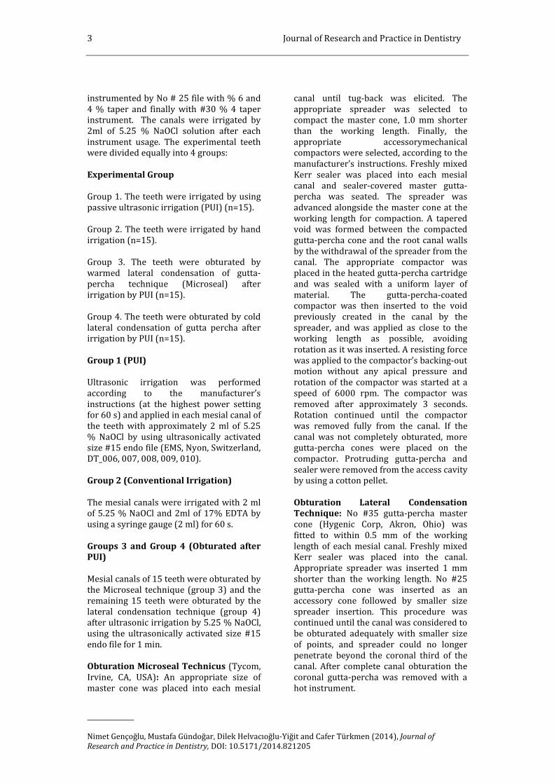
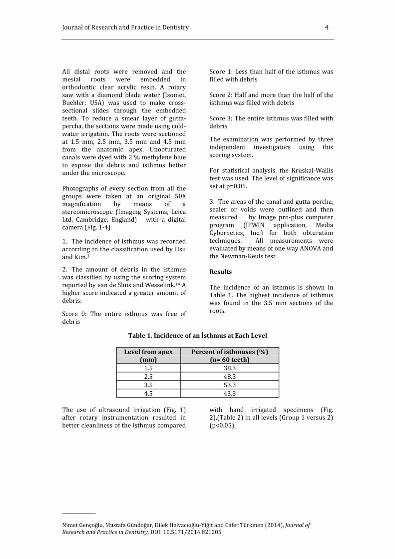
The use of ultrasound irrigation (Fig. 1)
after rotary instrumentation resulted in
better cleanliness of the isthmus compared
with hand irrigated specimens (Fig.
2),(Table 2) in all levels (Group 1 versus 2)
(p<0.05).
5 Journal of Research and Practice in Dentistry
_______________
Nimet Gençoğlu, Mustafa Gündoğar, Dilek Helvacıoğlu-Yiğit and Cafer Türkmen (2014), Journal of
Research and Practice in Dentistry, DOI: 10.5171/2014.821205
Fig. 1: Cross Section of a Root Canal Isthmus irrigated with PUI
Fig. 2: Cross Section of a Root Canal Isthmus Irrigated with Hand Irrigation

Journal of Research and Practice in Dentistry 6
_______________
Nimet Gençoğlu, Mustafa Gündoğar, Dilek Helvacıoğlu-Yiğit and Cafer Türkmen (2014), Journal of
Research and Practice in Dentistry, DOI: 10.5171/2014.821205
Table 2. Amount of Debris for Each Group
Tooth No. SL (mm) PUI SL (mm) Hand Irr.
1
1.5 1
2.5 0
3.5 1
1.5 1
2.5 3
3.5 3
2 1.5 0
2.5 0
3.5 0
1.5 2
2.5 3
3.5 3
3 1.5 0
2.5 0
3.5 0
1.5 2
2.5 3
3.5 3
4 1.5 0
2.5 0
3.5 0
1.5 3
2.5 3
3.5 3
5 1.5 0
2.5 0
3.5 0
1.5 3
2.5 1
3.5 1
6 1.5 0
2.5 0
3.5 1
1.5 3
2.5 3
3.5 3
7 1.5 2
2.5 2
3.5 2
1.5 1
2.5 1
3.5 1
8 1.5 2
2.5 0
3.5 0
1.5 1
2.5 3
3.5 1
9 1.5 1
2.5 0
3.5 0
1.5 3
2.5 3
3.5 3
10 1.5 1
2.5 0
3.5 0
1.5 1
2.5 1
3.5 1
11 1.5 2
2.5 1
3.5 2
1.5 3
2.5 3
3.5 3
12 1.5 2
2.5 1
3.5 1
1.5 3
2.5 3
3.5 3
13 1.5 2
2.5 0
3.5 0
1.5 2
2.5 2
3.5 3
14 1.5 0
2.5 0
3.5 0
1.5 2
2.5 2
3.5 3
15 1.5 0
2.5 0
3.5 0
1.5 3
2.5 3
3.5 3
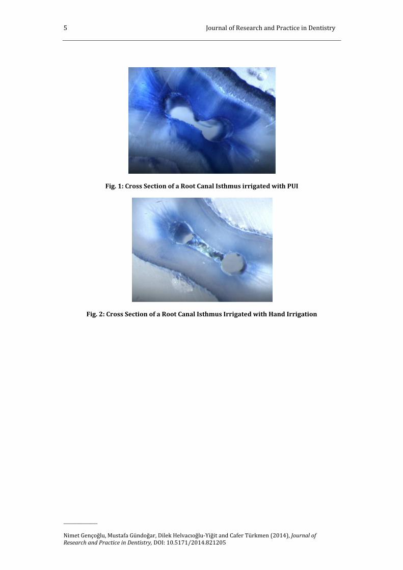
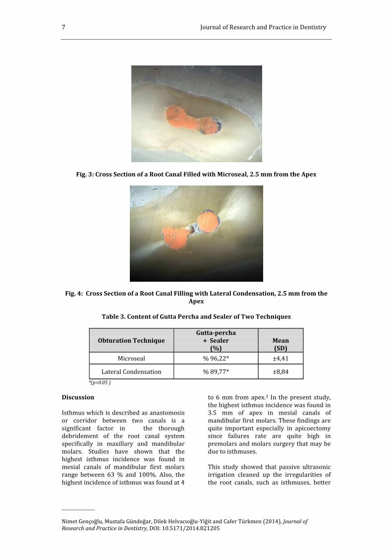
When the obturation techniques were
compared, the Microseal technique (Fig. 3)
was found to be superior to the lateral
condensation technique (Fig. 4) in
obturating isthmuses after irrigation by
ultrasonics. The gutta percha and sealer
contents in the two techniques are shown
in Table 3.
7 Journal of Research and Practice in Dentistry
_______________
Nimet Gençoğlu, Mustafa Gündoğar, Dilek Helvacıoğlu-Yiğit and Cafer Türkmen (2014), Journal of
Research and Practice in Dentistry, DOI: 10.5171/2014.821205
Fig. 3: Cross Section of a Root Canal Filled with Microseal, 2.5 mm from the Apex
Fig. 4: Cross Section of a Root Canal Filling with Lateral Condensation, 2.5 mm from the Apex
Table 3. Content of Gutta Percha and Sealer of Two Techniques
Obturation Technique
Gutta-percha + Sealer
(%)
Mean (SD)
Microseal % 96,22* ±4,41
Lateral Condensation % 89,77* ±8,84
*(p<0.05 )
Discussion
Isthmus which is described as anastomosis
or corridor between two canals is a
significant factor in the thorough
debridement of the root canal system
specifically in maxillary and mandibular
molars. Studies have shown that the
highest isthmus incidence was found in
mesial canals of mandibular first molars
range between 63 % and 100%. Also, the
highest incidence of isthmus was found at 4
to 6 mm from apex.3 In the present study,
the highest isthmus incidence was found in
3.5 mm of apex in mesial canals of
mandibular first molars. These findings are
quite important especially in apicoectomy
since failures rate are quite high in
premolars and molars surgery that may be
due to isthmuses.
This study showed that passive ultrasonic
irrigation cleaned up the irregularities of
the root canals, such as isthmuses, better
Journal of Research and Practice in Dentistry 8
_______________
Nimet Gençoğlu, Mustafa Gündoğar, Dilek Helvacıoğlu-Yiğit and Cafer Türkmen (2014), Journal of
Research and Practice in Dentistry, DOI: 10.5171/2014.821205
than hand irrigation. This finding can be
attributed to the action of the ultrasonically
activated irrigation solution within the
canal system. Isthmus cleanliness values
improved significantly from the 1.5 mm to
the 3 mm level when passive ultrasonic
irrigation was used after rotary
instrumentation.
The effect of irrigation time when using
ultrasonic irrigation is not clear in
literature. Sabin et al.15 reported that 30 s
to 1 min of ultrasonic activation was
sufficient to produce clean canals, whereas
Krell et al.16 recommended the use of
ultrasonic irrigation for 2 min. Van de Sluis
et al.14 found that a 3 min ultrasonic
irrigation was effective to remove dentine
debris. In the present study, 1 min
ultrasonic irrigation seemed to be effective
in cleaning isthmuses.
Tauber et al.17 and Goldman et al.18
reported that low power ultrasonication
was not effective when used for the
irrigation of the canals. Cameron19,
however, showed that the use of medium
power was effective in cleaning root canals.
In this study, the highest power setting was
used for ultrasonic irrigation according to
the manufacturer’s recommendation.
Two types of ultrasonic irrigation are
described in the literature: one type where
irrigation is combined with simultaneous
ultrasonic instrumentation (UI), and the
other without simultaneous
instrumentation, called passive ultrasonic
irrigation (PUI).20 Weller et al.9 showed
that UI is less effective in removing
simulated pulp tissue from the root canal
system, or the smear layer from the root
canal wall, than PUI. Ahmad et al.8
explained that this could be the result of a
reduction of acoustic streaming and
cavitation. In the present study, the EMS
Endo file was used to perform passive
ultrasonic irrigation and found to be
effective in cleaning the isthmuses of the
root canal.
Cuningham et al.21 have demonstrated that
ultrasonics can also improve the
disinfection of root canals. The studies
showed that passive ultrasonic irrigation is
significantly better than syringe irrigation
in the reduction of bacteria in the root
canals. Walmsley22 reported that this could
be because of the disruption of organic
tissues entering the streaming field that
was generated. Ahmad23 explained that
ultrasonically activated files generate a
mechanism that damages biological cells.
Lee et al. 1 found that a lower number of
colonies survived when ultrasonic
activation was used. Carver et al.24 also
found that ultrasonic irrigation following
hand/rotary instrumentation in vivo
decreased significantly the number of
bacteria to a greater extent than
hand/rotary instrumentation alone.
Despite these findings, it generally
accepted that no technique is able to
ensure complete canal disinfection.
Previous studies have shown that some
endodontic sealers are soluble25 and may
shrink slightly.26 Sealer dissolution may
trigger an increase in leakage along the
root filling over time. So, generally it is
preferred to minimize the amount of sealer
and maximize the volume of gutta-percha.
Eguchi et al.27 reported that lateral
condensation results in excessive amounts
of sealer and apical voids. Peters25 also
found voids, spreader tracts, incomplete
fusion of the gutta-percha cones, and lack
of surface adaptation in lateral
condensation technique. In our previous
study28, the gutta-percha/sealer content of
different warmed condensation techniques
were compared with lateral condensation
and all warmed condensation techniques
(included Microseal) were found superior
to lateral condensation technique. In the
present study, again Microseal technique
was found to be superior to lateral
condensation technique with regard to
gutta-percha-sealer content.
In literature, studies have been performed
concerning the microleakage effect of
different obturation techniques rather than
that of the gutta-percha content. Hwang et
al.29 investigated the microleakage effect of
Microseal, warm vertical condensation and
lateral condensation techniques on
obturation of isthmuses in multi-rooted
teeth, and found that the Microseal
technique was superior to other

9 Journal of Research and Practice in Dentistry
_______________
Nimet Gençoğlu, Mustafa Gündoğar, Dilek Helvacıoğlu-Yiğit and Cafer Türkmen (2014), Journal of
Research and Practice in Dentistry, DOI: 10.5171/2014.821205
techniques. Davalou et al.30 reported no
significant difference in apical leakage
between the System B and Microseal
techniques. Recently, Mazotti et al.31
compared Microseal to hybrid ENAC
ultrasonic and lateral condensation
technique and found the best results in
Microseal obturation. When the studies
were compared to literature, contradictory
results could be found between leakage
studies even when the same materials have
been studied. The lack of standardization of
the experimental techniques leads to
conflicting results. Also, the clinical
significance of leakage tests in vitro is
questionable, but incompletely filled canal
irregularities such as isthmuses with the
assumption that this meant apical leakage
was occurring. Also, reducing the ratio of
sealer to gutta-percha may improve the
long term seal provided by the root canal
filling.
Although these are in vitro results, they are
of significance because these factors cannot
easily be quantitatively determined in vivo.
Nevertheless, further clinical studies are
necessary to confirm these results and
evaluate their relevance to treatment
outcome.
References 1. Lee, S. J., Wu, M. K. & Wesselink, P. R.
(2004). “The Effectiveness of Syringe
Irrigation and Ultrasonics to Remove
Debris from Stimulated Irregularities
within Prepared Root Canal Walls,”
International Endodontic Journal 2004;
37:672-8.
2. Luebke, R. G., Glick, D. H. & Ingle, J. I.
(1964). “Indications and
Contraindications for Endodontic
Surgery,” Oral Surgery, Oral Medicine,
Oral Pathology 1964; 18:97-113.
3. Hsu, Y. Y. & Kim, S. (1997). “The
Resected Root Surface. The Issue of
Canal Isthmuses,” Dental Clinics of North
America 1997; 41:529-40.
4. Cambruzzi, J. V. & Marshall, F. J. (1983).
“Molar Endodontic Surgery,” Journal
(Canadian Dental Association) 1983;
1:61-6.
5. Vertucci, F. J. (1984). “Root Canal
Anatomy of the Human Permanent
Teeth,” Oral Surgery, Oral Medicine, Oral
Pathology 1984; 58:589-99.
6. Richman, M. J. (1957). 'The Use of
Ultrasonics in Root Canal Therapy and
Root Resection,' Journal of Dental
Medicine 1957; 12:12-8.
7. Martin, H., Cunningham, W. T. & Norris,
J. P. (1980). “A Quantitative Comparison
of the Ability of Diamond and K-Type
Files to Remove Dentin,” Oral Surgery,
Oral Medicine, Oral Pathology 1980;
50:566-8.
8. Ahmad, M., Pitt Ford, T. R. & Crum, L. A.
(1987). “Ultrasonic Debridement of
Root Canals: Acoustic Streaming and its
Possible Role,” Journal of Endodontics
1987; 13:490-9.
9. Weller, R. N., Brady, J. M. & Bernier, W.
E. (1980). “Efficacy of Ultrasonic
Cleaning,” Journal of Endodontics 1980;
6:740-3.
10. Stock, C. J. (1991). “Current Status of the
Use of Ultrasound in Endodontics,”
International Dental Journal 1991;
4:175-82.
11. Cheung, G. S. P. & Stock, C. J. R. (1993).
“In Vitro Cleaning Ability of Root Canal
Irrigants with and without Endosonics,”
International Endodontic Journal 1993;
26:334-43.
12. Ferreira, R. B., Alfredo, E., Porto de
Arruda, M., Silva Sousa, Y. T. & Sousa-
Neto, M. D. (2004). “Histological
Analysis of the Cleaning Capacity of
Nickel-Titanium Rotary
Instrumentation with Ultrasonic
Irrigation in Root Canals,” Australian
Endodontic Journal 2004; 30: 56-8.
13. Weller, R. N., Niemczyk, S. P. & Kim, S.
(1995). “Incidence and Position of the
Canal Isthmus. Part 1.Mesiobuccal Root
Journal of Research and Practice in Dentistry 10
_______________
Nimet Gençoğlu, Mustafa Gündoğar, Dilek Helvacıoğlu-Yiğit and Cafer Türkmen (2014), Journal of
Research and Practice in Dentistry, DOI: 10.5171/2014.821205
of the Maxillary First Molar,” Journal of
Endodontics 1995; 21:380-3.
14. Van de Sluis, L. W. M, Wu, M.- K. &
Wesselink, P. R. (2005). “A Comparison
between a Smooth Wire and a K-File in
Removing Artificially Placed Dentine
debris from Root Canals in Resin Blocks
during Ultrasonic
Irrigation,” International Endodontic
Journal 2005; 38:593-6.
15. Sabins, R. A., Johnson, J. D. & Hellstein, J.
W. (2003). “A Comparison of the
Cleaning Efficacy Short-Term Sonic and
Ultrasonic Passive Irrigation after Hand
Instrumentation in Molar Root
Canals,” Journal of Endodontics 2003;
29:674-8.
16. Krell, K. V., Johnson, R. J. & Madison, S.
(1988). “Irrigation Pattern during
Ultrasonic Canal Instrumentation. Part I.
K-Types Files,” Journal of Endodontics
1988; 14:65-8.
17. Tauber, R., Morse, D. R., Sinai, I. A. &
Furst, M. L. (1983). "A Magnifying Lens
Comparative Evaluation of Conventional
and Ultrasonically Energized
Filling,” Journal of Endodontics 1983;
9:269-74.
18. Goldman, M., White, R. R., Moser, C. R. &
Tenca, J. I. (1988). “A Comparison of
Three Methods of Cleaning and Shaping
the Root Canal in Vitro,” Journal of
Endodontics 1988; 14:7-12.
19. Cameron, J. A. (1982). “The Use of
Ultrasound in the Cleaning of Root
Canals: A Clinical Report,” Journal of
Endodontics 1982; 8: 471-3.
20. Van de Sluis, L. W. M., Versluis, M., Wu,
M. K. & Wesselink, P. R. (2007). “Passive
Ultrasonic Irrigation of the Root Canal:
A Review of the
Literature,” International Endodontic
Journal 2007; 40: 415-26.
21. Cunningham, W. T., Martin, H., Pelleu, G.
B. & Stoops, D. E. (1982). “A Comparison
of Antimicrobial Effectiveness of
Endosonic and Hand Root Canal
Therapy,” Oral Surgery, Oral Medicine,
Oral Pathology 1982;54:238-41.
22. Wamsley, A. D. (1987). “Ultrasound and
Root Canal Treatment: The Need for
Scientific Evaluation,” International
Endodontic Journal 1987; 20:105-11.
23. Ahmad, M. (1989). "Effect of Ultrasonic
Instrumentation on Bacteroides
Intermedium,” Dental Traumatology
1989; 5:83-6.
24. Carver, K., Nusstern, J. & Reader, A.
(2007). “Adding Ultrasonic Irrigation to
Endodontic Therapy,” Journal of
Endodontics 2007; 53:199-200.
25. Peters, D. D. (1986). “Two Year in Vitro
Solubility Evaluation of four Gutta-Perch
Sealer Obturation Techniques,” Journal
of Endodontics 1986; 12:139-45.
26. Weiner, B. H. & Schilder, H. (1971). “A
Comparative Study of the Important
Physical Properties of Various Root
Canal Sealers. II. Evaluations of
Dimensional Changes,” Oral Surgery,
Oral Medicine, Oral Pathology 1971;
32:928-36.
27. Eguchi, D. S., Peters, D., Hollinger, J. O. &
Lorton, L. (1985). “A Comparison the
Area of the Canal Space occupied by
Gutta-Percha Following Four Gutta-
Percha Obturation Techniques Using
Procosol Sealer,” Journal of Endodontics
1985; 11:66-75
28. Gencoglu, N. (2003). “Comprasion of 6
Different Gutta-Percha Techniques (part
II): Thermafil, JS Quick- Fill, Soft Core,
Microseal, System B, and Lateral
Condensation,” Oral Surgery, Oral
Medicine, Oral Pathology, Oral Radiology
and Endodontology 2003; 96:91-5.
29. Hwang, H. K., Jou, S. & Kim, S. (1998).
'Sealing Ability of Isthmuses by
Different Obturation Techniques,'
Journal of Endodontics 1998; 24: 283-5.
11 Journal of Research and Practice in Dentistry
_______________
Nimet Gençoğlu, Mustafa Gündoğar, Dilek Helvacıoğlu-Yiğit and Cafer Türkmen (2014), Journal of
Research and Practice in Dentistry, DOI: 10.5171/2014.821205
30. Davalou, S., Guttman, J. L. & Nunn, M. H.
(1999). “Assessment of Apical and
Coronal Root Canal Seals Using
Contemporary Endodontic Obturation
and Restorative Materials and
Techniques,” International Endodontic
Journal 1999; 32:388-96.
31. Mazotti, D., Sivieri-Araujo, G., Berbert, F.
L. & Bonetti-Filho, I. (2008). “In Vitro
Evaluation of the Obturation Ability,
Adaptation and Compaction of Gutta-
Percha in the Root Canal System
Employing Different Filling Techniques,”
Acta Odontologica Latinoamericana
2008; 21:3-9.