Embed Size (px)
Citation preview
176
Veterinary Practitioner Vol. 17 No. 2 December 2016
OCCURRENCE AND PATHOLOGY OF UPPER GASTROINTESTINALPARASITES IN CAMEL (CAMELUS DROMEDARIES)#
Mahendra Kumar1, Indu Vyas, Hemant Dadhich, Sunita Rani, Manisha Mathur, Sonia Sharma2,Karmveer Saini3 and Sanjay Kumar4
Department of Veterinary Pathology, College of Veterinary and Animal ScienceRajasthan University of Veterinary and Animal Sciences, Bikaner-334 001, Rajasthan, India
ABSTRACT
Samples of tongue, oesophagus and stomach compartments of 246 camels (Camelus dromedaries) from western Rajasthan wereexamined between February 2014 to January 2015. By gross and histopathological examination 21.78% of suspected samples werefound positive for parasites. Sarcocystis in tongue (5.12%), in oesophagus (10.25%) and Haemonchus species worms in abomasum(6.41%) were found in the upper gastrointestinal tract of camel. In sarcosporidiosis, different sized dark stained sarcocysts inbetween muscle bundles of oesophagus and tongue were seen with the mild cellular infilteration mainly of oeosinophilic granulocytessurrounding the lesions. Haemonchus longistipes infected abomasum showed thickened wall and oedematous folds with focalareas of haemorrhage with reddish thread like Haemonchus spp. worms between mucosal folds. Histologically, abomasum showedmarked haemorrhages and congestion between gastric glands and hyperplasia of gastric glands with infiltration of eosinophils,lymphocytes and macrophages. These lesions could reduce the productivity of the infested dromedary. In conclusion, strategicdeworming of camel using broad-spectrum anthelmintics is necessary to increase the productivity of camels.
Key words: Camel, parasites, occurrence, histopathology, Rajasthan
#1Part of M.V.Sc. Thesis and Corresponding author. Present address: VPO-Dewas,Teh-Fatehpur,Dist.-Sikar-332301 (Raj.). Email:[email protected], 9982715717; 2Veterinary Officer, Khaarbara, Bikaner; 3&4PG and PhD Scholar, Department of Veterinary Pathology,College of Veterinary and Animal Science, RAJUVAS, Bikaner-334001(Rajasthan).
IntroductionThe camel (Camelus dromedarious) has an important place
in the desert ecosystem. This species uniquely adapted to hotand desert travel, that’s why it is known as “Ship of the desert”. Thecamel tolerates high temperature, solar radiation and waterdeprivation. The temperature of skin remain cool due to coarseand well ventilated hairs on its back which allow evaporation totake place on the surface of the skin (Mathur et al., 2013). In thepresent context, the camel is not only a draught species but alsoused for racing, desert safari, milk, meat, leather and it’s hair isalso useful. Pathogenic diseases, poor nutrition and traditionalmanagement system have restricted their full utilization (Bekele,2002). Gastrointestinal parasites injure their hosts by a wide varietyof mechanisms, mainly reduction in voluntary food intake andloss of productivity. However, the clinical manifestation is subclinicalor asymptomatic in which animals appear normal but areperforming at below their full potential. Sarcocyst and Haemonchuslongistipes are common parasites of upper gastrointestinal tractof camel. Haemonchus longistipesis the most pathogenicnematode of camel that may be associated with clinical diseaseand can be fatal. Anaemia is one of the pathogenic effects ofgastrointestinal parasites. Moreover, few studies have beenconducted on GI helminths of camels (El Bihari, 1985; Abdul-Mogod, 2001; Bekele, 2002). Hence, the present study wasdesigned to provide preliminary information on theoccurrencealong with describing both gross and microscopicchanges caused by these parasites of camels in WesternRajasthan.
Materials and MethodsSampling and study area
For the present study, 246 samples of the upper gatro-
intestinal tract were collected during February 2014 to January2015 from carcasses of camels subjected to post-mortemexamination to veterinary clinics of various districts of westernRajasthan and the upper gastro-intestinal tract samples of deadcamels from Municipal Corporation. The tissue samples werealso collected from the carcasses of camel submitted to theDepartment of Veterinary Pathology, College of Veterinary andAnimal Science, Bikaner for routine post-mortem examinations.The samples received from the field veterinarians and BorderSecurity Force (BSF), in the Department of Veterinary Pathologywere also included in this study. All the samples were properlypreserved in 10 per cent formal saline after cutting in to Individualparts.
Histopathological examinationFor histopathological examination, processing of tissues
done by paraffin embedding using acetone and benzene technique(Lillie, 1965). The tissue sections of 4-6 micron were cut andstained with haematoxylin and eosin staining method as a routine.As far as possible the results were recorded by gross andmicrophotography.
Results and DiscussionIn the present investigation, a total number of 246 specimens
of upper gastro-intestinal tract of camel were examined irrespectiveof age, breeds and sex. Out of these, specimens suspected forabnormalities were further processed for histopathologicalexamination in which 21.78% cases were positive for parasites.Sarcosporidiosis was observed in 5.12% cases in tongue and10.25% cases in oesophagus. Nematodiasis was found in the6.41% cases in abomasum of upper gastrointestinal tract of camel.In sarcosporidiosis, different sized dark stained sarcocysts in
Accepted: 21.06.2016Received Revised : 11.04.2016
177
Veterinary Practitioner Vol. 17 No. 2 December 2016
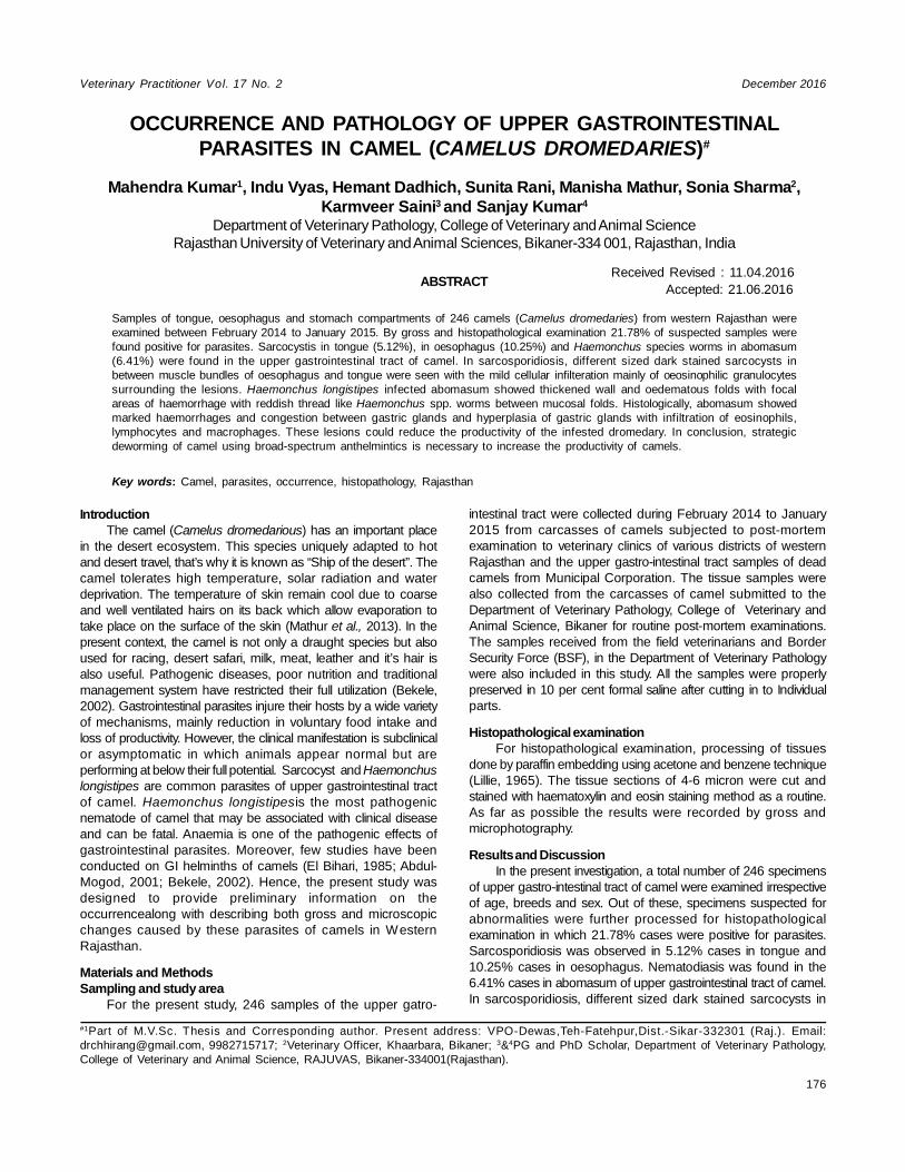
between muscle bundles of oesophagus were seen with themild cellular infilteration mainly of eosinophilic granulocytessurrounding the lesions (Fig. 1). Each sarcocyst was surroundedby thin layer of muscle fibres which contain numerous bradyzoites.
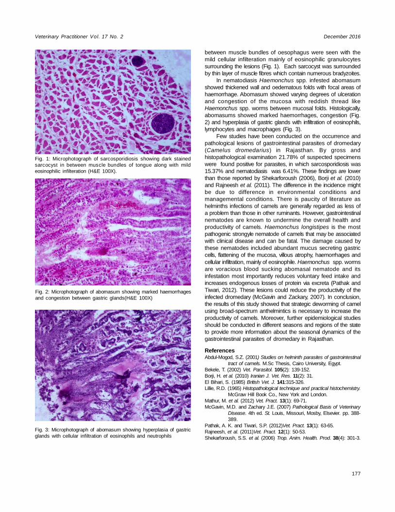
In nematodiasis Haemonchus spp. infested abomasumshowed thickened wall and oedematous folds with focal areas ofhaemorrhage. Abomasum showed varying degrees of ulcerationand congestion of the mucosa with reddish thread likeHaemonchus spp. worms between mucosal folds. Histologically,abomasums showed marked haemorrhages, congestion (Fig.2) and hyperplasia of gastric glands with infiltration of eosinophils,lymphocytes and macrophages (Fig. 3).
Few studies have been conducted on the occurrence andpathological lesions of gastrointestinal parasites of dromedary(Camelus dromedarius) in Rajasthan. By gross andhistopathological examination 21.78% of suspected specimenswere found positive for parasites, in which sarcosporidiosis was15.37% and nematodiasis was 6.41%. These findings are lowerthan those reported by Shekarforoush (2006), Borji et al. (2010)and Rajneesh et al. (2011). The difference in the incidence mightbe due to difference in environmental conditions andmanagemental conditions. There is paucity of literature ashelminths infections of camels are generally regarded as less ofa problem than those in other ruminants. However, gastrointestinalnematodes are known to undermine the overall health andproductivity of camels. Haemonchus longistipes is the mostpathogenic strongyle nematode of camels that may be associatedwith clinical disease and can be fatal. The damage caused bythese nematodes included abundant mucus secreting gastriccells, flattening of the mucosa, villous atrophy, haemorrhages andcellular infiltration, mainly of eosinophile. Haemonchus spp. wormsare voracious blood sucking abomasal nematode and itsinfestation most importantly reduces voluntary feed intake andincreases endogenous losses of protein via excreta (Pathak andTiwari, 2012). These lesions could reduce the productivity of theinfected dromedary (McGavin and Zackary, 2007). In conclusion,the results of this study showed that strategic deworming of camelusing broad-spectrum anthelmintics is necessary to increase theproductivity of camels. Moreover, further epidemiological studiesshould be conducted in different seasons and regions of the stateto provide more information about the seasonal dynamics of thegastrointestinal parasites of dromedary in Rajasthan.
ReferencesAbdul-Mogod, S.Z. (2001) Studies on helminth parasites of gastrointestinal
tract of camels. M.Sc Thesis, Cairo University, Egypt.Bekele, T. (2002) Vet. Parasitol. 105(2): 139-152.Borji, H. et al. (2010) Iranian J. Vet. Res. 11(2): 31.El Bihari, S. (1985) British Vet. J. 141:315-326.Lillie, R.D. (1965) Histopathological technique and practical histochemistry.
McGraw Hill Book Co., New York and London.Mathur, M. et al. (2012) Vet. Pract. 13(1): 69-71.McGavin, M.D. and Zachary J.E. (2007) Pathological Basis of Veterinary
Disease. 4th ed. St. Louis, Missouri, Mosby, Elsevier. pp. 388-389.
Pathak, A. K. and Tiwari, S.P. (2012)Vet. Pract. 13(1): 63-65.Rajneesh, et al. (2011)Vet. Pract. 12(1): 50-53.Shekarforoush, S.S. et al. (2006) Trop. Anim. Health. Prod. 38(4): 301-3.
Fig. 1: Microphotograph of sarcosporidiosis showing dark stainedsarcocyst in between muscle bundles of tongue along with mildeosinophilic infilteration (H&E 100X).
Fig. 2: Microphotograph of abomasum showing marked haemorrhagesand congestion between gastric glands(H&E 100X)
Fig. 3: Microphotograph of abomasum showing hyperplasia of gastricglands with cellular infiltration of eosinophils and neutrophils


![LCD-Array Kit MEAT 5.0 - Specificity - CHIPRON GmbH · Donkey: ACD-005-025 Goat: ACD-006-025 Camel: ACD-007-025 Buffalo: ACD-008-025 [Equus asinus ] [Capra hircus ] [Camelus dromedarius](https://img.pdfslide.net/doc/110x75/60608c3fab6e5a6d06647729/lcd-array-kit-meat-50-specificity-chipron-gmbh-donkey-acd-005-025-goat-acd-006-025.jpg)