Embed Size (px)
Citation preview

International Journal of Agricultural Technology 2013, Vol. 9(1): 151-164
151
Occurrence of toxigenic fungi and mycotoxins in some legume
seeds
Embaby, E.M.1*
, Mohamed Reda
2, Mosaad A. Abdel-Wahhab
3, Hassan
Omara2
and Asmaa M. Mokabel4
1Department of Plant Pathology, National Research Center, Dokki, Cairo, Egypt ,
2Department
of Botony, Faculty of Science, Benha University, 3Department of Food Toxicology &
Contaminants, National Research Center, Dokki, Cairo, Egypt, 4Agoza Hospital, Agoza, Giza,
Egypt
Embaby, E.M., Mohamed Reda, Mosaad A. Abdel-Wahhab, Hassan Omara and Asmaa M.
Mokabel (2013) Occurrence of toxigenic fungi and mycotoxins in some legume seeds.
International Journal of Agricultural Technology 9(1):151-164.
Abstract The current research as conducted to study the natural occurrence of toxigenic fungi
and mycotoxins contamination in three legume seeds (i.e. beans, pea and soybean) in great
Cairo governorate. The results indicated that four fungal genera were isolated from the
examined seeds. These isolated fungi included Aspergillus flavus, A. niger, A. parasiticus,
Fusarium moniliforme, F. oxysporum, Fusarium spp., Penicillium spp and Sclerotinia
sclerotiorum. Soybean seeds were found the higher percentage of fungal infection followed by
pea and beans seeds. Aspergillus niger was the common in beans and soybean, followed by A.
parasiticus. Whereas, A. parasiticus was the common in pea, followed by Fusarium spp. S.
sclerotiorum was found to be the lowest in all examined seeds. On the other hand, A.
parasiticus and F. moniliforme were capable to produce aflatoxins and fumonisin in significant
concentrations exceed the permit levels recommended by the Egyptian authorities. The fungal
infection with A. parasiticus, F. moniliforme decreased the chemical components of the tested
seeds (i.e. protein, fat, carbohydrates and ash). Furthermore, moisture content was found to be a
causative factor in fungal infection. It could be concluded that fungal infection of legume seeds
reduced its nutritive value as well as induced a health risk for the consumer.
Key words: Legume, beans, pea, soybean, fungi, mycotoxins, chemical components.
Introduction
Legumes "Fabaceae" is one of the most important plant in Egypt for local
consumption and exportation. Legumes are generally good sources of slow
release carbohydrates and are rich in proteins. Legumes are normally consumed
after processing, which not only improves palatability of foods but also
increases the bioavailability of nutrients, by inactivating trypsin and growth
* Corresponding author: Embaby, E.M.; e-mail: [email protected]
International Journal of Agricultural Technology 2013 Vol. 9(1): 151-164
Available online http://www.ijat-aatsea.com ISSN 2630-0192 (Online)

152
inhibitors and haemaglutinins (Tharanathan and Mahadevamma, 2003). It is
the most important source of plant protein in human food. Several fungi attack
the legume plants during growth, harvest and storage. While more than 25
different species of fungi are known to invade stored grains and legumes (Duan
et al., 2007), species of Aspergillus, Penicillum and Fusarium are responsible
for most spoilage and germ damage during storage. They cause reduction in
cooking or baking quality, and nutritive values, produce undesirable odors and
color, and change appearance of stored food grade grains and decrease
germinibility and total decay (Quenton et al., 2003 and Castillo et al., 2004). In
addition, they produce mycotoxins those are health hazard for man and animals,
make products unacceptable for edible purposes or lower their market grade.
Moreover, fungal infestation of seed coat decreases viability of seeds, or may
cause abnormal seedlings (Selcuk et al., 2008).
A large number of fungal species regularly associated with seeds and can
infect developing seeds and still attached to the mother plant (Neergaard, 1979,
Agrwal and Sinclair, 1993 and Mathur and Olga 2003). This has been
demonstrated by the isolation of fungi from seeds collected before seed-set.
Many of these fungi have no negative impact on seeds but there are also
many saprophytic and pathogenic fungi commonly isolated from seeds (Schafer
and Kotanen, 2004). These include the mainly saprophytic genera Mucor,
Rhizopus, Trichoderma, Cladosporium, Penicillium, Chaetomium and
Aspergillus as well as the mainly pathogenic genera Pythium and Alternaria.
Finally, Fusarium, Acremonium and Phoma contain both saprophytes and
pathogens (Schafer and Kotanen, 2004) While the fungal pathogens of growing
plants are comparatively well-investigated (Friberg et al., 2005), the knowledge
on fungal seed decay and its importance for plant demographic and community
processes is quite limited (Blaney and Kotanen, 2001). Five fungal genera i.e.
Alternaria, Aspergillus, Epicoccum, Fusarium and Trichoderma were isolated
from some legume seeds as beans, cowpea, and lupine (Embaby and Mona,
2006).
In recent years, there has been a notable increase in the occurrence of
chronic diseases caused by the consumption of food products contaminated
with mycotoxins (U.S. FDA/CFSAN, 2001). Mycotoxins are secondary
metabolites produced by toxigenic fungi in contaminated foods. Aflatoxins and
fumonisin are the most dangerous mycotoxins in tropical areas. They are
produced, respectively, by species of the genera Aspergillus and Fusarium
(Konietzny and Greiner, 2003). Regarding legumes in Egypt, very little
information exists with respect to its natural contamination with toxigenic fungi
and mycotoxin. Aflatoxin(s) were detected in some Aspergillus isolates while
Fumonisin was detected in some Fusarium isolates (Embaby and Mona, 2006).
International Journal of Agricultural Technology 2013, Vol. 9(1): 151-164
153
The main toxigenic species identified were Aspergillus flavus, A. fumigatus,
Fusarium graminearum and F. culumorum in all cereals and F. verticillioides
in maize (Tabuc et al., 2009).
Changes in the protein, reducing and non-reducing sugars were observed
in cowpea seeds infected with either A. nidulants and A. tereus (Maheshwari
and Mathur, 1987). Chemical composition (protein, lipid, carbohydrate, crude
fibre) of sesame and soybean seeds were influenced by A. flavus growth (Farag,
1990). Invasion of seeds by some pathogens may result in biochemical
deterioration and change in qualitity of seed nutrient as infected in soybean
seed with A. flavus (Agrwal and Sinclair, 1993). Fusarium moniliforme
decreased with time with increase in the relative humidity. Protein, total and
reducing sugar contents decreased gradually with increase in the RH values
(Lokesh and Hiremath, 1993). There was an increase in moisture content,
reduction in the fat and decrease in the available carbohydrates in all grain
cowpeas analyzed. Similarly, the energy content showed a significant (p<0.05)
decrease in all the grains (Kungu et al., 2003). Aspergillus flavus decrease
lipids and carbohydrate contents of wheat, soybean and faba-bean seeds. A.
flavus utilizes carbohydrates of seeds for its growth and aflatoxin production
(Aziz and Mahrous, 2004). The aim of the current study was to isolate and
identify the toxigenic fungi associated with some legumes included beans
(Phaseolus vulguris L.), pea (Pisum sativum L.) and soybean (Glycine max L.),
the ability of these fungi to produce mycotoxins and the effect of Aspergillus
parasiticus and Fusarium moniliforme on chemical content of seeds.
Materials and methods
Samples: Thirty samples of legume seeds, beans (Phaseolus vulguris L.),
pea (Pisum sativum L.) and soybean (Glycine max L.) were collected from the
local markets at great Cairo Governorates, Egypt.
Isolation: Purification and identification of all fungal association were
done. Seed samples were tested using two standard methods of isolation (i.e.
agar plate and blotter tests) as described by Neergaard (1979), Agarwal and
Sinclair, (1993) and and Mathur and Olga (2003). Seed samples were divided
into two groups; the first group was disinfected with sodium hypochlorite
solution (1%) for 2 min, while the second group was untreated (non-
disinfected). All seed samples were washed several times by sterilized water
(SW), then dried between two sterilized filter papers and plated on potato
dextrose agar (PDA) and/or in sterilized filter papers with enough moisture
(blotter test) in sterilized Petri dishes. Five seeds/dish and three dishes were
used as replicates for each treatment. All dishes were incubated for 5-7 days at
25 + 2°C. All fungal growth was transferred and purified using hyphal tip

154
and/or single spore techniques onto PDA medium in the presence of antibiotic
(Streptomycin). Developing fungi were cultured on PDA slants (5-7 days old)
then identified at Department of Plant Pathology, National Research Centre
(NRC), El-Dokki, Egypt based on cultural characteristics using specific media
and the available of literature according to Raper and Funel (1965) and Maren
and Johan (1988) for Aspergillus, Booth (1977) and Nelson et al. (1983) for
Fusarium, and Barent and Hunter (1977) for the genera of imperfect fungi and
Singh et al. (1991) for Aspergillus, Fusarium and Penicillium.
Mycotoxin production: Each isolate of Aspergillus and Fusarium was
grown in 500 ml flask containing 100 g of each autoclaved legume seeds with
enough moisture and incubated at 25 OC for 14 days for Aspergillus and 21
days for Fusarium isolates. The incubated seeds were extracted for aflatoxins
and fumonisins according to the method described by AOAC (2007).
Mycotoxins determination: Mycotoxins were determined at Department
of Food Toxicology and Contamination, National Research Centre (NRC).
Aflatoxins and fumonisin were determined by HPLC according to the methods
described by Hustchins and Hagler (1983) for aflatoxins and Shephard et al.
(1990) for fumonisin respectively.
Effect of Aspergillus and Fusarium on chemical content of seeds: The
chemical content (i.e. protein, carbohydrate, ash and moisture) of the inoculated
and control legume seeds were determined as described by AOAC (2007). The
results were calculated as percentage of losses or reduction in the infected seeds
compared to the control seeds.
Results
The results of the total fungal count (TFC), germination and the
percentage of infection for the three tested legume seeds using the two standard
methods (Blotter and PDA) as presented in Table 1. These results indicated that
the blotter method exhibited TFC and infection percentage in the non
disinfected and disinfected pea seeds that was higher than beans and soybean
seeds.
On the other hand, in PDA method, TFC and percentage of infection in
disinfected beans and pea seeds were the same and higher than soybean seeds,
whereas the higher TFC was found in non disinfected pea followed by
disinfected beans than soybean. Also the results showed thatm agar plate (PDA
medium) was better than blotter test which gave higher percentage of
germinated seeds. Germination of disinfected phaseolus seeds gave 53% in
blotter test and 73% in PDA medium while non-disinfected seeds gave 27% in
blotter and 53% in PDA methods, 20 and 27% were the results of infection
percent in disinfected and non-disinfected beans (phaseolus) seeds with blotter

International Journal of Agricultural Technology 2013, Vol. 9(1): 151-164
155
test and 80 and 87% of infection percent in disinfected and non-disinfected
seeds with agar plate method respectively.
Germination of disinfected and non-disinfected pea seeds resulted in 33
and 13 % in blotter test and 93 and 73 % of germination in disinfected and non-
disinfected pea seeds with PDA medium. The infection percent of disinfected
and non-disinfected pea seeds with blotter test were 67 and 80 % comparing
with 80 and 100 % with PDA test respectively.
On the other hand, the percentage of germinated soybean seeds resulted
in 12 and 13% with blotter test compared with 40 and 40 % of germination in
PDA test method with disinfected and non-disinfected seeds respectively.
Infection percentage of disinfected and non-disinfected soybean seeds showed
7 and 27 % with blotter test and 60 and 67% with PDA respectively.
Table 1. Total count(s), germination and infection percentage of some
disinfected and non disinfected legume seeds on blotter and agar plate methods
Seed
cro
ps
Blotter method PDA
Disinfected Non-disinfected Disinfected Non-disinfected
G I G I G I G I
N.G
% T.C
% N.G
% T.C
% N.G
% T.C
% N.G
% T.C %
Beans 8 53 3 20 4 27 4 27 11 73 12 80 8 53 13 87
Pea 5 33 10 67 2 13 12 80 14 93 12 80 11 73 15 100
soybean 3 20 1 7 2 13 4 27 6 40 9 60 6 40 10 67
G = Germination N.G = Number of Germinated seeds I = Infected seeds T.C= Total count
of fungi in 15seeds (5seeds/dish x 3 replicates)
Results indicated that the frequency of A. falvus was the most prominent
fungi in beans and pea seeds whereas, A. niger was the most prominent in
soybean seeds. Fusarium moniliforme was the lowest fugus found in beans and
soybean seeds while; Sclerotinia sclerotiorum was the lowest fungal isolates
that infected pea seeds as shown in Table 2 and Figs.1, 2 and3.
Fig. 1. a-Fusarium associated with non disinfected and disinfected beans seeds (blotter test
and PDA method). b-Aspergillus flavus with non disinfected beans seed.
a b

156
Fig. 2. a-Sclerotinia sclerotiorum and sclerotia associated with disinfected pea seeds (blotter
test). b-Fusarium with disinfected pea seeds.
Fig. 3. a- Aspergillus niger and A. flavus associated with disinfected soybean seed, b-
Penicillium sp. with bean seed.
Table 2. Frequency of some legume seed-borne Fungi
% Total Soybean Pea Beans Fungi
% T.C % T.C % T.C
21.3 27 7.8 10 7.1 9 6.3 8 Aspergillus
flavus 24.4 31 14.2 18 3.2 4 7.1 9 A. niger 8.7 11 3.9 5 1.6 2 3.2 4 A. parasiticus 4.7 6 0.8 1 3.2 4 0.8 1 Fusarium
moniliforme 11.0 14 5.5 7 1.6 2 3.9 5 F. oxysporum 15.7 20 4.7 6 4.7 6 6.3 8 Fusarium. spp 9.5 12 4.7 6 0.8 1 3.9 5 Penicillium spp
4.7 6 0.0 0 4.7 6 0.0 0 Sclerotinia
sclerotiorum
100% 127 41.7 53 26.8 34 31.5 40 Total
T.C = Total count of fungi.
a b
b a

International Journal of Agricultural Technology 2013, Vol. 9(1): 151-164
157
Results showed that mycotoxins concentration was produced by the
toxigenic fungi which isolated from legume seeds are presented in Table 3. It is
clearly demonstrated that both A. parasiticus (No.59) isolated from beans seeds
and F. moniliforme (No.8) isolated from soybean had the ability to produce
mycotoxins in significant concentrations. The total aflatoxins concentration was
196.58 µ/kg whereas, fumonisin concentration was198 mg/kg seeds. It is of
interest to mention that A. flavus isolated from the legume seeds was not able to
produce aflatoxin. (Fig 5. a,b) showing HPLC chromatogram for aflatoxin and
fumonisin. HPLC Chromatograph of Aflatoxins sample showing that, AFB1
eluted at 10.8 , AFB2 at 21.4, AFG1 at 9.0 and AFG2 at 14.3 min. HPLC
Chromatograph for Fumonisin showing that AFB1 eluted at 6.957min.
Table 3. Concentration of mycotoxins production by the toxigenic A.
parasiticus and F. moniliforme isolated from some legume seeds
Aflatoxins (µ/kg)
Fumonisins
(mg/kg)
AFB1 AFB2 AFG1 AFG2 Total
44.74 1.4 15.24 135.2 196.58 198
AFB1, 2 = Aflatoxin B1, B2
AFG1, 2 = Aflatoxin G1, G2
Fig. 5a. HPLC Chromatograph for Aflatoxins.

158
Fig. 5b. HPLC Chromatograph for Fumonisin.
The results showed that chemical composition of Aspergillus parasiticus
contaminated and healthy legume seeds are presented in Table 4. It indicated
that A. parasiticus infection resulted in 8.7, 2.0 and 3.4 % loss in protein
content in beans, pea and soybean respectively. Whereas, the loss in
carbohydrates reached 20.38, 18.0 and 6.1% for beans, pea and soybean
respectively. The loss in fat content in the same seeds reached 20.4, 2.29 and
11.9% meanwhile; the loss in ash reached 3.8, 2.7 and 4.74% for the same
seeds respectively. The main factor in A. parasiticus infection was found to be
higher in the infected seeds compared to the healthy seeds. These percentages
of moisture content in the infected seeds recorded 37.4, 52.7 and 28.8% which
higher than the healthy beans, pea and soybean respectively.
Table 4. Effect of infection with Aspergillus parasiticus on the chemical
composition of legume seeds
Seed crops Beans Pea Soybean
Chemical
composition
H I L% R% H I L% R% H I L% R%
Protein 31.1 22.4 8.7 27.9 32.2 30.2 2 6.2 45.0 41.6 3.4 7.5 Carbohydrate 38.7 18.32 20.38 52.6 30.95 12.88 18.0 58.3 30.7 24.6 6.1 19.86
Fat 21.9 1.5 20.4 93.1 3.99 1.7 2.29 57.3 22.8 10.9 11.9 52.1
Ash 7.2 3.4 3.8 52.7 5.0 2.3 2.7 54 6.1 1.36 4.74 77.7 Moisture 8.49 45.9 37.4 81.4 8.43 61.3 52.7 85.9 7.7 36.5 28.8 78.9
H= Healthy I= Infected L= Loss% R= Reduction%
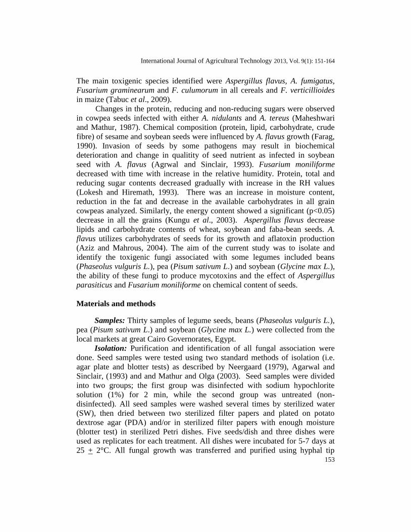
On the other hand, the effects of F. moniliforme infection on chemical
composition of legume seeds are presented in Table 5. It is clearly shown that
F. moniliforme infection also affected the nutritive values of the legume seeds.
The infection with this species resulted in the loss of the chemical components
International Journal of Agricultural Technology 2013, Vol. 9(1): 151-164
159
of beans, pea and soybean reached 4.3, 9.5 and 4.1% for protein, protein, 17.7,
13.59 and 5.5% for carbohydrates, 19.2, 1.09 and 6.3% for fat, 2.1, 0.8 and
0.8% for ash respectively. However, moisture content was found to be higher in
the infected seeds than the healthy seeds. These percentages reached 38.7,
43.87 and 22.4% higher in the infected seeds than the healthy beans, pea and
soybean respectively.
Table 5. Effect of infection with Fusarium moniliforme on the chemical
composition of legume seeds
Seed crops Beans Pea Soybean
Chemical
composition
H I L% R% H I L% R% H I L% R%
Protein 31.1 26.8 4.3 13.8 32.2 22.7 9.5 29.5 45.0 40.9 4.1 9.1
Carbohydrate 38.7 21.0 17.7 43.7 30.95 17.36 13.59 43.9 30.76 25.25 5.5 17.9
Fat 21.9 2.7 19.2 87.6 3.99 2.9 1.09 27.3 22.8 16.5 6.3 27.8 Ash 7.2 5.1 2.1 29.1 5.0 4.2 0.8 16 6.1 5.3 0.8 13.1
Moisture 8.49 47.2 38.7 82 8.43 52.3 43.87 83.8 7.77 30.1 22.4 74.4
H= Healthy I= Infected L=Loss% R= Reduction %
Discussion
There are many fungi associated with legume seeds. In the current study,
both blotter method and PDA method showed that pea seeds were found to be
highly infected with different fungal species followed by beans and soybean.
TFC recorded in disinfected legume seeds were lower than the non disinfected
seeds in both applied methods. Four major fungal genera were isolated
including A. parasiticus , A. fiavus, A. niger, Penicillium spp., F. moniliforme,
F. oxysporum, Fusarium spp. and S. sclerotiorum. Similar to the current
observation, Pepeljnjak and Cvetnic (1986) reported that the frequency of
Penicillium spp. and Aspergillus spp. was 67 and 33%, respectively in beans
samples. Moreover, Tseng et al. (1995) indicated that F. oxysporum, Fusarium
spp., F. solani, Ascochyta pisi, A. pinodes (Mycosphaerella pinodes), Phoma
medicaginis var pinodella, Alternaria alternata, F. poae,, F. sporotrichioides,
F. sambucinum (Gibberrella putiearis), F. culmorum, F. avenaceum (G.
avenacea), F. equiseti, S. sclerotiorum, Botrytis cinerea and Rhizoctonia solani
were found in beans samples collected from Ontario and Taiwan. They reported
that the fungi most frequently isolated from the diseased Ontario beans were
Alternaria (51.1%) Fusarium (18.0%), Rhizoctonia (65.1%) Penicillium
(5.2%), Rhizopus (3.2%) Sclerotinia (3.0%), Gliocladium (2.2%) and Mucar
(1.7%), however, Aspergillus, Penicillium, Euriotium, Rhizopus and Nularia
were the most fungal isolates from diseased Taiwan beans which recorded 48.5,
6.7, 5.3 and 2.4% frequently respectively.

160
In the current study, PDA medium was found to be a better method than
blotter test. Moreover, the total fungal count isolated on PDA medium was
found to be higher in both disinfected and non disinfected seeds compared with
blotter test. Similar results were reported by Neergaard (1979), Agarwal and
Sinclair, (1993), Mathur and Olga (2003) and Kumud et al. (2004). Also, El-
Nagerabi et al. (2000) found that Aspergillus was the most common genus
isolate followed by Rhizopus, Alternaria, Fusarium, Emericella, Drechslera,
Cladosporium, Pencillium and Pythium. In the same concern, Rauf (2002)
isolated A. alternata, Ascochyta spp, Colletotrichum spp. Fusarium spp., and
Macrophomina phaseolina from major legume crops in Pakistan. Moreover,
Henning (2005) reported that the main seed-transmitted pathogens affecting
soybean are Phomopsis sp, Fusarium semitectum, S. sclerotiorum, Sclerotium
rolfsii, Aspergillus spp. A. flavus which cause germination problems and
mycotoxins accumulation.
On the other hand, percentage of seed germination with PDA medium
was found to be higher in both disinfected and non-disinfected seeds than
blotter test method. Also, percentage seed germination of disinfected legume
seeds were found to be higher than the non-disinfected seeds in both applied
methods. Similar results were reported by Neergaard (1979), Agarwal and
Sinclair, (1993) and Mathur and Olga (2003), Kumud et al., (2004) and
Embaby and Mona (2006). Pathogenic seed-borne fungi caused decreased in
the germination ability and emergence weight of 1000 seeds, plant healthiness,
number of yielding plants and seed yield ( Czyzewska, 1983).
In the present study, the isolated fungal genera were tested for their
ability to produce mycotoxins. A. parasiticus and F. moniliforme were found to
have the ability for aflatoxins and fumonisin production in significant
concentrations exceed the save limits recommended by the Egyptian authorities
(Embaby and Mona, 2006). It is well documented that aflatoxins have a
carcinogenic effects (I.A.R.C, 1993) whereas; fumonisin causes lipid
peroxidation, sphingolipid disturbances and developmental toxicity as well as
its role as a cancer promoter (Abdel-Wahhab et al., 2004). In this regards,
Tseng et al. (1995, 1996) reported that aflatoxin B1, B2, G1 and G2 and
fumonisins were found in the infected beans that collected from Taiwan.
Furthermore, Ruiz et al. (1996) and Vaamonde et al. (2003) reported that A.
flavus isolated from green beans and soybean had capacity synthesize
aflatoxins.
The effects of fungal infection on chemical components of legume seeds
were also investigated. Our results showed that infection with the tested fungi
reduced all chemical components in the legume seeds including protein,
carbohydrates, fat and ash consequently reduced the nutritive values of the

International Journal of Agricultural Technology 2013, Vol. 9(1): 151-164
161
infected seeds. The same result reported by Embaby and Mona (2006) and
these results were in accordance with those reported by Lokesh and Hiremath
(1993) and Ushamalini et al. (1998) they reported that, biochemical content of
seed-borne fungi were changed by A. flavus, A. niger, F. oxysporum and
Macrophomina phaseolina. Also, fungal infection resulted in the decrease in
protein, total and reducing sugar contents. The reduction of these chemical
components in the infected legume seed may be due to the utilization of these
components by the fungi in its growth (Azize and Mahrous 2004). Inoculated
lupine seeds with F. oxysporum f. sp. lupine resulted in a considerable decline
in soluble carbohydrates between 24 and 72h. (Morkunas et al., 2005).
Moisture content was found to be higher in the infected seeds compared
to the healthy seeds. These percentages reached 38.7, 43.87 and 22.4% higher
in the infected seeds than the healthy beans, pea and soybean respectively.
Similar results were reported by Embaby and Mona (2006) and many
investigators reported that, the increase in moisture content in the legume seeds
reported in the current study which considered the causative factor in the
infection rate. It is well documented that a higher seed moisture content
increased fungal infection particularly, by Aspergillus spp., of which A. flavus
and A. niger were predominant at higher and lower moisture contents
respectively (Maheshwari and Mathur, 1987). F. moniliforme decreased with
time with increase in the relative humidity. Protein, total and reducing sugar
contents decreased gradually with increase in the RH values (Lokesh and
Hiremath, 1993). There was an increase in moisture content, reduction in the
fat and decrease in the available carbohydrates in all grain cowpeas analyzed.
Similarly, the energy content showed a significant (p<0.05) decrease in all the
grains (Kungu et al., 2003).
It concluded that legume seeds collected from Cairo and Kalubia
Governorates were found to be infected with numerous fungal genera. Moisture
content was found to be the most important factor in fungal infection. The
isolated fungi were capable to produce mycotoxins in significant concentrations
exceed the save limits recommended by the Egyptian authorities and may cause
health risk for consumers. Moreover, the fungal infection resulted in the
decrease of the chemical components of the seeds and consequently reduces its
nutritive value.
References
AOAC (2007). Association of Official Analytical Chemists. Official Methods of Analysis of
AOAC International 17th
ed., Nature Toxins. AOAC International, Arlington, Virginia,
USA, Chapter pp. 49.
162
Abdel-Wahhab, M.A., A.M. Hassan; H.A. Amer and K.M. Naguib (2004). Prevention of
fumonisin-induced maternal and developmental toxicity in rats by certain plant extracts.
Journal of Applied Toxicology 24:469 -474.
Agrwal, K.V. and B.J. Sinclair, (1993). Principles of Seed Pathology. Vol. I, 176PP. and Vol.
II, 186 PP. First Indian Reprint Jai Bhawan. India.
Aziz, N.H. and S.R. Mahrous (2004). Effect of gamma irradiationon aflatoxin B1 production by
Aspergillus flavus and chemical composition of three crop seeds Nahrunig Wiely Vclt
Verlag GMBtt & Co. Kga A,Weinheim, Germany 48:234- 238.
Barent, H.L. and B. Hunter, (1977). Illustrated genera of imperefect fungi. Burgess Publishing
Company, Minnesota, pp. 2412.
Blaney, C.S. and P.M. Kotanen (2001). Effects of fungal pathogens on seeds of native and
exotic plants: A test using congeneric pairs. Journal of Applied Ecology 38: 1104–1113.
Booth, C. (1977). The genus Fusarium. First published. In commonwealth Mycological
Institute, Kew, Surrey, England pp. 235.
Castillo, M.D., H.H.L. Gonzulez; E.J. Martinez; A.M.Pacin and S.L. Resnik. (2004). Mycoflora
and potential for mycotoxin production of freshly harvested black beans from
Argentinean main production area. Mycopathologia. Kluwer Academic Publishers
Dorderecht, Netherlands, pp. 107-112.
Czyzewska, S. (1983). The effect of pathogenic seed-borne fungi on green pea (Pisum sativum)
emergence. Seed Research in Horticulture 188:289-299.
Duan, C.X.; X.M. Wang; Z.D. Zhu and X.F. Wu (2007). Testing of seedborne fungi in wheat
germ plasm conserved in the National Crop Genebank of China Agricultural Science
6:682–687.
El–Nagerabi, S.A.F.; A.M. EL-Shafi and A.H. Abdalla (2000). Composition of mycoflora and
aflatoxins in pea seeds from the Sudan. Kuwait journal of Science and Engineering
27:109-122.
Embaby, E.M. and Mona M.abdel-Galil (2006). Seed borne fungi and mycotoxins associated
with some legume seeds in Egypt. Journal of Applied Sciences Research 2(11):1064-
1071.
Farag, R.S. (1990). Effects of fungal infection and agrochemicals on the chemical composition
of some seeds and aflatoxin production (a review). Bulletin of Fac. Of Agric. Cairo
Univ. 41(1):43-61.
Friberg, H., J. Lagerlöf and B. Rämert (2005). Influence of soil fauna on fungal plant pathogens in
agricultural and horticultural systems. Biocontrol Science and Technology (15):641–658.
Henning, A. (2005). Seed pathology and treatment. Documentos Embrapa Soja, pp. 264.
Hustchins, J.E. and W.M.Jr Hagler (1983). Rapid liquid chromatographic determination of
aflatoxins in heavily contaminated corn. Journal of Association of Official Analytical
Chemistry (66):1458–1465.
I.A.R.C. (1993). Aflatoxins. In IRAC monographs on the evaluation of carcinogenic risks to
humans, vol.56, IRAC, Lyon, France, pp. 243-395.
Konietzny, U. and R. Greiner (2003). The application of PCR in detection of mycotoxigenic
fungi in foods. Brazilian Journal of Microbiology 34:1–58.
Kumud, K., Jitendra, S. and R. Ved (2004). Seed-borne fungi of Cowpea, their parasitism and
control. Annals of plant protein science. Society of Plant Protection Science, New Delhi,
India. Rev. 12:80-82.
Kungu, J.K., Muroki, N. and A. Omwege (2003). Effect of storage on the quality and safety of
grains in Tharaka District, Kenya. African Journl of Food Agriculture, Nutrition and
Development. 3: 2 unpaginated.

International Journal of Agricultural Technology 2013, Vol. 9(1): 151-164
163
Lokesh, M.S. and R.V. Hiremath (1993). Effect of relative humidity on seed mycoflora and
nutritive value of red gram. Mysore Journal of Agricultural-Science, Dharwad, India
27:268-271.
Maheshwari, R. and K. Mathur (1987). Changes in Lobia seeds due to some Aspergilli -
Alexandria Journal of Agricultural Research 32(2):289-293.
Maren, A.K. and I.P. Johan (1988). A Laboratory guid to the common Aspergillus spp. and
their teleomprph. Commnwealth Scientific and Industrial, pp. 116.
Mathur, S.B. and Olga Kongsdal (2003). Common Laboratory Seed Health Testing Methods
for Detecting Fungi. First edition, International Seed Testing Association Published,
425PP. e-mail: [email protected] http://www.seedtest.org
Morkunas, I., J. Marzak, Stachowiak and M. Stabiecki (2005). Sucrose induced lupine defense
against F. oxysporum: Sucrose stimulated accumulation of iso-flavonoids as a defense
response of lupine to F. oxysporum. Plant Physiology and Biochemistry. Elsevier SAS,
Paris, France 43(4):363-373.
Neergaard, P. (1979). Seed pathology. London: MacMillan Press Ltd. London and Basingstok,
UK. Associated Companies in New York, Dublin, Melbourne, Johannesburg and Mad
ran, pp. 1191.
Nelson Toussoun and Marasas (1983). Fusarium spp. An Illustrated Manual for Identification.
Published by the Pennsylvania state university. Press University Park and London.
Pepeljnjak, S. and Z. Cvetnic (1986). Mycological and Mycotoxicological contamination of
grains in a wide Anephropathic area of SR Croatia. Akademija nauka I umjetnosti Bosne
i Hercegovine
Quenton, K., A.S. Theresa F.O. Walter, P.R. Johon; V.D.W. Liana and S.S. Gardon (2003).
Mycoflora and fumonisin Mycotoxin Associated with Cowpea seeds. Journal of
Agricultural and Food Chemistry 51:2188-2191.
Raper, K.B. and D.I. Funel (1965). The genus Aspergillus Williams and Wilkins Baltimore.
U.S.A.
Rauf, B.A. (2002). Seed-borne disease problem of legume in Pakistan. Pakistan Journal of
Scientific and Industrial Reseasrch 43:249-254.
Ruiz; J.A., Bentabol, A. Gallego, R.C. Angulo and M. Jordal (1996). Mycoflora and aflatoxin-
producing strains of Aspergillus flavus in greenhouse-cultivated green beans .Journal of
Food Protection 59(4):433-435.
Schafer, M. and P.M. Kotanen (2004). The influence of soil moisture on losses of buried seeds
to fungi. Acta Oecologica 24:255–263.
Selcuk, M., L. Oksuz and P. Basaran (2008). Decontamination of grains and legumes infected
with Aspergillus spp. and Penicillum spp. by cold plasma treatment. Bioresource
Technology 99:5104–5109.
Shephard, G.S., E.W. Sydenham, P.G. Thiel and W.C.A. Gelderblom (1990). Quantitive
determination of fumonisins B1 and B2 by high-performance liquid chromatography with
fluorescence detection. Journal of Liquid Chromatography 13: 2077–2087.
Singh, K.; C. Jenz, U. Thron and S.B. Mathur (1991). An Illustrated of some Seed-born
Aspergilli, Fusaria, Penicillia and their mycotoxins. First edition, Danish Government
Institute of Seed Pathology for Developing Countries. Ryvangs Alle 78. DK-2900
Hellerup, Denmark and Department of Biotechnology.
Tabuc, C., D. Marin, P. Guerre, T. Sesan, and J.D. Bailly (2009). Mold and mycotoxin content
of cereals in southeastern Romania. Institute of Biology and Animal Nutrition 72(3):
662-665.
Tharanathan, R.N. and S. Mahadevamma (2003). Grain legumes-a boon to human nutrition.
Trends in Food Science and Technology 14:507–518.

164
Tseng, T.C., J.C. Tu. and S.S. Tzean (1995). Mycoflora and mycotoxin in dry beans produced in
Taiwan and in Orntario, Canada. Botanical Bulletin of Academic Sinica 36(4):229-234.
Tseng, T.C., T.C. Tu and C.C. Soo (1996). Comparison of the profiles of seedborne fungi and the
occurrence of aflatoxins in mould – damaged beans and soybeans Microbios 84:105-116.
Ushamalini, C., K. Rajappn, and G. Kousalya (998). Change in constituents of cowpea due to
seed-borne fungi. Indian Phytopathology 51(3):258-260.
U.S. FDA/CFSAN (2001). Fumonisin Levels in Human Foods and Animal Feeds. Final
Guidance for Industry. Center for Veterinary Medicine
Vaamonde, G., A. Patriarca, V.F. Pinto and R.C.C. Degrossi (2003). Laboratorio de
Microbiologı´a de Alimentos, Departamento deuımica Organica, Area Bromatologıa,
Facultad de Ciencias Exactas y Naturales, Universidad de Buenos Aires, Ciudad
Universitaria, Pabellon II, 3j Piso, 1428. Buenos Aires, Argentina International Journal
of Food Microbiology 88:79– 84.
(Received 29 February 2012 ; accepted 30 December 2012)