Embed Size (px)
Citation preview
REVIEW Open Access
Ocular toxocariasis: a neglected parasiticdisease in EgyptNagwa Mostafa El-Sayed1* and Nagham Gamal Masoud2
Abstract
Background: Ocular toxocariasis is considered a parasitic disease of major socioeconomic importance. In spite ofthe high prevalence of human toxocariasis (up to 84%) among Egyptian patients, the incidence of oculartoxocariasis is underestimated. The recognition of this neglected disease would be the initial step to overcome it.Thus, this review gave updated information on the pathogenesis, clinical manifestations, diagnosis, and treatmentof ocular toxocariasis.
Results: Ocular toxocariasis is an important cause of unilateral vision impairment mostly in children and always inthe differential diagnosis of retinoblastoma. This disease exhibits various manifestations such as posterior polegranuloma, peripheral granuloma, or chronic endophthalmitis. Diagnosis of ocular toxocariasis can be carried out bythe ophthalmic examination and immunodiagnostic methods to reveal the specific antibodies in serum and ocularfluids. In addition, molecular diagnosis, medical imaging techniques, and histopathologic observation of Toxocaralarva in the surgically obtained specimens can be performed. Ocular toxocariasis can be treated either medically orsurgically. Regarding medical treatment, the ophthalmologists prefer to use steroids and anthelminthic drugs;however, there are no standardized parameters for doses, duration, and route of administration.
Conclusion: Clinical suspicion plays a leading role in the diagnosis of ocular toxocariasis, but always with otherdiagnostic methods. Accurate diagnosis and prompt treatment can minimize ocular morbidity.
Keywords: Ocular toxocariasis, Pathogenesis, Clinical manifestations, Diagnosis, Treatment, Egyptian studies
IntroductionOcular parasitic infections cause a significant ocular mor-bidity not because they are non-treatable, but mostly due todelay or misdiagnosis, frequently from unawareness of theresulting diseases. The parasites attain the eye by means ofdirect invasion through trauma or surgery, via extensionfrom neighboring infected tissues, or via hematogenous dis-semination to the eye. Ocular lesions may be due to directdamage caused by the parasites or indirect pathologycaused by toxic products of parasites and also may be dueto the immune response to infectious parasitism. Variationin the disease spectrum depends on the geographical loca-tion, the hygienic conditions, the living and eating habits ofthe individuals, and the contact with animals (El-Sayed andSafar 2015).
Toxocariasis is a parasitic infection caused by invadingthe tissues by larval stages of Toxocara canis or Toxo-cara cati. The adult worms of these nematodesparasitize the small intestines of dogs and cats, respect-ively (Fillaux and Magnaval 2013). The human diseaseoccurs by ingesting the viable Toxocara embryonatedeggs, commonly from contaminated raw vegetables orfrom polluted water sources as well as from contact withdomestic dogs and cats, especially puppies and kittens,which harbor eggs in their fur (Holland 2017).According to Toxocara larvae migration through tissues,
human toxocariasis is classified into visceral, cerebral, ocular,and covert toxocariasis (El-Sayed and Ramadan 2017). Ocu-lar toxocariasis is an important cause of posterior and dif-fuse uveitis and constantly in the differential diagnosis ofretinoblastoma (Cortez et al. 2011). It is more frequent inchildren with an age group that ranges from 3 to12 years(Fomda et al. 2007; Azira and Zeehaida, 2011; Zibaei et al.2014). The adult patients were also affected by ocular toxo-cariasis mostly in Asian populations where eating of raw
© The Author(s). 2019 Open Access This article is distributed under the terms of the Creative Commons Attribution 4.0International License (http://creativecommons.org/licenses/by/4.0/), which permits unrestricted use, distribution, andreproduction in any medium, provided you give appropriate credit to the original author(s) and the source, provide a link tothe Creative Commons license, and indicate if changes were made.
* Correspondence: [email protected]; [email protected] Parasitology Department, Research Institute of Ophthalmology,Giza, EgyptFull list of author information is available at the end of the article
Bulletin of the NationalResearch Centre
El-Sayed and Masoud Bulletin of the National Research Centre (2019) 43:146 https://doi.org/10.1186/s42269-019-0185-8
meat is usual (Jee et al. 2016). In 90% of cases, ocular toxo-cariasis is unilateral (Rubinsky-Elefant et al. 2010). It hasbeen estimated that ocular toxocariasis results in 5 to 20%of blindness secondary to uveitis (Arevalo et al. 2013).In Egypt, toxocariasis occurs widely in the poor commu-
nities with low standard of hygiene and sanitation. Bothstray and domestic dogs and cats play a pivotal role in thetransmission of Toxocara species providing environmentalcontamination, which perpetuates the spreading of the in-fection among Egyptian populations. It was estimated thatsoil contamination with Toxocara eggs is up to 30%(Farghly et al. 2016). Several studies were conducted todetermine the prevalence of human toxocariasis among theEgyptian population with a significantly high proportion ofchildren because of their playing habits and hygienestandards. The seroprevalence of anti-Toxocara antibodieswas 10.7% among children with renal troubles (Nada et al.1996), 6% among children with hepatomegaly in ZagazigCity (Hassan et al. 1996), 6.2% among children with respira-tory symptoms or pyrexia of unknown origin, 18% amongadults with pyrexia of unknown origin in Tanta City (Anto-nios et al. 2008), 23.3% among adult schizophrenic patients(El-Sayed and Ismail 2012), 84% among asthmatic childrenin Mansoura City (El-Tantawy et al. 2013a), and 48.5% inchildren with cryptogenic epilepsy (El-Tantawy et al.2013b). In spite of the high prevalence of human toxocaria-sis (up to 84%) among Egyptian patients, the incidence ofocular toxocariasis is undetermined. The potential under-reporting of ocular toxocariasis is related to the wide vari-ation in its clinical manifestations and the complexity in itsdiagnosis which necessitates clinical, radiological, and im-munodiagnosis, many of which are not available for routinediagnostic service for suspected patients. Also, there aresome difficulties in the serological diagnosis concerningToxocara antigen production or the highly expensive com-mercial diagnostic kits which are utilized only for researchstudies. The consequence of ocular toxocariasis and the po-tential under-reporting cases, based on seroprevalencestudies, make ocular toxocariasis worthy of more medicalawareness (Hare and Franco-Paredes 2014). This reviewdiscussed the pathogenesis, clinical manifestations, diagno-sis, and treatment of ocular toxocariasis. The recognition ofthis parasitic disease would be the initial step to overcomethis neglected infection.
Pathogenesis of ocular toxocariasisOcular toxocariasis is caused by the migration of Toxocaralarvae into the posterior segment of the eye. Based on stud-ies executed in animal models, the migration route of Toxo-cara larvae may follow several pathways. The most commonone is via the brain to the cerebrospinal fluid and then tothe choroid. The other possible pathways are via the brainto the optic nerve or via the arteries from the internal ca-rotid artery to the ophthalmic artery, retinal central artery,
or ciliary artery (Hayashi et al. 2003). Migrated larvae secreteenzymes, waste products, and cuticular components, whichcause tissue damage, necrosis, and marked inflammatory re-action, with eosinophils as the most component. These eo-sinophils release toxic proteins which contribute to thepathology and symptomatology of toxocariasis. The typicalpathological finding in ocular toxocariasis is eosinophilicgranulomas with central necrosis and remnants of Toxocaralarva surrounded by a mixed inflammatory infiltrate withnumerous eosinophils (Rubinsky-Elefant et al. 2010; Fillauxand Magnaval 2013; Das et al. 2016).Ocular toxocariasis is usually due to a single Toxocara
larva invading the eye. This results in a low-level immuneresponse which is insufficiently activated to kill the larva andallows its persistence in the eye for years. During this longperiod, it can migrate through ocular tissues causing mech-anical and immunopathological damage (Magnaval et al.2001). Thus, the pathogenesis of ocular toxocariasis in thehuman depends on the inflammatory reactions activated bythe existence of larva in the eye, the host immune response,the number of the larvae present in the eye, the incidence ofreinfection, and the host sensitization to the secreted or ex-creted products by the larvae (Fan et al. 2013).
Clinical manifestations of ocular toxocariasisClinical manifestations and severity of ocular toxocariasisrely on the primary anatomical site implicated, the num-ber of the larvae existing in the eye, and the immune reac-tion of the host (Azira and Zeehaida, 2011). The mostcommon symptoms of ocular toxocariasis include photo-phobia, floaters, leukocoria, strabismus, white pupillary re-flex, bloodshot conjunctiva, mild ocular pain, vitreousinflammation, and blindness of one eye which is recordedin about 80% of cases and is permanent in the most pa-tients. Occasionally, the retinal symptoms may be associ-ated with those due to involvement of the central nervoussystem. These symptoms range for about 12months. Inyoung cases, the eye infection may not be observed untilthey fail a school vision screening test (Rubinsky-Elefantet al. 2010; Azira and Zeehaida 2011).Ocular toxocariasis exhibits various manifestations such
as posterior pole granuloma, peripheral granuloma, orchronic endophthalmitis, and occasionally, it may presentwith atypical manifestations.
Posterior pole granulomaIt represents 25–50% of cases in ages between 4 and 14years old. Posterior pole granuloma appears as an oval,white lesion in the posterior pole of the retina (Fig. 1).The cause of this predilection to the posterior pole hasbeen proposed by the hematogenous spread of larvaethat may lodge in small, perifoveal end arteries. In theacute stage, Toxocara retinochoroiditis manifests clinic-ally as a hazy, ill-defined white lesion with overlaying
El-Sayed and Masoud Bulletin of the National Research Centre (2019) 43:146 Page 2 of 8
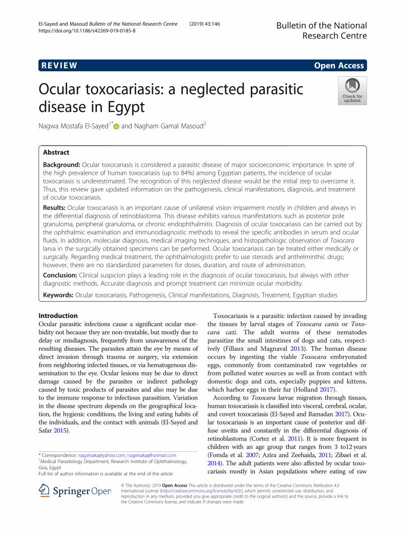
inflammatory cells in the vitreous. When the inflamma-tory reaction subsides, the lesion appears as a well-defined elevated mass ranging from one half to four discdiameters in size. A retinal pigment epithelium disturb-ance often surrounds the lesion with retinal folds ex-tending from the lesion (Fig. 1a) and epiretinalmembrane formation (Fig. 1b) (Singh et al. 2007; Ahn etal. 2014). Close examination may detect a dark gray cen-ter, which has been suggested to be remnants of thelarva as well as retinal blood vessels entering the granu-loma. In some cases, traction bands may extend fromthe lesion to the optic disc or to the macular area. In thecase of chronic granulomatous inflammation, large ret-inal vessels may infiltrate the mass and disappear into itssubstance, probably representing retinochoroidal anasto-mosis (Fig. 1c) (Cortez et al. 2011).The causes of vision loss may be by direct involvement
of the macula or optic disc, by the secondary formationof retinal folds or epiretinal membranes, or rarely by thedevelopment of choroidal neovascularization (Singh etal. 2007; Cortez et al. 2011; Ahn et al. 2014).
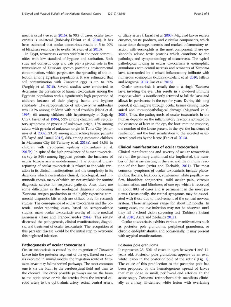
Peripheral granulomaOcular toxocariasis may present as an acute inflammationin the peripheral retina and ciliary body. About 20–40% ofthe infected eyes with toxocariasis manifest as a peripheralgranuloma. It is observed in cases with ages between 6and40 years old. The peripheral granuloma presents as ahazy, white, elevated mass in the peripheral fundus. It canbe associated with retinal folds that may extend from theperipheral mass to the optic nerve head or to other areasof the fundus. In some cases, the traction may lead toheterotopia of the macula (Fig. 2) (Cortez et al. 2011). Re-duced vision in patients with peripheral ocular toxocaria-sis is due to macular involvement by posteriorly extendingfalciform folds or exudate. In addition, amblyopia may de-velop in young patients with media opacities and/ormacular involvement (Singh et al. 2007).
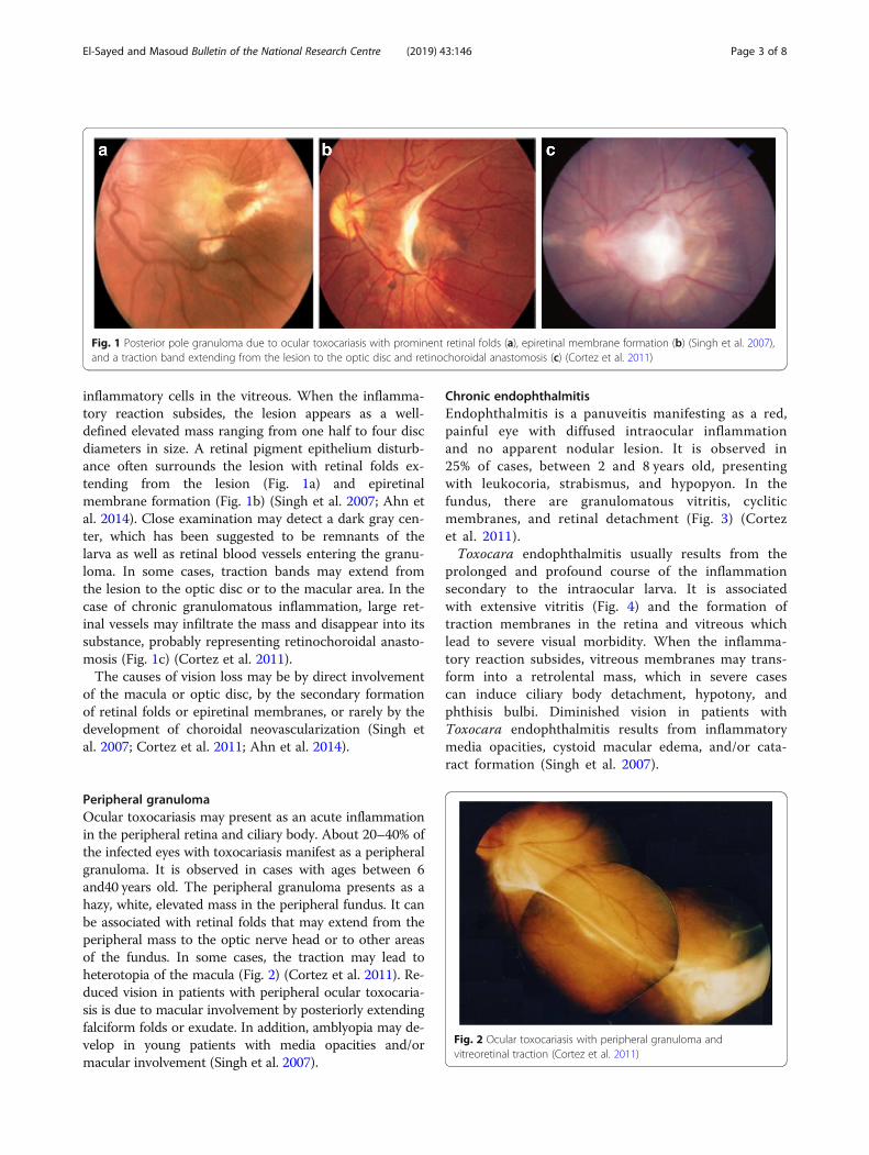
Chronic endophthalmitisEndophthalmitis is a panuveitis manifesting as a red,painful eye with diffused intraocular inflammationand no apparent nodular lesion. It is observed in25% of cases, between 2 and 8 years old, presentingwith leukocoria, strabismus, and hypopyon. In thefundus, there are granulomatous vitritis, cycliticmembranes, and retinal detachment (Fig. 3) (Cortezet al. 2011).Toxocara endophthalmitis usually results from the
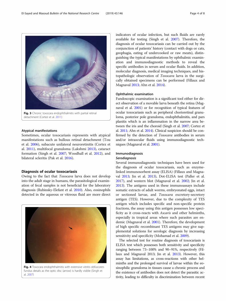
prolonged and profound course of the inflammationsecondary to the intraocular larva. It is associatedwith extensive vitritis (Fig. 4) and the formation oftraction membranes in the retina and vitreous whichlead to severe visual morbidity. When the inflamma-tory reaction subsides, vitreous membranes may trans-form into a retrolental mass, which in severe casescan induce ciliary body detachment, hypotony, andphthisis bulbi. Diminished vision in patients withToxocara endophthalmitis results from inflammatorymedia opacities, cystoid macular edema, and/or cata-ract formation (Singh et al. 2007).
Fig. 1 Posterior pole granuloma due to ocular toxocariasis with prominent retinal folds (a), epiretinal membrane formation (b) (Singh et al. 2007),and a traction band extending from the lesion to the optic disc and retinochoroidal anastomosis (c) (Cortez et al. 2011)
Fig. 2 Ocular toxocariasis with peripheral granuloma andvitreoretinal traction (Cortez et al. 2011)
El-Sayed and Masoud Bulletin of the National Research Centre (2019) 43:146 Page 3 of 8
Atypical manifestationsSometimes, ocular toxocariasis represents with atypicalmanifestations such as bullous retinal detachment (Touet al. 2006), subacute unilateral neuroretinitis (Cortez etal. 2011), multifocal granuloma (Lakshmi 2013), cataractformation (Singh et al. 2007; Woodhall et al. 2012), andbilateral scleritis (Pak et al. 2016).
Diagnosis of ocular toxocariasisOwing to the fact that Toxocara larva does not developinto the adult stage in humans, the parasitological examin-ation of fecal samples is not beneficial for the laboratorydiagnosis (Rubinsky-Elefant et al. 2010). Also, eosinophilsdetected in the aqueous or vitreous fluid are more direct
indicators of ocular infection, but such fluids are rarelyavailable for testing (Singh et al. 2007). Therefore, thediagnosis of ocular toxocariasis can be carried out by theconjunction of patients’ history (contact with dogs or cats,geophagia, eating of undercooked or raw meats), distin-guishing the typical manifestations by ophthalmic examin-ation and immunodiagnostic methods to reveal thespecific antibodies in serum and ocular fluids. In addition,molecular diagnosis, medical imaging techniques, and his-topathologic observation of Toxocara larva in the surgi-cally obtained specimens can be performed (Fillaux andMagnaval 2013; Ahn et al. 2014).
Ophthalmic examinationFundoscopic examination is a significant tool either for dir-ect observation of a movable larva beneath the retina (Mag-naval et al. 2001) or for recognition of typical features ofocular toxocariasis such as peripheral chorioretinal granu-loma, posterior pole granuloma, endophthalmitis, and parsplanitis which is an inflammation in the narrow area be-tween the iris and the choroid (Singh et al. 2007; Cortez etal. 2011; Ahn et al. 2014). Clinical suspicion should be con-firmed by the detection of Toxocara antibodies in serumand/or intraocular fluids using immunodiagnostic tech-niques (Magnaval et al. 2001).
ImmunodiagnosisSerodiagnosisSeveral immunodiagnostic techniques have been used forthe diagnosis of ocular toxocariasis, such as enzyme-linked immunosorbent assay (ELISA) (Fillaux and Magna-val 2013; Jin et al. 2013), Dot-ELISA test (Paller et al.2017), and western blot (Magnaval et al. 2002; Jin et al.2013). The antigens used in these immunoassays includesomatic extracts of adult worms, embryonated eggs, intactor sectioned larvae, and Toxocara excretory-secretoryantigen (TES). However, due to the complexity of TESantigen which includes specific and non-specific proteinfractions, the assay using this antigen possesses low speci-ficity as it cross-reacts with Ascaris and other helminths,especially in tropical areas where such parasites are en-demic (Magnaval et al. 2001). Therefore, the developmentof high specific recombinant TES antigens may give sup-plemental solutions for serologic diagnosis by increasingsensitivity and specificity (Mohamad et al. 2009).The selected test for routine diagnosis of toxocariasis is
ELISA test which possesses both sensitivity and specificityranging between 73–100% and 90–91%, respectively (Fil-laux and Magnaval 2013; Jin et al. 2013). However, thisassay has limitations, as cross-reactions with other hel-minths and the prolonged survival of larvae within the eo-sinophilic granuloma in tissues cause a chronic process andthe existence of antibodies does not detect the parasitic ac-tivity, leading to difficulty in discrimination between recent
Fig. 3 Chronic toxocara endophthalmitis with partial retinaldetachment (Cortez et al. 2011)
Fig. 4 Toxocara endophthalmitis with extensive vitritis obfuscatesfundus details as the optic disc (arrow) is hardly visible (Singh etal. 2007)
El-Sayed and Masoud Bulletin of the National Research Centre (2019) 43:146 Page 4 of 8
and chronic infection (Magnaval et al. 2001). Assays basedon the detection of IgG avidity have the ability to distin-guish between the active and chronic infection (Dziemianet al. 2008).Currently, the best choice serodiagnostic methods are
ELISA-IgG as a screening test and confirm the positivesamples by western blot test. Immunoblot analysis isusually used in research for separation and identificationof proteins. Western blot using crude antigen preparedfrom Toxocara canis larvae revealed antigenic proteinsof typical seven bands (24, 28, 30, 35, 132, 147, 200 kDa).The high specificity of this assay is related to its abilityto discriminate between high molecular weight bands(not specific and suggestive of cross-reactions with otherhelminths) and low molecular weight bands (24–35kDa), which have a high level of specificity (Magnaval etal. 2002; Jin et al. 2013).A remarkable issue when trying to evaluate serological
tests for human toxocariasis is that there is no referencetest or parasitological method to conclusively diagnosethis parasitic disease. Many authors observed that Toxo-cara antibodies may be undetectable or the titer may beless than the cutoff level in the sera of several patientshaving clinical manifestations of ocular toxocariasis (Ele-fant et al. 2006), probably due to the presence of lowparasite loads in them. Hence, the absence of serumantibodies does not exclude the diagnosis of ocular toxo-cariasis. In this condition, intraocular assay could be use-ful for confirming Toxocara infection diagnosis (DeVisser et al. 2008).
Intraocular assayIntraocular fluids include aqueous humor which is aclear liquid found in the space present between the cor-nea and the lens, and vitreous humor which is present inthe space between the lens and the retina. Detection ofspecific Toxocara antibodies in aqueous and/or vitreoussamples using different immunological methods assiststhe confirmation of ocular toxocariasis diagnosis (Fon-seca et al. 2019) and distinguishes it from retinoblastoma(De Visser et al. 2008). Some researchers reported casesof ocular toxocariasis via the detection of specific anti-Toxocara IgG antibodies only in aqueous and/or vitre-ous samples and not in sera (Fomda et al. 2007). Thesefindings underscore the hazards in diagnosing oculartoxocariasis based on the clinical and serological assaysalone. Therefore, it is recommended to detect Toxocaraantibodies via ELISA in both serum and aqueous/vitre-ous samples to increase the sensitivity of ocular toxocar-iasis diagnosis (De Visser et al. 2008).
Molecular diagnosisMolecular-based methods for Toxocara detection inclinical and environmental samples have been described
by several investigators (Fogt-Wyrwas et al. 2007; Tianand O’Hagan 2015). However, these methods are notwidely available as Toxocara organisms do not replicateinside the human host and sequestration of Toxocaralarvae within various tissues. Tian and O’Hagan (2015)reported a case of clinically suspected ocular toxocariasiswith a negative result via serological test; however, thediagnosis was proven by using polymerase chain reaction(PCR) on ocular fluids. Although the detection of Toxo-cara deoxyribonucleic acid (DNA) is a very sensitivemethod, it may be negative in patients who have a verylow larval burden, or if the larvae are sequestrated ordestroyed within granulomas and did not shed DNA-containing tissues into the aqueous or vitreous humor(Schneier and Durand 2011). Subsequently, recombinantDNA and mitochondrial markers can offer new insightfor the diagnosis of toxocariasis and may be helpful forepidemiological studies.
Diagnostic imaging techniquesMedical imaging techniques such as ocular ultra-sound, computed tomography, optical coherencetomography, fluorescein angiography, and magneticresonance imaging are used to detect granulomatouslesions related to Toxocara larva migration withinthe eye (Magnaval et al. 2001; Arevalo et al. 2013)and may be helpful in the differential diagnosis ofocular toxocariasis from other ocular diseases, par-ticularly retinoblastoma.Optical coherence tomography is a non-invasive im-
aging technique which uses coherent light to obtaincross-sectional images with high resolution. In oculartoxocariasis, posterior pole granuloma appears as ahighly reflective mass above the retinal pigment epithe-lium layer. Also, optical coherence tomography can re-veal the contributing factors which lead to vision loss, asthe presence of intraretinal and subretinal fluids(Hashida et al. 2014).B-scan ultrasonography uses the reflections of high-
frequency sound waves to construct a two-dimensional,cross-sectional view of the tissue. In toxocariasis, ultra-sonography can be utilized to reveal the optic discgranuloma, vitreous bands, retinal folds, and tractionalretinal detachment, and also, it is useful in ruling out thepresence of ocular tumor (Liu et al. 2017).
Histopathologic examinationIn surgically treated cases, the definitive diagnosis ofocular toxocariasis is via the histopathological identifica-tion of Toxocara larva or its fragments in the vitrectomyspecimens (Singh et al. 2007). Otherwise, the obtainingof biopsy materials from the eyes is difficult and risky.This method is a time-consuming, and occasionally, it isdifficult to identify Toxocara larvae due to their small
El-Sayed and Masoud Bulletin of the National Research Centre (2019) 43:146 Page 5 of 8

size and their extensive distribution (Rubinsky-Elefant etal. 2010; Fillaux and Magnaval 2013).
Treatment of ocular toxocariasisOcular toxocariasis can be treated either medically orsurgically. Treatment is usually based on the intensity ofsymptoms, the appearance of intraocular inflammation,the visual impairment, the macular involvement, and theoccurrence of ocular damage. It is noticeable that themost significant parameter of cure is the clinical re-sponse (Woodhall et al. 2012).
Medical treatmentThe goal of medical treatment is to prevent the oculardamage and visual loss. The choice of therapeutic medi-cations relies on the previous experience of the ophthal-mologist for toxocariasis treatment. Corticosteroids arethe mainstay medical treatment for ocular toxocariasisas they have the ability to decrease the release of localmediators of inflammation leading to the suppression ofinflammation, induce cell membranes stabilization, andprevent vitreous opacification and tractional retinal de-tachment. In spite of that, corticosteroids have limitedefficiency to deal with structural complications in theretina (Ahn et al. 2014). Corticosteroids are applied top-ically or infiltrated into periocular space, and/or givensystemically, based on the clinical condition. In mostcases, they result in notable amelioration (Cortez et al.2011). Oral prednisolone is the most commonly usedanti-inflammatory drug with an effective dosage of 1 mg/kg/day for a period of 1 month or more when needed;after that, the dose is reduced (Magnaval et al. 2001).Regarding antiparasitic medications, some ophthalmolo-
gists recommend anthelmintic using in addition to cortico-steroids while the others use these medications only whenthe response to corticosteroids is inefficient. Usage of theanthelmintic treatment for patients with ocular toxocariasismay induce an intraocular inflammation due to a hypersen-sitivity reaction to larval death inside the eye, leading to thepermanent damage of the eye (Schneier and Durand 2011).Anthelmintic treatment can be given, especially with thepresence of extraocular toxocariasis symptoms. Albenda-zole and diethylcarbamazine, the most commonly useddrugs, have larvicidal activity and are able to penetrate theblood-brain barrier. Other anthelmintic drugs such as thia-bendazole, mebendazole, and tinidazole are highly effectivein preventing the progression of Toxocara larvae to theneurotropic phase of infection (Magnaval et al. 2001;Schneier and Durand 2011). Seong et al. (2014) presented acase of ocular toxocariasis treated with albendazole (400mg twice daily) for 1month, and from day 13 of the treat-ment, oral triamcinolone was given. Also, Antonowicz et al.(2016) presented a case of a 6-year-old patient with steroid-dependent nephrotic syndrome having ocular toxocariasis.
It was observed that the lesions decreased after the treat-ment of the patient for 7 days with albendazole (15mg/kg/day) plus concomitant increase of prednisone dose to 1mg/kg/day.The effectiveness of anthelmintics in human toxocaria-
sis is difficult to determine because the comparativelysmall number of the treated cases, treatment is usuallystarted after varying lengths of the disease and differentresponses of both migrating and trapped Toxocara lar-vae. In addition, the immunopathological response maydiffer among patients and symptoms and signs mayundergo remission in treated and placebo-treated pa-tients. Therefore, the dose rates of drugs and length oftreatment differ among patients (Othman 2012).There is a new promising horizon of potential new
drugs for toxocariasis such as nitazoxanide, tribendimi-dine, and immunomodulators. Usage of glucan withbenzimidazoles gave better therapeutic effect in experi-mental animals (Othman 2012). Nowadays, naturalproducts and medicinal plants are under assessment forthe treatment of toxocariasis (Musa et al. 2011; El-Sayed2017). The value of such medicinal plants is related totheir therapeutic effect and their use as template mole-cules for the production of novel drugs.
Surgical managementSurgical interferences are required in cases with post-inflammatory complications such as vitreous opacification,retinal scars, bands, or detachment and formation of theepiretinal membrane with vitreomacular or optic nervetraction. Pars plana vitrectomy is the most common pro-cedure in ocular toxocariasis, especially for patients who donot respond to the medical treatment or have severe com-plications (Woodhall et al. 2012; Othman 2012). Successfuloutcome of surgery is obtained by inducing structuralmodification such as peeling of the membrane, removal ofthe vitreous opacification, or retinal reattachment whichleads to improvement in visual function (Magnaval et al.2001; Ahn et al. 2014). Surgery may preserve visual acuityin patients where fovea is not affected (Woodhall et al.2012; Othman 2012).Laser photocoagulation is recommended when Toxo-
cara larva is directly visualized inside the eye or in caseswith choroidal neovascular membrane. This proceduremay induce an inflammatory reaction; therefore, it needsto combine with steroid therapy (Azira and Zeehaida2011; Woodhall et al. 2012; Othman 2012). Cryotherapyis used to treat ocular granulomas. It is applied directlyto exudation parts at the pars plana by using a doublefreeze-thaw procedure followed by steroid therapy ad-ministration (Arevalo et al. 2013).In resistant cases of Toxocara endophthalmitis who are
not responsive to surgical or medical treatment, cyclo-sporine A may be effective. It is an immunosuppressive
El-Sayed and Masoud Bulletin of the National Research Centre (2019) 43:146 Page 6 of 8
drug and may relieve signs of uveitis with lower sideeffects. Local ocular injections result in more drug con-centration in the eye and prevent systemic adverse effects(Mora et al. 2006).
ConclusionIn Egypt, ocular toxocariasis is considered a neglected para-sitic disease. Research Institute of Ophthalmology (RIO) asa professional Egyptian institution should create websitesupport for Egyptian ophthalmologists and clinicians. Thiswebsite can be provided with standardized images of oculartoxocariasis lesions, diagnostic criteria, treatment regimens,and monitoring questionnaires to determine disease sever-ity and effectiveness of the treatment. By this manner,Egyptian ophthalmologists, clinicians, and epidemiologistscould use uniform protocols to determine the occurrenceof ocular toxocariasis and its prevalence more accurately. Inaddition, consciousness of the public population couldassist Toxocara parasite control in pets and animals andavoid the environmental contamination.
AcknowledgementsNot applicable
Authors’ contributionsNGM collected the scientific data and wrote the manuscript. NME-S revisedand edited the manuscript. All authors read and approved the finalmanuscript.
FundingNot applicable
Availability of data and materialsThe datasets used and/or analyzed during the current study are availablefrom the corresponding author on reasonable request.
Ethics approval and consent to participateNot applicable
Consent for publicationNot applicable
Competing interestsThe authors declare that they have no competing interests.
Author details1Medical Parasitology Department, Research Institute of Ophthalmology,Giza, Egypt. 2Faculty of Medicine, Ain Shams University, Cairo, Egypt.
Received: 29 May 2019 Accepted: 23 August 2019
ReferencesAhn SJ, Ryoo N-K, Woo SJ (2014) Ocular toxocariasis: clinical features, diagnosis,
treatment, and prevention. Asia Pac Allergy 4:134–141Antonios SN, Eid MM, Khalifa EA, Othman AA (2008) Seroprevalence study of
Toxocara canis in selected Egyptian patients. J Egypt Soc Parasitol 38:313–318Antonowicz A, Skrzypczyk P, Kępa B, Pańczyk-Tomaszewska M (2016) Ocular
toxocariasis in a boy with idiopathic nephrotic syndrome - a case report. PolMerkur Lekarski 41:192–195
Arevalo JF, Espinoza JV, Arevalo FA (2013) Ocular toxocariasis. J PediatrOphthalmol Strabismus 50:76–86
Azira NMS, Zeehaida M (2011) A case report of ocular toxocariasis. Asian Pac JTrop Biomed 1(2):164–165
Cortez RT, Ramirez G, Collet L, Giuliari GP (2011) Ocular parasitic diseases: areview on toxocariasis and diffuse unilateral subacute neuroretinitis. J PediatrOphthalmol Strabismus 48(4):204–212
Das D, Ramachandra V, Islam S, Bhattacharjee H, Biswas J, Koul A, Deka P, Deka A(2016) Update on pathology of ocular parasitic disease. Indian J Ophthalmol64:794–802
De Visser L, Rothova A, de Boer JH, van Loon AM, Kerkhoff FT, Canninga-van DijkMR, Weersink AY, de Groot-Mijnes JD (2008) Diagnosis of ocular toxocariasis byestablishing intraocular antibody production. Am J Ophthalmol 145(2):369–374
Dziemian E, Zarnowska H, Kołodziej-Sobocińska M, Machnicka B (2008)Determination of the relative avidity of the specific IgG antibodies in humantoxocariasis. Parasite Immunol 30(3):187–190
Elefant GR, Shimizu SH, Sanchez MC, Jacob CM, Ferreira AW (2006) A serologicalfollow-up of toxocariasis patients after chemotherapy based on thedetection of IgG, IgA, and IgE antibodies by enzyme linked immunosorbentassay. J Clin Lab Anal 20(4):164–172
El-Sayed NM (2017) Efficacy of Zingiber officinale ethanol extract on the viability,embryogenesis and infectivity of Toxocara canis eggs. J Parasit Dis 41(4):1020–1027
El-Sayed NM, Ismail KA (2012) Relationship between Toxocara canis infection andschizophrenia. Rawal Med J 37(2):155–161
El-Sayed NM, Ramadan ME (2017) Toxocariasis in children: an update on clinicalmanifestations, diagnosis and treatment. J Pediatr Infect Dis 12:222–227
El-Sayed NM, Safar EH (2015) Characterization of the parasitic induced lesions inthe posterior segment of the eye. Indian J Ophthalmol 63(12):881–887
El-Tantawy NL, El-Nahas HA, El-Assmy MM, Alsalem AM (2013a) Clinico-seroepidemiological evaluation of toxocariasis in asthmatic pediatric childrenin Mansoura city in Egypt. Arch Clin Microbiol 4:3
El-Tantawy NL, El-Nahas HA, Salem D, Salem N, Hasaneen BM (2013b)Seroprevalence of Toxoplasma gondii and Toxocara spp. in children withcryptogenic epilepsy. Am J Infect Dis Microbiol 1(5):92–95
Fan CK, Liao CW, Cheng YC (2013) Factors affecting disease manifestation oftoxocarosis in humans: genetics and environment. Vet Parasitol 193(4):342–352
Farghly AM, Mohamed SM, Abdel-Rahman SA, Mohammed FE, El-Bahaie ES, El-Shafey MA (2016) The relation between the prevalence of soil transmittedparasites in the soil and among school children in Zagazig district, SharkyiaGovernorate, Egypt. J Parasit Dis 40(3):1021–1029
Fillaux J, Magnaval JF (2013) Laboratory diagnosis of human toxocariasis. VetParasitol 193(4):327–336
Fogt-Wyrwas R, Jarosz W, Mizgajska-Wiktor H (2007) Utilizing a polymerase chainreaction method for the detection of Toxocara canis and T. cati eggs in soil. JHelminthol 81:75–78
Fomda BA, Ahmad Z, Khan NN, Tanveer S, Wani SA (2007) Ocular toxocariasis in a child:a case report from Kashmir, north India. Indian J Med Microbiol 25(4):411–412
Fonseca C, Silva AM, Freire S, Proença R (2019) Ocular toxocariasis: atypicalclinical course. BMJ Case Rep 12(4):e228717
Hare AQ, Franco-Paredes C (2014) Ocular larva migrans: a severe manifestation ofan unseen epidemic. Curr Trop Med Rep 1:69–73
Hashida N, Nakai K, Nishida K (2014) Diagnostic evaluation of ocular toxocariasisusing high-penetration optical coherence tomography. Case ReportsOphthalmol 5(1):16–21
Hassan MM, Farghaly AM, Gaber NS, Nageeb HF, Hegab MH, Galal N (1996) Parasiticcauses of hepatomegaly in children. J Egypt Soc Parasitol 26(1):177–189
Hayashi E, Akao N, Fujita K (2003) Evidence for the involvement of the opticnerve as a migration route for larvae in ocular toxocariasis of Mongoliangerbils. J Helminthol 77:311–315
Holland CV (2017) Knowledge gaps in the epidemiology of Toxocara: the enigmaremains. Parasitology 144(1):81–94
Jee D, Kim KS, Lee WK, Kim W, Jeon S (2016) Clinical features of oculartoxocariasis in adult Korean patients. Ocul Immunol Inflamm 24(2):207–216
Jin Y, Shen C, Huh S, Sohn WM, Choi MH, Hong ST (2013) Serodiagnosis oftoxocariasis by ELISA using crude antigen of Toxocara canis larvae. Korean JParasitol 51(4):433–439
Lakshmi KJB (2013) Multifocal granulomata in presumed Toxocara canis infectionin adult. World J Ophthalmol 3:38–41
Liu J, Li S, Deng G, Yang W, Chen W, Lu H (2017) Ultrasound biomicroscopicimaging in paediatric ocular toxocariasis. Br J Ophthalmol 101:1514–1517
Magnaval JF, Glickman LT, Dorchies P, Morassin B (2001) Highlights of humantoxocariasis. Korean J Parasitol 39:1–11
Magnaval JF, Malard L, Morassin B, Fabre R (2002) Immunodiagnosis of oculartoxocariasis using western-blot for the detection of specific anti-Toxocara IgG
El-Sayed and Masoud Bulletin of the National Research Centre (2019) 43:146 Page 7 of 8
and CAP for the measurement of specific anti-Toxocara IgE. J Helminthol76(4):335–339
Mohamad S, Azmi NC, Noordin R (2009) Development and evaluation of asensitive and specific assay for diagnosis of human toxocariasis by use ofthree recombinant antigens (TES-26, TES-30USM, and TES-120). J ClinMicrobiol 47(6):1712–1717
Mora P, Vecchi M, Barbera L, Toscani M, Orsoni JG (2006) Use of systemiccyclosporin A in a case of severe Toxocara uveitis. J Infect 52(5):e159–e161
Musa D, Senocak G, Borazan G, Altas M, Ozgonul A, Sogut O, Güldür ME (2011)Effects of Nigella sativa and albendazole alone and in combination inToxocara canis infected mice. J Pak Med Assoc 61:866–870
Nada SM, Abbaza BE, Mahmoud LA, Habeeb YS, Hussein HF, Amer OT (1996)Toxocariasis as a cause of renal disease in children in Sharkia Governorate,Egypt. J Egypt Soc Parasitol 26(3):709–717
Othman AA (2012) Therapeutic battle against larval toxocariasis: are we still farbehind? Acta Trop 124:171–178
Pak KY, Park SW, Byon IS, Lee JE (2016) Ocular toxocariasis presenting as bilateralscleritis with suspect retinal granuloma in the nerve fiber layer: a case report.BMC Infect Dis 16:426
Paller VGV, Besana CM, Valdez IKM (2017) Dot enzyme-linked immunosorbentassay (ELISA) for the detection of Toxocara infection using a rat model. JParasit Dis 41(4):933–939
Rubinsky-Elefant G, Hirata CE, Yamamoto JH, Ferreira MU (2010) Humantoxocariasis: diagnosis, worldwide seroprevalences and clinical expression ofthe systemic and ocular forms. Ann Trop Med Parasitol 104:3–23
Schneier AJ, Durand ML (2011) Ocular toxocariasis: advances in diagnosis andtreatment. Int Ophthalmol Clin 51(4):135–144
Seong S, Moon D, Lee DK, Kim HE, Oh HS, Kim SH, Kwon OW, You YS (2014) Acase of ocular toxocariasis successfully treated with albendazole andtriamcinolon. Korean J Parasitol 52(5):537–540
Singh A, Cunningham ET Jr, Stewart JM (2007) Detection and treatment of oculartoxocariasis. Rev Ophthalmol 14:55–58
Tian JX, O’Hagan S (2015) Toxocara polymerase chain reaction on ocular fluids inbilateral granulomatous chorioretinitis. Int Med Case Rep J 8:107–110
Tou N, Ibi K, Tawara A, Nakamura F, Hadeyama T (2006) A case of secondaryretinal detachment caused by ocular toxocariasis. Nippon Ganka GakkaiZasshi 110:415–420
Woodhall D, Starr MC, Montgomery SP, Jones JL, Lum F, Read RW, Moorthy RS(2012) Ocular toxocariasis: epidemiologic, anatomic, and therapeutic variationsbased on a survey of ophthalmic subspecialists. Ophthalmol 119:1211–1217
Zibaei M, Sadjjadi SM, Jahadi-Hosseini SH (2014) Toxocara cati larvae in the eyeof a child: a case report. Asian Pac J Trop Biomed 4(1):S53–S55
Publisher’s NoteSpringer Nature remains neutral with regard to jurisdictional claims inpublished maps and institutional affiliations.
El-Sayed and Masoud Bulletin of the National Research Centre (2019) 43:146 Page 8 of 8