Embed Size (px)
Citation preview
1 2017 Vol 3 Issue 1
President Message: Hunter Yuen Dear APSOPRS colleagues, It’s my greatest honor to take up the presidency for the Asia Pacific Society of Ophthalmic Plastic and Reconstructive Surgery APSOPRS. With the conjoint effort of all past presidents and councils, APSOPRS has been making great progress since its establishment in year 2000. Other than our regular biannual meeting, APSOPRS has been invited to give courses and lectures in a number of major international meetings such as WOC, AAO, APAO.…etc. A number of national oculoplastic societies in the Asia Pacific Region have been affiliated with APSOPRS. Few years ago, APSOPRS has been affiliated with ASOPRS and recently, APSOPRS is also affiliated with ESOPRS. I hope such affiliations can further enhance the collaboration between APSOPRS and other oculoplastic societies. Since Dr Hirohiko Kakizaki and his team took over the secretariat and established an online payment system, people can join APSOPRS as members and pay their membership fee more easily. The introduction of life membership has also allowed APSOPRS to enhance membership. Nevertheless, the APSOPRS account is still in Singapore, the secretariat is in Japan but the current president and president-elect are outside Japan. Certainly we can work by various electronic communication platforms, the current council will decide whether to stay with the current situation or explore other alternatives. One of my major tasks is to further enhance the member number; we will explore various ways to achieve this. In particular, our team will work with those Asia Pacific countries that have no or little members, and I hope we can get those places involved in our future activities. Team work is very important and the whole council will be working closely together. Specifically, our three vice presidents will be helping me to complete various tasks, Dr Kelvin Chong will help me to coordinate courses in various international meetings; Dr Gangadhara Sundar will be helping me for the membership issue and the connection with national oculoplastic societies; Dr Yasuhiro Takahashi will coordinate research in APSOPRS and recruit articles for iPlastic. Our treasurer Dr Sunny Shen will look after our account in Singapore and Dr Mary Rose Yan will be the secretary for the council.
APSOPRS President Yuen Kwok Lai Hunter (Hong Kong, SAR)
Immediate Past President Hirohiko Kakizaki (Japan)
President-elect Raoul Paolo D. Henson (Philippines)
APSOPRS Vice-Presidents Kelvin CHONG Kam Lung (Hong Kong, SAR) Gangadhara Sundar (Singapore)
Yasuhiko Takahashi (Japan)
Editor Audrey Looi (Singapore)
Editorial Board Ashok Grover (India)
Kelvin Chong (Hong Kong, SAR)
Yoon-Duck, Kim (South Korea)
Lily Li Dong Mei (China)
Raoul Henson (Philippines)
Sunny Shen (Singapore)
Official Newsletter of APSOPRS 2017 Volume 3 Issue 1
Asia-Pacific Society of Ophthalmic Plastic and Reconstructive Surgery

2 2017 Vol 3 Issue 1
We look forward to further development of the specialty of Oculoplastics in the Asia Pacific region with the active participation of all the members and affiliated national societies. The coming APSOPRS meeting will be held in Dec 2018 at Hong Kong, we hope you can mark your calendar and come to Hong Kong, which is one of the vibrant and dynamic city in the world. I would like to express my sincere gratitude to Dr Audrey Looi and the whole editorial team who has been working hard for this newsletter, and in particular for doing this again in the coming 2 years. I would like to thank the secretariat for their work in keeping up our society activities and website management. Last but not least, I would like to thank all APSOPRS members for the opportunity to serve the society, and I would like to appeal for your support for the upcoming activities.
Warmest regards, YUEN Kwok Lai Hunter MBChB, FRCOphth, FRCS(Ed), FCOphthHK, FHKAM (Ophthalmology), DipClinDerm (London) Clinical Associate Professor (Honorary), Department of Ophthalmology and Visual Sciences, The Chinese University of Hong Kong (CUHK) Consultant, HKEH
Editorial Note Dear friends and colleagues,
This issue of our iPlastic newsletter has been a pleasure to edit for the sole reason that, here in Singapore, we have finally achieved our aim of establishing our own Oculoplastic society, the Singapore Society of Ophthalmic, Plastic and Reconstructive Surgeons, SSOPRS. We began with social dinners and discussions to increase opportunities to get to know one another and our different perspectives better and gradually, the society took shape and achieved real form. Our first collaboration came in the recent formulation of Guidelines for Functional Ptosis Repair, Blepharoplasty and Browlifting procedures for the College of Ophthalmology and the Ministry of Health. We certainly look forward to further opportunities to work together to advance our sub-specialty.
In this issue, Dr Gangar Sundar and colleagues have contributed a useful piece on endoluminal nasolacrimal duct recanalization, detailing its history as well as the advantages and disadvantages. Given a propensity for repeat procedures, its popularity has not been widespread and its use limited to a few centres. Conventional dacryocystorhinostomy, whether external or endoscopic, remains the gold standard of care for patients with nasolacrimal duct obstruction. Patient selection and proper informed consent are key when this therapeutic option is offered. We also feature an eye-popping case of eyelash trichomegaly from Hong Kong and included a report from Japan concerning the rare manifestation of breast carcinoma in the eyelid. Dr Sunny Shen has also kindly contributed an educational piece on early and delayed orbital haemorrhage following orbital fracture repair.
In concluding, I would like to take this opportunity to thank our new President, Prof Hunter Yuen, for his shout out to all members to contribute scientific content for our newsletter. And would like to gently nudge all readers to send interesting reports so that all may benefit from this collaborative effort.
Best wishes,
Adj Assoc Prof. Audrey Looi MBBS, M.Med (Ophth), FRCS (Ed), FAMS Senior Consultant Oculoplastic Department Editor, APSOPRS Singapore National Eye Centre

3 2017 Vol 3 Issue 1
CONTENT PAGE Page Inaugural Singapore Society of Ophthalmic Plastic & Reconstructive Surgery (SSOPRS) 4 By Choo Chai Teck On the birth of a new Society By Geoffrey E. Rose Highlights from Singapore National Eye Centre Cadaveric Dissection Workshop 4-5 (28 Feb – 1 Mar 2017) and 32nd Asia Pacific Academy of Ophthalmology Congress (1 Mar – 5 Mar 2017) By Gillian Teh Case Highlights Dacryoendoscopy and Endoluminal Lacrimal Duct Recanalization (ELDR) – A perspective 9-11 By Gangadhara Sundar, Toru Suzuki, & Reynaldo Javate Unusual long curly eye lashes in a male with metastatic lung cancer 11-12 By Kwok Yuen Ting, & Yuen Kwok Lai Hunter
Eyelid Metastasis as the Primary Manifestation of Breast Carcinoma 13-14
By Yasuhiro Takahashi, Hirohiko Kakizaki Early and late orbital haemorrhage post orbital fracture repair 15-17 By Yong Kai-Ling, & Sunny Shen Guidelines 18 iPlastic e-Newsletter Paper Contribution Guidelines

4 2017 Vol 3 Issue 1
Inaugural Singapore Society of Ophthalmic Plastic & Reconstructive Surgery (SSOPRS)
Meeting 17 Feb 2017 SNEC Choo Chai Teck
Dear Friends and Colleagues It is my pleasure to announce the formation of SSOPRS to represent all our ophthalmic colleagues practicing oculoplastic surgery as their main subspecialty. The idea of having common subspecialty interest group started in NUH where the group first met in Aug 2013. Beginning as an oculoplastic interest group meeting which subsequently held regularly at colleagues’ residences ( Audrey, Jin Fong, Shawn, Geok Cheng), at Medical Alumni and SNEC. Many contributed in various ways to make the meeting fun and meaningful. Interesting, difficult and challenging cases were presented for discussion. Foods and drinks were generously put on the table by the hosts. The enthusiasm that fuelled the creation of society followed. The purpose of officially having a society is like forming a family with legitimate child. This is inevitably important as Singapore medicine advances. With the society, we would like to promote the interest of this subject (Eyelid / Lacrimal / Orbital / Oculofacial diseases) amongst the Ophthalmology community and to advance the science and art of ophthalmic plastic and reconstructive surgery in Singapore. We will enhance the collegiality between oculoplastic specialists and introduce junior specialists to senior colleagues of the field who can provide ready counsel and advice. We disseminate and share the knowledge and skills of oculoplastic surgery amongst members of other medical profession. Using this platform, we liaise with other national, regional and international oculoplastic societies, in the fields of education, research and other collaborations. Finally we always support our College of Ophthalmology oculoplastic subcommittee CME activities and play a crucial role in sharing the best oculoplastic practices. With the birth of a new Society, not only do we learn from each other, but also work toward creating a sense of belonging to this subspecialty community, and achieve the challenging tasks that lay ahead of us.
Let me thank all our founding members in advance in helping the creation of this family with everyone's views and support for this. I wish everyone the best of luck. CHOO Chai Teck
On the birth of a new Society Geoffrey E. Rose
Many thanks for the invitation to speak at the birth of a new Society, and it is a particular pleasure to reflect on the foundation of “SSOPRS” and congratulations to its first President, Dr Choo. Starting any society is like having a child: First the “idea” goes around for quite a while, with its worries and doubts; secondly, the “decision and start” tends to be rapid and all is underway before you know it (!!); then quite a long time goes by with much happening behind the scenes (or maybe, “under the bonnet”!) before – hey presto! – the baby is born! Is it a boy? Is it a girl? How much does she weigh? …..…. and then things go on, and on, and on, and on ………… Anyone who has had kids (or run a Society for that matter) knows that most of the hard work – the “ups” and “downs” – occur after the child is born. Like teenagers, some of the decisions will be seen as “contrary”, and sometimes they will seem totally irrational! However, stick in there! A family generally survives through thick and thin, through hard times and easy. So what helps a family survive – or, for that matter, a new Society? The answer is “love”, “understanding”, “mutual respect”, “tolerance” and – if you are a parent – probably a “sainthood” is rather helpful!!! The parent has to learn to deal with the child who announces, in anger, “I hate you!!” – as this is part of the normal childhood brain! It is no different for a Society: the father or mother must be both respected and respectful. The members of the Society must respect the President and his team – even if hard to agree with all decisions – and he or she must respect (and, above all, listen to) the members. Without leaves, the tree is nothing; without the members, the President and Council are naught. So why have a Society; indeed, why have a child when it seems such a precarious and fraught matter! The answer is clear: You learn from each other (both things to do, and things not to do); you get lots more done when you all pull in one direction (well, almost all – there are always 1 or 2 teenagers!!); and, especially, one can have the real fun that only comes from mutual respect and a common goal. There will be hard times and times of disagreement, but do not let these linger or fester! Most squabbles can be resolved by coming together as a family – preferably with a kindly parent, like Choo,

5 2017 Vol 3 Issue 1
to lead in a relaxed way. Above all, do not let professional jealousy get between you. Uncontrolled rivalry spoils everything -- the friendship, the free flow of knowledge, and the happiness that comes from working together; the happiness from the common aim. So, with a new Society like SSOPRS – stick together for protection, work together for growth, learn together for developing your minds, and – above all – play together for the fun of a family!! May I, as the current “father” for ESOPRS, congratulate you all on the birth of a new child (SSOPRS) and propose a toast to the health and growth of the new “baby”! May there be many more birthdays over the years to come ……… Geoffrey E. Rose After-dinner congratulatory speech at SNEC/APAO dinner, 1st March, 2017
Highlights from the Singapore National Eye Centre Cadaveric Dissection Workshop (28 Feb – 1 Mar 2017) and 32nd Asia-Pacific Academy of Ophthalmology Congress (1 Mar – 5 Mar 2017)
Organising Committee: Adjunct Associate Professor Audrey Looi, Dr Sunny Shen
Summary contributed by Dr Gillian Teh, Associate Consultant, Singapore National Eye Centre
Earlier this year, we had the privilege of welcoming participants from near and far to the Singapore National Eye Centre (SNEC) for our Cadaveric Dissection Workshop which was held just before the 32nd Asia-Pacific Academy of Ophthalmology Congress (APAO). The course was attended by eager young minds from South East Asia (Thailand, Malaysia, Indonesia, Myanmar, Philippines and Singapore), as well as slightly further afield from Bangladesh, Pakistan, Taiwan and Hong Kong. We even had participants from “Down Under” Australia – making us a truly intercontinental gathering of Ophthalmologists!
Dr Sunny Shen and Adj A/Prof Audrey Looi with participants from Thailand
We kicked off the meeting with the Cadaveric dissection workshop which was held over two days at the Academia Building on the Singapore General Hospital campus. Following that, the Oculoplastics program continued as part of the APAO congress at the Suntec Convention Centre.
The workshop started off with a series of lectures on Day 1 and the wetlab session on Day 2. We welcomed back to our shores Professors Geoffrey Rose and David Verity from Moorfields Eye Hospital in the United Kingdom, as lecturers in the pre- workshop course, and instructors in the cadaveric dissection segment. The last time these two giants were in town was for our OPRS Symposium in 2015, held in conjunction with the SNEC 25th Anniversary International Meeting. Needless to say, we were most excited to have them back to share their insights on orbital surgery.
We also had the great fortune of Professor Choi Woong Chul from Myoung Eye Plastic Surgery in Seoul, South Korea as a lecturer and instructor in our course. His expertise in blepharoplasty surgery and experience in dealing with problematic epicanthic folds proved a great source of wisdom for everyone.
Faculty dinner at the Majestic Restaurant after Day 1 of the cadaveric dissection course. Also the night Profs Rose and Verity

6 2017 Vol 3 Issue 1
tried durian during dessert (Prof Rose still reeling from the experience… judging from his expression) From L to R: Dr Yvonne Chung, Dr Morgan Yang, Adj A/Prof Seah Lay leng, Prof Choi Woong Chul, Prof Park Ju Wan , Prof Geoffrey Rose, Dr Hunter Yuen, Adj A/Prof Audrey Looi, Prof David Verity, Dr Choo Chai Teck, Dr Sunny Shen
Day 2 of the workshop saw our participants get into the thick of things with hands- on experience in the wet lab. The day commenced with Professors Rose and Verity showing us surgical approaches for tackling orbital lesions and tumours. These included the caruncular approach for medially sited lesions and a swinging eyelid approach for inferior orbital lesions. The spritely duo also showed the participants how to perform a “bone-swinging” lateral orbitotomy in dealing with trickier posterior lesions.
As per our previous cadaveric dissection course, we also had a panel of co-demonstrators at each dissection table to help guide the participants along. This included Dr Hunter Yuen, President of APSOPRS; Dr Sunny Shen; Dr Morgan Yang; Dr Yvonne Chung; Dr Cheng Jin Fong; Dr Llewellyn Lee; Dr E-Shawn Goh, Dr Lim Lee Hooi; Dr Elaine Chee; DrPetrina Tan and Dr Stephanie Young.
“Walking among giants”. Dr Sunny Shen and Adj A/Prof Audrey Looi with Prof David Verity and Prof Geoff Rose.
From L to R: Dr Hunter Yuen, Dr Choo Chai Teck, Dr Elaine Chee, Dr Sunny Shen, Adj A/Prof Audrey Looi, Prof Choi Woong Chul, Dr Park , Dr Lim Lee Hooi, Dr Nay Lin Oo
After a hearty lunch, the afternoon session was headed by Professor Choi who demonstrated his technique for minimally invasive upper lid blepharoplasty and, as promised, how to deal with problematic epicanthic folds in surgical correction of epiblepharon using his “redraping technique”.
Dr Sunny Shen and Dr Choo Chai Teck with Prof Choi, Dr Park and Dr Sonia Yuen.
7 2017 Vol 3 Issue 1
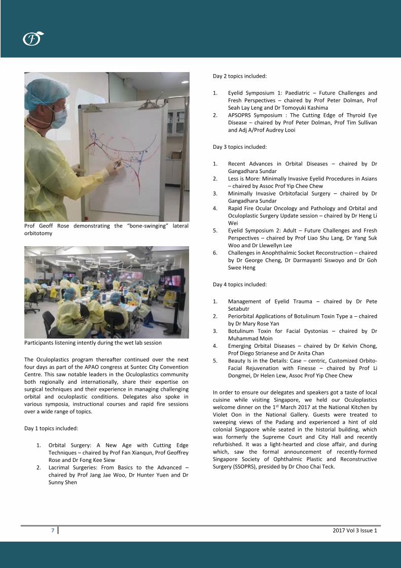
Prof Geoff Rose demonstrating the “bone-swinging” lateral orbitotomy
Participants listening intently during the wet lab session
The Oculoplastics program thereafter continued over the next four days as part of the APAO congress at Suntec City Convention Centre. This saw notable leaders in the Oculoplastics community both regionally and internationally, share their expertise on surgical techniques and their experience in managing challenging orbital and oculoplastic conditions. Delegates also spoke in various symposia, instructional courses and rapid fire sessions over a wide range of topics.
Day 1 topics included:
1. Orbital Surgery: A New Age with Cutting Edge Techniques – chaired by Prof Fan Xianqun, Prof Geoffrey Rose and Dr Fong Kee Siew
2. Lacrimal Surgeries: From Basics to the Advanced – chaired by Prof Jang Jae Woo, Dr Hunter Yuen and Dr Sunny Shen
Day 2 topics included:
1. Eyelid Symposium 1: Paediatric – Future Challenges and Fresh Perspectives – chaired by Prof Peter Dolman, Prof Seah Lay Leng and Dr Tomoyuki Kashima
2. APSOPRS Symposium : The Cutting Edge of Thyroid Eye Disease – chaired by Prof Peter Dolman, Prof Tim Sullivan and Adj A/Prof Audrey Looi
Day 3 topics included:
1. Recent Advances in Orbital Diseases – chaired by Dr Gangadhara Sundar
2. Less is More: Minimally Invasive Eyelid Procedures in Asians – chaired by Assoc Prof Yip Chee Chew
3. Minimally Invasive Orbitofacial Surgery – chaired by Dr Gangadhara Sundar
4. Rapid Fire Ocular Oncology and Pathology and Orbital and Oculoplastic Surgery Update session – chaired by Dr Heng Li Wei
5. Eyelid Symposium 2: Adult – Future Challenges and Fresh Perspectives – chaired by Prof Liao Shu Lang, Dr Yang Suk Woo and Dr Llewellyn Lee
6. Challenges in Anophthalmic Socket Reconstruction – chaired by Dr George Cheng, Dr Darmayanti Siswoyo and Dr Goh Swee Heng
Day 4 topics included:
1. Management of Eyelid Trauma – chaired by Dr Pete Setabutr
2. Periorbital Applications of Botulinum Toxin Type a – chaired by Dr Mary Rose Yan
3. Botulinum Toxin for Facial Dystonias – chaired by Dr Muhammad Moin
4. Emerging Orbital Diseases – chaired by Dr Kelvin Chong, Prof Diego Strianese and Dr Anita Chan
5. Beauty Is in the Details: Case – centric, Customized Orbito-Facial Rejuvenation with Finesse – chaired by Prof Li Dongmei, Dr Helen Lew, Assoc Prof Yip Chee Chew
In order to ensure our delegates and speakers got a taste of local cuisine while visiting Singapore, we held our Oculoplastics welcome dinner on the 1st March 2017 at the National Kitchen by Violet Oon in the National Gallery. Guests were treated to sweeping views of the Padang and experienced a hint of old colonial Singapore while seated in the historial building, which was formerly the Supreme Court and City Hall and recently refurbished. It was a light-hearted and close affair, and during which, saw the formal announcement of recently-formed Singapore Society of Ophthalmic Plastic and Reconstructive Surgery (SSOPRS), presided by Dr Choo Chai Teck.

8 2017 Vol 3 Issue 1
Dinner at the National Kitchen From L to R: Dr Mark Imperial, Dr Lim Lee Hooi, Dr Christine Therese Amurao Santos, Dr Sunny Shen, Dr Choo Chai Teck, Dr Raoul Paolo D Hensen, Adj A/Prof Audrey Looi
With friends from Taiwan and Thailand
Dr Sunny Shen, Prof David Verity, Dr Reynaldo Javate, Dr Gillian Teh, Adj A /Prof Audrey Looi
The Hong Kong contingent. From L to R: Dr Kelving Chong, Dr George Cheng, Adj A/Prof Audrey Looi, Dr Sunny Shen, Dr Hunter Yuen
The delegates also joined the main congress Gala Dinner on 3rd March 2017 at the Gardens by The Bay, where they enjoyed a sumptuous Chinese fusion meal under the glittering lights of the Flower Field Hall while taking in the views of the city skyline.
Enjoying a stroll in the gardens before the Gala Dinner.
With Prof Fan Xianqun and his team. None of this would have been possible without the nursing and administrative staff of SNEC who helped to coordinate the cadaveric dissection workshop, and the generosity of the sponsors

9 2017 Vol 3 Issue 1
and technical support staff involved in the smooth running of the wet lab session.
A special mention needs to go to our department secretary Miss Elaine Koek, who laboured long hours to help coordinate venues and logistics, to ensure everything went ahead without a hitch.
Last but not least, our sincere thanks and appreciation for the instructors and speakers of the cadaveric dissection course and Oculoplastics program for making our meeting a successful and fruitful experience for all participants!
Dacryoendoscopy and Endoluminal Lacrimal Duct Recanalization (ELDR) – A perspective
Gangadhara Sundar Toru Suzuki
Reynaldo Javate Lacrimal drainage surgeries have been performed for over a century. Conventional procedures that have stood the test of time include the external dacryocystorhinostomy (ExDCR), and over the past few decades, endonasal dacryocystorhinostomy (EnDCR). The latter may be performed either with or without a transnasal endoscopy and currently surpasses ExDCR in both patient acceptance and outcomes1. Alternative drainage procedures include transcanalicular laser DCR (TCLDCR), which has lost acceptance and popularity owing to poor outcomes. Endoluminal Lacrimal Duct Recanalization (ELDR) has been performed since the late 1990s with progressive refinements in techniques and thus improved outcomes. Although it is currently performed by a limited number of Lacrimal surgeons worldwide it is gaining attention and popularity with the advent of Lacrimal Microendoscopy which aids direct visualization of the location and nature of obstruction within the lacrimal drainage system. This procedure also meets the requirements of Natural Orifice Transluminal Endoscopic Surgery (NOTES), popular with some other surgical disciplines. A brief history of lacrimal endoscopy is highlighted in Table 1. Table 1. Brief history of lacrimal endoscopy
1989 Ebran, Matgret & Bechetoille
Clinical research in lacrimal system endoscopy
1997 Kuchar, Novak et al Clinical lacrimal microendoscopy with presaccal Er-YAG recanalization
2001 Hafliger, Piffaretti Lacrimal drainage system endoscopic examination and surgery
2010 Meyer-Rusenberg, Emmerich2
Modern lacrimal duct surgery
2010 Javate, Pamintuan, Cruz3.
Endoscopic lacrimal duct recanalization
The salient steps of Dacryoendoscopy and Endoluminal Lacrimal Duct Recanalization and their variations amongst the authors are highlighted below.
1. Preoperative examination and localization of obstruction:
Lacrimal irrigation and probing, Dacryocystography (TS),
Dacryoendoscopy, (Fig 1a, b), Intraoperative imaging (GS).
Fig 1a.
Fig 1b.
2. Informed consent process.
3. Anesthesia: Intraluminal lacrimal anesthesia, infratrochlear
nerve block or general anesthesia.
10 2017 Vol 3 Issue 1
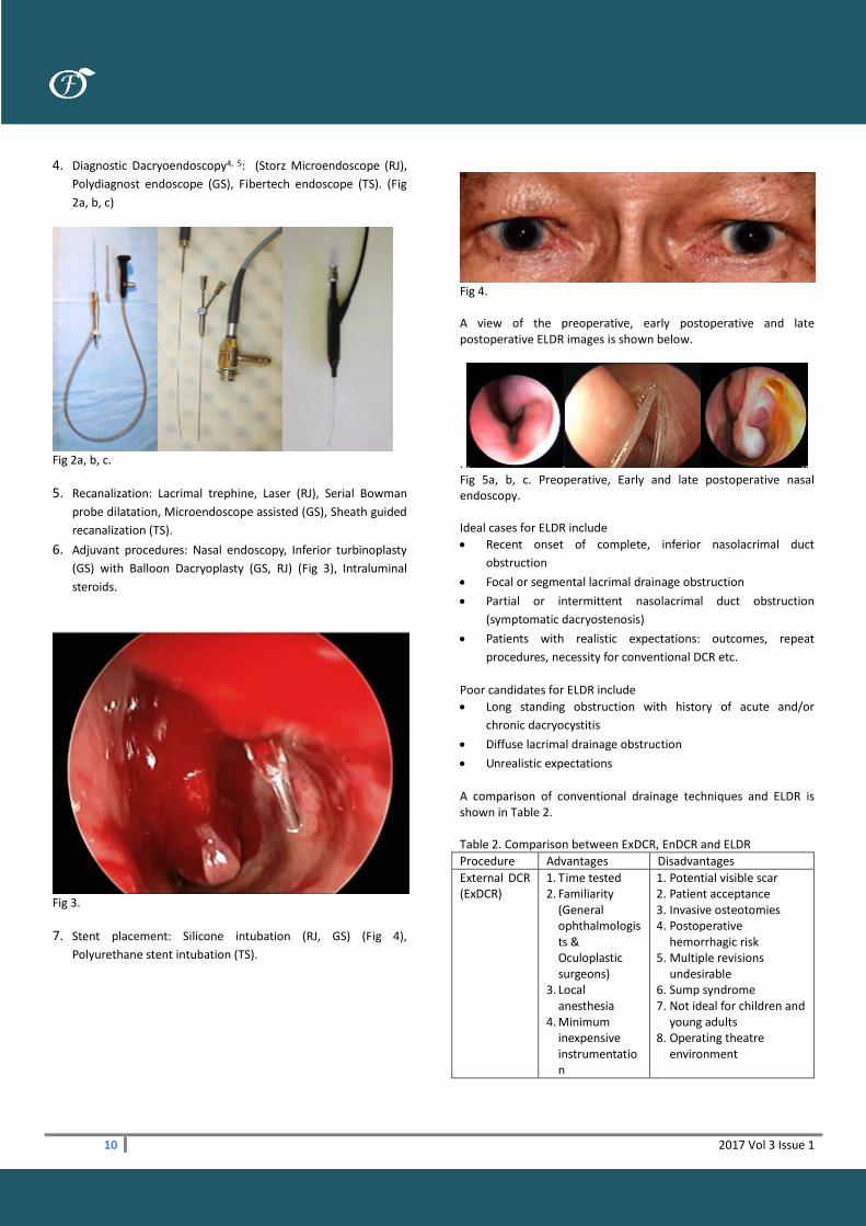
4. Diagnostic Dacryoendoscopy4, 5: (Storz Microendoscope (RJ),
Polydiagnost endoscope (GS), Fibertech endoscope (TS). (Fig
2a, b, c)
Fig 2a, b, c.
5. Recanalization: Lacrimal trephine, Laser (RJ), Serial Bowman
probe dilatation, Microendoscope assisted (GS), Sheath guided
recanalization (TS).
6. Adjuvant procedures: Nasal endoscopy, Inferior turbinoplasty
(GS) with Balloon Dacryoplasty (GS, RJ) (Fig 3), Intraluminal
steroids.
Fig 3.
7. Stent placement: Silicone intubation (RJ, GS) (Fig 4),
Polyurethane stent intubation (TS).
Fig 4. A view of the preoperative, early postoperative and late postoperative ELDR images is shown below.
. Fig 5a, b, c. Preoperative, Early and late postoperative nasal endoscopy. Ideal cases for ELDR include
Recent onset of complete, inferior nasolacrimal duct
obstruction
Focal or segmental lacrimal drainage obstruction
Partial or intermittent nasolacrimal duct obstruction
(symptomatic dacryostenosis)
Patients with realistic expectations: outcomes, repeat
procedures, necessity for conventional DCR etc.
Poor candidates for ELDR include
Long standing obstruction with history of acute and/or
chronic dacryocystitis
Diffuse lacrimal drainage obstruction
Unrealistic expectations
A comparison of conventional drainage techniques and ELDR is shown in Table 2. Table 2. Comparison between ExDCR, EnDCR and ELDR
Procedure Advantages Disadvantages
External DCR (ExDCR)
1. Time tested 2. Familiarity
(General ophthalmologists & Oculoplastic surgeons)
3. Local anesthesia
4. Minimum inexpensive instrumentation
1. Potential visible scar 2. Patient acceptance 3. Invasive osteotomies 4. Postoperative
hemorrhagic risk 5. Multiple revisions
undesirable 6. Sump syndrome 7. Not ideal for children and
young adults 8. Operating theatre
environment

11 2017 Vol 3 Issue 1
5. Chronic dacryocystitis, Mucocele
6. Minimal follow up
Endonasal DCR (EnDCR)
1. Elegant 2. Absence of scar 3. Suitable for all
NLD obstruction & ages
1. Learning curve / expertise
2. Expensive instrumentation
3. Postoperative hemorrhagic risk
4. General anesthesia/deep sedation
5. Operating theatre environment
Endoluminal Lacrimal Duct Recanalization (ELDR) with Dacryoendoscopy6
1. Minimally invasive
2. Intraluminal (NOTES)
3. Repeatability 4. Patient
acceptance 5. Identifying
specific location of obstruction
6. Identifying specific pathology causing obstruction
7. Office procedure
1. Long term evidence 2. Lacrimal
endoscope(Expensive) 3. Closer follow up 4. Dacryocystitis, chronic
diffuse NLD obstruction
Conclusion Although ELDR has limited awareness and thus global practice, recent advances in techniques and technology in the field of dacryology, patient desire for minimally invasive procedures, reduced morbidity, cost and improved outcomes have propelled this technique of lacrimal drainage obstruction recanalization. We predict that the future of lacrimal drainage surgery will be less expensive and invasive, offered at much earlier stages than is now provided (i.e., acute, chronic dacryocystitis) and possibly routinely as an outpatient procedure. References:
1. Jawaheer L, MacEwen CJ, Anijeet D. Endonasal versus external
dacryocystorhinostomy for nasolacrimal duct obstruction.
Cochrane Database Syst Rev. 2017 Feb 24;2:CD007097.
2. Emmerich KH, Meyer-Rüsenberg HW, Simko P. Endoscopy of the lacrimal ducts. Ophthalmologe. 1997 Oct;94(10):732-5. German.
3. Javate RM, Pamintuan FG, Cruz RT. Efficacy of endoscopic
lacrimal duct recanalization using microendoscope. Ophthal
Plast Reconstr Surg 2010; 26:330-333.
4. Inoue E, Suzuki T, Sasaki T. The beginners’ guidebook for
Dacryoendoscopy. Medical View 2016. ISBN 978-4-7583-1620-
0.
5. Ali MJ. Dacryoendoscopic examination of the lacrimal system.
In Principles and Practice of Lacrimal Surgery. Ed Ali MJ.
Springer 2015 ISBN 978-81-322-2019-0.
6. Sundar G. Lacrimal trauma and its management In Principles
and Practice of Lacrimal Surgery. Ed Ali MJ. Springer 2015 ISBN
978-81-322-2019-0.
Declaration:
None of the authors have a financial conflict of interest with the
products and devices mentioned.
Unusual long curly eye lashes in a male with metastatic lung cancer
Kwok Yuen Ting Yuen Kwok Lai Hunter
Case summary
A 58-years-old man, non-smoker, first presented to neurosurgeon
with diplopia and headache. MRI brain disclosed an extradural
tumor at left retrobulbar region involving the skull base. He
underwent craniotomy and tumour resection. Pathological exam
revealed metastatic adenocarcinoma with mutation in epidermal
growth factor receptor (EGFR) gene (L858R mutation positive),
suggestive of metastatic carcinoma with pulmonary origin.
Subsequent PET scan showed a right lower lobe lung carcinoma
and extensive skeletal metastases. He was given radiotherapy and
erlotinib (Tarceva) as palliative treatment.
On examination, he was noted to have bilateral long, thick and
curly eye lashes (Figure 1&2) affecting the upper and lower lids.
Such trichomegaly of eyelashes is likely secondary to erlotinib use.
There were no misdirected lashes or signs of ocular irritation.
There was no compressive optic neuropathy.
Discussion
Eyelash trichomegaly is a rare condition which is characterized not
just by increase in length but also the thickness, pigmentation and
curling of the eyelashes. It can be congenital, noted in conditions
like Oliver-McFarlane syndrome, Cornelia de Lange syndrome and

12 2017 Vol 3 Issue 1
familial trichomegaly, or acquired1,2. It is known to be associated
with human immunodeficiency syndrome, uveitis or connective
tissue diseases e.g. dermatomyositis or systemic lupus
erythematosus. There are many trichomegaly-inducing drugs,
including prostaglandin analogues, EGFR inhibitors3 like the
monoclonal antibodies (cetuximab, panitumumab) or tyrosine
kinase inhibitors (erlotinib4, getitinib), anticonvulsants (phenytoin,
topiramate), immunosuppressants (cyclosporine, tarcolimus) and
more. Among all, application of topical prostaglandin analogues
(latanoprost, bimatoprost) as an anti-glaucomatous agent is the
most common cause of eyelash trichomegaly. Such property
makes prostaglandin analogue a very effective in treatment of
hypotrichosis. Latisse® (bimatoprost 0.03% solution) is a FDA-
approved medication for hypotrichosis and it is very popular as a
cosmetic product. But long term use of prostaglandin analogues
may give rise to prostaglandin associated periorbitopathy (PAP)
which included periocular pigmentation, fat atrophy with
subsequent deep upper lids sulcus, tight eyelids, ptosis and
enophthalmos (sunken eye). Patient should be warned of these
side effects when put on prostaglandin analogues.
Trichomegaly itself is a benign condition, but it can cause ocular
irritation and corneal abrasion if trichiasis occurs. There is also
cosmetic and psychological concern. Patients with eyelash
trichomegaly should be examined by ophthalmologist under slit
lamp biomicroscopy. If symptomatic, conservative management
with frequent trimming or epilation, together with lubricants is
usually successful. Electroepilation or cryoablation may be
considered for recurrent misdirected lashes with ocular irritation.
Erlotinib and other EGRF inhibitors are widely used in patients
with non-small-cell lung cancer. They are well known for their
cutaneous side effect, most commonly as a papulopustular rash
over face and torso. EGFR is also found in hair follicles and thus
alters the normal hair growth. There are several cases of lash
trichomegaly associated with EGFR inhibitors use reported. These
patients may have eyebrow trichomegaly and facial hypertrichosis
as well. In a recent Asian study3, they found 2% of patient would
have trichomegaly as a side effect after EGFR inhibitor treatment.
Usually these drug-induced trichomegaly appear 2-5 months after
treatment and subside after cessation of the medication.
Fig 1: Clinical photo showing eyelashes trichomegaly of all 4 lids, the eyelashes were unusually long, thick and curly. There was also papulopustular rash over patient’s face. Fig 2: The lateral views.
(Fig 2a: The left eye, Fig 2b: The right eye)
REFERENCE
1. Paul L, Cohen P, Kurzrock R. Eyelash trichomegaly: review of
congenital, acquired, and drug-associated etiologies for
elongation of the eyelashes. International Journal of
Dermatology. 2012;51(6):631-646
2. Kaur S, Mahajan B. Eyelash trichomegaly. Indian Journal of
Dermatology. 2015;60(4):378.
3. Vachiramon V, Chanprapaph K, Pongcharoen P. Cutaneous
adverse events of epidermal growth factor receptor inhibitors:
A retrospective review of 99 cases. Indian J Dermatol Venereol
Leprol. 2015;81(5):547.
4. Jeon S, Ryu J, Choi G et al. Erlotinib Induced Trichomegaly of
the Eyelashes. Tuberc Respir Dis. 2013;74(1):37.
5. Paul L, Cohen P, Kurzrock R. Eyelash trichomegaly: review of
congenital, acquired, and drug-associated etiologies for
elongation of the eyelashes. International Journal of
Dermatology. 2012;51(6):631-646
6. Kaur S, Mahajan B. Eyelash trichomegaly. Indian Journal of
Dermatology. 2015;60(4):378.
7. Vachiramon V, Chanprapaph K, Pongcharoen P. Cutaneous
adverse events of epidermal growth factor receptor inhibitors:
A retrospective review of 99 cases. Indian J Dermatol Venereol
Leprol. 2015;81(5):547.
8. Jeon S, Ryu J, Choi G et al. Erlotinib Induced Trichomegaly of
the Eyelashes. Tuberc Respir Dis. 2013;74(1):37.

13 2017 Vol 3 Issue 1
Eyelid Metastasis as the Primary Manifestation
of Breast Carcinoma Yasuhiro Takahashi Hirohiko Kakizaki
ABSTRACT A 74-year-old woman without a previous history of breast carcinoma presented with a subcutaneous mass in the left lower eyelid. Before biopsy, the patient showed a subcutaneous, non-tender, non-erythematous induration in the left lower eyelid with mild eyelid edema. Biopsy with histopathology and immunohistochemistry were all consistent with metastatic invasive lobular carcinoma from the breast. A breast surgeon found palpable masses in the left breast and left palpable maxillary lymph node. The pathological findings from biopsy specimens of these lesions were similar to the findings of the eyelid lesion. The patient was treated using hormonal therapy. Twenty-two months after biopsy of the eyelid lesion, the patient was alive and well. Keywords: Eyelid; metastasis; breast; hormonal therapy INTRODUCTION: Metastatic tumors to the eyelids are rare, accounting for less than 1% of all malignant eyelid tumors.1-4 The most common primary origin of metastatic eyelid tumors is the breast (35-65%).1,5 Although the primary breast carcinoma is usually diagnosed before periocular manifestations, eyelid metastasis rarely occurs in the absence of a history or clinical evidence of an overt primary breast carcinoma.2,3,5-7 Here, we present a patient with a metastatic eyelid tumor from the breast. The patient had no previous history of breast carcinoma. Case report: This retrospective observational case report adhered to the tenets of the 1964 Declaration of Helsinki. Written informed consent for publication of this report and any accompanying images was obtained from the patient. A 74-year-old woman presented with a subcutaneous mass in the left lower eyelid. The patient had taken levothyroxine for Hashimoto disease and underwent recession of the inferior rectus muscle for restrictive hypotropia by thyroid-associated inferior rectus myopathy. The patient did not have a previous history of malignant tumors. At the examination, the patient had a subcutaneous, non-tender, non-erythematous induration below the pretarsal area of the left lower eyelid (Figure 1). Eyelid edema was found around the tumor. Excisional biopsy of the mass was performed under local
anesthesia. Pathological examination of the biopsy specimen revealed infiltration of tumor cells with round nuclei between muscle fibers (Figure 2A). Immunohistochemistry showed cytokeratin (AE1/AE3), CK7, CK20, and estrogen receptor positivity and progesterone receptor, E-cadherin, and LCA negativity (Figure 2B). These findings were all consistent with metastatic invasive lobular carcinoma from the breast. A breast surgeon found palpable masses in the left breast and left palpable maxillary lymph node. The pathological findings from biopsy specimens of these lesions were similar to the findings of the eyelid lesion. On immunohistochemistry, the tumors were negative for HER2 and had an MIB-1 labelling index of 20%. The patient was administered anastrozole, after which the size of the eyelid lesion reduced and the results of blood tests for tumor markers were stable. However, 1 year after biopsy of the eyelid lesion, the patient presented with similar eyelid thickening in the upper eyelid on both sides and in the lower eyelid on the right side; the patient declined to undergo biopsy. Therefore, her hormonal therapy was changed to fulvestrant, after which her tumor marker levels decreased. Twenty-two months after initial biopsy of the eyelid lesion, the patient was alive and well without additional metastatic lesions. DISCUSSION: We report a patient with eyelid metastasis as the primary manifestation of breast carcinoma. Previous studies reported that 15–45% of patients presented with metastatic eyelid lesions manifesting before diagnosis of the primary site.2,3,7 Breast carcinoma is often identified at an early stage, whereas lung cancer, which is also a common primary lesion of eyelid metastasis,3 frequently shows poorly-defined subjective symptoms and is uncommonly diagnosed at an early stage.8 These characteristics may contribute to the rarity of eyelid metastasis as the primary manifestation of breast carcinoma. Although eyelid induration with mild edema shown in our patient is the most common form of metastatic eyelid tumor from the breast,9 eyelid edematous lesions need to be differentiated from inflammatory diseases.4 Our patient had thyroid eye disease, which can cause eyelid edema; however, thyroid eye disease commonly presents bilaterally without induration.9 Although chalazion, cellulitis, and idiopathic orbital inflammation are considered as differential diagnoses of unilateral eyelid edema,4 these entities are commonly accompanied by eyelid erythema. In addition, the lesion in our patient was below the lower pretarsal area, unlike chalazion. Eyelid metastasis frequently occurs in the advanced stages of malignant tumors after the disease has already spread systemically.1 Even with systemic chemo- and/or hormonal therapies, the prognosis is poor, with a mean survival of 9.7 months.4 In our patient, metastasis to the axillary lymph node was found after the diagnosis of breast carcinoma. However, hormonal therapy was effective and the patient was alive 22 months after

14 2017 Vol 3 Issue 1
biopsy of the eyelid tumor, although the patient had what appeared to be metastatic eyelid tumors on the right eyelids.
In conclusion, we report a patient with eyelid metastasis as the primary manifestation of breast carcinoma. Because eyelid metastasis is sometimes part of systemic metastasis from the breast, metastasis to other organs needs to be explored and systemic chemo- and/or hormonal therapies should be considered
REFERENCES: 1. Zhang GJ, Adachi I, Yin DF, Narabayashi M, Tokue Y, Watanabe T, Kaneko A, Tsuda H, Abe K. Eyelid metastasis from breast carcinoma showing marked response to chemotherapy. Jpn J Clini Oncol 1995;25:10–15. 2. Bianciotto C, Dermirci H, Shields CL, Eagle RC Jr., Shields JA. Metastatic tumors to the eyelid: Report of 20 cases and review of the literature. Arch Ophthalmol 2009;127:999–1005. 3. Mansour AM, Hidayat AA. Metastatic eyelid disease. Ophthalmology 1987;94:667–670. 4. Rongioletti F. Monocle and mask-like metastasis: two cases of eyelid metastasis from breast carcinoma—role of isotopic phenomenon? Int J Dermatol 2015;54:e136–e139. 5. Rubio FA, Pizarro A, Robana G, Mayor M, Buron I, Contreras E, Casedo M. Eyelid metastasis as the presenting sign of recurrent carcinoma of the breast. Br J Dermatol 1997;137:1026–1027. 6. Weimar VM, Ceilley RI, Hurwitz N. Breast carcinoma metastatic to the eyelids. Arch Dermatol 1978;114:461. 7. Hood CI, Font RL, Zimmerman LE. Metastatic mammary carcinoma in the eyelid with histiocytoid appearance. Cancer 1973;31:793–800. 8. Tamura M, Tada T, Tsuji H, Tamura M, Yoshimoto M, Takahashi K, Tada K, Tanabe M, Kutomi G, Sekine Y, Kasumi F. Clinical study on the metastasis to the eyes from breast cancer. Breast Cancer 2004;11:65–68. 9. Riley FC. Metastatic tumors of the eyelids. Am J Ophthalmol 1970;69:259–264. FIGURE LEGENDS: Figure 1. Facial photo before biopsy of the eyelid lesion. A subcutaneous, non-erythematous induration with mild edema is present in the left lower eyelid (arrows).
Figure 2. Pathological results from the biopsy specimen of the eyelid lesion.
A. Infiltration of tumor cells with round nuclei between muscle fibers (hematoxylin-eosin, original magnification ×200).

15 2017 Vol 3 Issue 1
B. Immunohistochemistry shows the tumor cells are positive for estrogen receptor (immunohistochemistry, original magnification ×100).
Early and late orbital haemorrhage
post orbital fracture repair Yong Kai-Ling Sunny Shen
Case 1 A 21-year-old male sustained right orbital floor and medial wall fractures after an assault. CT scan showed prolapse of the medial rectus into ethmoid sinus, but no entrapment. (Figure 1) The patient underwent right orbital floor and medial orbital wall repair with Medpore Titan orbital floor and wall implant ®, 1 week after the presentation. Post-operative Day 1 CT scan showed good placement of the implant. (Figure 2) The patient returned on Post-op Day 5 with sudden onset of right eye proptosis, swelling and blurring of vision. Examination showed subconjunctival haemorrhage, chemosis and increased intraocular pressure (IOP) to 35mmHg. His visual acuity (VA) dropped to 6/18 but there was no sign of optic neuropathy. There was limitation of extraocular movement in all directions of gaze, with hyperglobus. CT scan revealed a retrobulbar haemorrhage adjacent to the orbital implant. (Figure 3) Despite medical treatment, the patient’s IOP remained high and subsequently underwent surgical exploration and blood clot evacuation under general anaesthesia. Post-operatively his VA recovered to 6/6 with improvement in diplopia and globe position. (Figure 4, 5) Follow-up imaging showed resolution of the haemorrhage and good placement of implant. Case 2 A 49-year-old female with known history of right orbital floor fracture, repaired uneventful 12 years ago with a silicone implant, presented with a 2-week history of right lower eyelid bruising, swelling, discomfort and double vision on up gaze. Examination showed mild right periorbital swelling and bluish-brown discolouration of the lower eyelid skin (Figure 6). Elevation and depression of the right globe was limited with 1mm of hyperglobus. There was no history of new injury, no tenderness nor proptosis. The patient was treated for possible orbital infection and was prescribed a 1-week course of oral ciprofloxacin, but there were no improvements in her symptoms. Subsequent CT orbits revealed a right infra-orbital hyper-intense dome-shaped lesion measuring 2.3 x 0.6 cm in the subperiosteal space, elevating the globe (Figure 7). A right orbital floor implant was seen above the orbital floor (which appeared intact) and below the lesion. Clinical findings and imaging studies were suggestive of the presence of a fluid collection in the right inferior subperiosteal space, which warranted surgical exploration and drainage.
An exploration and drainage was performed using a trans-conjunctival approach under general anaesthesia. On incision of the periosteum along the inferior orbital rim, a small amount of chocolate-coloured fluid was released. The remaining 1 mL was aspirated and sent for histological examination and bacteriology studies (Figure 8). Upon reflection of the orbital periosteum, a large silicone implant was seen anchored to the orbital rim with sutures. On release of these sutures, the implant was seen encapsulated within a fibrous capsule. The implant was freed from the capsule and adjacent tissues, and subsequently removed from the orbit. Some yellowish, powdery deposits were seen on the surface of the silicone implant. The previous fracture site appeared well covered with fibrous tissue, providing adequate support for the orbital contents. Cytopathological analysis of the aspirate revealed blood, scattered inflammatory cells and a few aggregates of foamy histiocytic cells. Cultures of the aspirate grew coagulase-negative Staphyloccocus sensitive to ciprofloxacin, while cultures from the silicone implant were negative for microorganisms. The patient completed a 14-day course of oral ciprofloxacin and made an uneventful recovery. Resolution of the lid swelling, proptosis and globe displacement occurred 2 weeks post-surgery. Hess chart showed minimal limitation of right depression while binocular single vision charting revealed diplopia mainly on extreme elevation. Discussion Complications of orbital floor fracture repair include residual diplopia, paraesthesia, enophthalmos, lower eyelid ectropion, infection, orbital implant extrusion and loss of vision.1 Retrobulbar haemorrhage is a rare complication, estimated at 0.15 to 3.2% and is the most common cause of visual loss after orbital fracture repair.1,2,3 The acute form usually occurs within 24 hours after surgery,3 but may occur up to first 10 days after surgery.3,4,5,6 Late haemorrhage has been reported to occur from 3 months to 20 years post implantation.7 Signs and symptoms of acute retrobulbar haemorrhage include pain, decreased vision, diplopia, pupillary defect, limited extraocular movement and increased IOP.2,8 Permanent visual loss from compartment syndrome, optic neuropathy, retinal ischaemia may occur if not treated promptly.3,9 Risk factors of retrobulbar haemorrhage include severe trauma and anti-coagulants use.1 CT confirmation should be done if there is any clinical suspicion.2 Treatment includes medical or surgical decompression. An urgent lateral canthotomy and inferior cantholysis should be performed if initial medical treatment is insufficient, and definitive surgical exploration and decompression may be required for severe case.1,9 Loss of vision from optic nerve compression is rare in late haemorrhage.7 The underlying mechanism of retrobulbar haemorrhage is not well-described, and the aetiology of early and late retrobulbar haemorrhage is likely to be different.7 It was suggested that late haemorrhage may arise from low-grade infection and inflammation, resulting in rupture of blood vessels along the fibrous capsule with intracapsular haemorrhage.7 For early haemorrhage (24 hour to 10 days), as represented in our first

16 2017 Vol 3 Issue 1
case, underlying pathogenesis include clot lysis and resultant haemorrhage into the orbital soft tissues.
1. Gosau M, Schöneich M, Draenert FG, Ettl T, Driemel O, Reichert TE.. Retrospective analysis of orbital floor fractures--complications, outcome, and review of literature. Clin Oral Investig. 2011 Jun; 15(3):305-13
2. Cheon JS, Seo BN, Yang JY, Son KM. Retrobulbar hematoma in blow-out fracture after open reduction. Arch Plast Surg. 2013 Jul;40(4):445-9.
3. Girotto JA, Gamble WB, Robertson B, et al. Blindness after reduction of facial fractures. Plast Reconstr Surg 1998;102:1821–1834
4. Shew M, Carlisle MP, Lu GN, Humphrey C, Kriet JD. Surgical Treatment of Orbital Blowout Fractures: Complications and Postoperative Care Patterns. Craniomaxillofac Trauma Reconstr. 2016 Nov;9(4):299-304.
5. Nicholson DH, Guzak SW. Visual loss complicating repair of orbital floor fractures. Arch Ophthalmol 1971;86(4):369–375
6. Ord RA. Post-operative retrobulbar haemorrhage and blindness complicating trauma surgery. Br J Oral Surg 1981;19(3):202–207
7. Gilhotra JS, McNab AA, McKelvie P, O'Donnell BA. Late orbital haemorrhage around alloplastic orbital floor implants: a case series and review. Clin Exp Ophthalmol. 2002 Oct;30(5):352-5.
8. Ord RA. Post-operative retrobulbar haemorrhage and blindness complicating trauma surgery. Br J Oral Surg. 1981 Sep;19(3):202-7
9. Chen CH, Chen CT, Huang F. Retrobulbar Hematoma as a Rare Complication After Secondary Correction of Enophthalmos. Craniofac Surg. 2009 May;20(3):963-7.
10. Chan CG, Shen S, Seah LL. Delayed orbital haemorrhage with possible infectious aetiology after silicone implantation. Ann Acad Med Singapore. 2009 Mar;38(3):271-3.
11. Mauriello JA, Flanagan JC, Peyster RG. An unusual late complication of orbital floor fracture repair. Ophthalmology 1984; 91: 102−7.
Figure 1. CT scan showing right medial wall and orbital floor fracture.
Figure 2. Post-operative day 1 CT scan showing good placement of Medpore Titan implant

17 2017 Vol 3 Issue 1
Figure 3. Post-operative day 5 CT scan showing retrobulbar haemorrhage adjacent to the orbital implant with resultant proptosis and hyperglobus.
Figure 4. Hess/Binocular Single Vision charts of patient at pos-op month 2 (top), post-op month 4 (middle) and post-op month 6 (bottom) showing improvement of diplopia.
Figure 5. Post-operative month 6 photos of patient showing improvement of hyperglobus and enophthalmos.
Figure 6. Patient with right mild right periorbital swelling and bluish brown discolouration of the lower lid skin with hyperglobus.
Figure 7. Coronal CT – hyper-intense lesion in the subperiosteal space, elevating and indenting the right globe (black arrow).
Figure 8. Subperiosteal chocolate-coloured aspirate.

18 2017 Vol 3 Issue 1
Guidelines Below are the formats for the different categories of articles: Invited Articles (no more than 1600 words; include images where appropriate) Case Highlights This refers to the written presentation of an interesting or challenging case in the following format:
History (no more than 100 words)
Examination (no more than 150 words; include clinical photos)
Investigations (include imaging where appropriate)
Management (no more than 100 words; include pathology images where appropriate)
Discussion (no more than 200 words; including challenges encountered in diagnosis or management) Operative Pearls This refers purely to advice that allows the reader to improve on his or her intraoperative technique and can include immediate post-op advice (no more than 600 words; include images where appropriate) Meetings or Social Visits All meetings organized by APSOPRS members as well as social visits to each other’s' centers are eligible for inclusion in the newsletter (no more than 1000 words for the main APSOPRS biennial meeting and no more than 500 words for other meetings or visits). Philosophical Notes No more than 800 words; include images where appropriate


![€¦ · and endoscopic dacryocystorhinostomy (DCR) was first performed by Caldwell in 1893, but was soon abandoned due to difficult visualization and numerous complications]" However,](https://img.pdfslide.net/doc/110x75/5ffe6ce26ff5f92c4f1c0757/and-endoscopic-dacryocystorhinostomy-dcr-was-first-performed-by-caldwell-in-1893.jpg)