Embed Size (px)
Citation preview
Oligophenylenevinylene Phane Dimers: Probing the Effect of ContactSite on the Optical Properties of Bichromophoric Pairs
Shujun Wang,† Guillermo C. Bazan,*,† Sergei Tretiak,‡ and Shaul Mukamel*,‡
Contribution from the Department of Chemistry, UniVersity of California,Santa Barbara, California 93106, and the Department of Chemistry and Rochester Theory Center forOptical Science and Engineering, UniVersity of Rochester, Rochester, New York 14627
ReceiVed May 13, 1999. ReVised Manuscript ReceiVed NoVember 19, 1999
Abstract: Paracyclophane derivatives have been prepared that may be considered models of bichromophoriccontacts in the solid. The optical properties of these compounds give insight into how bringing two chromophoresinto close proximity affects the photophysics of the pair. Thus, reaction of 4,7,12,15-tetrabromo[2.2]-paracyclophane (4,7,12,15-Br4Pc) with excess 4-tert-butylstyrene using Pd(OAc)2 under phase transfer conditionsaffords 4,7,12,15-tetra(4-tert-butylstyryl)[2.2]paracyclophane (3RD). The connectivity of3RD models a contactbetween two distyrylbenzene molecules across the central ring. Reaction of 4,7,12,15-Br4Pc with 4-(4-tert-butylstyryl)styrene (TBSS) under similar conditions gives 4,7,12,15-tetra(4-(4′-tert-butylstyryl)styryl)[2.2]-paracyclophane (5RD). In 5RD two oligophenylenevinylene units containing five phenyl rings are connectedvia their central ring. Similar reaction protocols gave 2,5-dimethyl-1,4-di(4-tert-butylstyryl)benzene (3R) and2,5-dimethyl-1,4-bis[4-(4′-tert-butylstyryl)styryl]benzene (5R). Molecules3R and5R serve to give the opticalproperties of the monomeric units. Comparison against the properties of3R and5R shows that the absorptionand emission data of3RD and5RD are consistent with considerable delocalization between the two subunitsacross the paracyclophane bridge. The observed trends in the optical properties of these compounds are analyzedusing collective electronic oscillators (CEO) representing the changes induced in the electronic density matrixupon optical excitation. Comparison of the CEO of the paracyclophane dimers with the corresponding monomersusing two-dimensional plots provides an efficient method for tracing the origin of the various optical andelectronic transitions by identifying the underlying changes in charge densities and bond orders. The electronicdescription of3RD and5RD, in which the interchromophore contact is across the central ring, is considerablydifferent from the description of paracyclophane dimers of similar chromophores that are connected via theterminal ring. Essentially no delocalization is observed for the “termini” dimers.
Introduction
Chromophore-chromophore interactions are a critical con-sideration for designing technologically relevant optoelectronicmaterials. The charge transport properties of organic materialsdepend on the relative orientation and distance betweenindividual molecules in the solid. Proper molecular alignmentover long distances allows for better charge transport and loweroperating voltages in organic transistors.1 For polymer-basedlight-emitting diodes, ordered regions lead to excimer formationand a reduced electroluminescence quantum yield.2,3 Becausemultiple environments are often present in solid samples, it isdifficult to ascertain to a high level of precision how theenvironment surrounding an individual chromophore affectsproperties of interest.4 The study of polychromophores in dilute
solution is complicated by intrachain contacts that lead to energytransfer or excimer formation.5 Well-defined molecules that arerepresentative of chromophore aggregates in the solid and thatcan be studied in the absence of interactions with otherchromophores give insight into the effect of through-spacedelocalization on the photophysics of the aggregate.
The importance of the optical response of chromophoreaggregates extends into various branches of chemical science.Significant attention has been devoted to studies of clusters insupersonic beams,6,7 J-aggregates of cyanine dyes,8 supramo-lecular structures,9 and biological complexes (photosyntheticantennae and reaction centers).10 It is possible to treat aggre-gates as giant molecules and employ methods of quantum
† University of California.‡ University of Rochester.(1) (a) Servet, B.; Horowitz, G.; Ries, S.; Lagorsse, O.; Alnot. P.; Yassar,
A.; Deloffre, F.; Srivastava P.; Hajlaoui, R.; Lang, P.; Garnier, F.Chem.Mater.1994, 6, 1809. (b) Dodalabalapur, A.; Torsi, L.; Katz, H. E.Science1995, 268, 270.
(2) (a) Conwell, E.Trends Polym. Sci.1997, 5, 218. (b) Cornil, J.; dosSantos, D. A.; Crispin, X.; Silbey, R.; Bredas, J. L.J. Am. Chem. Soc.1998, 120, 1289.
(3) (a) Lee, C. H.; Yu, G.; Moses, D.; Heeger, A. J.Synth. Met.1995,69 (1-3), 429. (b) Gettinger, C. L.; Heeger, A. J.; Drake, J. M.; Pine, D.J. J.Chem. Phys.1994, 101, 1673. (c) Kohler, A.; Gruner, J.; Friend, R.H.; Mullen, K.; Scherf, U.Chem. Phys. Lett.1995, 243, 456.
(4) (a) Guillet, J.Polymer Photophysics and Photochemistry; CambridgeUniversity Press: Cambridge, U.K., 1985. (b)Photophysics of Polymers;Hoyle, C. E., Torkelson, J. M., Eds.; ACS Symp. Ser. 358; AmericanChemical Society: Washington, DC, 1987. (c) Winnik, F. M.Chem. ReV.1993, 93, 587.
(5) Miao, Y.-J.; Herkstroeter, W. G.; Sun, B. J.; Wong-Foy, A. G.; Bazan,G. C. J. Am. Chem. Soc. 1995, 117, 11407.
(6) (a) Easter, D. C.; Whetten, R. L.; Wessel, J. E.J. Chem. Phys.1991,94, 3347. (b) Easter, D. C.; Khoury, J. T.; Whetten, R. L.J. Chem. Phys.1992, 97, 1681. (c) Easter, D. C.; Baronavski, A. P.; Whetten, R. L.J.Chem. Phys.1993, 99, 4942.
(7) (a) Syage, J. A.; Wessel, J. E.J. Chem. Phys.1988, 89, 5962. (b)Wessel, J. E.; Syage, J. A.J. Phys. Chem.1990, 94, 737.
(8) Mukamel, S., Chemla, D. S., Eds.; Special Issue, Confined Excitationsin Molecular and Semiconductor Nanostructures.Chem. Phys.1996, 210.
1289J. Am. Chem. Soc.2000,122,1289-1297
10.1021/ja991611y CCC: $19.00 © 2000 American Chemical SocietyPublished on Web 02/03/2000
chemistry to calculate their electronic structure. These ap-proaches are limited to small aggregates.11,12An important goalof computational chemistry is to trace the electronic states andspectra of aggregates to those of their basic building blocks,the monomers. By doing so it should become possible to get abetter microscopic insight into the nature of their electronicexcitations and to predict qualitative features of complex largesystems using simple, readily available information. Predictingqualitative features of complex multicomponent ensembles fromreadily available information should then become possible onisolated molecules.
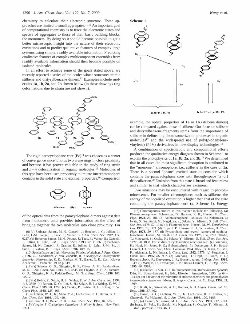
In an effort to achieve some of the goals stated above, werecently reported a series of molecules whose structures mimicstilbene and distyrylbenzene dimers.13 Examples include mol-ecules1a, 1b, 2a, and2b shown below (in these drawings ringdeformations due to strain are not shown).
The rigid paracyclophane core (Pc)14 was chosen as a centerof convergence since it holds two arene rings in close proximityand because it has proven valuable in the study of ring strainandπ-π delocalization in organic molecules.15 Molecules ofthis type have been used previously to imitate interchromophorecontacts in the solid state and excimer properties.16 Comparison
of the optical data from the paracyclophane dimers against datafrom monomeric units provides information on the effect ofbringing together the two molecules into close proximity. For
example, the optical properties of1a or 1b (stilbene dimers)can be compared against those of stilbene. Our focus on stilbeneand distyrylbenzene fragments stems from the importance ofstilbene in delineating photoisomerization processes in organicmolecules17 and the widespread use of poly(p-phenylene-vinylene) (PPV) derivatives in new display technologies.18
A combination of spectroscopic and computational effortsproduced the qualitative energy diagram shown in Scheme 1 toexplain the photophysics of1a, 1b, 2a, and2b.19 We determinedthat in all cases the most significant absorption is attributed tothe “monomer” chromophore, i.e., stilbene in the case of1a.There is a second “phane” excited state to consider whichcontains the paracyclophane core with through-space (π-π)delocalization.20 Emission from this state is broad and featurelessand similar to that which characterizes excimers.
Two situations may be encountered with regard to photolu-minescence. For smaller chromophores such as stilbene, theenergy of the localized excitation is higher than that of the statecontaining the paracyclophane core (a, Scheme 1). Energy
(9) (a) Berberan-Santos, M. N.; Canceill, J.; Brochon, J.-C.; Jullien, L.;Lehn, J.-M.; Pouget, J.; Tauc, P.; Valeur, B.J. Am. Chem. Soc.1992, 114,6227. (b) Berberan-Santos, M. N.; Pouget, J.; Tauc, P.; Valeur, B.; Canceill,J.; Jullien, L.; Lehn, J.-M.J. Phys. Chem.1993, 97, 11376. (c) Berberan-Santos, M. N.; Canceill, J.; Gratton, E.; Jullien, L.; Lehn, J.-M.; So, J.;Sutin, J.; Valeur, B.J. Phys. Chem.1996, 100, 15.
(10) Special Issue on Light-Harvesting Physics Workshop.J. Phys. Chem.B 1997, 101. Sundstro¨m, V.; van Grondelle, R. InAnoxygenic PhotosyntheticBacteria; Blankenship, R. E., Madiga, M. T., Baner, C. E., Eds.; KluwerAcademic: Dordrecht, 1995; p 349.
(11) (a) Scholes, G. D.; Ghiggino, K. P.; Oliver, A. M.; Paddon-Row,M. N. J. Am. Chem. Soc.1993, 115, 4345. (b) Clayton, A. H. A.; Scholes,G. D.; Ghiggino, K. P.; Paddon-Row,: M. N.J. Phys. Chem.1996, 100,10912.
(12) (a) Hobra, P.; Selzle, H. L.; Schlag, E. W.J. Am. Chem. Soc.1994,116, 3500. (b) Bo¨rsen, K. O.; Lin, S. H.; Selzle, H. L.; Schlag, E. W.J.Chem. Phys.1989, 90, 1299. (c) Ca´rsky, P.; Selzle, H. L.; Schlag, E. W.Chem Phys.1988, 125, 166.
(13) Oldham, W. J., Jr.; Miao, Y.-J.; Lachicotte, R. J.; Bazan, G. C.J.Am. Chem. Soc.1998, 120, 419.
(14) Cram, D. J.; Bauer, R. H.J. Am. Chem. Soc.1959, 81, 5971.(15) Voegtle, F.Cyclophane Chemistry; J. Wiley & Sons: New York,
1993.
(16) Chromophores studied in this manner include the following. (a)Phenanthrenophane: Schweitzer, D.; Hausser, K. H.; Haenel, M.Chem.Phys. 1978, 29, 181. (b) Anthracenophane: Ishikawa, S.; Nakamura, J.;Iwata, S.; Sumitami, M.; Nagakura, S.; Sakata, Y.; Misumi, S.Bull. Chem.Soc. Jpn.1979, 52, 1346. (c) Fluorenophane: Haenel, M. W.TetrahedronLett.1976, 36, 3121. (d) Colpa, J. P.; Hausser K. H.; Schweitzer, D.Chem.Phys.1978, 29, 187. (d) Pyrenophane and several isomers of naphtha-lenophane: Haenel, M.; Staab, H. A.Chem. Ber.1973, 106, 2203. Otsubo,T.; Mizogami, S.; Osaka, N.; Sakata, Y.; Misumi, S.Bull. Chem. Soc. Jpn.1977, 50, 1858. For studies of cycloaddition reactions see: (e) Grieiving,H.; Hopf, H.; Jones, P. G.; Bubenitscheck, P.; Desvergne, J. P.; Bouas-Laurent, J. J. Chem. Soc., Chem. Commun. 1994, 1075. (f) Okada, Y.; Ishii,F.; Akiyama, I.; Nishimura, J.Chem. Lett.1992, 1579. (g) Ferguson, J.Chem. ReV. 1986, 86, 957. (h) Grieiving, H.; Hopf, H.; Jones, P. G.;Bubenitscheck, P.; Desvergne, J. P.; Bouas-Laurent,Liebigs. Ann.1995,1949. (i) Marquis, D.; Desvergne, J. P.; Bouas-Laurent, H.J. Org. Chem.1995, 60, 7984.
(17) (a) Saltiel, J.; Sun, Y.-P. InPhotochromism, Molecules and Systems;Durr, H., Bouas-Laurent, H., Eds.; Elsevier: Amsterdam, 1990; pp 64-164. (c) For a review of the relevance of stilbene chemistry and photophysicsto materials science see: Meier, H.Angew. Chem., Int. Ed. Engl.1992, 31,1399.
(18) Kraft, A.; Grimsdale, A. C.; Holmes, A. B.Angew. Chem., Int. Ed.Engl. 1998, 37, 402.
(19) Bazan, G. C.; Oldham, W. J., Jr.; Lachicotte, R. J.; Tretiak, S.;Chernyak, V.; Mukamel. S.J. Am. Chem. Soc.1998, 120, 9188.
(20) (a) Canuto, S.; Zerner, M. C.J. Am. Chem. Soc.1990, 112, 2114.(b) Iwata, S.; Fuke, K.; Sasaki, M.; Nagakura, S.; Otsubo, T.; Misumi, S.J. Mol. Spectrosc.1973, 46, 1.
Scheme 1
1290 J. Am. Chem. Soc., Vol. 122, No. 7, 2000 Wang et al.
migration21 after photon absorption transfers the excitation fromthe localized “monomer”, and emission takes place from the”phane“ state. The latter carries a very weak oscillator strengthto the ground state. Population of the ”phane" state via energytransfer thus results in a relatively long-lived excited state.
The second situation occurs when the energy of the “mono-mer” state is lower than that of the corresponding “phane” state(b, Scheme 1). This is the case for distyrylbenzene (2a,b). Underthese circumstances there is no driving force for energymigration and the excitation remains localized at the initiallygenerated state. Except for the spectral shift due to thePcmoiety, there is therefore negligible difference between thespectra of the parent compounds and the dimers2a,b.
In this contribution we report on the synthesis, spectroscopy,and quantum chemical analysis of paracyclophane dimers thatbring together oligophenylenevinylene chromophores into con-tact by way of the central ring. These molecules provide infor-mation on how the electronic structure of these model chro-mophores changes by making a contact across the central ring.We will refer to this interaction as “criss-cross” to differentiateit from the “terminal” contact probed by molecules1a,b and2a,b. These efforts are aimed at understanding the effect ofcontact regiochemistry on the photophysics of the pair. We willfind that the localization and the type of excitation are quitedifferent in the criss-cross dimers, relative to their terminicounterparts.
Results and Discussion
Synthesis and Characterization.Bromination ofPc by useof liquid bromine and a catalytic amount of iodine gives equalamounts of the isomeric tetrabromides 4,7,12,15-tetrabromo-[2.2]paracyclophane (4,7,12,15-Br4Pc) and 4,5,12,13-tetrabromo-[2.2]paracyclophane (4,5,12,13-Br4Pc) in 90% yield.22 Sepa-ration by repeated crystallization in acetone affords the desired4,7,12,15-Br4Pc in 40% yield.
The stilbenoid arms are constructed by Heck reaction23 un-der the phase transfer conditions reported by Jeffrey (excessK2CO3 and BrNBu4 in DMF).24 For example, reaction of anexcess of 4-tert-butylstyrene with 4,7,12,15-Br4Pc in thepresence of Pd(OAc)2 for 4 days affords the 4-fold coupledproduct 4,7,12,15-tetra(4-tert-butylstyryl)[2.2]paracyclophane(3RD for the three-ring dimer) in 55% yield (eq 1). As the
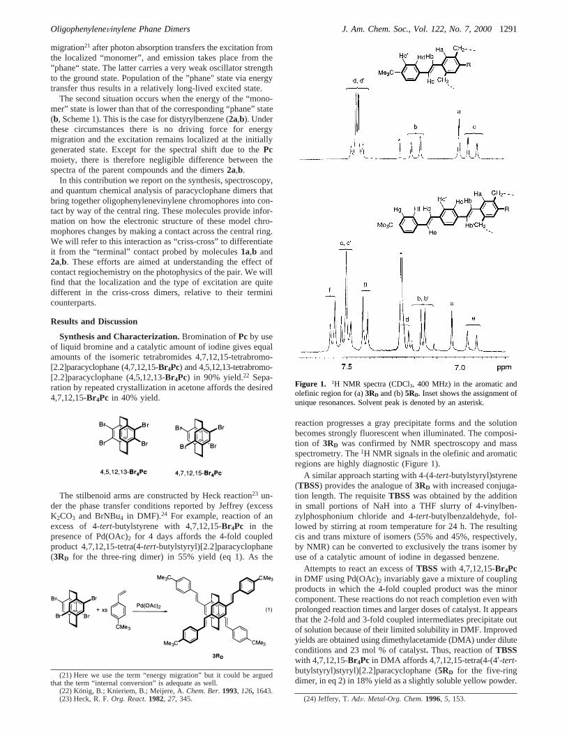
reaction progresses a gray precipitate forms and the solutionbecomes strongly fluorescent when illuminated. The composi-tion of 3RD was confirmed by NMR spectroscopy and massspectrometry. The1H NMR signals in the olefinic and aromaticregions are highly diagnostic (Figure 1).
A similar approach starting with 4-(4-tert-butylstyryl)styrene(TBSS) provides the analogue of3RD with increased conjuga-tion length. The requisiteTBSS was obtained by the additionin small portions of NaH into a THF slurry of 4-vinylben-zylphosphonium chloride and 4-tert-butylbenzaldehyde, fol-lowed by stirring at room temperature for 24 h. The resultingcis and trans mixture of isomers (55% and 45%, respectively,by NMR) can be converted to exclusively the trans isomer byuse of a catalytic amount of iodine in degassed benzene.
Attempts to react an excess ofTBSS with 4,7,12,15-Br4Pcin DMF using Pd(OAc)2 invariably gave a mixture of couplingproducts in which the 4-fold coupled product was the minorcomponent. These reactions do not reach completion even withprolonged reaction times and larger doses of catalyst. It appearsthat the 2-fold and 3-fold coupled intermediates precipitate outof solution because of their limited solubility in DMF. Improvedyields are obtained using dimethylacetamide (DMA) under diluteconditions and 23 mol % of catalyst. Thus, reaction ofTBSSwith 4,7,12,15-Br4Pc in DMA affords 4,7,12,15-tetra(4-(4′-tert-butylstyryl)styryl)[2.2]paracyclophane (5RD for the five-ringdimer, in eq 2) in 18% yield as a slightly soluble yellow powder.
(21) Here we use the term “energy migration” but it could be arguedthat the term “internal conversion” is adequate as well.
(22) Konig, B.; Knieriem, B.; Meijere, A.Chem. Ber.1993, 126, 1643.(23) Heck, R. F.Org. React.1982, 27, 345. (24) Jeffery, T.AdV. Metal-Org. Chem.1996, 5, 153.
Figure 1. 1H NMR spectra (CDCl3, 400 MHz) in the aromatic andolefinic region for (a)3RD and (b)5RD. Inset shows the assignment ofunique resonances. Solvent peak is denoted by an asterisk.
OligophenyleneVinylene Phane Dimers J. Am. Chem. Soc., Vol. 122, No. 7, 20001291
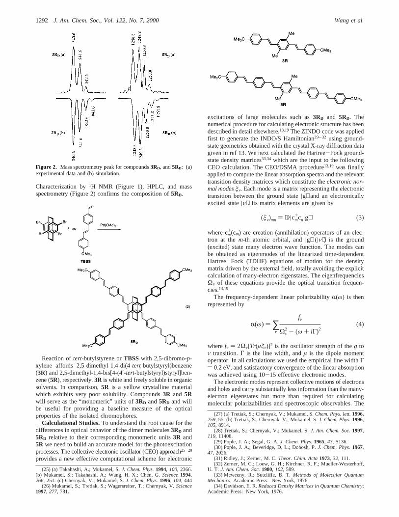
Characterization by1H NMR (Figure 1), HPLC, and massspectrometry (Figure 2) confirms the composition of5RD.
Reaction oftert-butylstyrene orTBSS with 2,5-dibromo-p-xylene affords 2,5-dimethyl-1,4-di(4-tert-butylstyryl)benzene(3R) and 2,5-dimethyl-1,4-bis[4-(4′-tert-butylstyryl)styryl]ben-zene (5R), respectively.3R is white and freely soluble in organicsolvents. In comparison,5R is a yellow crystalline materialwhich exhibits very poor solubility. Compounds3R and 5Rwill serve as the “monomeric” units of3RD and5RD and willbe useful for providing a baseline measure of the opticalproperties of the isolated chromophores.
Calculational Studies.To understand the root cause for thedifferences in optical behavior of the dimer molecules3RD and5RD relative to their corresponding monomeric units3R and5R we need to build an accurate model for the photoexcitationprocesses. The collective electronic oscillator (CEO) approach25-28
provides a new effective computational scheme for electronic
excitations of large molecules such as3RD and 5RD. Thenumerical procedure for calculating electronic structure has beendescribed in detail elsewhere.13,19The ZINDO code was appliedfirst to generate the INDO/S Hamiltonian29-32 using ground-state geometries obtained with the crystal X-ray diffraction datagiven in ref 13. We next calculated the Hartree-Fock ground-state density matrices33,34 which are the input to the followingCEO calculation. The CEO/DSMA procedure13,19 was finallyapplied to compute the linear absorption spectra and the relevanttransition density matrices which constitute theelectronic nor-mal modesêν. Each mode is a matrix representing the electronictransition between the ground state|g⟩ and an electronicallyexcited state|ν⟩. Its matrix elements are given by
where cm+(cm) are creation (annihilation) operators of an elec-
tron at them-th atomic orbital, and|g⟩ (|ν⟩) is the ground(excited) state many electron wave function. The modes canbe obtained as eigenmodes of the linearized time-dependentHartree-Fock (TDHF) equations of motion for the densitymatrix driven by the external field, totally avoiding the explicitcalculation of many-electron eigenstates. The eigenfrequenciesΩν of these equations provide the optical transition frequen-cies.13,19
The frequency-dependent linear polarizabilityR(ω) is thenrepresented by
wherefν ) 2Ων[Tr(µêν)]2 is the oscillator strength of theg toν transition.Γ is the line width, andµ is the dipole momentoperator. In all calculations we used the empirical line widthΓ) 0.2 eV, and satisfactory convergence of the linear absorptionwas achieved using 10-15 effective electronic modes.
The electronic modes represent collective motions of electronsand holes and carry substantially less information than the many-electron eigenstates but more than required for calculatingmolecular polarizabilities and spectroscopic observables. The
(25) (a) Takahashi, A.; Mukamel, S.J. Chem. Phys.1994, 100, 2366.(b) Mukamel, S.; Takahashi, A.; Wang, H. X.; Chen, G.Science1994,266, 251. (c) Chernyak, V.; Mukamel, S.J. Chem. Phys.1996, 104, 444
(26) Mukamel, S.; Tretiak, S.; Wagersreiter, T.; Chernyak, V.Science1997, 277, 781.
(27) (a) Tretiak, S.; Chernyak, V.; Mukamel, S.Chem. Phys. lett.1996,259, 55. (b) Tretiak, S.; Chernyak, V.; Mukamel, S.J. Chem. Phys.1996,105, 8914.
(28) Tretiak, S.; Chernyak, V.; Mukamel, S.J. Am. Chem. Soc.1997,119, 11408.
(29) Pople, J. A.; Segal, G. A.J. Chem. Phys.1965, 43, S136.(30) Pople, J. A.; Beveridge, D. L.; Dobosh, P.J. Chem. Phys.1967,
47, 2026.(31) Ridley, J.; Zerner, M. C.Theor. Chim. Acta1973, 32, 111.(32) Zerner, M. C.; Loew, G. H.; Kirchner, R. F.; Mueller-Westerhoff,
U. T. J. Am. Chem. Soc.1980, 102, 589.(33) Mcweeny, R.; Sutcliffe, B. T.Methods of Molecular Quantum
Mechanics; Academic Press: New York, 1976.(34) Davidson, E. R.Reduced Density Matrices in Quantum Chemistry;
Academic Press: New York, 1976.
Figure 2. Mass spectrometry peak for compounds3RD, and5RD: (a)experimental data and (b) simulation.
(êν)mn ) ⟨ν|cm+cn|g⟩ (3)
R(ω) ) ∑ν
fν
Ων2 - (ω + iΓ)2
(4)
1292 J. Am. Chem. Soc., Vol. 122, No. 7, 2000 Wang et al.
diagonal elements (êν)nn represent the net charge induced onthe n-th atomic orbital by an external field, whereas (êν)mn m* n is the dynamical bond order representing the joint amplitudeof finding an electron on orbitalmand a hole on orbitaln. Two-dimensional representation of electronic modes allows us tointerpret and visualize electronic motions in terms of collectivedynamics of the electronic density matrix.
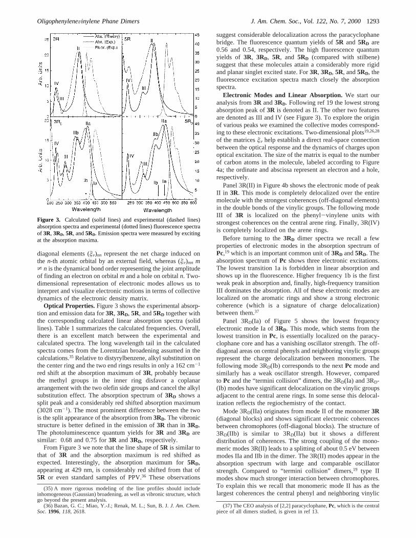
Optical Properties. Figure 3 shows the experimental absorp-tion and emission data for3R, 3RD, 5R, and5RD together withthe corresponding calculated linear absorption spectra (solidlines). Table 1 summarizes the calculated frequencies. Overall,there is an excellent match between the experimental andcalculated spectra. The long wavelength tail in the calculatedspectra comes from the Lorentzian broadening assumed in thecalculations.35 Relative to distyrylbenzene, alkyl substitution onthe center ring and the two end rings results in only a 162 cm-1
red shift at the absorption maximum of3R, probably becausethe methyl groups in the inner ring disfavor a coplanararrangement with the two olefin side groups and cancel the alkylsubstitution effect. The absorption spectrum of3RD shows asplit peak and a considerably red shifted absorption maximum(3028 cm-1). The most prominent difference between the twois the split appearance of the absorption from3RD. The vibronicstructure is better defined in the emission of3R than in3RD.The photoluminescence quantum yields for3R and 3RD aresimilar: 0.68 and 0.75 for3R and3RD, respectively.
From Figure 3 we note that the line shape of5R is similar tothat of 3R and the absorption maximum is red shifted asexpected. Interestingly, the absorption maximum for5RD,appearing at 429 nm, is considerably red shifted from that of5R or even standard samples of PPV.36 These observations
suggest considerable delocalization across the paracyclophanebridge. The fluorescence quantum yields of5R and 5RD are0.56 and 0.54, respectively. The high fluorescence quantumyields of 3R, 3RD, 5R, and 5RD (compared with stilbene)suggest that these molecules attain a considerably more rigidand planar singlet excited state. For3R, 3RD, 5R, and5RD, thefluorescence excitation spectra match closely the absorptionspectra.
Electronic Modes and Linear Absorption. We start ouranalysis from3R and3RD. Following ref 19 the lowest strongabsorption peak of3R is denoted as II. The other two featuresare denoted as III and IV (see Figure 3). To explore the originof various peaks we examined the collective modes correspond-ing to these electronic excitations. Two-dimensional plots19,26,28
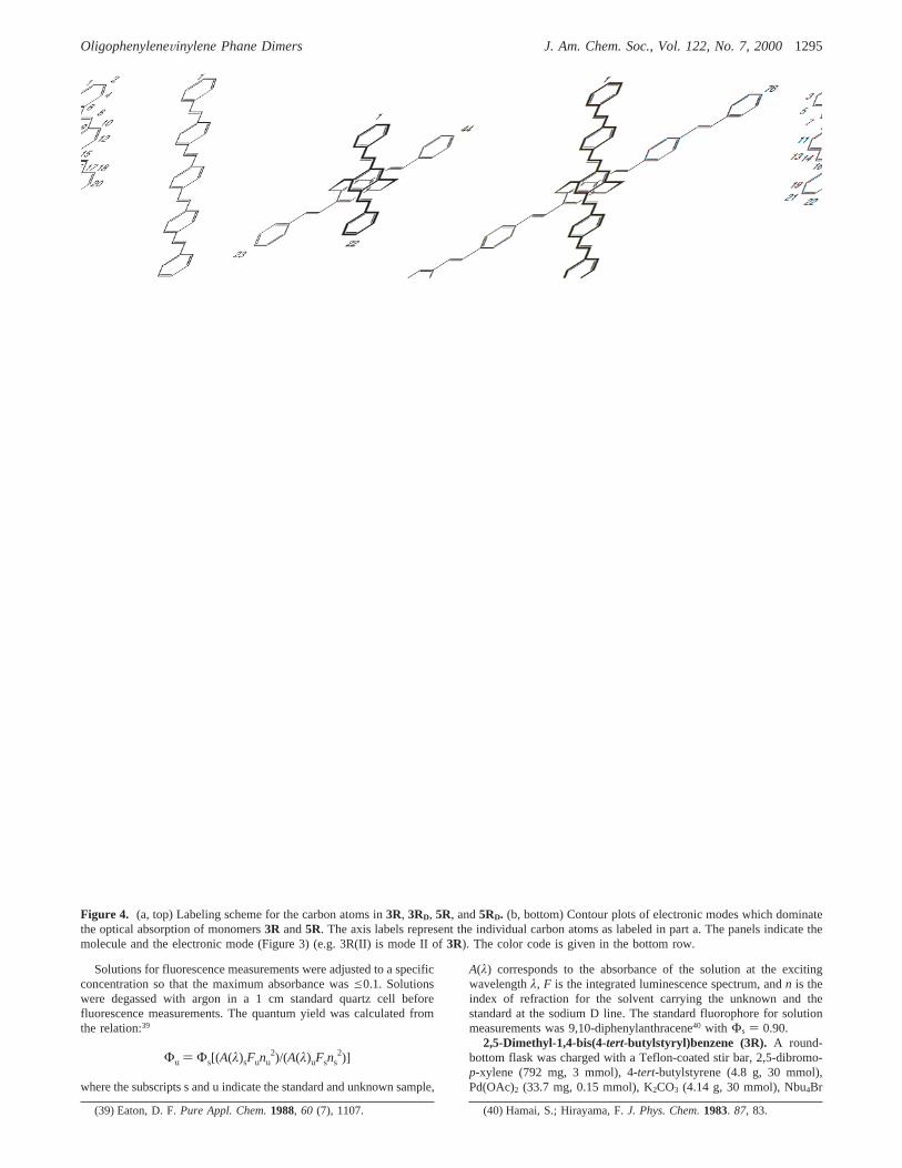
of the matricesêν help establish a direct real-space connectionbetween the optical response and the dynamics of charges uponoptical excitation. The size of the matrix is equal to the numberof carbon atoms in the molecule, labeled according to Figure4a; the ordinate and abscissa represent an electron and a hole,respectively.
Panel 3R(II) in Figure 4b shows the electronic mode of peakII in 3R. This mode is completely delocalized over the entiremolecule with the strongest coherences (off-diagonal elements)in the double bonds of the vinylic groups. The following modeIII of 3R is localized on the phenyl-vinylene units withstrongest coherences on the central arene ring. Finally, 3R(IV)is completely localized on the arene rings.
Before turning to the3RD dimer spectra we recall a fewproperties of electronic modes in the absorption spectrum ofPc,19 which is an important common unit of3RD and5RD. Theabsorption spectrum ofPc shows three electronic excitations.The lowest transition 1a is forbidden in linear absorption andshows up in the fluorescence. Higher frequency 1b is the firstweak peak in absorption and, finally, high-frequency transitionIII dominates the absorption. All of these electronic modes arelocalized on the aromatic rings and show a strong electroniccoherence (which is a signature of charge delocalization)between them.37
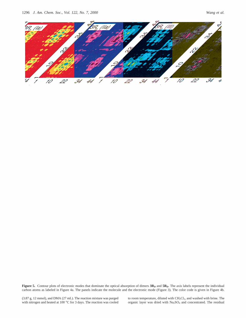
Panel 3RD(Ia) of Figure 5 shows the lowest frequencyelectronic mode Ia of3RD. This mode, which stems from thelowest transition inPc, is essentially localized on the paracy-clophane core and has a vanishing oscillator strength. The off-diagonal areas on central phenyls and neighboring vinylic groupsrepresent the charge delocalization between monomers. Thefollowing mode 3RD(Ib) corresponds to the nextPc mode andsimilarly has a weak oscillator strength. However, comparedto Pc and the “termini collision” dimers, the 3RD(Ia) and 3RD-(Ib) modes have significant delocalization on the vinylic groupsadjacent to the central arene rings. In some sense this delocal-ization reflects the regiochemistry of the contact.
Mode 3RD(IIa) originates from mode II of the monomer3R(diagonal blocks) and shows significant electronic coherencesbetween chromophores (off-diagonal blocks). The structure of3RD(IIb) is similar to 3RD(IIa) but it shows a differentdistribution of coherences. The strong coupling of the mono-meric modes 3R(II) leads to a splitting of about 0.5 eV betweenmodes IIa and IIb in the dimer. The 3R(II) modes appear in theabsorption spectrum with large and comparable oscillatorstrength. Compared to “termini collision” dimers,19 type IImodes show much stronger interaction between chromophores.To explain this we recall that monomeric mode II has as thelargest coherences the central phenyl and neighboring vinylic
(35) A more rigorous modeling of the line profiles should includeinhomogeneous (Gaussian) broadening, as well as vibronic structure, whichgo beyond the present analysis.
(36) Bazan, G. C.; Miao, Y.-J.; Renak, M. L.; Sun, B. J.J. Am. Chem.Soc.1996, 118, 2618.
(37) The CEO analysis of [2,2] paracyclophane,Pc, which is the centralpiece of all dimers studied, is given in ref 13.
Figure 3. Calculated (solid lines) and experimental (dashed lines)absorption spectra and experimental (dotted lines) fluorescence spectraof 3R, 3RD, 5R, and5RD. Emission spectra were measured by excitingat the absorption maxima.
OligophenyleneVinylene Phane Dimers J. Am. Chem. Soc., Vol. 122, No. 7, 20001293
groups, i.e., the electron-hole pair “spends time” in the middleof the molecule. The interaction between excitons through thecenter (“central collision” dimers) is much stronger than thatthrough the ends (“termini collision” dimers) of monomers.
The high-frequency modes 3RD(III) and 3RD(IV) arise from3R(III) and 3R(IV) monomeric modes, respectively. Theymanifest themselves with strong intensities in the absorptionspectra, show the same localization properties and, again, havesignificant optical coherences between the monomers. We thusconclude that the electronic excitations of the dimer3RD
originate from the excitations of itsPcand3R units with stronginteraction between monomers compared to the “termini col-lision” dimers.
We next turn to5R and5RD. The second row in Figure 4bshows the dominant electronic modes of5R. They have basicallythe same properties as the corresponding modes of the shortermolecule3R. The delocalized mode II is red-shifted. TransitionIII exhibits delocalization to the two phenyl-vinylene units atthe molecular ends and is therefore red-shifted as well comparedto 3R(III). In contrast, the 5R(IV) mode is completely localizedat the phenyls and does not shift.
As shown in Figure 5, the electronic modes of5RD originatefrom the correspondent excitations ofPcand5R units and showa strong interaction between the monomers fragments similarto that observed in3RD. The principal difference between3RD
and 5RD is the frequency position of Ia and Ib peaks. Inmolecule3RD transition Ia corresponds to the lowest frequencytransition. In longer dimers5RD, the state IIa is significantlyred-shifted since it is delocalized, whereas the state Ia does notshift and, consequently, the state IIa is the lowest energy state.
An indirect confirmation of our predicted ordering of the Iaand IIa states in the3RD and 5RD dimers is provided bycomparing the monomer and dimer fluorescence line shapes.The fluorescence spectra of5R and 5RD both have distinctvibronic structure, which suggests that the emitting state hasthe same nature, namely the 1Bu state of the monomer. Incontrast, the fluorescence profile of3RD is featureless and doesnot show the vibrational bands of3R. It closely resembles theemission of paracyclophane19 and suggests that the3RD emissionoriginates from the phane state.
Conclusion
Figure 4b shows that the lowest energy absorption band in3R (II) leads to electron delocalization over a large portion ofthe molecule. Also, the strongest coherences are localized overthe olefins. For5R the outer rings participate less in (II). Withincreasing excitation frequency the electronic modes becomemore localized on the repeat units. In other words, they becomemore localized on the individual aromatic rings.
It follows from our calculations that the Ia state (which maybe traced to thePc core) in3RD is nearly isoenergetic to stateIIa, which is more delocalized. An important difference betweenIa and IIa is in the distribution of electronic coherences acrossthe electronic mode. Ia and IIa have the strongest coherencesin the off-diagonal and diagonal directions, respectively. This
further leads to the vanishing oscillator strength of Ia and thestrong transition dipole of IIa. Therefore, while the distributionof electron density appears similar in these two states, Ia isforbidden and IIa is fully allowed. For5RD the IIa state issubstantially lower in energy than the corresponding Ia state. Itis more likely that the emitting state is directly related to thisstate. Otherwise the overall electronic description is similar.
The splitting of spectra in molecular aggregates is usuallyinterpreted using the Frenkel Exciton model, which assumesthat the electron density is localized within each unit. Theresulting Davydov splitting originates from purely electrostatic(e.g. dipole-dipole and higher multipole) interactions amongchromophores.38 The IIa-IIb splitting observed in the spectrais reminiscent of Davydov splittings. However, our CEOanalysis shows that the interaction between monomers is notelectrostatic, but involves electron exchange. This is clearly seenby the off-diagonal blocks between monomers in Figure 5,which represent a charge separation of an electron and a holebetween the monomers. These off-diagonal coherences shouldvanish identically for the Davydov Model. The strong couplingthrough the paracyclophane core provides a conduit for electrondelocalization. Differences in the optical properties of the “arms”and the “dimers” are a consequence of strong electroniccoherence between the two fragments brought about by theirclose proximity. The Davydov model cannot be used forcomputing these splittings.
Finally, these results highlight the effect of contact locationon the strength of delocalization between fragments. In the“terminal” contact pairs,19 the optical properties could be neatlyexplained in terms of excitations that migrated to the phane state(short arms) or that remained localized in the chromophores(longer arms). Choosing a trajectory such that the inner ringsare brought into close proximity prevents an analogous dissec-tion of parts. The optical properties and electronic descriptionof the criss-cross delocalized pairs indicate that the excitationis delocalized across the entire molecule. In a sense there isstrong mixing of the phane and the “antenna” or “chromophore”states.
Experimental Section
General Details.1H and13C NMR spectra were recorded on a VarianUnity 400 NMR spectrometer. UV-vis absorption spectra weremeasured on a Shimadzu UV-2401 PC diode array spectrophotometerand photoluminescence spectra on a Spex Fluoromax-2 spectrometer.High-resolution mass spectrometry was performed on a VG-70SEDouble Focusing System with FAB ionization sources. Purification offinal products was done with Chromatotron form Harrison ResearchCompany. Chromatotron rotor was coated with Merk TLC grade 7749silica gel. Reagents were obtained from Aldrich and used as received.The fluorescence quantum yield standard 9,10-diphenylanthracene wasobtained from Aldrich and was purified by column chromatographyon silica using petroleum ether as eluent.
(38) (a) Kasha, M.; Rawls, H. R.; El-Bayoumi, M. A.Pure Appl. Chem.1965, 11, 371. (b) Silinish, E. A.; Capek, V.Organic Molecular Crystals;American Institute of Physics: New York, 1994. (c) Pope, M.; Swenberg,C. E. Electronic Processes in Organic Crystals; Clarendon Press: NewYork, 1982.
Table 1. Computed Frequencies (in eV and cm-1)
Pc 3R 3RD 5R 5RD
Ia 3.95 (31857) 2.87 (23147) 2.85 (22985)Ib 4.77 (38470) 3.27 (26373) 3.17 (25566)IIa 3.50 (28227) 3.03 (24437) 3.27 (26373) 2.78 (22421)IIb 3.56 (28711) 3.21 (25889)III 4.96 (40002) 4.39 (35405) 3.95 (31857) 3.87 (31212)IV 5.5 (44358) 5.38 (43390) 5.37 (43309) 5.35 (43148) 5.33 (42986)
1294 J. Am. Chem. Soc., Vol. 122, No. 7, 2000 Wang et al.
Solutions for fluorescence measurements were adjusted to a specificconcentration so that the maximum absorbance wase0.1. Solutionswere degassed with argon in a 1 cm standard quartz cell beforefluorescence measurements. The quantum yield was calculated fromthe relation:39
where the subscripts s and u indicate the standard and unknown sample,
A(λ) corresponds to the absorbance of the solution at the excitingwavelengthλ, F is the integrated luminescence spectrum, andn is theindex of refraction for the solvent carrying the unknown and thestandard at the sodium D line. The standard fluorophore for solutionmeasurements was 9,10-diphenylanthracene40 with Φs ) 0.90.
2,5-Dimethyl-1,4-bis(4-tert-butylstyryl)benzene (3R). A round-bottom flask was charged with a Teflon-coated stir bar, 2,5-dibromo-p-xylene (792 mg, 3 mmol), 4-tert-butylstyrene (4.8 g, 30 mmol),Pd(OAc)2 (33.7 mg, 0.15 mmol), K2CO3 (4.14 g, 30 mmol), Nbu4Br
(39) Eaton, D. F.Pure Appl. Chem.1988, 60 (7), 1107. (40) Hamai, S.; Hirayama, F.J. Phys. Chem.1983. 87, 83.
Figure 4. (a, top) Labeling scheme for the carbon atoms in3R, 3RD, 5R, and5RD. (b, bottom) Contour plots of electronic modes which dominatethe optical absorption of monomers3R and5R. The axis labels represent the individual carbon atoms as labeled in part a. The panels indicate themolecule and the electronic mode (Figure 3) (e.g. 3R(II) is mode II of3R). The color code is given in the bottom row.
Φu ) Φs[(A(λ)sFunu2)/(A(λ)uFsns
2)]
OligophenyleneVinylene Phane Dimers J. Am. Chem. Soc., Vol. 122, No. 7, 20001295
(3.87 g, 12 mmol), and DMA (27 mL). The reaction mixture was purgedwith nitrogen and heated at 100°C for 3 days. The reaction was cooled
to room temperature, diluted with CH2Cl2, and washed with brine. Theorganic layer was dried with Na2SO4 and concentrated. The residual
Figure 5. Contour plots of electronic modes that dominate the optical absorption of dimers3RD and5RD. The axis labels represent the individualcarbon atoms as labeled in Figure 4a. The panels indicate the molecule and the electronic mode (Figure 3). The color code is given in Figure 4b.
1296 J. Am. Chem. Soc., Vol. 122, No. 7, 2000 Wang et al.
liquid (containing a small amount of DMA) was precipitated withhexanes and collected by filtration. The crude product was purifiedwith a Chromatotron on a 2 mmsilica-coated rotor (1:9 CHCl3/hexanes)to yield 0.7 g (55%) of a white solid.13C NMR (100 MHz, CDCl3) δ150.9, 135.7, 135.2, 133.6, 129.4, 127.2, 126.5, 125.8, 125.6, 34.8,31.5, 19.8 ppm.1H NMR (400 MHz, CDCl3) δ 7.41 (d,J ) 8.3 Hz,4H), 7.43 (s, 2H), 7.40 (d,J ) 8.3 Hz, 4H), 7.28 (d,J ) 16.1 Hz, 2H),7.00 (d,J ) 16.1 Hz, 2H), 2.43 (s, 6H), 1.35 (s, 18H) ppm. Exactmass (FAB, NBA) for M+ (C32H38): calculated 422.2974; found422.2958.
2,5-Dimethyl-1,4-bis[4-(4′-tert-butylstyryl)styryl]benzene (5R).Around-bottom flask was charged with a Teflon-coated stir bar, 2,5-dibromo-p-xylene (153 mg, 0.58 mmol), 4-trans-4,4′-tert-butylvinyl-stilbene (915 mg, 3.5 mmol), Pd(OAc)2 (6.5 mg, 0.03 mmol), K2CO3
(800 mg, 5.8 mmol), NBu4Br (748 mg, 2.3 mmol), and DMA (8 mL).The reaction mixture was purged with nitrogen and heated at 100°Cfor 3 days. The reaction was cooled to room temperature, diluted withCH2Cl2, and washed with brine. The organic layer was dried withNa2SO4 and concentrated. The residual liquid (containing a smallamount of DMA) was precipitated with hexanes and collected byfiltration. The crude product was purified with a Chromatotron on a 2mm silica-coated rotor (1:5 CHCl3/hexanes) to yield 91 mg (25%) ofyellow solid.1H NMR (400 MHz, CDCl3) δ 7.52 (s, 8H), 7.47 (d,J )8.5 Hz, 4H), 7.46 (s, 2H), 7.39 (d,J ) 8.5 Hz, 4H), 7.33 (d,J ) 16.1Hz, 2H), 7.10 (dd,J ) 16.6 Hz, 4H), 7.04 (d,J ) 16.1 Hz, 2H), 2.43(s, 6H), 1.35 (s, 18H) ppm. The compound was too insoluble to obtainan accurate13C NMR spectrum. Exact mass (FAB, NBA) for M+
(C48H50): calculated 626.3912; found 626.3901.4,7,12,15-Tetra(4-tert-butylstyryl)[2.2]paracyclophane (3RD). A
round-bottom flask was charged with a Teflon-coated stir bar, 4,7,-12,15-tetrabromoparacyclophane (200 mg, 0.39 mmol), 4-tert-butyl-styrene (1.84 g, 11.5 mmol), Pd(OAc)2 (12 mg, 0.054 mmol), K2CO3
(539 mg, 3.9 mmol), NBu4Br (502 mg, 1.56 mmol), and DMA (14mL). The reaction mixture was purged with nitrogen and heated at100°C for 4 days. The reaction was cooled to room temperature, diluted
with CH2Cl2, and washed with brine. The organic layer was dried withNa2SO4 and concentrated. The residual liquid (containing a smallamount of DMA) was precipitated with methanol and collected byfiltration. The crude product was purified with Chromatotron on 2 mmsilica-coated rotor (1:9 CHCl3/hexanes) to yield 146 mg (44%) of yellowsolid. 1H NMR (400 MHz, CDCl3) δ 7.45 (dd,J ) 8.6 Hz, 16H), 7.20(d, J ) 16.1 Hz, 4H), 6.94 (d,J ) 16.1 Hz, 4H), 3.57 (m, 4H), 2.87(m, 4H), 1.40 (s, 36H) ppm.13C NMR (100 MHz, CDCl3) δ 150.6,137.7, 136.6, 134.9, 128.2, 127.9, 126.3, 125.6, 124.7, 34.6, 33.0, 31.3ppm. Exact mass (FAB, NBA) for M+ (C64H72): calculated 840.5649;found 840.5634.
4,7,12,15-Tetra[4-(4′-tert-butylstyryl)styryl][2.2]paracyclo-phane (5RD). A round-bottom flask was charged with a Teflon-coatedstir bar, 4,7,12,15-tetrabromoparacyclophane (200 mg, 0.39 mmol),trans-4,4′-tert-butylvinylstilbene (1.39, g, 5.3 mmol), Pd(OAc)2 (20 mg,0.09 mmol), K2CO3 (539 mg, 3.9 mmol), NBu4Br (502 g, 1.56 mmol),and DMA (25 mL). The reaction mixture was purged with nitrogenand heated at 100°C for 4 days. The reaction was cooled to roomtemperature, diluted with CH2Cl2, and washed with brine. The organiclayer was dried with Na2SO4 and concentrated. The residual liquid wasprecipitated with methanol and collected by filtration. The crude productwas purified with a Chromatotron on 2 mm silica-coated rotor (3:7CHCl3/hexanes) to yield 88 mg (18%) of a yellow solid.1H NMR (400MHz, CDCl3) δ 7.56 (d,J ) 8.5 Hz, 8H), 7.50 (dd,J ) 8.5 Hz, 16H),7.42 (d,J ) 8.5 Hz, 8H), 7.23 (d,J ) 16.0 Hz, 4H), 7.16 (dd,J )16.3 Hz, 8H), 7.03 (s, 4H), 6.95 (d,J ) 16.0 Hz, 4H), 3.62 (m, 4H),2.92 (m, 4H), 1.40 (s, 36H) ppm. The compound was too insoluble toobtain an accurate13C NMR spectrum. Exact mass (FAB, NBA) forM+ (C96H96): calculated 1248.7512; found 1248.7499.
Acknowledgment. Financial support from NSF (DMR9500627, CHE-9814061, and PHYS 94-15583) and the Officeof Naval Research is gratefully acknowledged.
JA991611Y
OligophenyleneVinylene Phane Dimers J. Am. Chem. Soc., Vol. 122, No. 7, 20001297