Embed Size (px)
Citation preview
On Factors Influencing the Outcome of VariousMethods Using Endosseous Implants for
Reconstruction of the Atrophic Edentulous andPartially Dentate Maxilla
by
Jonas P Becktor
Department of Biomaterials, Institute for Clinical Sciences,Sahlgrenska Academy at Göteborg University
andThe Department of Oral and Maxillofacial Surgery,
Maxillofacial Unit, Halmstad Hospital,SWEDEN
Göteborg 2006
I

This PhD thesis represents number 33 in a series of investigations on implants, hard tissue
and the locomotor apparatus originating from the Department of Biomaterials/Handicap Research,
Institute for Clinical Sciences at Sahlgrenska Akademy, Göteborg University, Sweden.
1. Anders R Eriksson DDS, 1984. Heat-induced Bone Tissue Injury. An in vivo investigationof heat tolerance of bone tissue and temperature rise in the drilling of cortical bone. Thesisdefended 21.2.1984. Ext. examin.: Docent K.-G. Thorngren.
2. Magnus Jacobsson MD, 1985. On Bone Behaviour after Irradiation. Thesis defended29.4.1985. Ext. examin.: Docent A. Nathanson.
3. Fredrik Buch MD, 1985. On Electrical Stimulation of Bone Tissue. Thesis defended28.5.1985. Ext. examin.: Docent T. Ejsing-Jörgensen.
4. Peter Kälebo MD, 1987. On Experimental Bone Regeneration in Titanium Implants. Aquantitative microradiographic and histologic investigation using the Bone Harvest Chamber.Thesis defended 1.10.1987. Ext. examin.: Docent N.Egund.
5. Lars Carlsson MD, 1989. On the Development of a new Concept for Orthopaedic ImplantFixation. Thesis defended 2.12.1989. Ext. examin.: Docent L.-Å. Broström.
6. Tord Röstlund MD, 1990. On the Development of a New Arthroplasty. Thesis defended19.1.1990. Ext. examin.: Docent Å. Carlsson.
7. Carina Johansson Techn Res, 1991. On Tissue Reactions to Metal Implants. Thesisdefended 12.4.1991. Ext. examin.: Professor K. Nilner.
8. Lars Sennerby DDS, 1991. On the Bone Tissue Response to Titanium Implants. Thesisdefended 24.9.1991. Ext. examin.: Dr J.E. Davis.
9. Per Morberg MD, 1991. On Bone Tissue Reactions to Acrylic Cement. Thesis defended19.12.1991. Ext. examin.: Docent K. Obrant.
10. Ulla Myhr PT, 1994. On Factors of Importance for Sitting in Children with Cerebral Palsy.Thesis defended 15.4.1994. Ext. examin.: Docent K. Harms-Ringdahl.
11. Magnus Gottlander MD, 1994. On Hard Tissue Reactions to Hydroxyapatite-CoatedTitanium Implants. Thesis defended 25.11.1994. Ext. examin.: Docent P. Aspenberg.
12. Edward Ebramzadeh MSCeng, 1995. On Factors Affecting Long-Term Outcome of TotalHip Replacements. Thesis defended 6.2.1995. Ext. examin.: Docent L. Linder.
13. Patricia Campbell BA, 1995. On Aseptic Loosening in Total Hip Replacement: the Role ofUHMWPE Wear Particles. Thesis defended 7.2.1995. Ext. examin.: Professor D. Howie.
14. Ann Wennerberg DDS, 1996. On Surface Roughness and Implant Incorporation. Thesisdefended 19.4.1996. Ext. examin.: Professor P.-O. Glantz.
15. Neil Meredith BDS MSc FDS RCS, 1997. On the Clinical Measurement of Implant Stabilityand Osseointegration. Thesis defended 3.6.1997. Ext. examin.: Professor J. Brunski.
16. Lars Rasmusson DDS, 1998. On Implant Integration in Membrane-Induced and GrafterBone. Thesis defended 4.12.1998. Ext. examin.: Professor R. Haanaes.
17. Thay Q Lee MSc, 1999. On the Biomechanics of the Patellofemoral Joint and PatellarResurfacing in Total Knee Arthroplasty. Thesis defended 19.4.1999. Ext. examin.: DocentG. Nemeth.
18. Anna Karin Lundgren DDS, 1999. On Factors Influencing Guided Regeneration andAugmentation of Intramembraneous Bone. Thesis defended 7.5.1999. Ext. examin.:Professor B. Klinge.
19. Carl-Johan Ivanoff DDS, 1999. On Surgical and Implant Related Factors InfluencingIntegration and Function of Titanium Implants. Experimental and Clinical Aspects. Thesisdefended 12.5.1999. Ext. examin.: Professor B. Rosenquist.
II
20. Bertil Friberg DDS MDS, 1999. On Bone Quality and Implant Stability Measurements.Thesis defended 12.11.1999. Ext. examin.: Docent P. Åstrand.
21. Åse Allansdotter Johnsson MD, 1999. On Implant Integration in Irradiated Bone. AnExperimental Study of the Effects of Hyberbaric Oxygenation and Delayed ImplantPlacement. Thesis defended 8.12.1999. Ext. examin.: Docent K. Arvidsson-Fyrberg.
22. Börje Svensson DDS, 2000. On Costochondral Grafts Replacing Mandibular Condyles inJuvenile Chronic Arthritis. A Clinical, Histologic and Experimental Study. Thesis defended22.5.2000. Ext. examin.: Professor Ch. Lindqvist.
23. Warren Macdonald BEng, MPhil, 2000. On Component Integration in Total Hip Arthroplasty:Pre-Clinical Evaluations. Thesis defended 1.9.2000. Ext. examin.: Dr A.J.C. Lee.
24. Magne Røkkum MD, 2001. On Late Complications with HA Coated Hip Asthroplasties.Thesis defended 12.10.2001. Ext. examin.: Professor P. Benum.
25. Carin Hallgren Höstner DDS, 2001. On the Bone Response to Different Implant Textures.A 3D analysis of roughness, wavelength and surface pattern of experimental implants.Thesis defended 9.11.2001. Ext. examin.: Professor S. Lundgren.
26. Young-Taeg Sul DDS, 2002. On the Bone Response to Oxidised Titanium Implants: Therole of microporous structure and chemical composition of the surface oxide in enhancedosseointegration. Thesis defended 7.6.2002. Ext. examin.: Professor J.-E. Ellingsen.
27. Victoria Franke Stenport DDS, 2002. On Growth Factors and Titanium Implant Integrationin Bone. Thesis defended 11.6.2002. Ext. examin.: Associate Professor E. Solheim.
28. Mikael Sundfeldt MD, 2002. On the Aetiology of Aseptic Loosening in Joint Arthroplasties,and Routes to Improved cemented Fixation. Thesis defended 14.6.2002. Ext. examin.:Professor N Dahlén.
29. Christer Slotte DDS, 2003. On Surgical Techniques to Increase Bone Density and Volume.Studies in the Rat and the Rabbit. Thesis defended 13.6.2003. Ext. examin.: ProfessorC.H.F. Hämmerle.
30. Anna Arvidsson MSc, 2003. On Surface Mediated Interactions Related to Chemo-mechanical Caries Removal. Effects on surrounding tissues and materials. Thesis defended28.11.2003. Ext. examin.: Professor P. Tengvall.
31. Pia Bolind DDS, 2004. On 606 retrieved oral and cranio-facial implants. An analysis ofconsecutively received human specimens. Thesis defended 17.12. 2004. Ext. examin:Professor A. Piattelli.
32. Patricia Miranda Burgos DDS, 2006. On the influence of micro-and macroscopic surfacemodifications on bone integration of titanium implants.Thesis defended 1.9. 2006. Ext.examin: Professor A. Piattelli.
33. Jonas P Becktor DDS, 2006. On factors influencing the outcome of various techniquesusing endosseous implants for recosntruction of the atrophied edentulous and partiallydentate maxilla. To be defended 17.11.2006. Ext exam: Professor K. F. Moos
III
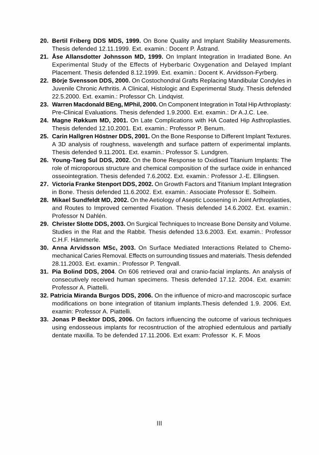
Surgical reconstruction of the severely resorbed edentulous maxilla often requires acombination of bone grafts and dental implants. Different methods have been used during the yearswhere donor site, type of bone graft, healing period, timing of implant placement and implant surfacehave varied. The overall objective of this research work is to evaluate the clinical outcome of suchmethods when used on a routine basis at one oral & maxillofacial surgery clinic at a county hospitalin Sweden. The purpose is also to evaluate the influence of various factors on implant failure.
In Paper I, one group of 64 grafted patients with 437 implants and one group of 118non-grafted patients with 683 implants were retrospectively evaluated and compared with regard toimplant and prosthesis survival. The former patients had received bone grafts from the iliac crestwith simultaneous or delayed (6 months) placement of dental implants with a minimally rough surface(machined/turned). More implant losses were seen in grafted than in non-grafted patients after amean follow-up of 5 to 6 years, 25% versus 16%, respectively. Most of the implants were lost beforeloading. There was no difference in prosthesis survival rate. A correlation between the bone volumeof the residual jaw bone prior to bone grafting and implant failure rate was seen in the anteriormaxilla. There was no difference in implant failure rate between one-stage and two-stage bonegrafting and implant placement procedures.
The influence of the type of occlusal support on early implant failure in grafted maxillaewas evaluated in Paper II. Ninety (90) patients previously treated with bone grafts from the iliaccrest and 643 machined/turned implants were included in the retrospective study. The total failurerate was 18%. In comparison, few failures (6.2%) were seen in patients with a removable mandibulardenture and the highest failure rate (43.8%) was seen in patients with unilateral occlusal support.
Sixteen patients previously treated with 31 zygomatic implants and 74 regular implantsin the anterior maxilla as an alternative to bone grafting of the atrophic maxilla were evaluated inPaper III. All implants had a minimally rough surface. Three (4.1%) regular implants were lost andthree (9.7%) zygomatic implants had to be removed due to recurrent sinusitis after a mean follow upperiod of 4 years. All patients received and maintained a fixed bridge.
Paper IV evaluated 17 patients subjected to maxillary sinus floor augmentation withblocks of bone from the iliac creast and simultaneous or delayed (6 months) placement of 69machined/turned implants. After a mean follow up period of 4 years, 8.7% of the implants had beenlost. All failures occurred prior to loading of the fixed prostheses. More implants were lost in grafted(10.4%) than in non-grafted (4.8%) areas. Less implants were lost when using a two-stage approachthan when using a one-stage technique, 6% versus 18%, respectively.
In a prospective study including 61 patients (Paper V), the use of particlated mandibularbone for maxillary sinus floor augmentation and delayed placement of three types of surface modifiedimplants (oxidized, blasted, blasted+acid etched) was evaluated. The majority of patients were treatedunder local anaesthesia. Two of 180 implants were lost from placement to delivery of the finalprosthesis.
It is concluded that more implant failures occur in grafted than in non-grafted maxillae.The bone volume of the residual anterior crest and the occlusal support depending on the type ofmandibular occlusion seems to influence the outcome of grafting procedures in the edentulousmaxilla. Delayed placement of dental implants in bone grafts seems preferable, at least in partiallydentate patients. The use of surface modified implants and particulated mandibular bone may beone way to further improve the results of sinus grafting procedures. The use of zygomatic implantsis a viable alternative to bone grafting in the treatment of the severely resorbed maxilla.
Keywords: clinical studies, dental implants, maxilla, bone grafting, zygomatic implants
ISBN-10: 91-628-6992-2, ISBN-13: 978-91-628-6992-2
Correspondence: Jonas P Becktor, Dept Biomaterials, Inst Clin Sciences, Sahlgrenska Academyat Göteborg University, PO Box 412, SE-405 30 Göteborg, SWEDEN
IV
ABSTRACT

LIST OF PAPERS
I. Becktor JP, Isaksson S, Sennerby L. Survival Analysis ofEndosseous Implants in Grafted and Nongrafted EdentulousMaxillae.Int J Oral Maxillofac Implants 2004;19(1):107–115.
II. Becktor JP, Eckert SE, Isaksson S, Keller EE.The Influence ofMandibular Dentition on Implant failures in Bone-graftedEdentulous Maxillae.Int J Oral Maxillofac Implants 2002;17(1):69-77.
III. Becktor JP, Isaksson S, Abrahamsson P, Sennerby L.Evaluation of 31 Zygomatic Implants and 74 Regular DentalImplants Used in 16 Patients for Prosthetic Reconstruction of theAtrophic Maxilla with Cross-Arch Fixed BridgesClin Implant Dent Relat Res 2005;7(3):159-65.
IV. Becktor JP, Isaksson S, Sennerby L. Endosseous Implants andBone Augmentation in the Partially Dentate Maxilla:An Analysis of 17 Patients with a Follow-Up of 29 to 101 Months.Int J Oral Maxillofac Implants 2006, Accepted
V. Becktor JP, Hallström H, Isaksson S, Sennerby L. Aprospective clinical and radiographic analysis of 180 implantsplaced in partially dentate maxilla after maxillary sinus flooraugmentation with particulated autogenous bone from themandibular ramus/corpus.In manuscript
V
INTRODUCTION 1Audit and quality assessment 1Background 1Bone biology and implant integration 3Bone biology 3Bone cells 3Intramembranous and endochondral bone formation: 5Bone turnover: 5Bone healing: 6Osteoinduction and osteoconduction: 6Osseointegration and implants: 7Influence of maxillary growth and anatomy on implant installation 9Maxillary growth: 9Congenital maxillary edentulism: 9Acquired maxillary edentulism: 9Bone graft to the maxilla and implant installation: 10Historical review: 10Free autogenic bone grafts: 11Biologic factors: 11Embryology: 12Donor sites: 13Inlay bone graft: 13Onlay bone graft 15Block and/or particulated bone: 15Vascularised bone grafts 16Allogenic and xenogenic bone grafts: 17Healing period 18Surgeon’s experience 20AIMS 23MATERIALS AND METHODS 24Subjects 24Drop-outs 26Surgery 27Prosthodontics 30Examinations and Follow-up 30Radiographic Examination 30Statistics 31RESULTS 33Paper I 33Paper II 38Paper III 40Paper IV 42Paper V 44DISCUSSION 46CONCLUSIONS 57ACKNOWLEDGEMENTS 58REFERENCES 60
CONTENTS
VI
1
INTRODUCTION
Audit and quality assessmentNew public management (NPM) has been introduced to the world of public
health lately. The ideas are coming from the cooperate world and are modifiedto fit the establishments of public health (Hood and Dunleavy, 1994). The idea isto be capable of evaluating the different structures at a hospital, such as cultural/social, institutional/organizational and down to the individual level. The hospitalsshould regulate themselves by systematic compulsory training, education andcollegial discipline (Starr and Immergut, 1987). One of the ingredients of NPMis quality assessment, audit, which should provide a way of measuring anddescribing the public health from a quality point of view. ”We have always beenworking with quality in our department, we just did not have the tools andknowledge to systemize it”.
In the ”Audit bill” which was passed in Sweden in 1997,(Socialstyrelsen1996-00-116, Stockholm, 1996) it was required that ”right things will be done theright way” to acquire productivity and efficiency in the organization.
The material in the present thesis has been collected throughout the dailywork at the Department of Oral and Maxillofacial Surgery, Maxillofacial Unit, atthe County Hospital, Halmstad, Sweden. One may consider the present thesisas representative of one form of NPM, where the audit of treatment is evaluatedand thereby leading to a research based improved development.
BackgroundTotal or partial edentulism of the maxilla can be of different aetiologies;
agenesis, periodontal disease, infections, caries, malignancies or trauma.Despite of the different aetiologies, oral rehabilitation of these patients currentlyinvolves installation of endosseous implants and good long-term clinical resultshave been demonstrated (Adell et al., 1990a; Jemt and Lekholm, 1995; Tolmanand Laney, 1992).
Conventional removable prostheses retained by remaining teeth and/orthe residual alveolar crest and a tooth supported dental bridge with cantileversin the edentulous regions have been the treatment of choice for many years. Incases of low patient acceptability and risk for prosthetic mechanical failures, theuse of endosseous implants is currently well documented and considered aroutine treatment for prosthetic reconstruction of the edentulous and partially
2
dentate maxilla (Branemark et al., 1977; Owall and Cronstrom, 2000; Randowet al., 1986). The use of titanium implants for rehabilitation of edentulism wasfirst introduced by Brånemark et al. (1969) and later by Schroeder et al.(1976).Acceptable long-term implant survival rates with minimal marginal bone losshave been presented in patients with sufficient jaw bone volumes (Adell et al.,1990a; Henry et al., 1996; Jemt and Lekholm, 1993; Roos et al., 1997; Tolman,1995). However, an adequate amount of jaw bone to allow sufficient numbersand sizes of implants seems to be a requirement for achieving good results.Moreover, the quality/density of the jaw bone is an important factor for implantsurvival (Friberg et al., 1991; Sennerby and Roos, 1998; van Steenberghe et al.,1990).
In subjects with insufficient jaw bone volume the problem may be solvedby using shortand/or thin implants or by tilting the implants into regions wherebone is present. However, this approach may sometimes result in difficulties ofmanaging the prosthetic treatment (Aparicio et al., 2001; Krekmanov, 2000;Mattsson et al., 1999).
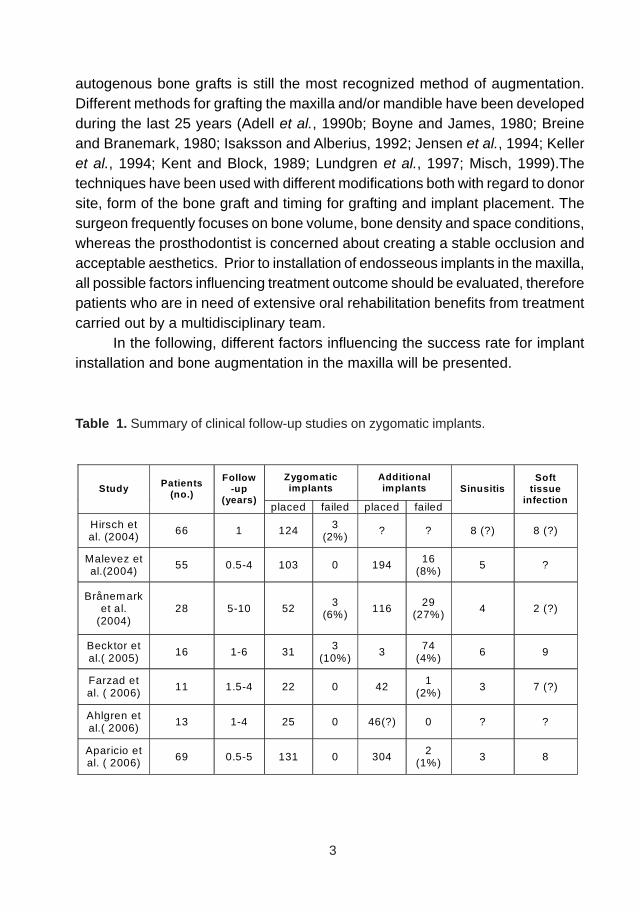
The insertion of specially designed long implants, zygomatic implants, hasalso been used to overcome problems with insufficient bone volume in theposterior maxilla. The placement of dental implants in the zygomatic bone iswell known from preprosthetic surgery following ablative tumour surgery (Higuchi,2000; Parel et al., 2001) and has also been used in conjunction with regularimplants in patients with severe atrophy and resorption of the posterior maxillaas an alternative to bone augmentation (Bedrossian et al., 2002; Branemark etal., 2004; Higuchi, 2000; Hirsch et al., 2004; Malevez et al., 2004). (Table 1)Another technique is the pterygomaxillary implant that was first described byTulasne (1992). This implant is placed in the maxillary tuberosity region, and issupposed to involve the pterygoid plate to gain acceptable implant stability (Bahat,1992; Balshi et al., 1999; Tulasne, 1992). Reviews of the literature reveal anincreased implant failure rate in situations with inadequate bone volume andthe insertion of either zygomatic or pterygomaxillary implants could thus be analternative treatment (Esposito et al., 1998a; Esposito et al., 1998b; Tong et al.,1998).
The atrophied maxilla constitutes a challenging therapeutic problem andbone augmentation is often essential to enable placement of sufficient numberand sizes of implants. Bone augmentation is required when the width and thevertical height of the residual alveolar ridge in the edentulous or partially dentatepatient is insufficient for placing implants with acceptable size, which is necessaryfor optimal functional and aesthetic prosthetic reconstruction. The use of
3
autogenous bone grafts is still the most recognized method of augmentation.Different methods for grafting the maxilla and/or mandible have been developedduring the last 25 years (Adell et al., 1990b; Boyne and James, 1980; Breineand Branemark, 1980; Isaksson and Alberius, 1992; Jensen et al., 1994; Kelleret al., 1994; Kent and Block, 1989; Lundgren et al., 1997; Misch, 1999).Thetechniques have been used with different modifications both with regard to donorsite, form of the bone graft and timing for grafting and implant placement. Thesurgeon frequently focuses on bone volume, bone density and space conditions,whereas the prosthodontist is concerned about creating a stable occlusion andacceptable aesthetics. Prior to installation of endosseous implants in the maxilla,all possible factors influencing treatment outcome should be evaluated, thereforepatients who are in need of extensive oral rehabilitation benefits from treatmentcarried out by a multidisciplinary team.
In the following, different factors influencing the success rate for implantinstallation and bone augmentation in the maxilla will be presented.
Table 1. Summary of clinical follow-up studies on zygomatic implants.
Zygomatic implants
Additional implants Study Patients
(no.)
Follow-up
(years) placed failed placed failed
Sinusitis Soft
tissue infection
Hirsch et al. (2004)
66 1 124 3 (2%)
? ? 8 (?) 8 (?)
Malevez et al.(2004)
55 0.5-4 103 0 194 16 (8%)
5 ?
Brånemark et al.
(2004) 28 5-10 52
3 (6%) 116
29 (27%) 4 2 (?)
Becktor et al.( 2005)
16 1-6 31 3
(10%) 3
74 (4%)
6 9
Farzad et al. ( 2006)
11 1.5-4 22 0 42 1
(2%) 3 7 (?)
Ahlgren et al.( 2006)
13 1-4 25 0 46(?) 0 ? ?
Aparicio et al. ( 2006) 69 0.5-5 131 0 304
2 (1%) 3 8
4
Bone biology and implant integration
Bone biology
A characteristic of all bones is a dense outer sheet of compactbone and a central medullary cavity. The cavity is filled with bone marrow, whichis interrupted by a network of bone trabeculae. Mature bone, irrespective if corticalor cancellous, is histologically identical, in that it consists of microscopic layersor lamellae. Three distinct types of layering are recognised: circumferential,concentric and interstitial. Circumferential lamellae (i) enclose the entire adultbone, forming its outer perimeter. Concentric lamellae (ii) make the bulk ofcompact bone, and represent the basic metabolic unit of bone, the osteon. Theosteon is a cylinder of bone, with a central Haversian canal, lined by layers ofbone cells that cover the bone surface; each canal houses minimally one capillary.Haversian canals are interconnected by Volkmann canals, channels that alsocontain blood vessels, thus creating a rich vascular network through corticalbone. Interstitial lamellae (iii) are interspersed between adjacent concentriclamella and fill the spaces between them.
The periosteum is surrounding the outer aspect of every compactbone and the internal surfaces of compact and trabecular bone is covered bythe endosteum. In general the periosteum is more active in bone formationthan the endosteum, particularly in young individuals.
Bone formation occurs by three main mechanisms: endochondral,intramembranous and sutural. Endochondral bone formation takes place whencartilage is replaced by bone, intramembranous bone formation occurs directlywithin the mesenchyme and sutural growth takes place at the sutural margins.
Bone cells
Two cell lineages are present in bone: (i); osteogenic cells, whichform and maintain bone and (ii); osteoclasts which resorb bone. Osteogeniccells (i) include osteoprogenitors, preosteoblasts, osteoblasts, osteocytes andbonelining cells. Osteoblasts synthesize collagenous and noncollagenous bonematrix proteins that may accumulate as an uncalcified matrix called osteoid thatacts as a scaffold for the deposition of apatite crystals of bone. They arise frompluripotent stem cells, which are of mesenchymal origin. In addition to osteoid,ostoblasts secrete a variety of cytokines that regulates cell metabolism.Osteoblasts produce several different forms of bone morphogenetic proteins(BMP). Although the interaction between these growth factors is very complex,
5
they increase the rapidity of bone formation and bone healing. Hormones arealso a important factor for bone metabolism.
Transformation of an osteoblast into an osteocytes occurs whenthe osteoblast stops synthesising matrix (osteoid) and it becomes buried withinthe calcified tissues. Woven bone have more osteocytes than lamellar bone.Adjacent osteocytes maintain contact through channels called canaliculi, thatalso connect with nearby capillaries. Osteoclasts are large multinucleated cellsthat resorb bone and their origin is hematopoietic. Recruitment of bone formingcells and bone resorbing cells is of great importance during bone growth andbone healing. Osteoblasts and osteocytes, although of opposing fractions, actas coupled cells, i.e. their actions are dependent of one another.
Intramembranous and endochondral bone formation
Intramembranous bone formation occurs directly within themesenchyme, the mesenchymal cells proliferate and condense simultaneouslywith an increase in vascularity at these sites of condensed mesenchyme whereosteoblasts differentiate and begin to produce osteoid. The interval betweenosteoid deposition and mineralization in woven bone is 1-3 days. Once begun,intramembranous bone formation proceeds rapidly, and the first deposited boneis termed woven bone. A continual process occurs where woven bone istransformed into lamellar bone. Consequently, woven bone is seen during earlybone formation during growth and healing whereas lamellar bone is the moremature bone characterized by tightly packed osteons.
The formation of endochondral bone takes place through thedifferentiation of the mesenchymal cells into cartilage producing cells, forming acartilage template of the future bone. The cartilage template will becomehypotrophic, calcify, and then be replaced by bone tissue. The initially producedbone has a primitive and irregular appearance which also is the case forintramembranous woven bone, before it remodels into lamellar bone (Alberiuset al., 1992; Rabie et al., 1996; Zins and Whitaker, 1983)
Bone turnover
Bone remodelling is a substitution of the bone tissue withoutchanging its architecture in contrast to surface modelling that changes the shapeof bone due to resorption and/or appositional growth. Remodelling occurs
6
throughout life by the coordinated action by osteoclasts and osteoblasts, in ahealthy individual, this turnover is in steady state, i.e. the amount of lost bone isbalanced by bone formation.
Bone healing
Jaw bone healing, e.g. after a fracture or implant placement, occurs in twophases, initial repair and secondary remodelling (Schenk et al., 1994). Initially,as a result of vascular disruption, a haematoma forms between and around thebone segments. The haematoma is converted into a clot and bony necrosisoccurs at the end of the fracture segments. Ingrowth of vasoformative cells andcapillaries for the restoration of blood supply, angiogenesis, followed by migrationof granulocytes, monocytes, lymphocytes and pluripotent stem cells occur inthe traumatized area. After 1-3 days the clot is replaced by granulation tissue,which consists of inflammatory cells, fibroblasts, collagen and invading capillaries.The granulation tissue is converted into a collagen matrix with continuousingrowths of capillaries. Woven bone is rapidly formed by osteoblasts, whichhave either been differentiated from mesenchymal stem cells or activated liningcells. Because of poor mineralization and organization of this bone, itsbiomechanical properties are poor. The second phase, secondary remodelling,consists of replacement of the woven bone with ordered lamellar bone, which isdirected by osteoblastic and osteoclastic activities. A complete regeneration ofa wound, where all areas of woven bone have been replaced by lamellar bone isseldom seen in adults. Incomplete healing occurs with ingrowth of fibrous tissue.This can be due to lack of sufficient blood supply, pressure and instability (Schenket al., 1994). Stability of the immature bone is important in the early stage ofwound healing, if this is not established the mesenchymal stem cells maydifferentiate into fibroblasts instead of osteoblasts (Hjørting-Hansen et al., 1990;Phillips and Rahn, 1988).
Osteoinduction and osteoconduction
Osteoinduction is when primitive undifferentiated and pluripotent cells arestimulated by an inductive agent to develop into bone-forming cells andosteogenesis is induced. Osteoconduction is when bone grows in a matrix oron a surface. An osteoconductive surface permits bone growth on its surfaceand down into pits and pores and it is suggested that the bone is conformed toa materials surface (Albrektsson and Johansson, 2001).
7
Osseointegration and implants
Per-Ingvar Brånemark placed his first clinical oral implant in 1965 and theterm osseointegration was established in 1977 (Brånemark et al. 1977). Althoughearly trials with the Brånemark system of osseointegration were unsuccessful,significant improvements and thorough documentation of the clinical outcomeled to their general acceptance of the osseointegration technique (Brånemarket al., 1977). Osseointegration is histologically defined in Dorland’s IllustratedMedical Dictionary as the direct anchorage of an implant by the formation ofbony tissue around the implant without the growth of fibrous tissue at the bone-implant interface.
Different dental implant systems are available on the market. Jokstad etal. reported 220 different implant brands produced by about 80 manufactures.The implants vary in shape, material, dimension and surface structure (Jokstadet al., 2003). In the past, the most common implants where produced eitherwith a machine turned technique resulting in a minimally rough surface (machined/turned) or with a plasma spraying approach producing a rough surface. Today,the market is dominated by implants with moderate surface roughness, i.e.blasted, acid-etched, oxidized, plasma-sprayed and hydroxylapatite coated ones,which have been developed to allegedly improve the clinical performance.
The importance of implant surface properties for successfulosseointegration has been known for some time (Albrektsson et al., 1981).However, the exact role of the surface properties of titanium implants during theformation of osseointegration is still under discussion (Albrektsson, 1983;Wennerberg, 1996). Interests in the surface oxide properties of titanium implantshave increased with the development of methods to characterize such surfaces.Moreover, the influence of surface modification of titanium implants on the tissueresponses is an important and common topic in implant research. Implants withrough surfaces are claimed to promote faster and earlier bone healing and therebybe more suitable for earlier loading than has previously been the standard formany years. Ivanoff et al.(2003) evaluated the human bone tissue response totwo surfaces (oxidized and turned) implants on twenty patients who receivedone test and one control micro-implant each during implant surgery. Surfaceroughness and enlargement were greater for the oxidized implants than for theturned implants. Histomorphometric evaluations demonstrated significantlyhigher bone-to-implant contact and bone density in the threaded region for theoxidized implants (Ivanoff et al., 2003). However, rougher surfaces may havetheoretical clinical drawbacks such as being more prone to marginal bone
8
resorption and/or increased ion release, which has been found in bone tissue inthe surrounding area of titanium implants and it has been hypothesized that thiscould be damaging to osteogenesis (Osborn et al., 1990; Tsutsui et al., 1999).However, Wennerberg et al. (2004) showed no correlation between increasingroughness and ion release, neither in vitro nor in vivo.
In the contact zone between implant and bone, the ”tissues” have no directcontact to the bulk titanium, but rather to a thin oxide layer of the metal. Thisthin oxide layer was shown to be in ’contact’ with remodelled mineralized bone(Sennerby et al., 1992). Studies of implants that have been retrieved from patientshave demonstrated that both the thickness and the nature of the thin oxide layerchanged during implantation. Successfully osseointegrated titanium implantsshowed an increase in oxide thickness of up to 200 nm (Sundgren et al., 1985).However, in the case of failed titanium implants that were retrieved from patients,there were no changes in the oxide thickness or oxide composition during aperiod of function of up to eight years (Esposito et al., 1999).
It is likely that the surface of a transmucosal implant part should have asmooth surface in order to establish a mucosal seal and to avoid soft tissuereactions (Sawase et al., 2000). Previous publications have indicated thatabutment surface roughness is positively correlated with increased accumulationof subgingival plaque (Quirynen et al., 1990). Experimental studies have shownthat plaque accumulation may lead to inflammatory lesions in the adjacentmucosa and bone resorption, with subsequent risk of implant failure(Abrahamsson et al., 1998; Lindhe et al., 1992). However, Wennerberg et al.(2003) presented a statistically significant difference only between patientsregarding the amount of accumulated plaque on the abutment surfaces andinflammatory cells, but no difference between the surface modifications in relationto plaque accumulation or number of inflammatory cells, although their studieswere limited to a healed situation and a follow up time of only one month(Wennerberg et al., 2003).
9
Influence of maxillary growth and anatomy on implant installation
Maxillary growth
The facial skeleton is formed by intramembranous ossification andcomprises six different anatomical bones: Maxillary bone, Palatine bone,Zygomatic bone, Vomer, Ethmoid bone and Nasal bone. Vomer is a single boneand the remaining five bones are pairs. The maxilla consists of four processes:Processus frontalis, processus zygomaticus, processus palatinus and processusalveolaris. The four processes meet the facial skeleton in different sutures fromwhere vertical and sagittal growth displacement of the maxilla occurs. Growthof the alveolar process occurs sagitally, vertically and transversely by eruptionof the dentition and additionally there is apposition posteriorly to the maxillarytuberosity. The maxilla also increases in height by relocation of the nasal floorand transversely by differentiated growth of the midpalatine suture (Bjork 1964;Bjork and Skieller, 1977).
Congenital maxillary edentulism
In areas of the maxilla with multiple missing teeth, growth of the alveolarprocess will not occur. Accordingly, sufficient bone for implant installation willnot be present. Unfavourable anatomy of the maxillary sinus may furtherdecrease the amount of jaw bone and thereby complicate implant installation inthe congenital fully or partially edentulous maxilla.
Due to the fact that implants are osseointegrated, they will not take part inthe growth mechanism of the alveolar process, it has been recommended not toplace implants in growing individuals (Thilander et al., 2001). The continuouslyerupting dentition in growing individuals will lead to infraocclusion of the implants.
Acquired maxillary edentulism
Acquired maxillary edentulism shows morphological alteration of thejawbone anatomy and reduces the mastication capability with time. Resorptionof the alveolar process and the maxillary basal bone (Cawood and Howell, 1988;Tallgren, 1972) and pneumatization of the maxillary sinus, lead to an unfavourableanatomy and thereby constituting a therapeutic problem. The same morphologicalalteration of the jaw bone anatomy is also present in the partially dentate patient,and it is most likely that future demands for implant-based reconstructions will
10
come from partially edentulous patients, of whom a number will need boneaugmentation (Meskin and Brown, 1988; Weintraub et al., 1985).
Bone grafting to the maxilla and implant installation
Historical review
In the 19th century, Ollier (1867) considered the periosteum of majorimportance for successful bone grafting and accepted only autogenous bonegrafts for clinical use, because this was the only type of graft that survivedtransplantation. Barth (1895) questioned the conclusions made by Ollier andreported that the periosteum seldom survived transplantation. The importantfactor for regeneration of a bone defect was suggested to be the osteogenicproperty of the host bone. He also believed that the transplanted bone alwayswas resorbed and replaced by the host. Accordingly, there should be no differencebetween autogenous, allogenous and xenogenous bone grafts (Barth, 1895).Bull (1928) supported Barth´s theory, but concluded that the replacement phasewas shortest for the autogenous graft. Baschkirzew & Petrov (1912)experimented by inserting different types of bone into muscular tissue and noticedthat neither the periosteum nor the osteocytes were necessary for bone formation.In vital bone, without periosteum or marrow and transplanted into muscular tissue,osteocytes were differentiated from the surrounded connective tissue cells(Baschkirzew and Petrov, 1912).
Several reviews have been published (Albreksson, 1979; Chase andHerndon, 1955; Puranen, 1966; Ray, 1956; Urist, 1960) based on the abovementioned basic principles. Today, it is generally accepted that autogenousbone is the best grafting material and that osteogenic cells from periosteum,endosteum and bone marrow, all may take part in the process of bone graftincorporation and healing.
Numerous surgical procedures for implanting allogenous materials tocompensate for loss bone and teeth, such as subperiosteal and blade implantswere used for many years, if with dubious results. The first scientificallydocumented rehabilitation of edentulism with osseointegrated implants wasdescribed by Brånemark et al. (1969). Today, when following the same principles,the use of osseointegrated oral implants is considered to be a well documented,safe procedure with predictable outcomes.
11
Free autogenic bone grafts
The process of healing and incorporation of free autogenic bone is ofutmost importance for clinical success. Due to osteoinductive andosteoconductive capacities, they are superior to both allografts and xenografts.Osteoinduction is described as a process where mesenchymal cells within thedonor tissue has the potential to initiate new bone formation under influence ofBMP (Urist, 1960). Osteoconduction is a three dimensional process, where thedonor tissue acts as a scaffold for ingrowth of capillaries, perivascular tissueand osteoprogenitor cells from the recipient bed into the donor tissue (Urist,1960).
Abundant factors and biological processes have to take place before anautogenic transplant is successfully incorporated in the recipient bed. Primarily,the surgical technique has an important influence on the success rate.Furthermore, the level of incorporation depends on biologic factors associatedwith the graft and on factors associated with the recipient site. The importantfactors for healing are similar in all the different types of autogenous grafts tothe maxilla such as; sinus inlays, alveolar/maxillary onlays, block and/orparticulated bone and vascularized bone grafts.
Biologic factors
Revascularization is crucial for graft healing and is characterised bymicrovascularisation initially occurring in a layer of about one mm of the graftsurface, which is in direct contact with the recipient bed. Microanastomosesmay restore circulation and are responsible for survival of osteoprogenitor cellsin the graft. The revascularization process differs between cortical and cancellousbone grafts due to different morphologies. Cortical bone is densely packed andcancellous bone porous with marrow tissue in between the bone trabeculae,because of this difference, vascular ingrowth has been demonstrated to occur30% more rapidly into cancellous compared to cortical bone grafts (Albrektsson,1980).
In the osteoinductive graft preosteoblasts may survive transplantation andthese proliferating cells will form a bridge between the surface of the donor andthe recipient site, which in turn will enhance the amount and pace of remodelling.Furthermore growth factors and proteins will influence the osteoinductive processduring healing of the autogenic bone graft. BMP has demonstrated to enhancebone healing (Urist, 1960; Urist, 1965) and autogenic bone enriched with BMP

12
and BMP alone will lead to enhanced bone regeneration (Marukawa et al., 2001).Another factor that has been demonstrated to have an osteoinductive influenceon bone grafting, is autogenic bone enriched with platelet-rich plasma (PRP)which is suggested to increase bone regeneration (Wiltfang et al., 2004).
Embryology
The embryological origin of the bone graft has been suggested to play arole in the success of the bone augmentation procedure. It has been proposedbased on animal studies, that intramembranous bone block grafts have a betterresistance towards volumetric bone block graft resorption compared toendochondral bone grafts (Smith and Abramson, 1974; Zins and Whitaker, 1983).Alberius et al. (1992) showed in an animal study that intramembranous bonegrafts healed better compared to endochondral grafts and indicated that abiological difference exists between the two types of bone grafts. Rabie etal.(1996) reported that intramembranous bone grafts healed through anosteogenic ossification route where preosteoblasts, osteoblasts, and osteocyteswere observed with no cartilage intermediate stage, while in endochondral bonegrafts, chondroblasts and chondrocytes were observed and healing occurredthrough an endochondral ossification route. Kusiak et al. (1985) suggested inan animal study that intramembranous onlay bone grafts become earlierrevascularized than endochondral grafts and thereby maintain volume and viabilityto a greater extent. Sullivan & Szwajkun (1991) found that endochondral graftshad quantitatively greater revascularization than intramembranous grafts.Differences in graft architecture were theorized to account for the difference inrevascularization in endochondral and membranous bone grafts.
Chen et al. (1994) demonstrated that calvarial bone grafts maintainedvolume better than iliac bone grafts. The osteoclastic activity andrevascularization were greater in the cancellous portion of calvarial and iliacbone grafts. Because calvarial bone grafts contain more cortical bone, theirsuperior volume maintenance can be understood by the architectural influenceon revascularization and resorption. The revascularization process differsbetween cortical and cancellous bone grafts because of the differentmorphologies. Cortical bone is densely packed and cancellous bone porous,with marrow tissue in between the bone trabeculae.
13
Donor sites
Autogenous grafts are often used due to their osteoconductive andosteoinductive capacities (Urist, 1980). They can be harvested from differentsites in the body e.g.: the iliac crest, the calvaria, the ribs, the mandible (Kondellet al., 1996; Loukota et al., 1992; Lundgren et al., 1996). The most appropriateprocedure to use depends on the amount of bone needed and surgicalpreference.
To harvest large amounts of bone, extra oral sites such as the iliac cresthas often been used. Postoperative morbidity as bruising, swelling, pain andfunctional problems at the donor site is more often seen using extra- than intra-oral donor sites. The extra oral approach will also produce a permanentcutaneous scar, and usually involves general anaesthesia with days ofhospitalization (Beirne, 1986; Cricchio and Lundgren, 2003; Raghoebar et al.,1999).
Harvesting of bone from intra oral sites such as mandibular ramus/bodyor symphysis shows acceptable donor site morbidity (Hirsch and Ericsson, 1991;Misch, 1999; Nkenke et al., 2001; Nkenke et al., 2002). More over, the procedurecan be made in local anaesthesia and no hospitalization is needed.
Inlay bone grafts
Boyne et al.(Boyne and James, 1980) described a procedure wherebyparticulated cancellous bone and bone marrow harvested from the iliac crest,was grafted to the floor of the maxillary sinuses below the mucous membranethrough a fenestration of the lateral maxillary sinus wall. This method has sinethen been frequently used, either with particulated bone or bone blocks andimmediate or delayed implant placement with or without the combination of onlay(Blomqvist et al., 1996; Jensen et al., 1994; Johansson et al., 1999; Raghoebaret al., 2001b)(Table 2a & b).
The use of interpositional bone blocks in conjunction with a Le Fort Iprocedure was originally described by Keller et al. (1987) and by Sailer (1989).This approach has shown to have advantages when used in combination withcorrection of class III malocclusions (Isaksson, 1994).
It has been suggested that a delayed approach, where the bone graft isallowed to heal prior to implant placement, ought to result in higher implantsurvival (Lundgren et al., 1997; Rasmusson et al., 1999). However, clinical
14
N
pat. Graft Bone graft technique Implant survival Failures
Follow-up
Literature: Donor site Onlay: block /
particulated
Inlay: block /
particulated
Implant surface
1-stage / 2-
stage
falures/placed/survival rate
Before loading
After loading
Years (mean)
(Adell et al., 1990b)
23 Iliac crest Block no BS turned
1-stage
33 124 74% ? ? 2-9
(Isaksson and
Alberius, 1992)
8 Iliac crest block no BS
turned 1-
stage 8 46 83% 75% 25% 2-3
(Donovan et al., 1994) 10 calvarial Block no
BS turned
both 1 44 98% ? ? 1.5
(Jemt and Lekholm,
1995) 16 Iliac crest block no
BS turned
1-stage 16 83 82% ? ? 5
(Astrand et al., 1996)
17 Iliac crest Block no BS turned
1-stage
23 92 75% ? ? 3
(Kondell et al., 1996) 14 rib Block no
BS turned
1-stage 20 75 74% 80% 20% 4-6
(van Steenberghe et al., 1997)
13 Iliac crest block block BS
turned 1-
stage 12 93 87% ? ? 10
(Lundgren et al., 1997) 20 Iliac crest Block Block
BS turned
2-stage
23 136 83% 35% 65% 2
(Kondell et al., 1996) 14 rib Block no
BS turned
1-stage
20 75 74% 80% 20% 4-6
(Nystrom et al., 2002) 30 Iliac crest Block no
BS turned
1-stage
45 177 75% 69% 31% 5
(Johansson et al., 1999) 39
Iliac(n=28) chin(n=11)
no block BS
turned 1-
stage 47 254 81% 68% 32% 3
(Wannfors et al., 2000)
20 Iliac crest no block BS
turned 1-
stage 20 148 86% 65% 35% 1
(Wannfors et al., 2000) 20 Iliac crest no particulated
BS turned
2-stage
10 140 93% 80% 20% 1
(Widmark et al., 2001) 16 Iliac crest block block
BS turned
1&2-stage
25 101 75% 56% 44% 5
(Raghoebar et al.,
2001b) 75 Iliac crest no block
BS turned
1&2-stage
30 326 91% 67% 33% 1-10
(Becktor et al., 2004) 40 Iliac crest block block
BS turned
1-stage
63 260 76% 92% 8% 2-9
(Becktor et al., 2004) 24 Iliac crest block block
BS turned
2-stage 45 177 75% 93% 7% 2-9
(Sjostrom et al., 2005) 29 Iliac crest block block
BS turned
2-stage
17 222 92% 76% 24% 1
(Thor et al., 2005)
19 Iliac crest Block /
particulated particulated TiUnite
2-stage
2 152 99% 100% 0% 1
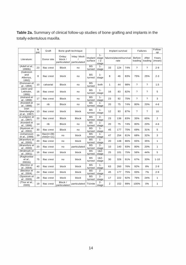
Table 2a. Summary of clinical follow-up studies of bone grafting and implants in the
totally edentulous maxilla.
15
follow-up studies have shown similar results as compared with a simultaneousapproach (Lekholm et al., 1999; Schliephake et al., 1997).
Onlay bone grafts
Adell et al. (Adell et al., 1990b) presented five-year follow-up results withan onlay bone grafting technique using iliac bone, of the shape of a horseshoe,and simultaneous placement of implants. They reported a survival rate ofapproximately 72%. Isaksson et al. (1992) presented a study, wheremanagement of the atrophic maxilla was accomplished by using two segmentsof onlay iliac bone blocks, constructed to meet in the midline and fixed to maxillawith immediate implant insertion. The implants were followed for 32-64 monthsand had a survival rate of 83% was observed. Both studies are consistent withthe findings of other authors using similar techniques (Albrektsson, 1988; Kelleret al., 1999; Lekholm et al., 1999; Nystrom et al., 2004). (Table 2a & b )
Block and/or particulated bone
In 1980, Breine & Brånemark (Breine and Branemark, 1980) reported twodifferent reconstructive procedures for patients with severe jaw atrophy:1; Reconstruction of 14 maxillas and 4 mandibles with placement of 5-6 implantsand packing of autogenic onlay grafts, consisting of chips of cancellous boneand marrow from the upper tibial metaphysis, to form a new alveolar bone. Onlyabout 25% of the the originally installed implants remained integrated.2; Reconstruction of 8 maxillas and one mandible, with autogenic grafts fromthe proximal tibial metaphysis, containing two incorporated implants in eachgraft, and fixed with one additional implant in each graft, providing permanentsupport for bridge constructions with an implant survival of approximately 60%.In a study with a split-mouth design, Thor et al. (2005) placed particulated bonemixed with PRP on one side in the anterior maxilla and onlay block grafts on theother side. Implants were placed in the grafted bone after 6 months of healing.The two sides were evaluated and compared after one year of loading. Noimplants were lost, the marginal bone level showed no significant differences; aresonance frequency analysis (RFA) revealed higher implant stability in theparticulated bone mixed with PRP. Although there were no obvious positiveeffects of PRP on bone graft healing, the handling of the particulated bone graftswas improved (Thor et al., 2005). Johansson et al. (2001) evaluated thevolumetric changes of onlay block bone grafts and bilateral particulate bone
16
grafts to the maxillary sinus of the severely atrophic edentulous maxilla over 6months. The area of each graft was measured and the volume calculated withthe help of computerized tomography. The volume of the inlay and onlay graftswas reduced by an average of 49.5 and 47%, respectively, of the initial volume.The same author, measured cutting torques during the placement of self-tappingdental implants in non-grafted bone and in bone grafts, in 2-stages, either asblocks or in a milled particulate form, in 40 edentulous maxillae. Significantlylower cutting torque values were assessed in grafted regions than in non-graftedregions, irrespective of grafting technique. Lower values were also seen forimplants placed in block grafts compared with implants placed grafts in particulateform (Johansson et al., 2004). (Table 2a & b)
Vascularised bone grafts
Vascularised bone flap methods are mostly used in management andreconstruction of oral malignancies and have resulted in improvements of thetreatment results (Urken et al., 1991; Vaughan et al., 1992). Surgical ablation oforal tissues, radiotherapy and microvascular tissue reconstruction often precedesthe oral rehabilitation. Occlusal rehabilitation often includes fixed or removalprostheses supported by dental implants. However, management in the head
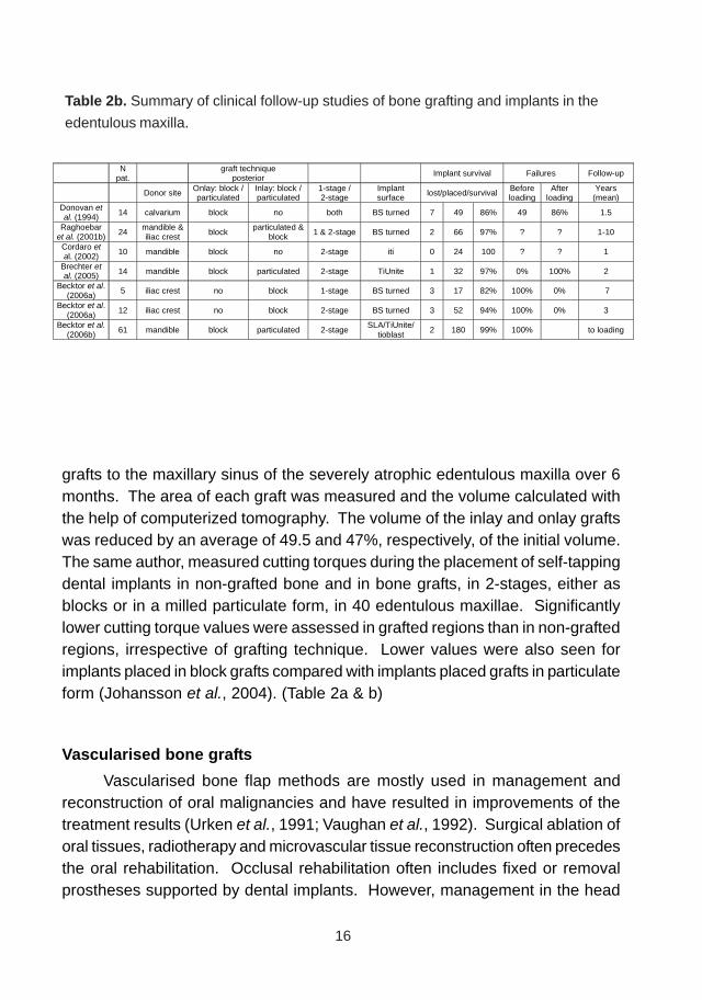
Table 2b. Summary of clinical follow-up studies of bone grafting and implants in the
edentulous maxilla.
N pat. graft technique
posterior Implant survival Failures Follow-up
Donor site Onlay: block / particulated
Inlay: block / particulated
1-stage / 2-stage
Implant surface lost/placed/survival Before
loading After
loading Years
(mean) Donovan et al. (1994) 14 calvarium block no both BS turned 7 49 86% 49 86% 1.5
Raghoebar et al. (2001b) 24
mandible & iliac crest block
particulated & block 1 & 2-stage BS turned 2 66 97% ? ? 1-10
Cordaro et al. (2002) 10 mandible block no 2-stage iti 0 24 100 ? ? 1
Brechter et al. (2005) 14 mandible block particulated 2-stage TiUnite 1 32 97% 0% 100% 2
Becktor et al. (2006a) 5 iliac crest no block 1-stage BS turned 3 17 82% 100% 0% 7
Becktor et al. (2006a) 12 iliac crest no block 2-stage BS turned 3 52 94% 100% 0% 3
Becktor et al. (2006b) 61 mandible block particulated 2-stage
SLA/TiUnite/ tioblast 2 180 99% 100% to loading
17
and neck cancer patient is demanding both surgically and prosthodontically.Factors contributing to the failure of oral implants include medical compromise,smoking, bone quality, use of bone grafts, radiation therapy, and poor oral hygiene(Esposito et al., 1998c).
Different opinions regarding irradiated bone and implant treatment exist.Irradiated jaw bone has been regarded as a contraindication to implant treatment(Gurlek et al., 1998; Kluth et al., 1988). The use of hyperbaric oxygen therapyin irradiated tissues in conjunction with implantat treatment has beenrecommended (Granstrom et al., 1993; Ueda et al., 1993). On the contrary,other studies report acceptable implant survival results in irradiated bone withouthyperbaric oxygen therapy (Eckert et al., 1996; Niimi et al., 1998). Some authorsstate that implants placed before radiotherapy will osseointegrate moresuccessfully than after radiotherapy (Urken et al., 1989), whereas others believethat the bone perfusion has recovered sufficiently 12 months after radiotherapy(Keller, 1997). The greater success of implant placement in native bone orvascularized bone flaps rather than free bone grafts has also been discussed inthe literature (Foster et al., 1999). In an animal study, showed histologically thatnew bone formation and bone union was observed and completed after 8-12weeks in the vascularized tibial grafts and bone formation was clearly delayed innon-vascularized tibial grafts. Dental implants in vascularized tibial graft seemednot to have any negative effect on revascularization (Kobayashi et al., 2005).
Shaw et al. (2005) presented data from 386 implants in 81 patients whoreceived microvascular free flap reconstruction after surgical ablation of oralsquamous cell carcinoma. The patients lost 15% of the implants after a medianfollow-up of four years. Radiotherapy did not seem to jeopardize implant survival,and hyperbaric oxygen had no demonstrable benefit. Despite some persistentsoft tissue problems and implant loss, most patients reached a successfulprosthetic and functional outcome (Shaw et al., 2005).
Allogenic and xenogenic bone grafts
Allograft is by definition a bone graft containing living cells, derived froman individual of the same species. They are hardly recommended because theyinitiate a cell mediated immune response: an allograft may only survive if thedonor is a parent or a sibling (Urist, 1980). A substitute to an allograft is an allo-implant which is bone tissue derived of an individual of the same species andwhich contains no viable cells. Allo-implants are prepared by freezing, freezedrying, irradiation or sterilization of the tissue. Autolysed antigen –extracted
18
allogenic (AAA) bone will incorporate better than freeze-dried or irradiated bone.The principles for incorporation of allo-implants and autografts are similar, sinceboth osteoinductive and osteoconductive responses have been described, evenif several other reports also describe the alloimplant to lack osteoinductiveproperties and to show very modest osteoconductive responses (Pinholt et al.,1994; Solheim et al., 2001; Urist, 1980).
Although autografts are superior to allografts, allografts are used to someextent in oral and maxillofacial reconstruction but commonly so in orthopaedicsurgery. In subjects with large bone defects, autographs are either not availablein sufficient quantities or their use accompanied by high morbidity at the donorsite (Gocke, 2005; Simion et al., 2001).
Xenografts are defined as bone derived from living tissue from anotherspecies whereas xeno-implants are bone grafts where all living cells and proteinshas been extracted. Consequently a xeno-implant can only be osteoconductiveand is replaced by new bone very slowly or to a small extent only (Jensen et al.,1996).
Recently, Hallman et al reported an implant survival rate of 86%, for implantsplaced in grafted areas after three years of prosthetic loading. They used amixture of deproteinized bovine bone and autogenous bone in patients formaxillary sinus inlay augmentation (Hallman et al., 2005). Accordingly cadavericbone allo-implant and bone substitutes are inferior to autogenous bone graftdue to lacking of osteogenic cells and a significant amount of osteoinductivegrowth factors. The combination of autologous cancellous bone marrow andAAA bone is called a composite bone graft. The use of these grafts is oftenmandatory in small children with insufficient iliac crest and tibia bone. Theirsuccess depends on absolute, uninterrupted internal fixation.
Healing periodEarly occlusal rehabilitation evolved from the concept of balanced
articulation, which can be defined as bilateral, simultaneous, anterior and posteriorocclusal contacts of the teeth in centric and eccentric positions. Bilateralarticulation, or balance, as the occlusal scheme of choice has a long history incomplete denture construction (Kurth, 1954).
Repeated trauma to the implant and/or local bone during the healing periodis considered to be a causative factor for implant failure (Brunski, 1993; Espositoet al., 1999; Pilliar et al., 1986). This trauma could be induced by the use of aprosthesis that transmits forces to the underlying bone (Atwood, 1971). A
19
combination syndrome has been described, in which the anterior maxillaundergoes residual ridge resorption in response to trauma from retainedmandibular anterior teeth with a lack of stable posterior mandibular occlusion(Kelly, 1972). A provisional maxillary denture opposed by a mandibular dentitionthat creates force concentration rather than force distribution could induce furthertrauma to the reconstructed maxilla. Force concentration is likely to occur whenocclusal instability is likely such as the presence of unilateral edentulism orretained anterior teeth only. Conversely, an intact mandibular arch, completelyrestored dentition or any other distribution of teeth that allows a broad distributionof forces should prove more favorable to the maxilla. Uhthoff et al. (1973)described that premature loading of an implant may result in a permanent softtissue encapsulation and Albreksson et al. (1981) recommended a minimumhealing period before loading of 3-4 months (Uhthoff, 1973). A literature reviewby Esposito et al. (1998a) reported a pooled failure rate of 15% after three yearsof loading in grafted edentulous and partially edentulous patients. Most of theimplant failures occurred during the healing period, at the second stage surgeryand the period immediately prior to connection of the permanent prosthesis(Esposito et al., 1998a). Trauma could be induced by the use of a prosthesisduring the healing period that transmits forces to the underlying bone (Atwood,1971). The reasons for this concern is mostly depending on the absence of aperiodontal ligament supporting the implants and the observation that non-axialforces will create areas of high stress concentration instead of uniformcompression along the implant to bone interface. The non-axial loading of amechanical device assembled with screw joints, such as dental implants, willinduce more mechanical failures (Rangert et al., 1989). However, evidence islacking, regarding the effect of non-axial load or overload on the osseointegratedinterface between bone and implant. The implant shape and surface textureindicate that the load will be transferred to bone by compression in some areasand as tension and shear in other areas (Jemt et al., 2000). The load of occlusionis seldom vertical and mastication distributes both non-axial and axial loadingand the damaging effects of bruxism are created through lateral friction betweenthe occlusal surfaces of maxilla and mandible. Thus, the resultant forces arenot vertical. There is limited evidence available and it does not demonstratethat non-axial loading is unfavourable to the osseointegrated interface (Asikainenet al., 1997).
The proprioception of the periodontal ligament is missing when the naturalteeth are lost and could be an important consideration in the replacement of
20
natural teeth with dental implants. The perception has been demonstrated to beextraordinarily different between natural teeth and implants (average 3.8-gpressure for natural teeth tested horizontally vs. 580-g horizontal force for implantsin the anterior mandible) (Mericske-Stern et al., 1993). Despite these findings,patients with extensive implant-supported restorations seem, to function wellwithout the benefit of periodontal proprioceptive nerve endings. The presenceof proprioceptive nerve endings in periosteum, muscles of mastication, oralmucosa, and the temporomandibular joints may to some extent compensate forthose lost from the missing periodontal ligament (Van Loven et al., 2000).
Surgeon’s experienceWhen a new method is described the difficulties in the beginning are
commonly addressed by excluding the first cases in the results, claiming a”learning curve”, the latter cases showing better but also more realistic results(Adell et al., 1990b; Nystrom et al., 2002). It has been demonstrated that agentle surgical technique with a minimal trauma to the graft, will lead to a fasterremodelling and revascularization. In addition, the degree of stability of the graftis important. Bone is heat sensitive and a temperature of more than 47ºCcombined with an exposure time of more than 1 minute has been shown toresult in an impaired bone healing (Eriksson and Albrektsson, 1983). To minimizeincreased temperature in the bone due to drilling, intense cooling with salinesolution, a graded series of drills and well sharpened instruments have beenrecommended (Eriksson, 1984). The integration of titanium implants inautogenous free bone grafts is dependent on the state of the bone graft.Revascularization of the bone graft is very important for its incorporation andremodelling (Albreksson, 1979). This is dependent on gentle surgery, minimaltrauma to the donor and recipient sites and the preservation of as much bloodsupply as possible (Bell, 1969; Eriksson, 1984). The potential of the bone graftto respond to the surgical trauma from installation of the implant will most likelyinfluence the quality of osseointegration and the stability of the implant. Thedegree of osseointegration, (bone to implant contact) is supposed to increasewhen the implant is installed six months after the bone graft procedure comparedwith simultaneous implant installation (Lundgren et al., 1999). These findingssupport the notion of the importance of a well vascularized and incorporatedbone graft to an optimal bone to implant contact.
Lambert et al. (1997) defined a learning curve for dental implant placement.Implants placed by inexperienced surgeons failed twice as often as those placedby experienced surgeons. It was suggested that surgeons with little or no previous

21
experience must expect a definite learning curve (Lambert et al., 1997). However,another study reported no differences in implant survival rate as a function ofthe level of training of the resident surgeon (Melo et al., 2006).
Albrektsson emphasized the importance of surgical skill of the surgeonwhen analyzing the reasons for implant success and failure (Albrektsson, 2001).

22
23
AIMS
• To analyze and compare the survival rate of endosseous implants placedin the maxilla of patients (i) subjected to bone augmentation proceduresprior to or in conjunction with implant placement and (ii) in routine patientswithout bone augmentation.
• To analyze the influence of the mandibular dentition on implantperformance in the maxilla prior to attachment of the definitive prosthesiswhen reconstruction is possible only with the use of autogenous bone-grafting techniques.
• To evaluate the clinical outcome of zygomatic implant treatment anddiscuss whether the treatment with zygomatic implants could be analternative to the bone grafting and implant procedures in patients withedentulous maxillae.
• To analyze the survival rate of endosseous implants placed in the partiallydentate maxilla treated with sinus inlay block bone-grafts harvested fromthe iliac crest.
• To describe the surgical technique when using particulated bone fromthe mandible for maxillary sinus floor augmentation prior to the placementof surface modified implants. The purpose was also to report on the
clinical outcome from bone grafting to delivery of the final prosthesis
24
MATERIALS AND METHODS
Subjects
Paper IThe study included 216 consecutively treated patients with
edentulous maxillae, rehabilitated with minimally rough (machined/turned)endosseous implants (Brånemark System, Nobel Biocare AB, Gothenburg,Sweden) with or without bone augmentation. Of a total of 216 patients and 1357implants, 34 patients with 237 implants were withdrawn as described below.
The patient material were divided into two retrospective patient groups: (i)the Graft Group included 64 patients with 437 implants and (ii) the Non-GraftGroup included 118 patients with 683 implants, that had been consecutivelytreated during a period from 1990 to 1996 (Table 3). In addition, the retrospectivepatient groups were also prospectively followed using a standardized clinicaland radiographic study-design. Routine implant treatment was commenced ifthe remaining bone volume was evaluated as adequate. The Graft Group,included 64 patients, 22 male and 42 female. Because of advanced horizontaland vertical bone loss of the alveolar processes as well as extensivepneumatization of the maxillary sinuses, the patients were considered to haveinsufficient bone volume for routine implant treatment. A one-stage graftingtechnique was used from 1990 to 1994 and a two-stage grafting technique wasused from 1994 to 1996.The Non-Graft Group included 118 patients, 72 maleand 46 female, were judged from clinical and radiographic examinations to havesufficient bone volume for implant treatment.
Table 3. Paper I. Distribution of Placed Implants With Regard to Number, Length andDiameter. Corrected values on gray background.
25
Paper IIThe present study was conducted as a retrospective investigation
of consecutively treated patients from two oral and maxillofacial surgerydepartments. A total of 101 consecutively treated patients were included, allwith edentulous maxillae that underwent treatment planning to receiveendosseous implants in conjunction with autogenous bone grafting. All patientshad insufficient bone volume and autogenous bone augmentation was required.The group of patients was treated from 1990 to 1996.Of a total of 101 patients, 11 subjects were excluded. The remaining 90 patients(31 men, 59 women) with 643 implants(Brånemark System, Nobel Biocare AB,Gothenburg, Sweden) were examined retrospectively according to the studyprotocol.
Paper IIIThe study included 16 patients, 6 males and 10 females with
edentulous maxillae, consecutively treated with 74 endosseous dental implants(Brånemark Implant System, Nobel Biocare AB, Gothenburg, Sweden, or AstraTech Implant Dental System, Astratech AB, Gothenburg, Sweden) and 31zygomatic implants (Brånemark System, Nobel Biocare AB, Gothenburg,Sweden) from 1998 to 2002. The patients were retrospectively evaluated andprospectively followed, using a standardized clinical and radiographic study-design. Because of advanced horizontal and vertical bone loss of the alveolarprocesses as well as extensive pneumatization of the maxillary sinuses, thepatients were considered to have insufficient bone volume for routine implanttreatment. The patients were treated with zygomatic implants as an alternativebone grafting.
Paper IVA number of 17 partially dentate patients, 4 males and 13 females,
were subjected to bone augmentation procedures prior to or in conjunction withimplant placement. Bone volumes were regarded as insufficient for implanttreatment unless a bone grafting procedure was performed. A total of 69 implants(Brånemark System, Nobel Biocare AB, Gothenburg, Sweden) were placed inthe patients. All patients were consecutive admissions treated from 1990 to1996. The retrospective patient group was also prospectively followed using astandardized clinical and radiographic study-design.
26
Paper VThe study group included 61 patients, 23 males and 38 females.
All patients were partially dentate. Because of advanced horizontal and verticalbone loss of the alveolar processes and/or extensive pneumatization of themaxillary sinuses, the patients were considered to have insufficient bone volumefor routine implant treatment. All patients were consecutive admissions treatedfrom 1998 to 2004. The patients were subjected to a bone augmentationprocedure using autogenous bone grafts and implant treatment using 180 surfacemodified implants: 119 Straumann implants with blasted/acid-etched surface(SLA, Straumann AG, Basel Switzerland) 38 Brånemark System implants withoxidized surface (TiUnite, Nobel Biocare AB, Gothenburg, Sweden) and 23 AstraTech implants with blasted surface (TioBlast, AstraTech AB, Gothenburg, Sweden)were placed.The patient group was prospectively followed using a standardizedclinical and radiographic study-design.
Drop-outsIn paper I, a total of 34 patients (15.7%) with 237 implants (17.5%)
were drop-outs. In the Graft Group, 19 patients with 133 implants were withdrawnas a result of; (i) combined infection and dehiscence of the wound related to theuse of a non resorbable membrane (n=3), (ii) patients had moved from the area(n=7), (iii) reduced health (n=3) or (iv) patients had deceased (n=6). In the Non-Graft Group, 15 patients with 104 implants were withdrawn because, (i) patientshad moved from the area (n=6), (ii) reduced health (n=7) or (iii) patients haddeceased (n=2).
In paper II, 11 of 110 subjects (10,9%) were drop-outs; (i) threepatients with combined infection and dehiscence of the wound due to the use ofa non resorbable membrane, (ii) two patients with maxillary discontinuity due togun shot wounds and resection of malignancy, (iii) three patients who movedbefore treatment was completed and (iv) three patients were deceased.
In paper III, sixteen patients received zygomatic implant treatmentand were included in the study. All patients were contacted for a furtherprospective follow-up examination. Of 16 patients, 14 presented, one patientwas deceased, and another patient was hospitalized in another city. Subsequently,14 patients underwent clinical and radiographic examination according to theprospective follow-up protocol.
There were no drop-outs in paper IV and V.
27
Surgery
Papers I, II and IVBone augmentation was performed in a hospital operating room
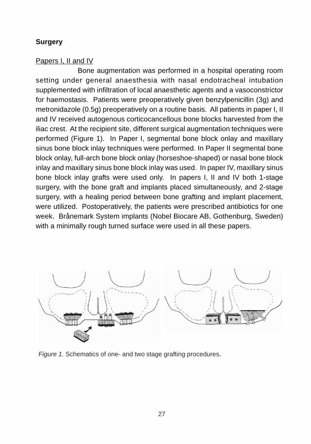
setting under general anaesthesia with nasal endotracheal intubationsupplemented with infiltration of local anaesthetic agents and a vasoconstrictorfor haemostasis. Patients were preoperatively given benzylpenicillin (3g) andmetronidazole (0.5g) preoperatively on a routine basis. All patients in paper I, IIand IV received autogenous corticocancellous bone blocks harvested from theiliac crest. At the recipient site, different surgical augmentation techniques wereperformed (Figure 1). In Paper I, segmental bone block onlay and maxillarysinus bone block inlay techniques were performed. In Paper II segmental boneblock onlay, full-arch bone block onlay (horseshoe-shaped) or nasal bone blockinlay and maxillary sinus bone block inlay was used. In paper IV, maxillary sinusbone block inlay grafts were used only. In papers I, II and IV both 1-stagesurgery, with the bone graft and implants placed simultaneously, and 2-stagesurgery, with a healing period between bone grafting and implant placement,were utilized. Postoperatively, the patients were prescribed antibiotics for oneweek. Brånemark System implants (Nobel Biocare AB, Gothenburg, Sweden)with a minimally rough turned surface were used in all these papers.
Figure 1. Schematics of one- and two stage grafting procedures.
28
Paper IIISurgery was performed under general anaesthesia with nasal
endotracheal intubation supplemented with infiltration of local anaesthetic agentswith a vasoconstrictor for haemostasis. Patients were preoperatively givenbensylpenicillin (3g) and metronidazole (0.5g) preoperatively on a routine basis.A crestal incision was made extending from the second molar bilaterally. Avestibular releasing incision was made at the posterior extent of the incision inthe maxillary second molar region. A muco-periosteal elevation revealed thenasal apertures and the piriform rim to the inferior aspect of the infraorbitalforamina and laterally of the buttress and body of the zygoma, bilaterally.
A round bur was then used to create a lateral window, 5 x 10, mm inthe lateral wall of the maxillary sinus. The sinus mucosa was then carefullyreflected and protected through the preparation of the zygomatic implant site. Aretractor was placed over the superior aspect of the zygomatic arch to enable acorrect orientation of the implant site preparation. The zygomatic implants headwere placed palatal and as close as possible to the alveolar crest, in the regionof the second premolar and first molar. After penetrating the maxillary bone intothe maxillary sinus, the preparation went through the cortical layer of the anterior-superior part of the zygomatic bone. The implant sites were then enlarged.Implant size was decided and final placement of the implant was accomplishedusing the standard protocol. The zygomatic implant was placed using low speeduntil the tip of the implant engaged the zygomatict bone and finalized manuallyuntil the implant was optimally seated. All 31 zygomatic implants had a stableand ridgid primary stability at the installation and were dressed with a coverscrew.
Patients obtained simultaneous placement of additional endosseousimplants in the anterior region of the maxilla (Brånemark Implant System, NobelBiocare AB, Gothenburg, Sweden, or Astra Tech Implant Dental System,Astratech AB, Gothenburg, Sweden).(Branemark, 1985) The wound was closedwith a continuous, absorbable 4-0 sutures. Postoperatively, the patients wereprescribed antibiotics for one week.
Paper VIn the first three patients of paper V, the bone augmentation was
performed under general anaesthesia with nasal endotracheal intubationsupplemented with infiltration of local anaesthetic agents. The majority of theremaining patients were treated in local anesthesia, using Lidocain / Adrenalin
29
2% in combination with Bupivacain / Adrenalin 5% with or without per oral sedationwith flunitrazepam (0.5 – 1.0 g) one hour preoperatively.
From the retromolar to the second or first molar area a 20-30 mmincision was made in the facial vestibule on the external oblique ridge of themandible. The lateral aspect of the mandible was exposed and the localizationof the osteotomy was marked with a 1mm fissure bur. The osteotomy wasstarted anterior to the coronoid process, cutting along the anterior border of theramus, medially to the external oblique ridge, finishing in the mandibular body inthe molar region. The length of the anterior and posterior vertical cuts wasdetermined by the size of the graft required. The inferior osteotomy, whichconnects the vertical cuts, was made with a diamond disc that creates a 2-mmdepth in the cortical bone. With adequate osteotomies through the cortical layer,the splitting of the bone block was done with careful bending movements usinga chisel. Following removal of the bone, sharp edges around the ramus/bodywere smoothed off with a round burr. The wound was rinsed with saline solution,haemostatic dressing (collagen) was placed into the donor area and the woundwas sutured using resorbable sutures. In all 61 patients the harvested bonewas kept in saline solution or blood until it was particulated with a surgical bonemill. In eight out of the 61 patients, a part of the harvested bone was kept as abone block and trimmed and used as onlay bone graft.
The approach for the posterior maxilla was made by a crestalincision along the alveolar process. The alveolar crest was subsequently exposedby raising a buccal pedicled mucoperiosteal flap and a bony window wasestablished on the lateral aspect of the maxillary sinus. The sinus membranewas carefully elevated and the particulated bone was positioned in contact withthe floor of the maxillary sinus. In eight cases the alveolar crest had to bewidened and some of the harvested bone was then used as a block. The boneblock was trimmed and fixed with titanium osteosynthesis screws (7 to 15mm inlength and 2mm in diameter) on the lateral aspect of the alveolar crest. After ahealing period of five to 21 months (mean 7.2) the implant placement was carriedout. In total, 180 implants were placed. Three different implant systems withmoderately rough surfaces were used, 119 Straumann implants with blasted/acid-etched surface (SLA, Straumann AG, Basel Switzerland) 38 BrånemarkSystem implants with oxidized surface(TiUnite, Nobel Biocare AB, Gothenburg,Sweden) and 23 Astra Tech implants with blasted surface (TioBlast, AstraTechAB, Gothenburg, Sweden) were placed. The implants were in lengths from 8 to15 mm (mean: 11.5mm) and in diameters from 3.3 to 4.8 mm (mean: 3.9mm).
30
Effort was made not to perforate into the maxillary sinus with thedrills or the implants, assuring that the implants were covered with grafted boneat the apical part. A non-submerged technique was used for Straumann implantsand a submerged technique for the other implant systems. The implants wereallowed to heal for 3 to 6 months prior to abutment connection and prosthetictreatment.
ProsthodonticsIn papers I, II and III, conventional dentures were relined 1 to 3
weeks after bone grafting and /or implant surgery and at abutment connection.No dentures were used in papers IV and V. Fabrication of gold-acrylic fixedprostheses, and in cases with overdenture therapy using a bar and clips, followedthe standard procedures for the different implant systems.
Examinations and Follow-upIn papers I, III and IV, data were collected from the time of bone
augmentation or implant treatment until the last follow-up and retrospectivelyanalyzed according to a research protocol. All patients were contacted for aprospective follow-up examination and subsequently underwent clinical andradiographic examination according to the prospective follow-up protocol.
All available data in paper II, such as clinical records andradiographs, were documented from the time of bone augmentation or implanttreatment until the last follow-up. This material was retrospectively analyzedaccording to a study protocol to confirm understanding of the material.
In paper IV, data were collected from the time of bone augmentationtill the day of delivery of definitive prosthesis and was prospectively analyzedaccording to a research protocol.
Radiographic ExaminationThe retrospective radiographic examinations had not been
consistently performed at the time of the abutment connection surgery and atthe annual controls in papers I, II, III and IV. Radiographs used in papers I andIV were taken at the prospective follow up examination. An intraoral radiographicparalleling technique (Hollender and Rockler, 1980) was utilized at the time ofthe prospective patient follow-up. The distance from the implant-abutmentjunction to the marginal bone at mesial and distal surfaces of each implant wasrecorded. Linear measurements were performed to the closest one mm.
31
In paper III, preoperatively, panoramic images supplemented withintra oral radiographs were used to evaluate the bone volume of the maxilla.Computed tomograms were used to determine whether the anatomy would allowinstallation of zygomatic implants and to eliminate the risk of undiagnosedpathological lesions.
Preoperative classification according to Cawood & Howell(Cawoodand Howell, 1991), was performed in all papers, retrospectively with the help oflateral and panoramic radiographs. The lateral radiographs were used todetermine the height in the anterior maxilla. The panoramic radiographs wereused for the classification in the region of the posterior maxilla where 5mm orless of bone in height corresponded to class V and VI, 6mm – 12mm to class III-IV and 12mm or more to Class I-II.
The radiographic evaluation in paper IV consisted of one pre-surgical panoramic radiograph and one post-surgical panoramic radiograph,taken after the bone grafting and implant installation.A standardized natural headposition was used while obtaining panoramic radiographs. The radiographswere taken with the same equipment at the radiographic department, and thesame film/focus distance was used. All radiographs were taken at optimalexposure and anatomical landmarks were clearly visualized. All radiographswere hand-traced on acetate paper by one examiner. Reference lines were drawnthrough structures like top of the alveolar crest, nasal bones and the floor of themaxillary sinus directly on the pre-surgical radiograph with a sharp soft pencil.The post-surgical radiograph was superimposed on anatomical structures andthe implant sites were evaluated pre- and post-surgically with regards to implantposition and its vertical bone volume including grafted and residual bone.
StatisticsIn papers I, II and IV life table analyses were performed to calculate
the cumulative implant survival rate (CSR). The Wilcoxon rank sum test wasused in paper II, to test differences in implant survival rates between the Non-Graft Group and the Graft Group, with the relative frequency of implant loss ineach patient as the calculation unit and to test the difference between the groupsin specific regions (incisor, canine, premolar and molar) with the implant as theunit. The Pearson Chi-Square test was used to compare the Non-Graft Groupand the Graft Group with respect to a variety of explanatory variables. The levelof statistical significance was set at the 5% level.
32
In paper II the experimental unit in the analysis was an individualimplant, of which there were several within each patient. The outcome of interestwas the occurrences of implant failure at any time during the healing period,including second-stage surgery, up until the time of prosthesis attachment.Various risk factors were evaluated for their association with failure. Theassociation between each risk factor and failure was summarized using an oddsratio (OR) and corresponding 95% confidence interval (CI). An OR of 1 indicatesno association between a risk factor and the occurrence of an implant failure.The binary response (failure versus no failure) was modelled using a logisticregression model. Robust estimates of the ORs and corresponding tests ofsignificance were obtained based on generalized estimating equation methodsto account for the correlation between implants within a patient. The correlationsamong the outcomes for each patient were modelled as exchangeablecorrelations. The SAS procedure PROC GENMOD (SAS Institute, Cary, NC)was used to perform the analysis.33 All calculated P values were 2-sided, and Pvalues less than .05 were considered statistically significant.
In paper IV, the test between groups were performed with Fisher’sexact text for dichotomous variables, Chi-square test for non-ordered categoricalvariables and Mantel Haenszel’s test for ordered categorical variables. All testswere two-tailed and conducted at 5% significance level
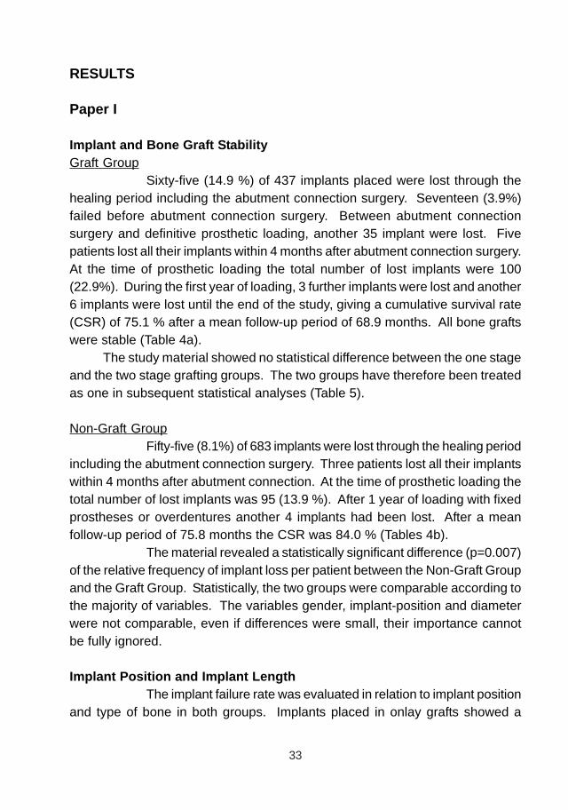
33
RESULTS
Paper I
Implant and Bone Graft StabilityGraft Group
Sixty-five (14.9 %) of 437 implants placed were lost through thehealing period including the abutment connection surgery. Seventeen (3.9%)failed before abutment connection surgery. Between abutment connectionsurgery and definitive prosthetic loading, another 35 implant were lost. Fivepatients lost all their implants within 4 months after abutment connection surgery.At the time of prosthetic loading the total number of lost implants were 100(22.9%). During the first year of loading, 3 further implants were lost and another6 implants were lost until the end of the study, giving a cumulative survival rate(CSR) of 75.1 % after a mean follow-up period of 68.9 months. All bone graftswere stable (Table 4a).
The study material showed no statistical difference between the one stageand the two stage grafting groups. The two groups have therefore been treatedas one in subsequent statistical analyses (Table 5).
Non-Graft GroupFifty-five (8.1%) of 683 implants were lost through the healing period
including the abutment connection surgery. Three patients lost all their implantswithin 4 months after abutment connection. At the time of prosthetic loading thetotal number of lost implants was 95 (13.9 %). After 1 year of loading with fixedprostheses or overdentures another 4 implants had been lost. After a meanfollow-up period of 75.8 months the CSR was 84.0 % (Tables 4b).
The material revealed a statistically significant difference (p=0.007)of the relative frequency of implant loss per patient between the Non-Graft Groupand the Graft Group. Statistically, the two groups were comparable according tothe majority of variables. The variables gender, implant-position and diameterwere not comparable, even if differences were small, their importance cannotbe fully ignored.
Implant Position and Implant LengthThe implant failure rate was evaluated in relation to implant position
and type of bone in both groups. Implants placed in onlay grafts showed a
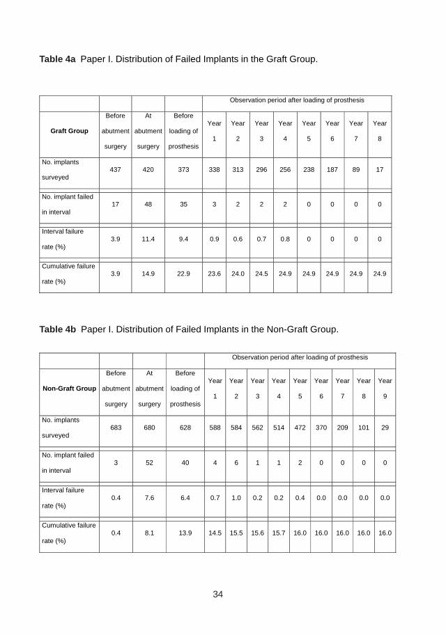
34
Observation period after loading of prosthesis
Graft Group
Before
abutment
surgery
At
abutment
surgery
Before
loading of
prosthesis
Year
1
Year
2
Year
3
Year
4
Year
5
Year
6
Year
7
Year
8
No. implants
surveyed 437 420 373 338 313 296 256 238 187 89 17
No. implant failed
in interval 17 48 35 3 2 2 2 0 0 0 0
Interval failure
rate (%) 3.9 11.4 9.4 0.9 0.6 0.7 0.8 0 0 0 0
Cumulative failure
rate (%) 3.9 14.9 22.9 23.6 24.0 24.5 24.9 24.9 24.9 24.9 24.9
Observation period after loading of prosthesis
Non-Graft Group
Before
abutment
surgery
At
abutment
surgery
Before
loading of
prosthesis
Year
1
Year
2
Year
3
Year
4
Year
5
Year
6
Year
7
Year
8
Year
9
No. implants
surveyed 683 680 628 588 584 562 514 472 370 209 101 29
No. implant failed
in interval 3 52 40 4 6 1 1 2 0 0 0 0
Interval failure
rate (%) 0.4 7.6 6.4 0.7 1.0 0.2 0.2 0.4 0.0 0.0 0.0 0.0
Cumulative failure
rate (%) 0.4 8.1 13.9 14.5 15.5 15.6 15.7 16.0 16.0 16.0 16.0 16.0
Table 4a Paper I. Distribution of Failed Implants in the Graft Group.
Table 4b Paper I. Distribution of Failed Implants in the Non-Graft Group.
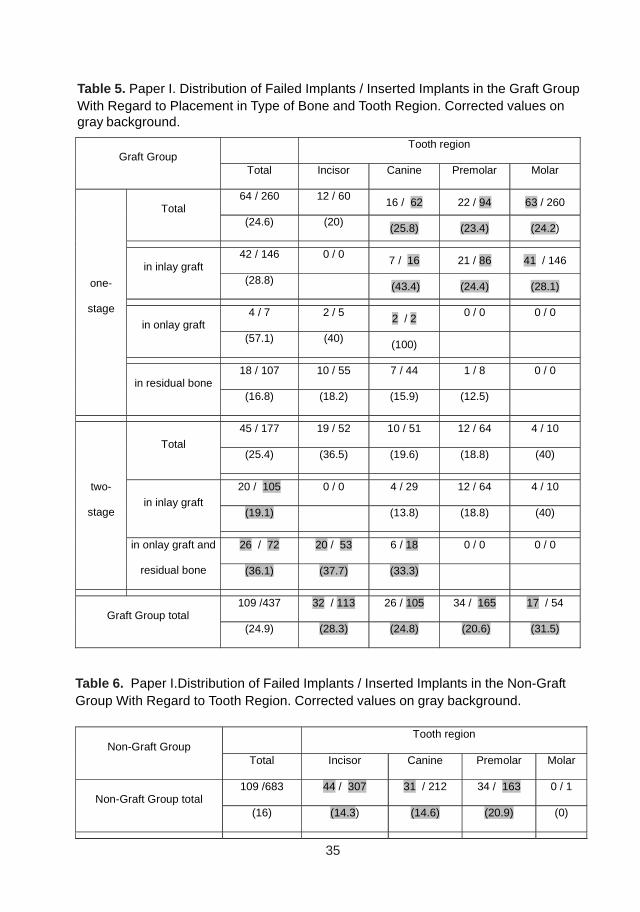
35
Tooth region Graft Group
Total Incisor Canine Premolar Molar
64 / 260 12 / 60 16 / 62 22 / 94 63 / 260 Total
(24.6) (20) (25.8) (23.4) (24.2)
42 / 146 0 / 0 7 / 16 21 / 86 41 / 146 in inlay graft
(28.8) (43.4) (24.4) (28.1)
4 / 7 2 / 5 2 / 2 0 / 0 0 / 0 in onlay graft
(57.1) (40) (100)
18 / 107 10 / 55 7 / 44 1 / 8 0 / 0
one-
stage
in residual bone (16.8) (18.2) (15.9) (12.5)
45 / 177 19 / 52 10 / 51 12 / 64 4 / 10
(25.4) (36.5) (19.6) (18.8) (40) Total
20 / 105 0 / 0 4 / 29 12 / 64 4 / 10
(19.1) (13.8) (18.8) (40) in inlay graft
26 / 72 20 / 53 6 / 18 0 / 0 0 / 0
two-
stage
in onlay graft and
residual bone (36.1) (37.7) (33.3)
109 /437 32 / 113 26 / 105 34 / 165 17 / 54 Graft Group total
(24.9) (28.3) (24.8) (20.6) (31.5)
Table 5. Paper I. Distribution of Failed Implants / Inserted Implants in the Graft GroupWith Regard to Placement in Type of Bone and Tooth Region. Corrected values ongray background.
Tooth region Non-Graft Group
Total Incisor Canine Premolar Molar
109 /683 44 / 307 31 / 212 34 / 163 0 / 1 Non-Graft Group total
(16) (14.3) (14.6) (20.9) (0)
Table 6. Paper I.Distribution of Failed Implants / Inserted Implants in the Non-GraftGroup With Regard to Tooth Region. Corrected values on gray background.
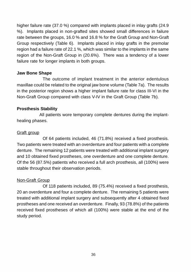
36
higher failure rate (37.0 %) compared with implants placed in inlay grafts (24.9%). Implants placed in non-grafted sites showed small differences in failurerate between the groups, 16.0 % and 16.8 % for the Graft Group and Non-GraftGroup respectively (Table 6). Implants placed in inlay grafts in the premolarregion had a failure rate of 22.1 %, which was similar to the implants in the sameregion of the Non-Graft Group in (20.6%). There was a tendency of a lowerfailure rate for longer implants in both groups.
Jaw Bone ShapeThe outcome of implant treatment in the anterior edentulous
maxillae could be related to the original jaw bone volume (Table 7a). The resultsin the posterior region shows a higher implant failure rate for class III-VI in theNon-Graft Group compared with class V-IV in the Graft Group (Table 7b).
Prosthesis StabilityAll patients wore temporary complete dentures during the implant-
healing phases.
Graft groupOf 64 patients included, 46 (71.8%) received a fixed prosthesis.
Two patients were treated with an overdenture and four patients with a completedenture. The remaining 12 patients were treated with additional implant surgeryand 10 obtained fixed prostheses, one overdenture and one complete denture.Of the 56 (87.5%) patients who received a full arch prosthesis, all (100%) werestable throughout their observation periods.
Non-Graft GroupOf 118 patients included, 89 (75.4%) received a fixed prosthesis,
20 an overdenture and four a complete denture. The remaining 5 patients weretreated with additional implant surgery and subsequently after 4 obtained fixedprostheses and one received an overdenture. Finally, 93 (78.8%) of the patientsreceived fixed prostheses of which all (100%) were stable at the end of thestudy period.
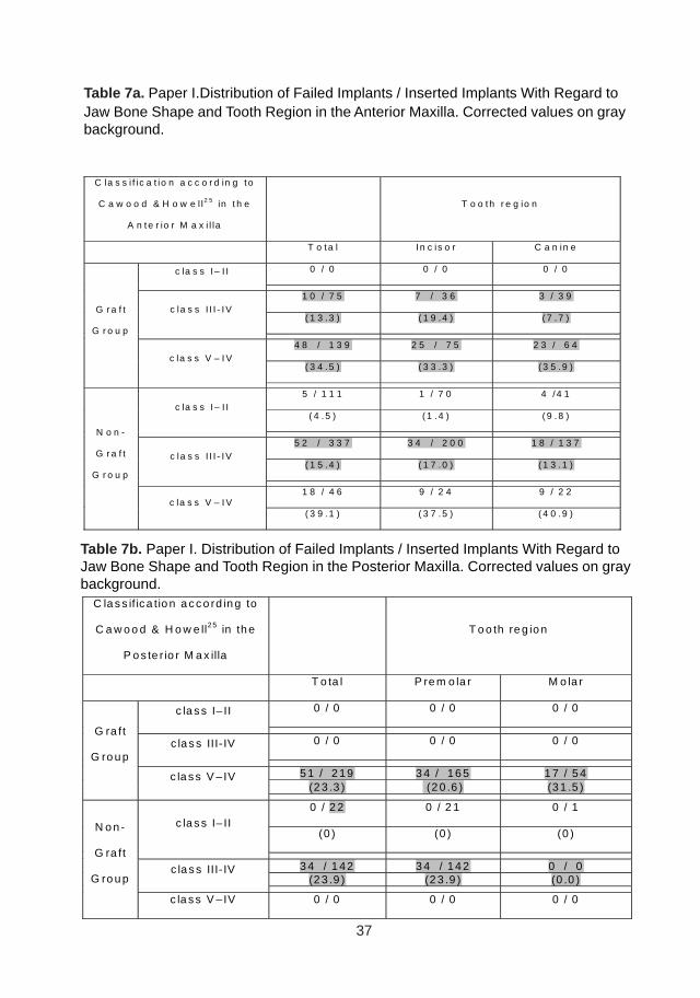
37
C la s s if ic a tio n a c c o rd in g to
C a w o o d & H o w e ll2 5 in th e
P o s te r io r M a x illa
T o o th re g io n
T o ta l P re m o la r M o la r
0 / 0 0 / 0 0 / 0 c la s s I– II
0 / 0 0 / 0 0 / 0 c la s s III- IV
5 1 / 2 19 3 4 / 16 5 1 7 / 5 4
(2 3 .3 ) (2 0 .6 ) (3 1 .5 )
G ra ft
G ro u p
c la s s V – IV
0 / 2 2 0 / 2 1 0 / 1
(0 ) (0 ) (0 ) c la s s I– II
3 4 / 1 42 3 4 / 14 2 0 / 0
(2 3 .9 ) (2 3 .9 ) (0 .0 ) c la s s III- IV
N o n -
G ra ft
G ro u p
c la s s V – IV 0 / 0 0 / 0 0 / 0
Table 7a. Paper I.Distribution of Failed Implants / Inserted Implants With Regard toJaw Bone Shape and Tooth Region in the Anterior Maxilla. Corrected values on graybackground.
C la s s i f ic a t io n a c c o r d in g t o
C a w o o d & H o w e l l2 5 in t h e
A n te r io r M a x i l la
T o o th r e g io n
T o ta l I n c is o r C a n in e
0 / 0 0 / 0 0 / 0 c la s s I – I I
1 0 / 7 5 7 / 3 6 3 / 3 9
( 1 3 .3 ) ( 1 9 .4 ) ( 7 .7 ) c la s s I I I - IV
4 8 / 1 3 9 2 5 / 7 5 2 3 / 6 4
( 3 4 .5 ) ( 3 3 .3 ) ( 3 5 .9 )
G r a f t
G r o u p
c la s s V – I V
5 / 1 1 1 1 / 7 0 4 / 4 1
( 4 .5 ) ( 1 .4 ) ( 9 .8 ) c la s s I – I I
5 2 / 3 3 7 3 4 / 2 0 0 1 8 / 1 3 7
( 1 5 .4 ) ( 1 7 .0 ) ( 1 3 .1 ) c la s s I I I - IV
1 8 / 4 6 9 / 2 4 9 / 2 2
N o n -
G r a f t
G r o u p
c la s s V – I V ( 3 9 .1 ) ( 3 7 .5 ) ( 4 0 .9 )
Table 7b. Paper I. Distribution of Failed Implants / Inserted Implants With Regard toJaw Bone Shape and Tooth Region in the Posterior Maxilla. Corrected values on graybackground.
38
Paper II
Implant and Bone Graft StabilityOf the 643 consecutively placed implants in 90 patients, 81 implants
(12.6%) were lost during the period from implant placement until and includingabutment connection surgery. At the time of definitive prosthesis placement, anadditional 37 implants were lost, resulting in a total of 118 lost implants (18.4%).During the first year of functional loading with the definitive prosthesis, anadditional 2 implants failed to maintain osseointegration, and 8 implants werelost subsequent to that time. The overall mean patient follow-up was 64.2 months.All bone grafts were stable. The follow-up period ranged from 22 to 105 months,with a mean of 64.2 months.
Mandibular DentitionThe type of mandibular dentition was significantly associated with
implant failure during the period from implant placement until the time of definitiveprosthesis placement (Figure 2)(Table 8 a-c). In the unilateral occlusal supportgroup, 28 of 64 implants were lost, indicating the highest rate of implant failure(43.8%). Among the implants placed opposing limited occlusal support, 14 of70 were lost (20.0%). Patients with stable occlusal contact, defined as mandibularbilateral occlusal support or an implant supported fixed prosthesis, demonstratedimplant failure rates of 16.9% (62 of 366) and 14.3% (10 of 70), respectively.The lowest implant failure rate was observed in the patients who wore amandibular removable denture, with a failure rate of 6.2% (4 of 65 implantsfailed). No failures were observed in the no dentition group, which consisted of1 patient with 8 implants.
Figure 2. Showing examples of different unilateral (left) and bilateral (right) occlusal
support in the mandible.
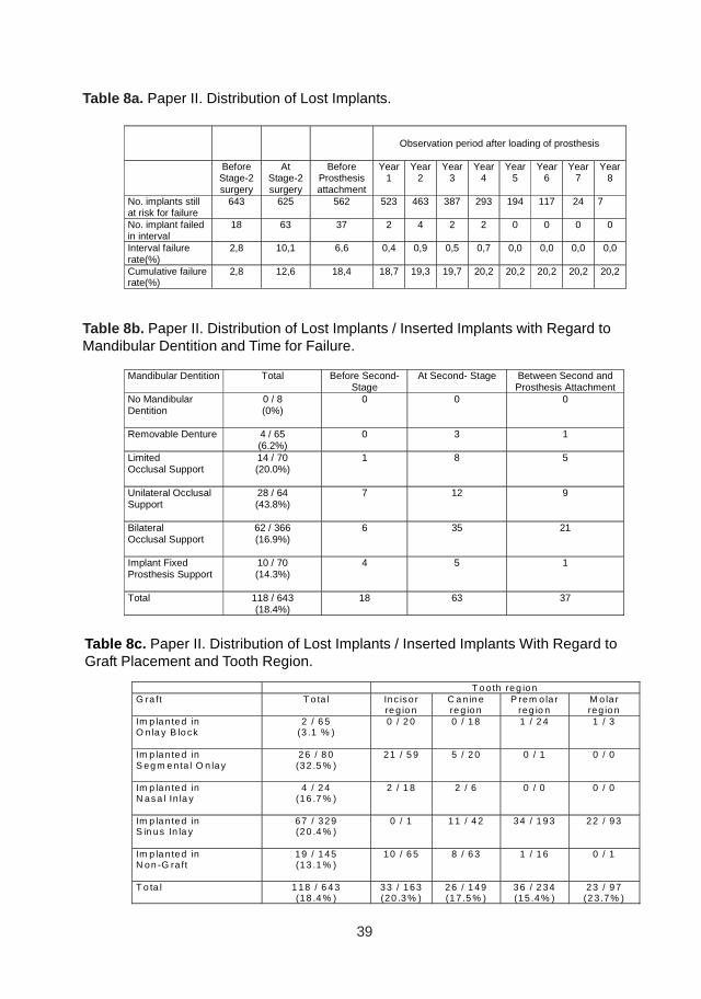
39
Observation period after loading of prosthesis
Before
Stage-2 surgery
At Stage-2 surgery
Before Prosthesis attachment
Year 1
Year 2
Year 3
Year 4
Year 5
Year 6
Year 7
Year8
No. implants still at risk for failure
643 625 562 523 463 387 293 194 117 24 7
No. implant failed in interval
18 63 37 2 4 2 2 0 0 0 0
Interval failure rate(%)
2,8 10,1 6,6 0,4 0,9 0,5 0,7 0,0 0,0 0,0 0,0
Cumulative failure rate(%)
2,8 12,6 18,4 18,7 19,3 19,7 20,2 20,2 20,2 20,2 20,2
Mandibular Dentition Total Before Second-Stage
At Second- Stage Between Second and Prosthesis Attachment
No Mandibular Dentition
0 / 8 (0%)
0 0 0
Removable Denture
4 / 65 (6.2%)
0 3 1
Limited Occlusal Support
14 / 70 (20.0%)
1 8 5
Unilateral Occlusal Support
28 / 64 (43.8%)
7 12 9
Bilateral Occlusal Support
62 / 366 (16.9%)
6 35 21
Implant Fixed Prosthesis Support
10 / 70 (14.3%)
4 5 1
Total 118 / 643 (18.4%)
18 63 37
T o o th reg ion G ra ft T o ta l Inc iso r
re g io n C a n in e re g io n
P rem o la r reg io n
M o la r reg io n
Im p la n ted in O n la y B lo ck
2 / 6 5 (3 .1 % )
0 / 2 0 0 / 1 8 1 / 2 4 1 / 3
Im p la n ted in S eg m e nta l O n la y
26 / 8 0 (32 .5 % )
21 / 5 9 5 / 2 0 0 / 1 0 / 0
Im p la n ted in N asa l In la y
4 / 2 4 (16 .7 % )
2 / 1 8 2 / 6 0 / 0 0 / 0
Im p la n ted in S in us In la y
67 / 329 (20 .4 % )
0 / 1 1 1 / 4 2 34 / 1 93 22 / 93
Im p la n ted in N on -G ra ft
19 / 145 (13 .1 % )
10 / 6 5 8 / 6 3 1 / 1 6 0 / 1
T o ta l 1 18 / 6 4 3 (18 .4 % )
33 / 163 (20 .3 % )
26 / 1 49 (17 .5 % )
36 / 2 34 (15 .4 % )
23 / 97 (2 3 .7% )
Table 8a. Paper II. Distribution of Lost Implants.
Table 8b. Paper II. Distribution of Lost Implants / Inserted Implants with Regard toMandibular Dentition and Time for Failure.
Table 8c. Paper II. Distribution of Lost Implants / Inserted Implants With Regard toGraft Placement and Tooth Region.
40
Paper III
Implant StabilityThree zygomatic implants in three different patients were removed,
after the prosthetic loading (Table 9). Recurrent acute and chronic sinusitisoccurred and despite monthly long treatment of local infection of the sites of thezygomatic implants, they had to be removed. Three (4.1%) of the 74 additionaldental implants were lost between the abutment connection surgery and definitiveprosthetic loading. After a mean follow-up period of 46.4 months (3 years and10 months) the overall percentage of functioning implants, including the zygomaticimplants, was 94.3 % (99/105).
Implant Position and Implant LengthAll 31 zygomatic implants were installed in the second premolar/
first molar region. The palatal location, of the zygomatic implants, was measuredwith the distance from the nearest buccal cusp on the prosthesis to the centre ofthe gold screw with a mean distance of 11.2 mm (range: 4-15). The zygomaticinterimplant distance was on average 21.7 mm (range: 13-29). The length ofthe zygomatic implants varied between the patients.
The additional 74 dental implants were all distributed in the anteriorregion of the maxilla.
ComplicationsThere were no records of any complications during the implant
surgery, during implant healing phase or at the abutment connection surgery.After the abutment connection surgery, 10 of 16 patients had
problems with oral hygiene at the zygomatic implant site. After professionalhelp from a dental hygienist, 7 patients had improved their hygiene and threepatients are still in this phase. Gingivitis was registered in nine patients and fiveof these patients presented with fistulas and local infections around theirzygomatic implant and four of these five patients had fistulas bilaterally. Thelocal infection was treated with antibiotics and in some cases excision of thefistulas. Sinusitis was a problem for six patients. Three patients had sinusitisbilaterally and another three unilaterally. This occurred both early and later inthe period after the abutment connection surgery. They were treated by an ear,nose, and throat-specialist, with antibiotics and sinus rinses. Three patientshad one zygomatic implant removed due to recurrent infections in the maxillarysinus. One patient was in treatment of her sinusitis throughout the observationperiods.

41
Tab
le9.
Pap
er II
I. S
umm
ary
of p
atie
nts
and
resu
lts
Pat
. S
ex
Age
(Yrs
)
Zyg
. im
pl.
leng
th
right
/left
(mm
)
Fol
low
-
up
(mo.
)
Dis
tanc
e
betw
een
zyg,
impl
ants
,
(mm
)
Dis
tanc
e bu
cc.
cusp
to z
yg.
impl
. rig
ht/le
ft
(mm
)
Den
tal
impl
ant
type
Den
t.
impl
.
plac
ed/
faile
d
Poo
r or
al h
ygie
ne
at z
yg. i
mpl
. in
time
perio
d (m
o.)
Loca
l inf
ectio
n at
the
zyg.
impl
. in
time
perio
d (m
o.)
Sin
usiti
s
sym
ptom
s in
time
perio
d
(mo.
)
Pal
. per
iimpl
. pro
be
at r
ight
/ le
ft zy
g.
impl
. with
ant
ral
com
mun
icat
ion
at th
e la
st fo
llow
up
1 M
71
45
/45
65
13
10/1
0 B
S
4/0
NO
Y
ES
5-6
N
O
YE
S /
YE
S
2 F
76
45
/45
69
21
10/1
2 A
S
5/0
YE
S 0
-1
YE
S 1
6-
NO
N
O /
NO
3 F
62
45
/40
62
- -/
10
BS
4/
1 Y
ES
0-1
6 Y
ES
2-1
4 Y
ES
-
/ YE
S
4 M
44
45
/45
63
24
5/7
BS
4/
0 Y
ES
0-1
2 Y
ES
3-1
9 N
O
NO
/ N
O
5 F
68
40
/30
57
18
15/9
B
S
5/1
YE
S 0
- N
O
NO
N
O /
YE
S
6 M
57
45
/40
58
- 8/
- B
S
4/0
NO
N
O
YE
S 3
3-37
Y
ES
/ -
7 M
77
45
/45
45
24
12/1
0 B
S
4/0
NO
N
O
NO
Y
ES
/ N
O
8 M
59
50
/45
55
25
4/8
BS
4/
0 N
O
NO
N
O
NO
/ N
O
9 F
63
40
/40
48
24
10/8
A
S
4/1
NO
N
O
YE
S 1
-6
YE
S /
YE
S
10
F
56
50/4
5 50
19
9/
6 A
S
4/0
YE
S 0
- Y
ES
4-1
0 N
O
NO
/ N
O
11
M
76
40/4
0 38
-
-/-
BS
4/
0 Y
ES
0-1
2 Y
ES
1-1
2 N
O
- / -
12
F
75
40/4
0 28
24
9/
8 A
S
6/0
YE
S 0
-10
NO
N
O
NO
/ N
O
13
F
49
40/4
0 34
29
9/
7 A
S +
BS
5/
0 Y
ES
0-3
Y
ES
1-4
Y
ES
1-6
NO
/ N
O
14
F
29
40/4
0 28
-
6/-
BS
4/
0 Y
ES
1-2
Y
ES
1-2
Y
ES
1-6
N
O /
-
15
F
59
45/-
9
- -/
B
S
6/0
YE
S 0
- Y
ES
1-6
N
O
- / -
16
F
56
40/3
5 34
18
9/
12
BS
4/
0 N
O
NO
Y
ES
18-
N
O /
NO
42
Prosthesis StabilityAll patients wore temporary complete dentures during the implant-
healing phase (6-8 months). All 16 patients received a fixed prosthesis that wasstable throughout the observation period.
Paper IV
Implant and Bone Graft StabilitySix (8.7 %), of the 69 implants placed were lost. All implant failures
occurred during the period from abutment connection surgery to delivery of thedefinitive prosthesis. No implants failed during prosthetic loading, giving a CSRof 91.3% after a mean follow-up period of 53.1 months (Table 10a).
The study showed better implant survival rate in the two-stagegrafting group (49/52, 94%) compared to the one-stage grafting group (14/17,82%).All bone grafts were stable and supported 48 implants of which five implantsfailed (10.4%). In the residual bone, 21 implants were installed of which onefailed (4.8%)(Table 10b).
Prosthesis StabilityAll patients received fixed prostheses, which were all stable
throughout the observation periods.
Radiographic ExaminationThe marginal bone level was on average 2.2 mm (SD: 1.01) from
the reference point after a mean follow up of 53.1 months. All (17) patients werefound to belong to class V and VI in the posterior part of the maxillae, using theCawood and Howell classification(Cawood and Howell, 1988).
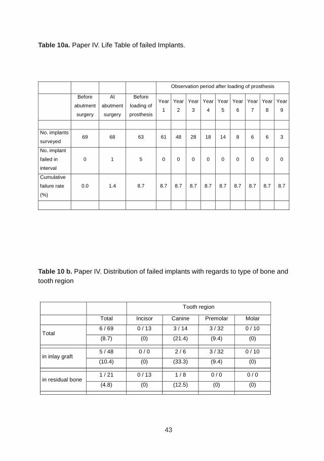
43
Table 10a. Paper IV. Life Table of failed Implants.
Table 10 b. Paper IV. Distribution of failed implants with regards to type of bone andtooth region
Observation period after loading of prosthesis
Before
abutment
surgery
At
abutment
surgery
Before
loading of
prosthesis
Year
1
Year
2
Year
3
Year
4
Year
5
Year
6
Year
7
Year
8
Year
9
No. implants
surveyed 69 68 63 61 48 28 18 14 8 6 6 3
No. implant
failed in
interval
0 1 5 0 0 0 0 0 0 0 0 0
Cumulative
failure rate
(%)
0.0 1.4 8.7 8.7 8.7 8.7 8.7 8.7 8.7 8.7 8.7 8.7
Tooth region
Total Incisor Canine Premolar Molar
6 / 69 0 / 13 3 / 14 3 / 32 0 / 10 Total
(8.7) (0) (21.4) (9.4) (0)
5 / 48 0 / 0 2 / 6 3 / 32 0 / 10 in inlay graft
(10.4) (0) (33.3) (9.4) (0)
1 / 21 0 / 13 1 / 8 0 / 0 0 / 0 in residual bone
(4.8) (0) (12.5) (0) (0)
44
Paper V
Implant and Bone Graft StabilityThe 61 patients received 52 unilateral and 9 bilateral sinus inlay
bone grafts. Eight patients also received an onlay bone graft in addition to thesinus inlay bone graft. All bone grafts, were stable and supported 146 implantsof which two implants failed (1.4%) giving an early survival rate of 98.6%. Theimplant failures were in the canine and premolar position, both engaged in graftedsinus inlay bone. In the residual bone, 34 implants were installed of which noimplants failed. Only two (1.1%), one Straumann implant and one Brånemarkimplant, of the 180 implants placed were lost resulting in an implant survival rateof 98.9%.
Prosthesis StabilityAll patients received a fixed prostheses after an implant healing
period of 2 to 17 months (mean 5.7).
Radiographic ExaminationsThe radiographic examinations showed an average residual vertical
bone height of 6.5mm (37 implants) in first premolar region, 3.8mm (56 implants)in the second premolar region, 3.5mm (46 implants) in the first molar region,and 2.6 mm (7 implants) in the second molar region. The average implantlength was 12 mm in the first premolar region and 11 mm in the second premolar,first and second molar regions (Figure 3).
ComplicationsThe postoperative symptoms at the donor site were similar to the
symptoms after surgical removal of teeth. Paraesthesia of the inferior alveolarnerve was seen in one patient, who completely recovered after less then 2months. There were no records of injured teeth, heavy bruising, bleeding orswelling from either the donor or the recipient site. At the recipient site threepatients had local postoperative infections with shallow fistulas. This wasmanaged with antibiotics and was cured after 2-3 weeks and had no negativeeffect on bone graft or implant survival. It was noted, in three patients, duringthe implants installation, that the new bone was softer than the residual bone.However, this had no negative influence on implant primary stability but one outof ten implants installed in soft bone, failed.
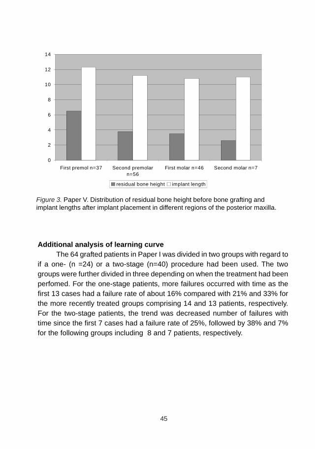
45
Additional analysis of learning curveThe 64 grafted patients in Paper I was divided in two groups with regard to
if a one- (n =24) or a two-stage (n=40) procedure had been used. The twogroups were further divided in three depending on when the treatment had beenperfomed. For the one-stage patients, more failures occurred with time as thefirst 13 cases had a failure rate of about 16% compared with 21% and 33% forthe more recently treated groups comprising 14 and 13 patients, respectively.For the two-stage patients, the trend was decreased number of failures withtime since the first 7 cases had a failure rate of 25%, followed by 38% and 7%for the following groups including 8 and 7 patients, respectively.
Figure 3. Paper V. Distribution of residual bone height before bone grafting andimplant lengths after implant placement in different regions of the posterior maxilla.
0
2
4
6
8
10
12
14
First premol n=37 Second premolarn=56
First molar n=46 Second molar n=7
residual bone height implant length

46
DISCUSSION
Bone grafting techniqueIn papers I and IV, edentulous and partially dentate patients were
treated with bone block grafts using the iliac crest as the donor site. In paper I,approximately 25 % (109 / 437) of the implants placed in the Graft Group failedwhilst 16 % (109 / 683) of the implants placed in the Nongraft Group were lostafter a mean follow-up of 68.9 and 75.8 months respectively. The difference inimplant failure rate was statistically significant. A failure rate of 17 % was seenin implants placed in the residual bone in the Graft Group (18 / 107) comparableto the implant failures in the Nongraft Group of 16 % (109 / 683). In paper IV,approximately 10 % (5/48) of the implants placed in augmented bone failedwhilst 5 % (1/21) of the implants placed in residual bone were lost. In paper I,the inlay bone grafting technique, showed comparable implant survival rate inthe premolar region as in the same region in the Non-Graft Group. Analysis ofthe patient material suggested that the more jaw bone volume present in anteriorregions at the start of treatment, the better the implant survival rates in bothgroups.
In paper V, the possibility of achieving improved results compared topapers I and IV, by using a combination of particulated mandibular bone foraugmentation and delayed placement of surface modified implants wasdemonstrated. The survival rate was 98.9% (2/180) for all implants and 98.6%(2/148) for implants placed in grafted areas after a mean follow-up of 12.8 months,which was until the delivery of fixed prostheses. A previous study, carried out inthe same clinic, comparing implant survival in grafted and in residual bone,showed an implant survival rate of 96% in 76 partially dentate patients treatedduring the same time period as paper I and IV of the present thesis, but withoutbone augmentation procedures and with a mean follow-up of 53,9 months(Johansson and Ekfeldt 2003).
Different outcomes may be expected when comparing edentulousand dentate jaws as discussed by Ragoebar et al. (2001b), who found a 97%versus 90.8% survival rate in dentate and edentulous patients respectively. (Table2a & b) This is in line with our experiences where an overall implant survivalrate of 91.3% was found in 17 partially dentate patients in paper IV, which indicateda better clinical outcome with bone grafting procedures in the partially dentatepatient compared with totally edentulous cases as in paper I. Interestingly, inboth paper I and IV, implant failure occurred mainly from abutment connectionto delivery of the final prosthesis. This indicates that many implants were not
47
well integrated and/or were sensitive to repeated manipulation and trauma fromattaching and de-attaching abutments and impression copings and provisionalprostheses during the prosthetic phase. Another negative factor could be theshort healing periods of the grafts, which were on average 4.9 months in paperI and 4,8 months in paper IV, possibly leading to an immature quality of the bonegraft and thereby impaired osseointegration of the implants.
It was noted in paper IV, that more implants failed when placedsimultaneously with the bone graft than if a two-stage procedure was used. Thiswas not seen in paper I. However, two-stage implants showed a lower survivalrate in inlays but higher in onlay bone grafts and vice versa, which suggests thatthe results were not conclusive. Wannfors et al. reported a doubled risk forimplant failure when using a one-stage technique compared with a two-stageapproach (Wannfors et al., 2000). (Table 2 & 3)
Biologically, a two-stage surgery is preferable, sincerevascularization, maturation and incorporation of the grafted bone areaccomplished before the implants are inserted (Lundgren et al., 1999). If theresidual bone height beneath the maxillary sinus is 4 to 5 mm and of goodquality, initial stability of the implants can probably be achieved using bothapproaches. However, in cases with insufficient bone volume where primaryimplant stability can not be achieved, delayed implant placement is preferred(Block and Kent, 1997; Lundgren et al., 1999). On the other hand, simultaneousplacement is less invasive, more cost-effective, and more time-efficient.
An analysis of implant failure rates in the grafted patients in Paper I,revealed more failures with time for one-stage procedures and less failures withtime for two-stage procedures. Thus, a learning curve, which may be expectedfor complex surgery like bone grafting, could be seen only for the two-stageprocedures. The increasing failure rate with time for the one-stage proceduresmay relate to a higher degree of cautiousness in the first cases.
Bone graft origin and donor siteIn papers I and IV, endochondral bone from the iliac crest was used
as bone grafts. There were no graft failures reported and the implant survivalwas 75.1 (Graft Group) in the edentulous cases and 91,3 % in the partial dentatecases. In paper V, intramembranous bone from the mandibular ramus/corpuswas used as bone graft. There were no graft failures reported and the implantsurvival was 98.9 % in the partially dentate cases.
The embryological origin of the bone graft has been suggested toplay a role in the success of the bone augmentation procedure. It has been
48
proposed, that intramembranous bone, has a better resistance towards boneresorption, shows better incorporation, becomes earlier revascularized and hencemaintain volume and viability to a greater extent than endocondral bone (Alberiuset al., 1992; Rabie et al., 1996; Smith and Abramson, 1974; Zins and Whitaker,1983). On the other hand, it has been suggested that the revascularizationprocess would be better in endochondral bone due to the difference in graftarchitecture. The endochondral bone graft consists mostly of cancellous porousbone, with marrow tissue in between the bone trabeculae in which therevascularisation easier can take part, while intramembranous bone consists ofmore densely packed cortical bone (Albrektsson, 1980; Sullivan and Szwajkun,1991).
In papers I, II and IV, the majority of implant failures were earlylosses, since more than 90 % of the failed implants occurred before loading,which most likely could be related to the bone graft inability to integrate theimplants. This may be explained by the biomechanical properties of theendochondral bone grafts as well as the healing capacity of the bone. Anothernegative factor could be the short healing period of the bone graft leading to animmature quality of the bone graft and thereby impaired osseointegration.In papers I, II and IV, all bone grafted patients were treated with bone graftsfrom the iliac crest. It is possible to harvest large amounts of bone from the iliaccrest, but postoperative morbidity with bruising, swelling, pain and functionalproblems from the donor site are occasionally seen. This approach may alsoproduce a permanent cutaneous scar and the procedure usually involves generalanaesthesia with days of hospitalization (Beirne, 1986; Cricchio and Lundgren,2003; Raghoebar et al., 1999).
The advantages of intra oral sitesfor bone harvesting, such as themandibular ramus/body h, which was used in paper V, are the use of localanaesthesia, reduced operating time, no need for postoperative hospitalizationand less morbidity at the donor sites (Clavero and Lundgren, 2003; Hirsch andEricsson, 1991; Raghoebar et al., 2001a).
Recent reports have been focusing on the morbidity after harvestingof bone from the mandibular symphysis region. Problems that include superficialsensory impairment with hyper and hypoesthesia and teeth that lost their pulpsensitivity have been described. These studies and others suggested that themorbidity when harvesting bone from the mandibular symphysis is moreadvanced than harvesting from the retromolar region (Hallman et al., 2002;Hallman et al., 2005; Nkenke et al., 2001; Nkenke et al., 2002; Raghoebar et al.,2001a). According to earlier findings (Paper V), very few complications with
49
intra-oral bone harvesting occurred at the lateral ramus/corpus of the mandible.Paraesthesia of the inferior alveolar nerve was seen in one patient, whocompletely recovered after less then 2 months. There were no records of heavybruising, bleeding or swelling from either the donor or the recipient site.
Different kinds of grafting material have been evaluated for maxillarysinus floor augmentation (Hallman et al., 2005; Wiltfang et al., 2003). Recently,Hallman et al. (2005) reported an implant survival rate of 86%, for implantsplaced in grafted areas after three years of prosthetic loading using a mixture ofdeproteinized bovine bone and autogenous bone in patients subjected to maxillarysinus inlay augmentation. One obvious advantage of using an allografts orxenografts in combination with or instead of autogenous bone is that a minor/nodonor site is needed, since morbidity after bone harvesting, for instance on theiliac crest and chin, is not negligible. Paper V describes a bone grafting methodthat produces sufficient volume of bone with a minimum of per- and post-operativediscomfort. Recently, several bone-collecting instruments have come on themarket. The ”Bone Trap” is connected on the suction and bone chips createdwhile drilling are collected (Widmark and Ivanoff, 2000). A so-called ”SafeScraper” is a device that shaves the outer surface of the cortical bone at therecipient site or at the donor site and thereby creates a bone chips that can beused as graft material.
Lundgren et al. demonstrated bone formation in the maxillary sinusby creating a secluded compartment by elevating the sinus membrane, placingimplants and replacing the bone window of the entrance through thelateral sinuswall according to the principle of guided tissue regeneration (Lundgren et al.,2004; Palma et al., 2006; Zuccati and Bocchieri, 2003). The studies showedthat the maxillary sinus has agreat potential for healing and bone formationwithout the use of additional bone grafts or bone substitutes. The method revealedto be a predictable technique for bone augmentation of the maxillary sinus floor.
With an increasing demand for bone augmentation procedures it isimportant that uncomplicated techniques which results in a minimum of morbidityand predictable outcomes are developed and tested (Meskin and Brown, 1988;Weintraub et al., 1985).
Implant surface and integrationImplants with rough surface are claimed to promote faster and earlier
bone healing and thereby supposed to be more suitable for earlier loading thanhas been the standard for many years. Ivanoff et al. (2003), evaluated thehuman bone tissue response to two implant surfaces (oxidized or turned) (TiUnite,
50
Brånemark System). Surface roughness and enlargement were greater for theoxidized implants than for the turned implants. Histomorphometric evaluationdemonstrated significantly higher bone-to-implant contact and bone responsefor the oxidized implants, placed in the maxilla or in the mandible and significantlymore bone was found inside the threaded area for the oxidized implants placedboth in the mandible and maxilla. The implants used in papers I, II and IV had allmachined turned and minimally rough surfaces, which may also have contributedto the higher failure rate in grafted bone compared to the surface modified dentalimplant survival outcome in paper V. One could speculate that the longer implanthealing periods in paper I and IV (mean: 8.8 and 7.2, respectively) were beappropriate since machined turned implants were used.
Histology of micro-implants has demonstrated a stronger boneresponse to surface modified implants as compared with turned implants (Ivanoffet al., 2001; Ivanoff et al., 2003) which confirms the findings from numerousanimal investigations (Wennerberg, 1996). Brechter et al. (2005) evaluated200 surface modified implants used in various bone reconstruction procedures.Of the 200 implants, 199 were considered osseointegrated at the time of abutmentsurgery. At the 12-month post-loading follow-up, another two implants wereconsidered not stable. Thus, three implants (1.5%) were considered as failures.The same team had previously reported on a failure rate of 8% when usingturned implants (Sjöström et al. 2005), which may indicate a better outcomewith surface modified implants (Brechter et al., 2005). Also the results in paperV, showed a similar low failure rate, which is different to the high early failurerates reported in papers I, II and III. However, comparative clinical trials areneeded to statistically establish possible differences.
Nevertheless, rougher surfaces may have theoretical clinicaldrawbacks such as being more prone to marginal bone resorption and/orincreased ion release which has been found in bone tissue in the surroundingarea of titanium implants (Osborn JF, 1990) and it has been hypothesized thatthis could be damaging to osteogenesis (Tsutsui et al., 1999).
The healing periodIn paper I and II the majority of implant failures were early losses,
since more than 90 % of the failed implants were lost before loading, whichmost likely could be related to the bone grafts inability to integrate the implants.Another factor for early implant failure in bone-grafted patients might be thepoor bone to implant interface and its sensitiveness to trauma. It is thought, that
51
trauma interferes with early integration, a situation that is exacerbated due to alimited amount of bone at the interface of the implant and the host bone (Espositoet al., 1999; Nystrom et al., 1993). A heterogeneous interface with areas ofhighly vascularized connective tissue and portions of bone were described.Where bone was present, there was always evidence of bone detachment fromthe implant surface by erythrocytes due to bleeding. This could indicate thatmanipulation of the implants at the abutment surgery and during the restorativetreatment phase, might cause bleeding due to mechanical disruption of the bone/implant interface (Aspenberg and Herbertsson, 1996). ”Reosseointegration” ofimplants with rotation mobility may be possible, if the implants are kept unloadedfor a longer healing period but this situation is likely subject to further atraumatichealing (Aspenberg and Herbertsson, 1996; Ivanoff et al., 1997).
Szmukler-Moncler et al. (Szmukler-Moncler et al., 1998) reportedthat the critical level of micromotion on the implant was not zero as generallyinterpreted, instead, the tolerated micromotion threshold was found to liesomewhere between 50 and 150 microns. Above this level, healing would undergofibrous repair rather than the desired osseous regeneration. However, inimmediate functional loading, the implant survival is believed to be successfuldue to that a large number of implants are spread out, which creates a stiff andinflexible prosthesis that controls the forces applied to the bone-implant interface(Balshi et al., 2005).
In paper II, aII patients were using a removable prosthesis in themaxilla during the healing period after the bone graft procedure and implantsurgery prior to definitive prosthesis attachment. The results from this paper,suggest that the type of mandibular dentition, occlusion and bite force might beof significance for early implant failures. Occlusal loading during the healingperiod could result in inadequate tissue healing and thereby impairedosseointegration. Furthermore, according to a parallel study, it was revealedthat also in edentulous maxillae without bone graft, the type of mandibulardentition, might be of significance to early implant failures (Abrahamsson et al.,2003).
In a study with similar bone grafting and implant treatment proceduresas used in our studies, the final survival rate was 88 % when special care wastaken to minimize the occlusal trauma to the edentulous maxillae during thehealing periods, by not using prostheses in the maxillae (Jensen et al., 1994).In another study, a special splint was fabricated to prevent occlusal trauma tothe anterior graft site during the first 2 –3 months of the healing period, and after
52
this a new removable prostheses was handed out, revealing a 80 to 90 % implantsurvival (Lundgren et al., 1997).
The higher survival rate forthe grafted partially dentate patients inpaper IV as compared with the edentulous patients in paper I, may be due tothat no removable dentures were used in study IV. Moreover, in partially dentatepatients occlusal forces on the definitive prosthetic construction are reducedand merely transferred to the natural dentition.
It has been suggested that fixed prostheses create a favourableforce distribution to dental implants (Barzilay et al., 1996; Randow et al., 1999;Tarnow et al., 1997). Applying this hypothesis to the edentulous maxilla, it maybe prudent to consider fixed immediate loading of transitional implants in thelabile bone of an autogenous graft rather than allowing force transmission througha provisional prosthesis that depends on the soft tissue of the residual ridge andthe hard palate for retention, support and stability of the prosthesis. Furtherstudies will be necessary before this routinely can be used clinically.
The correlation between bite force and implant failure during thehealing period is not well described in the literature. It is known that the maximalbite force is greater with implant supported prostheses increases than withremovable dentures. Carr and Laney (1987) reported a significant improvementof bite force in patients whom changed from conventional dentures to implantsupported prostheses in the mandibular arch. The mean maximal bite forcewas 59.6 N in the case of conventional removable dentures and 112.9 N for thepatients who had received a mandibular implant fixed prosthesis. These findings,together with the results of paper II, suggest that the unfavourable concentrationof forces and/or the magnitude of force could jeopardize the integration processof implants. Future studies addressing these issues may help to determine ifthere is an association between bite force and implant failures.According to the findings of paper II, it seems reasonable to suggest that onecausative factor for early implant failure might be the traumatic influence fromthe opposing arch during the healing period of bone-grafts and implants. Thetrauma caused by a provisional maxillary denture opposed by a mandibulardentition that creates force concentration rather than force distribution couldinduce further trauma to the maxilla.
Based on the observation that implant failure is higher when theopposing occlusion is not well distributed, it may be suggested that patientssubjected to bone grafting and implant placement should refrain from wearingdentures during the healing period. Especially, if the mandibular dentition revealsa limited or unilateral occlusion. Another suggestion is to restore the mandible
53
to bilateral occlusion prior to bone grafting and implant treatment in the edentulousmaxilla. This may ensure a broad distribution of teeth and stable occlusion,which should distribute forces favourably to the maxilla and thereby encourageimplant survival. However, It is impossible to attribute the failure rates to theeffect of mandibular dentition alone, as more uncontrolled and unknownparameters very well may play an additional role in implant failure.
Different outcomes may be expected when comparing edentulousand dentate jaws as discussed by Raghoebar et al. (2001b), who found a 97%survival rate in dentate and 90.8% survival in edentulous patients respectively.This is in line with paper IV, where an overall implant survival rate of 91.3% wasfound in 17 partially dentate patients, which indicated a better clinical outcomewith bone grafting procedures in the partially dentate patient compared withtotally edentulous cases as previously reported. Interestingly, in both paper Iand IV, implant failure occurred mainly from abutment connection to delivery ofthe final prosthesis. This indicates that the implants were not well integratedand were sensitive to repeated manipulation when attaching and de-attachingabutments and impression copings during the prosthetic phase
It may be wise to place the implant deeper within the graft in aneffort to avoid trauma to the implant from the provisional prosthesis. However,this will create a reduced cortical bone/implant contact with less favorable primarystability. It is possible that an internal space screw during the healing period,instead of the conventional cover screw, might have minimized the risk of traumato the implant.
Alternative treatments to bone graftingThe use of alternative implant sites and tilted implants have been
advocated to reduce the necessity of bone grafting procedures. The placementof dental implants in the zygomatic bone has been used in conjunction withregular implants in patients with severe resorption of the maxilla (Bedrossian etal., 2002; Branemark et al., 2004; Higuchi, 2000; Hirsch et al., 2004; Malevez etal., 2004). However, few studies include long-term evaluation of the soft andbone tissue reactions to zygomatic implants. In paper III, six out of 16 patientshad been treated for recurrent sinusitis. Three of these patients had onezygomatic implant removed due to the infection in the maxillary sinus. Anotheralternative technique for bone grafting is the pterygomaxillary implant (Bahat,1992; Balshi et al., 1999; Tulasne, 1992). Sorni et al. (2005) reported, in a
54
review of the literature, a survival rate from 86.3% to 97.2 for the pterygomaxillaryimplant and reported of no major complications during implant surgery.
The objective of paper III was to evaluate the clinical outcome ofzygomatic implant treatment in the severely atrophied maxilla as an alternativeto bone grafting procedures in patients with edentulous maxillae. There wereno adverse complications associated with the zygomatic implant surgery and allpatients had an uneventful healing period after the implant placement until theabutment connection surgery, similar to conventional implant surgery. After theabutment connection surgery, the complications were at an unacceptable level.However, oral hygiene was a problem at the zygomatic abutment only. Withhelp and instructions from a dental hygienist, most of the patients improvedtheir oral hygiene. It seems that the posterior, palatal localization of the zygomaticimplant creates difficulties in upholding hygiene and extra professional assistancemay be required. Even though effort was made to place the zygomatic implantas close as possible to the alveolar crest, this was not possible to accomplish inthis material due to the anatomy and degree of atrophy. The distance from thenearest buccal cusp on the prosthesis to the zygomatic implant, was measuredwith a mean distance of 11.2 mm and the zygomatic interimplant distance hadan average of 21.7 mm. An optimal dental implant placement is often consideredto result in a screw hole in the center of the prosthetic crown, which would mean2-4 mm from the buccal cusp. Considering the localization of the zygomaticimplant in this study, one can understand the difficulties, managing an optimaloral hygiene.
All zygomatic implants became osseointegrated and a survival rateof 100% would have been the case if not three zygomatic implants had beenremoved because of recurrent sinusitis. Nine (9/16) patients had problems withlocal infections at the periimplant site of their zygomatic implants with mucositisand/or fistulae, which is in line with the findings of other researchers (Table 1).The reason for the sinusitis observed in this study can be attributed to severalfactors. The internal threaded abutment screw chamber of the zygomatic implantseems to create a communication from the oral cavity into the maxillary sinus,which may result in sinusitis. Another causative factor may be the lack ofosseointegration, bone-to-implant contact, at the marginal level in the palatalarea and the functional loading, resulting in transversal mobility of the long coronalpart of the zygomatic implant. This could implicate a higher risk of communicationbetween the maxillary sinus and the oral cavity and thereby causing sinusitis.The prosthetic survival rate of 100% in this clinical report is encouraging for thetreatment of this patient population. Treatment with the zygomatic implant in
55
extensive maxillary defects looks very promising and could, in some cases, bethe only treatment solution (Parel et al., 2001; Reichert, 1999; Schmidt et al.,2004). However, the risk of soft tissue problems and sinusitis should not beunderestimated, especially if other treatment options are at hand. Furtherprospective long-term clinical studies are required focusing on the health of themaxillary sinus. Zygomatic implant treatment is promising and has someadvantages over bone grafting procedures. However, the frequent soft tissuecomplications at the abutment level and the development of sinusitis call formore studies.
In growing individuals who are lacking development of the alveolarprocess due to either trauma, congenital missing teeth, infra occluded teeth orother pathological conditions, autotransplantation to the affected region often isa very reliable treatment option in order to prevent extensive bone augmentationlater. If the treatment is successful, the autotransplantated tooth, in connectionwith tooth eruption, will lead to continue development of the alveolar process(Zachrisson et al., 2004). Furthermore in adults with loss of vertical bone heightas a result of periodontal disease hard tissue grafting can be difficult to overcome.In subjects where the failing teeth or failed tooth is still present, an alternativemethod can be utilized to generate vertical bone height. Pre-implant orthodonticextrusion can be exerted on the hopeless tooth and thereby induce bone andsoft tissue formation in the future implant site (Salama and Salama, 1993; Zuccatiand Bocchieri, 2003).The same principle can also be used by moving remainingteeth into an atrophied alveolar crest, whereby new bone is formed behind theorthodontically translated tooth. Behind the translated tooth there often will besufficient bone for implant installation without additional bone augmentation.This treatment approach is often indicated if the patient besides lacking teeth,also has a malocclusion
Reconstruction of the maxilla with distraction osteogenesis is atreatment option of choice. The method is new and the distraction osteogenesisdevices are mostly very costly and are still under development. Case reportshave shown promising results, however, the morbidity, cost, and time requiredfor the treatment must be weighed against the benefits of alternative approaches(Jensen et al., 2004; Laster et al., 2005).
The placement of short implants could be one therapeutic optionthat reduces the need for augmentation therapy in the maxilla. In a retrospectivestudy, it was demonstrated that the use of short implants may be considered forprosthetic rehabilitation of the severely resorbed maxilla as an alternative tomore complicated surgical techniques (Renouard and Nisand, 2005).

56
Diagnostic imaging has grown in recent years and a specializedtechnique has become available for the preoperative planning of oral implantplacement with the help of computer tomography. This imaging technologyprovides 3D and cross-sectional views of the jaws. This will provide us withinformation to plan a case more accurate and thereby decide in what degreebone grafting is needed (Guerrero et al., 2006).
57
CONCLUSIONS
• There is a lower overall implant survival rate in grafted than in non-grafted maxillae after a mean follow-up period of 5 to 6 years. Themajority of implant failures occur before prosthetic loading. It seemsthat the more jaw bone volume present in anterior regions at the start oftreatment, the better the implant survival rates in both groups. Moreover,the implant survival rate is similar in grafted posterior edentulous maxillaeof class V-IV and in non-grafted posterior edentulous maxillae of classIII-IV.
• The type of mandibular dentition has an influence on the outcomeof reconstructive surgery using endosseous implants and bone graftingin the edentulous maxilla. An unfavourable concentration of forces in themaxilla, such as in cases of an unilateral occlusal support, may contributeto the increased risk of implant failure. If grafting is required in theedentulous maxilla, every effort should be made to create a favourableocclusion in the mandible, with attention being paid to broad distributionof occlusal contacts.
• The zygomatic implant, when placed in conjunction with premaxillaryimplants, shows an acceptable outcome with regard to implant andprosthetic survival rates. However, other complications, such as sinusitis,not related to implant and prosthesis stability, may lead to the removal ofzygomatic implants.
• The use of endosseous implants and sinus inlay block bone-graftsharvested from the iliac crest for reconstruction of the partially dentatemaxilla results in a satisfactory clinical outcome after a mean follow-upof more than 4 years.
• The use of particulated manibular bone grafts for maxillary flooraugmentation and delayed placement of surface modified dental implantsresults in low morbidity and few implant failures from placement to delveryof the final prosthesis.
58
ACKNOWLEDGEMENTS
I would like to express my sincere gratitude to all people, who have been
involved in the work with this thesis, in particular:
Sten Isaksson, my Head tutor, for sharing material and knowledge, for his
constant support, guidance and never failing enthusiasm and for his way of
creating a friendly and creative atmosphere.
Lars Sennerby, my assistant tutor, for generous support, enthusiasm and
for creating a pleasant research atmosphere.
My research associates and co- authors, Eugene E. Keller, Steven E.
Eckert, Peter Abrahamsson and Hadar Hallström for stimulating collaborations.
All clinicians, at the Maxillofacial Unit, Halmstad, Sweden and MAYO Clinic,
Rochester, MN, USA, who have been part of the clinical trials.
All staff at the Maxillofacial Unit and at the Dento-Maxillofacial Radiology
Division, Halmstad, Sweden for their never ending help during the clinical trials.
My dearest, Karin, Jonathan, Mia and August, for their understanding and
ever lasting patience and encouragement.
This work has been supported by grants from Research & Development Council,
Halmstad Hospital, Sweden and Hjalmar Svensson Foundation.
59
REFERENCES
Abrahamsson I, Berglundh T, Glantz PO, Lindhe J (1998). The mucosalattachment at different abutments. An experimental study in dogs. J ClinPeriodontol 25(9):721-7.
Abrahamsson P, Becktor JP, Isaksson S, Blomqvist J (2003). The influence ofmandibular dentition on implant failures in the edentulous maxillae. SwedishDental Journal 95(11):42-48.
Adell R, Eriksson B, Lekholm U, Branemark PI, Jemt T (1990a). Long-termfollow-up study of osseointegrated implants in the treatment of totally edentulousjaws. Int J Oral Maxillofac Implants 5(4):347-59.
Adell R, Lekholm U, Grondahl K, Branemark PI, Lindstrom J, Jacobsson M(1990b). Reconstruction of severely resorbed edentulous maxillae usingosseointegrated fixtures in immediate autogenous bone grafts. Int J OralMaxillofac Implants 5(3):233-46.
Ahlgren F, Storksen K, Tornes K (2006). A study of 25 zygomatic dental implantswith 11 to 49 months’ follow-up after loading. Int J Oral Maxillofac Implants21(3):421-5.
Alberius P, Dahlin C, Linde A (1992). Role of osteopromotion in experimentalbone grafting to the skull: a study in adult rats using a membrane technique. JOral Maxillofac Surg 50(8):829-34.
Albreksson T (1979). Healing of bonegrafts. Gothenburg, University ofGothenburg.
Albrektsson T (1980). Repair of bone grafts. A vital microscopic and histologicalinvestigation in the rabbit.1-12.
Albrektsson T, Branemark PI, Hansson HA, Lindstrom J (1981). Osseointegratedtitanium implants. Requirements for ensuring a long-lasting, direct bone-to-implant anchorage in man. Acta Orthop Scand 52(2):155-70.
Albrektsson T (1983). Direct bone anchorage of dental implants. J Prosthet Dent50(2):255-61.
Albrektsson T (1988). A multicenter report on osseointegrated oral implants. JProsthet Dent 60(1):75-84.
60
Albrektsson T (2001). Is surgical skill more important for clinical success thanchanges in implant hardware? Clin Implant Dent Relat Res 3(4):174-5.
Albrektsson T, Johansson C (2001). Osteoinduction, osteoconduction andosseointegration. Eur Spine J 10 Suppl 2(S96-101.
Aparicio C, Perales P, Rangert B (2001). Tilted implants as an alternative tomaxillary sinus grafting: a clinical, radiologic, and periotest study. Clin ImplantDent Relat Res 3(1):39-49.
Aparicio C, Ouazzani W, Garcia R, Arevalo X, Muela R, Fortes V (2006). Aprospective clinical study on titanium implants in the zygomatic arch for prostheticrehabilitation of the atrophic edentulous maxilla with a follow-up of 6 months to5 years. Clin Implant Dent Relat Res 8(3):114-22.
Asikainen P, Klemetti E, Vuillemin T, Sutter F, Rainio V, Kotilainen R (1997).Titanium implants and lateral forces. An experimental study with sheep. ClinOral Implants Res 8(6):465-8.
Aspenberg P, Herbertsson P (1996). Periprosthetic bone resorption. Particlesversus movement. J Bone Joint Surg Br 78(4):641-6.
Astrand P, Nord PG, Branemark PI (1996). Titanium implants and onlay bonegraft to the atrophic edentulous maxilla: a 3-year longitudinal study. Int J OralMaxillofac Surg 25(1):25-9.
Atwood DA (1971). Reduction of residual ridges: a major oral disease entity. JProsthet Dent 26(3):266-79.
Bahat O (1992). Osseointegrated implants in the maxillary tuberosity: report on45 consecutive patients. Int J Oral Maxillofac Implants 7(4):459-67.
Balshi SF, Wolfinger GJ, Balshi TJ (2005). A prospective study of immediatefunctional loading, following the Teeth in a Day protocol: a case series of 55consecutive edentulous maxillas. Clin Implant Dent Relat Res 7(1):24-31.
Balshi TJ, Wolfinger GJ, Balshi SF, 2nd (1999). Analysis of 356 pterygomaxillaryimplants in edentulous arches for fixed prosthesis anchorage. Int J Oral MaxillofacImplants 14(3):398-406.
Barth A (1895). Histologische Untersuchungen über Knochenimplantationen.Beitr. path. Anat. 17(65-142.
61
Barzilay I, Graser GN, Iranpour B, Natiella JR, Proskin HM (1996). Immediateimplantation of pure titanium implants into extraction sockets of Macacafascicularis. Part II: Histologic observations. Int J Oral Maxillofac Implants11(4):489-97.
Baschkirzew N, Petrov N (1912). Beiträge zur freien Knochenüberpflazung.Dtsch. Z. Chir. 113(490-531.
Becktor JP, Isaksson S, Sennerby L (2004). Survival analysis of endosseousimplants in grafted and nongrafted edentulous maxillae. Int J Oral MaxillofacImplants 19(1):107-15.
Becktor JP, Isaksson S, Abrahamsson P, Sennerby L (2005). Evaluation of 31zygomatic implants and 74 regular dental implants used in 16 patients forprosthetic reconstruction of the atrophic maxilla with cross-arch fixed bridges.Clin Implant Dent Relat Res 7(3):159-65.
Becktor JP, Isaksson S, Sennerby L (2006b). Endosseopus implants and boneaugmnetation in the partially dentae maxilla. An analysis of 17 patients with afollow-up of 29 to 101 months. Int J Oral Maxillofac Implants, Accepted
Becktor JP, Hallström H, Isaksson S, Sennerby L (2006b). The use of particulatedbone grafts from the mandible for maxillary sinus floor augmentation prior toplacement of surface modified implants. Results from bone grafting to deliveryof the final fixed prosthesis. In manuscript
Bedrossian E, Stumpel L, 3rd, Beckely ML, Indresano T (2002). The zygomaticimplant: preliminary data on treatment of severely resorbed maxillae. A clinicalreport. Int J Oral Maxillofac Implants 17(6):861-5.
Beirne OR (1986). Comparison of complications after bone removal from lateraland medial plates of the anterior ilium for mandibular augmentation. Int J OralMaxillofac Surg 15(3):269-72.
Bell WH (1969). Revascularization and bone healing after anterior maxillaryosteotomy: a study using adult rhesus monkeys. J Oral Surg 27(4):249-55.
Bjork A (1964). Sutural Growth of the Upper Face Studied by the Implant Method.Rep Congr Eur Orthod Soc 40(49-65.
Bjork A, Skieller V (1977). Growth of the maxilla in three dimensions as revealedradiographically by the implant method. Br J Orthod 4(2):53-64.
62
Block MS, Kent JN (1997). Sinus augmentation for dental implants: the use ofautogenous bone. J Oral Maxillofac Surg 55(11):1281-6.
Blomqvist JE, Alberius P, Isaksson S (1996). Retrospective analysis of one-stage maxillary sinus augmentation with endosseous implants. Int J OralMaxillofac Implants 11(4):512-21.
Boyne PJ, James RA (1980). Grafting of the maxillary sinus floor with autogenousmarrow and bone. J Oral Surg 38(8):613-6.
Branemark PI, Adell R, Breine U, Hansson BO, Lindstrom J, Ohlsson A (1969).Intra-osseous anchorage of dental prostheses. I. Experimental studies. Scand JPlast Reconstr Surg 3(2):81-100.
Branemark PI, Hansson BO, Adell R, Breine U, Lindstrom J, Hallen O, OhmanA (1977). Osseointegrated implants in the treatment of the edentulous jaw.Experience from a 10-year period. Scand J Plast Reconstr Surg Suppl 16(1-132.
Branemark PI, Grondahl K, Ohrnell LO, Nilsson P, Petruson B, Svensson B,Engstrand P, Nannmark U (2004). Zygoma fixture in the management ofadvanced atrophy of the maxilla: technique and long-term results. Scand J PlastReconstr Surg Hand Surg 38(2):70-85.
Branemark PI, Zarb G, Albrektsson T (1985). Tissue-Integrated Prostheses.Osseointegration in Clinical Dentistry Chicago, IL: Quintessence Publishing.
Brechter M, Nilson H, Lundgren S (2005). Oxidized titanium implants inreconstructive jaw surgery. Clin Implant Dent Relat Res 7 Suppl 1(S83-7.
Breine U, Branemark PI (1980). Reconstruction of alveolar jaw bone. Anexperimental and clinical study of immediate and preformed autologous bonegrafts in combination with osseointegrated implants. Scand J Plast ReconstrSurg 14(1):23-48.
Brunski JB (1993). Avoid pitfalls of overloading and micromotion of intraosseousimplants [interview]. Dent Implantol Update 4(10):77-81.
Bull C (1928). Experimentelle Studien über Knochentransplantation undKnochenregeneration. Skrifter Norske Videnskaps-Akademi 9):1-105.
63
Carr AB, Laney WR (1987). Maximum occlusal force levels in patients withosseointegrated oral implant prostheses and patients with complete dentures.Int J Oral Maxillofac Implants 2(2):101-8.
Cawood JI, Howell RA (1988). A classification of the edentulous jaws. Int J OralMaxillofac Surg 17(4):232-6.
Cawood JI, Howell RA (1991). Reconstructive preprosthetic surgery. I. Anatomicalconsiderations. Int J Oral Maxillofac Surg 20(2):75-82.
Chase SW, Herndon CH (1955). The fate of autogenous and homogenous bonegrafts. J Bone Joint Surg Am 37-A(4):809-41.
Chen NT, Glowacki J, Bucky LP, Hong HZ, Kim WK, Yaremchuk MJ (1994). Theroles of revascularization and resorption on endurance of craniofacial onlay bonegrafts in the rabbit. Plast Reconstr Surg 93(4):714-22; discussion 723-4.
Clavero J, Lundgren S (2003). Ramus or chin grafts for maxillary sinus inlay andlocal onlay augmentation: comparison of donor site morbidity and complications.Clin Implant Dent Relat Res 5(3):154-60.
Cordaro L, Amade DS, Cordaro M (2002). Clinical results of alveolar ridgeaugmentation with mandibular block bone grafts in partially edentulous patientsprior to implant placement. Clin Oral Implants Res 13(1):103-111
Cricchio G, Lundgren S (2003). Donor site morbidity in two different approachesto anterior iliac crest bone harvesting. Clin Implant Dent Relat Res 5(3):161-9.
Donovan MG, Dickerson NC, Hanson LJ, Gustafson RB (1994). Maxillary andmandibular reconstruction using calvarial bone grafts and Branemark implants:a preliminary report. J Oral Maxillofac Surg 52(6):588-94.
Eckert SE, Desjardins RP, Keller EE, Tolman DE (1996). Endosseous implantsin an irradiated tissue bed. J Prosthet Dent 76(1):45-9.
Eriksson AR, Albrektsson T (1983). Temperature threshold levels for heat-inducedbone tissue injury: a vital-microscopic study in the rabbit. J Prosthet Dent50(1):101-7.
Eriksson AR (1984). An in vivo investigation of heat tolerance of bone tissue andtemperature rise in the drilling of cortical bone (Ph. D.). Gothenburg, Universityof Gothenburg.
64
Esposito M, Hirsch JM, Lekholm U, Thomsen P (1998a). Biological factorscontributing to failures of osseointegrated oral implants. (I). Success criteriaand epidemiology. Eur J Oral Sci 106(1):527-51.
Esposito M, Hirsch JM, Lekholm U, Thomsen P (1998b). Biological factorscontributing to failures of osseointegrated oral implants. (II). EtiopathogenesisBiological factors contributing to failures of osseointegrated oral implants. (I).Success criteria and epidemiology. Eur J Oral Sci 106(3):721-64.
Esposito M, Hirsch JM, Lekholm U, Thomsen P (1998c). Biological factorscontributing to failures of osseointegrated oral implants. (II). Etiopathogenesis.Eur J Oral Sci 106(3):721-64.
Esposito M, Thomsen P, Ericson LE, Lekholm U (1999). Histopathologicobservations on early oral implant failures. Int J Oral Maxillofac Implants14(6):798-810.
Farzad P, Andersson L, Gunnarsson S, Johansson B (2006). Rehabilitation ofseverely resorbed maxillae with zygomatic implants: an evaluation of implantstability, tissue conditions, and patients’ opinion before and after treatment. Int JOral Maxillofac Implants 21(3):399-404.
Foster RD, Anthony JP, Sharma A, Pogrel MA (1999). Vascularized bone flapsversus nonvascularized bone grafts for mandibular reconstruction: an outcomeanalysis of primary bony union and endosseous implant success. Head Neck21(1):66-71.
Friberg B, Jemt T, Lekholm U (1991). Early failures in 4,641 consecutively placedBranemark dental implants: a study from stage 1 surgery to the connection ofcompleted prostheses. Int J Oral Maxillofac Implants 6(2):142-6.
Gocke DJ (2005). Tissue donor selection and safety. Clin Orthop Relat Res435):17-21.
Granstrom G, Tjellstrom A, Branemark PI, Fornander J (1993). Bone-anchoredreconstruction of the irradiated head and neck cancer patient. Otolaryngol HeadNeck Surg 108(4):334-43.
Guerrero ME, Jacobs R, Loubele M, Schutyser F, Suetens P, van SteenbergheD (2006). State-of-the-art on cone beam CT imaging for preoperative planningof implant placement. Clin Oral Investig 10(1):1-7.
65
Gurlek A, Miller MJ, Jacob RF, Lively JA, Schusterman MA (1998). Functionalresults of dental restoration with osseointegrated implants after mandiblereconstruction. Plast Reconstr Surg 101(3):650-5; discussion 656-9.
Hallman M, Hedin M, Sennerby L, Lundgren S (2002). A prospective 1-yearclinical and radiographic study of implants placed after maxillary sinus flooraugmentation with bovine hydroxyapatite and autogenous bone. J Oral MaxillofacSurg 60(3):277-84; discussion 285-6.
Hallman M, Sennerby L, Zetterqvist L, Lundgren S (2005). A 3-year prospectivefollow-up study of implant-supported fixed prostheses in patients subjected tomaxillary sinus floor augmentation with a 80:20 mixture of deproteinized bovinebone and autogenous bone Clinical, radiographic and resonance frequencyanalysis. Int J Oral Maxillofac Surg 34(3):273-80.
Henry PJ, Laney WR, Jemt T, Harris D, Krogh PH, Polizzi G, Zarb GA, HerrmannI (1996). Osseointegrated implants for single-tooth replacement: a prospective5-year multicenter study. Int J Oral Maxillofac Implants 11(4):450-5.
Higuchi KW (2000). The zygomaticus fixture: an alternative approach for implantanchorage in the posterior maxilla. Ann R Australas Coll Dent Surg 15(28-33.
Hirsch JM, Ericsson I (1991). Maxillary sinus augmentation using mandibularbone grafts and simultaneous installation of implants. A surgical technique. ClinOral Implants Res 2(2):91-6.
Hirsch JM, Ohrnell LO, Henry PJ, Andreasson L, Branemark PI, Chiapasco M,Gynther G, Finne K, Higuchi KW, Isaksson S, Kahnberg KE, Malevez C, NeukamFW, Sevetz E, Urgell JP, Widmark G, Bolind P (2004). A clinical evaluation of theZygoma fixture: one year of follow-up at 16 clinics. J Oral Maxillofac Surg 62(9Suppl 2):22-9.
Hjørting-Hansen E, Worsaae N, Lemons JE (1990). Histologic response afterimplantation of porous hydroxylapatite ceramic in humans. Int J Oral MaxillofacImplants 5(3):255-63.
Hollender L, Rockler B (1980). Radiographic evaluation of osseointegratedimplants of the jaws. Experimental study of hte influence of radiographictechniques on the measurement of the relation between the implant and bone.Dentomaxillofac Radiol 9(2):91-5.
Hood C, Dunleavy P (1994). From Old Public Managment to New PublicManagment. Public Managment and Money July-September):9-16.
66
Isaksson S, Alberius P (1992). Maxillary alveolar ridge augmentation with onlaybone-grafts and immediate endosseous implants. J Craniomaxillofac Surg20(1):2-7.
Isaksson S (1994). Evaluation of three bone grafting techniques for severelyresorbed maxillae in conjunction with immediate endosseous implants. Int JOral Maxillofac Implants 9(679-688.
Ivanoff CJ, Sennerby L, Lekholm U (1997). Reintegration of mobilized titaniumimplants. An experimental study in rabbit tibia. Int J Oral Maxillofac Surg26(4):310-5.
Ivanoff CJ, Hallgren C, Widmark G, Sennerby L, Wennerberg A (2001). Histologicevaluation of the bone integration of TiO(2) blasted and turned titaniummicroimplants in humans. Clin Oral Implants Res 12(2):128-34.
Ivanoff CJ, Widmark G, Johansson C, Wennerberg A (2003). Histologic evaluationof bone response to oxidized and turned titanium micro-implants in humanjawbone. Int J Oral Maxillofac Implants 18(3):341-8.
Jemt T, Lekholm U (1993). Oral implant treatment in posterior partially edentulousjaws: a 5-year follow-up report. Int J Oral Maxillofac Implants 8(6):635-40.
Jemt T, Lekholm U (1995). Implant treatment in edentulous maxillae: a 5-yearfollow-up report on patients with different degrees of jaw resorption. Int J OralMaxillofac Implants 10(3):303-11.
Jemt T, Lekholm U, Johansson CB (2000). Bone response to implant-supportedframeworks with differing degrees of misfit preload: in vivo study in rabbits. ClinImplant Dent Relat Res 2(3):129-37.
Jensen J, Sindet-Pedersen S, Oliver AJ (1994). Varying treatment strategies forreconstruction of maxillary atrophy with implants: results in 98 patients. J OralMaxillofac Surg 52(3):210-6; discussion 216-8.
Jensen OT, Leopardi A, Gallegos L (2004). The case for bone graft reconstructionincluding sinus grafting and distraction osteogenesis for the atrophic edentulousmaxilla. J Oral Maxillofac Surg 62(11):1423-8.
Jensen SS, Aaboe M, Pinholt EM, Hjorting-Hansen E, Melsen F, Ruyter IE (1996).Tissue reaction and material characteristics of four bone substitutes. Int J OralMaxillofac Implants 11(1):55-66.
67
Johansson B, Wannfors K, Ekenback J, Smedberg JI, Hirsch J (1999). Implantsand sinus-inlay bone grafts in a 1-stage procedure on severely atrophied maxillae:surgical aspects of a 3-year follow-up study. Int J Oral Maxillofac Implants14(6):811-8.
Johansson B, Grepe A, Wannfors K, Hirsch JM (2001). A clinical study of changesin the volume of bone grafts in the atrophic maxilla. Dentomaxillofac Radiol30(3):157-61.
Johansson B, Back T, Hirsch JM (2004). Cutting torque measurements inconjunction with implant placement in grafted and nongrafted maxillas as anobjective evaluation of bone density: a possible method for identifying earlyimplant failures? Clin Implant Dent Relat Res 6(1):9-15.
Jokstad A, Braegger U, Brunski JB, Carr AB, Naert I, Wennerberg A (2003).Quality of dental implants. Int Dent J 53(6 Suppl 2):409-43.
Keller EE, Van Roekel NB, Desjardins RP, Tolman DE (1987). Prosthetic-surgicalreconstruction of the severely resorbed maxilla with iliac bone grafting and tissue-integrated prostheses. Int J Oral Maxillofac Implants 2(3):155-65.
Keller EE, Eckert SE, Tolman DE (1994). Maxillary antral and nasal one-stageinlay composite bone graft: preliminary report on 30 recipient sites. J OralMaxillofac Surg 52(5):438-47; discussion 447-8.
Keller EE (1997). Placement of dental implants in the irradiated mandible: aprotocol without adjunctive hyperbaric oxygen. J Oral Maxillofac Surg 55(9):972-80.
Keller EE, Tolman DE, Eckert S (1999). Surgical-prosthodontic reconstructionof advanced maxillary bone compromise with autogenous onlay block bone graftsand osseointegrated endosseous implants: a 12-year study of 32 consecutivepatients. Int J Oral Maxillofac Implants 14(2):197-209.
Kelly E (1972). Changes caused by a mandibular removable partial dentureopposing a maxillary complete denture. J Prosthet Dent 27(2):140-50.
Kent JN, Block MS (1989). Simultaneous maxillary sinus floor bone grafting andplacement of hydroxylapatite-coated implants. J Oral Maxillofac Surg 47(3):238-42.
68
Kluth EV, Jain PR, Stuchell RN, Frich JC, Jr. (1988). A study of factors contributingto the development of osteoradionecrosis of the jaws. J Prosthet Dent 59(2):194-201.
Kobayashi E, Ishihara O, Mataga I (2005). Effects of the placement of endosseousimplants in vascularized bone grafts on bone union in beagle dogs. Int J OralMaxillofac Surg 34(6):659-67.
Kondell PA, Nordenram A, Moberg LE, Eliasson S, Nyberg B (1996).Reconstruction of the resorbed edentulous maxilla using autogenous rib graftsand osseointegrated implants. Clin Oral Implants Res 7(3):286-90.
Krekmanov L (2000). Placement of posterior mandibular and maxillary implantsin patients with severe bone deficiency: a clinical report of procedure. Int J OralMaxillofac Implants 15(5):722-30.
Kurth LE (1954). Balanced occlusion. J Prosthet Dent 4(150-167.
Kusiak JF, Zins JE, Whitaker LA (1985). The early revascularization ofmembranous bone. Plast Reconstr Surg 76(4):510-6.
Lambert PM, Morris HF, Ochi S (1997). Positive effect of surgical experiencewith implants on second-stage implant survival. J Oral Maxillofac Surg 55(12Suppl 5):12-8.
Laster Z, Rachmiel A, Jensen OT (2005). Alveolar width distraction osteogenesisfor early implant placement. J Oral Maxillofac Surg 63(12):1724-30.
Lekholm U, Wannfors K, Isaksson S, Adielsson B (1999). Oral implants incombination with bone grafts. A 3-year retrospective multicenter study using theBranemark implant system. Int J Oral Maxillofac Surg 28(3):181-7.
Lindhe J, Berglundh T, Ericsson I, Liljenberg B, Marinello C (1992). Experimentalbreakdown of peri-implant and periodontal tissues. A study in the beagle dog.Clin Oral Implants Res 3(1):9-16.
Loukota RA, Isaksson SG, Linner EL, Blomqvist JE (1992). A technique forinserting endosseous implants in the atrophic maxilla in a single stage procedure.Br J Oral Maxillofac Surg 30(1):46-9.
Lundgren S, Moy P, Johansson C, Nilsson H (1996). Augmentation of the maxillarysinus floor with particulated mandible: a histologic and histomorphometric study.Int J Oral Maxillofac Implants 11(6):760-6.
69
Lundgren S, Nystrom E, Nilson H, Gunne J, Lindhagen O (1997). Bone graftingto the maxillary sinuses, nasal floor and anterior maxilla in the atrophic edentulousmaxilla. A two-stage technique. Int J Oral Maxillofac Surg 26(6):428-34.
Lundgren S, Rasmusson L, Sjostrom M, Sennerby L (1999). Simultaneous ordelayed placement of titanium implants in free autogenous iliac bone grafts.Histological analysis of the bone graft-titanium interface in 10 consecutivepatients. Int J Oral Maxillofac Surg 28(1):31-7.
Lundgren S, Andersson S, Gualini F, Sennerby L (2004). Bone reformation withsinus membrane elevation: a new surgical technique for maxillary sinus flooraugmentation. Clin Implant Dent Relat Res 6(3):165-73.
Malevez C, Abarca M, Durdu F, Daelemans P (2004). Clinical outcome of 103consecutive zygomatic implants: a 6-48 months follow-up study. Clin Oral ImplantsRes 15(1):18-22.
Marukawa E, Asahina I, Oda M, Seto I, Alam MI, Enomoto S (2001). Boneregeneration using recombinant human bone morphogenetic protein-2 (rhBMP-2) in alveolar defects of primate mandibles. Br J Oral Maxillofac Surg 39(6):452-9.
Mattsson T, Kondell PA, Gynther GW, Fredholm U, Bolin A (1999). Implanttreatment without bone grafting in severely resorbed edentulous maxillae. J OralMaxillofac Surg 57(3):281-7.
Melo MD, Shafie H, Obeid G (2006). Implant survival rates for oral andmaxillofacial surgery residents: a retrospective clinical review with analysis ofresident level of training on implant survival. J Oral Maxillofac Surg 64(8):1185-9.
Mericske-Stern R, Hofmann J, Wedig A, Geering AH (1993). In vivomeasurements of maximal occlusal force and minimal pressure threshold onoverdentures supported by implants or natural roots: a comparative study, Part1. Int J Oral Maxillofac Implants 8(6):641-9.
Meskin LH, Brown LJ (1988). Prevalence and patterns of tooth loss in U.S.employed adult and senior populations, 1985-86. J Dent Educ 52(12):686-91.
Misch CM (1999). The harvest of ramus bone in conjunction with third molarremoval for onlay grafting before placement of dental implants. J Oral MaxillofacSurg 57(11):1376-9.
70
Niimi A, Ueda M, Keller EE, Worthington P (1998). Experience withosseointegrated implants placed in irradiated tissues in Japan and the UnitedStates. Int J Oral Maxillofac Implants 13(3):407-11.
Nkenke E, Schultze-Mosgau S, Radespiel-Troger M, Kloss F, Neukam FW (2001).Morbidity of harvesting of chin grafts: a prospective study. Clin Oral ImplantsRes 12(5):495-502.
Nkenke E, Radespiel-Troger M, Wiltfang J, Schultze-Mosgau S, Winkler G,Neukam FW (2002). Morbidity of harvesting of retromolar bone grafts: aprospective study. Clin Oral Implants Res 13(5):514-21.
Nystrom E, Kahnberg KE, Albrektsson T (1993). Treatment of the severelyresorbed maxillae with bone graft and titanium implants: histologic review ofautopsy specimens. Int J Oral Maxillofac Implants 8(2):167-72.
Nystrom E, Ahlqvist J, Legrell PE, Kahnberg KE (2002). Bone graft remodellingand implant success rate in the treatment of the severely resorbed maxilla: a 5-year longitudinal study. Int J Oral Maxillofac Surg 31(2):158-64.
Nystrom E, Ahlqvist J, Gunne J, Kahnberg KE (2004). 10-year follow-up of onlaybone grafts and implants in severely resorbed maxillae. Int J Oral MaxillofacSurg 33(3):258-62.
Ollier L (1867). Traité experimental et clinique de la régénération des os et de laproduction artificielle du tissu osseux. Paris: V. Masson et Fils.
Osborn J, Willich P, Meenen N (1990). The release of titanium into human bonefrom titanium implant coated with plasma-sprayed titanium. In: Clinical ImplantMaterials. Advances in Biomaterial. SU Heimke G, Lee AJC editor. Amsterdam:Elsevier Science Publishers B. V., pp. 75-80.
Osborn JF WP, Meenen N (1990). The release of titanium into human bonefrom titanium implant coated with plasma-sprayed titanium. In: Clinical ImplantMaterials. Advances in Biomaterial. SU Heimke G, Lee AJC editor. Amsterdam:Elsevier Science Publishers B. V., pp. 75-80.
Owall B, Cronstrom R (2000). First two-year complications of fixed partialdentures, eight units or more. Swedish Guarantee Insurance claims. Acta OdontolScand 58(2):72-6.
71
Palma VC, Magro-Filho O, de Oliveria JA, Lundgren S, Salata LA, Sennerby L(2006). Bone reformation and implant integration following maxillary sinusmembrane elevation: an experimental study in primates. Clin Implant Dent RelatRes 8(1):11-24.
Parel SM, Branemark PI, Ohrnell LO, Svensson B (2001). Remote implantanchorage for the rehabilitation of maxillary defects. J Prosthet Dent 86(4):377-81.
Phillips JH, Rahn BA (1988). Fixation effects on membranous and endochondralonlay bone-graft resorption. Plast Reconstr Surg 82(5):872-7.
Pilliar RM, Lee JM, Maniatopoulos C (1986). Observations on the effect ofmovement on bone ingrowth into porous-surfaced implants. Clin Orthop 208):108-13.
Pinholt EM, Haanaes HR, Donath K, Bang G (1994). Titanium implant insertioninto dog alveolar ridges augmented by allogenic material. Clin Oral ImplantsRes 5(4):213-9.
Puranen J (1966). Reorganization of fresh and preserved bone transplants. Anexperimental study in rabbits using tetracycline labelling.Suppl 92:1-75.
Quirynen M, Marechal M, Busscher HJ, Weerkamp AH, Darius PL, vanSteenberghe D (1990). The influence of surface free energy and surfaceroughness on early plaque formation. An in vivo study in man. J Clin Periodontol17(3):138-44.
Rabie AB, Dan Z, Samman N (1996). Ultrastructural identification of cells involvedin the healing of intramembranous and endochondral bones. Int J Oral MaxillofacSurg 25(5):383-8.
Raghoebar GM, Batenburg RH, Timmenga NM, Vissink A, Reintsema H (1999).Morbidity and complications of bone grafting of the floor of the maxillary sinusfor the placement of endosseous implants. Mund Kiefer Gesichtschir 3 Suppl1(S65-9.
Raghoebar GM, Louwerse C, Kalk WW, Vissink A (2001a). Morbidity of chinbone harvesting. Clin Oral Implants Res 12(5):503-7.
Raghoebar GM, Timmenga NM, Reintsema H, Stegenga B, Vissink A (2001b).Maxillary bone grafting for insertion of endosseous implants: results after 12-124 months. Clin Oral Implants Res 12(3):279-86.
72
Randow K, Glantz PO, Zoger B (1986). Technical failures and some relatedclinical complications in extensive fixed prosthodontics. An epidemiological studyof long-term clinical quality. Acta Odontol Scand 44(4):241-55.
Randow K, Ericsson I, Nilner K, Petersson A, Glantz PO (1999). Immediatefunctional loading of Branemark dental implants. An 18-month clinical follow-upstudy. Clin Oral Implants Res 10(1):8-15.
Rangert B, Jemt T, Jorneus L (1989). Forces and moments on Branemarkimplants. Int J Oral Maxillofac Implants 4(3):241-7.
Rasmusson L, Meredith N, Cho IH, Sennerby L (1999). The influence ofsimultaneous versus delayed placement on the stability of titanium implants inonlay bone grafts. A histologic and biomechanic study in the rabbit. Int J OralMaxillofac Surg 28(3):224-31.
Ray RD (1956). Bone grafting: transplants and implants.177-86.
Reichert TE, Kunkel, M., Wahlmann, U., Wagner, W. (1999). The zygomaticusimplant - indications and first clinical experiences. Zeitschrift für ZahnärztlicheImplantologie 15(65-70.
Renouard F, Nisand D (2005). Short implants in the severely resorbed maxilla: a2-year retrospective clinical study. Clin Implant Dent Relat Res 7 Suppl 1(S104-10.
Roos J, Sennerby L, Lekholm U, Jemt T, Grondahl K, Albrektsson T (1997). Aqualitative and quantitative method for evaluating implant success: a 5-yearretrospective analysis of the Branemark implant. Int J Oral Maxillofac Implants12(4):504-14.
Sailer HF (1989). A new method of inserting endosseous implants in totallyatrophic maxillae. J Craniomaxillofac Surg 17(7):299-305.
Salama H, Salama M (1993). The role of orthodontic extrusive remodeling in theenhancement of soft and hard tissue profiles prior to implant placement: asystematic approach to the management of extraction site defects. Int JPeriodontics Restorative Dent 13(4):312-33.
Sawase T, Wennerberg A, Hallgren C, Albrektsson T, Baba K (2000). Chemicaland topographical surface analysis of five different implant abutments. Clin OralImplants Res 11(1):44-50.
73
Schenk RK, Buser D, Hardwick WR, Dahlin C (1994). Healing pattern of boneregeneration in membrane-protected defects: a histologic study in the caninemandible. Int J Oral Maxillofac Implants 9(1):13-29.
Schliephake H, Neukam FW, Wichmann M (1997). Survival analysis ofendosseous implants in bone grafts used for the treatment of severe alveolarridge atrophy. J Oral Maxillofac Surg 55(11):1227-33; discussion 1233-4.
Schmidt BL, Pogrel MA, Young CW, Sharma A (2004). Reconstruction ofextensive maxillary defects using zygomaticus implants. J Oral Maxillofac Surg62(9 Suppl 2):82-9.
Schroeder A, Pohler O, Sutter F (1976). [Tissue reaction to an implant of atitanium hollow cylinder with a titanium surface spray layer]. SSO SchweizMonatsschr Zahnheilkd 86(7):713-27.
Sennerby L, Thomsen P, Ericson LE (1992). A morphometric and biomechaniccomparison of titanium implants inserted in rabbit cortical and cancellous bone.Int J Oral Maxillofac Implants 7(1):62-71.
Sennerby L, Roos J (1998). Surgical determinants of clinical success ofosseointegrated oral implants: a review of the literature. Int J Prosthodont11(5):408-20.
Shaw RJ, Sutton AF, Cawood JI, Howell RA, Lowe D, Brown JS, Rogers SN,Vaughan ED (2005). Oral rehabilitation after treatment for head and neckmalignancy.459-70, 2005 Jun.
Simion M, Jovanovic SA, Tinti C, Benfenati SP (2001). Long-term evaluation ofosseointegrated implants inserted at the time or after vertical ridge augmentation.A retrospective study on 123 implants with 1-5 year follow-up. Clin Oral ImplantsRes 12(1):35-45.
Sjostrom M, Lundgren S, Nilson H, Sennerby L (2005). Monitoring of implantstability in grafted bone using resonance frequency analysis. A clinical studyfrom implant placement to 6 months of loading. Int J Oral Maxillofac Surg34(1):45-51.
Smith JD, Abramson M (1974). Membranous vs endochondrial bone autografts.Arch Otolaryngol 99(3):203-5.
Socialstyrelsen 1996-00-116, Stockholm (1996).
74
Solheim E, Pinholt EM, Talsnes O, Larsen TB, Kirkeby OJ (2001).Revascularisation of fresh compared with demineralised bone grafts in rats.Scand J Plast Reconstr Surg Hand Surg 35(2):113-6.
Sorni M, Guarinos J, Garcia O, Penarrocha M (2005). Implants in anatomicalbuttresses of the upper jaw. Med Oral Patol Oral Cir Bucal 10(2):163-8.
Starr P, Immergut E (1987). Health care and the boundaries of politics Cambridge:Cambridge University Press.
Sullivan WG, Szwajkun PR (1991). Revascularization of cranial versus iliac crestbone grafts in the rat. Plast Reconstr Surg 87(6):1105-9.
Sundgren JE, Bodo P, Lundstrom I, Berggren A, Hellem S (1985). Auger electronspectroscopic studies of stainless-steel implants. J Biomed Mater Res 19(6):663-71.
Szmukler-Moncler S, Salama H, Reingewirtz Y, Dubruille JH (1998). Timing ofloading and effect of micromotion on bone-dental implant interface: review ofexperimental literature. J Biomed Mater Res 43(2):192-203.
Tallgren A (1972). The continuing reduction of the residual alveolar ridges incomplete denture wearers: a mixed-longitudinal study covering 25 years. JProsthet Dent 27(2):120-32.
Tarnow DP, Emtiaz S, Classi A (1997). Immediate loading of threaded implantsat stage 1 surgery in edentulous arches: ten consecutive case reports with 1- to5-year data. Int J Oral Maxillofac Implants 12(3):319-24.
Thilander B, Odman J, Lekholm U (2001). Orthodontic aspects of the use of oralimplants in adolescents: a 10-year follow-up study. Eur J Orthod 23(6):715-31.
Thor A, Wannfors K, Sennerby L, Rasmusson L (2005). Reconstruction of theseverely resorbed maxilla with autogenous bone, platelet-rich plasma, andimplants: 1-year results of a controlled prospective 5-year study. Clin ImplantDent Relat Res 7(4):209-20.
Tolman DE, Laney WR (1992). Tissue-integrated prosthesis complications. IntJ Oral Maxillofac Implants 7(4):477-84.
75
Tolman DE (1995). Reconstructive procedures with endosseous implants ingrafted bone: a review of the literature. Int J Oral Maxillofac Implants 10(3):275-94.
Tong DC, Rioux K, Drangsholt M, Beirne OR (1998). A review of survival ratesfor implants placed in grafted maxillary sinuses using meta-analysis. Int J OralMaxillofac Implants 13(2):175-82.
Tsutsui T, Kawaguchi H, Fujino A, Sakai A, Kaji H, Nakamura T (1999). Exposureof macrophage-like cells to titanium particles does not affect bone resorption,but inhibits bone formation. J Orthop Sci 4(1):32-8.
Tulasne J (1992). Osseointegrated Fixtures in the Pterygoid Region. In: AdvancedOsseointegrated Surgery: Applications in the Maxillofacial Region. P Worthingtonand PI Branemark editors. Carol Stream, Illinois: Quintessence Publishing.
Ueda M, Kaneda T, Takahashi H (1993). Effect of hyperbaric oxygen therapy onosseointegration of titanium implants in irradiated bone: a preliminary report. IntJ Oral Maxillofac Implants 8(1):41-4.
Uhthoff HK (1973). Mechanical factors influencing the holding power of screwsin compact bone. J Bone Joint Surg Br 55(3):633-9.
Urist M (1960). Bone: transplants, implants, derivatives and substitutes - A surveyof research of the past decade. Amer. Acad. Orthop. Surg. 17(184-95.
Urist M (1965). Bone: formation by autoinduction. Science 150(893-899.
Urist M (1980). Bone transplants and implants. In: Fundamental and clinicalbone physiology. MR Urist editor. Philadelphia: Lippincott, Williams and Wilkins,pp. 331-368.
Urken ML, Buchbinder D, Weinberg H, Vickery C, Sheiner A, Biller HF (1989).Primary placement of osseointegrated implants in microvascular mandibularreconstruction. Otolaryngol Head Neck Surg 101(1):56-73.
Urken ML, Buchbinder D, Weinberg H, Vickery C, Sheiner A, Parker R, SchaeferJ, Som P, Shapiro A, Lawson W, et al. (1991). Functional evaluation followingmicrovascular oromandibular reconstruction of the oral cancer patient: acomparative study of reconstructed and nonreconstructed patients. Laryngoscope101(9):935-50.
76
Van Loven K, Jacobs R, Swinnen A, Van Huffel S, Van Hees J, van SteenbergheD (2000). Sensations and trigeminal somatosensory-evoked potentials elicitedby electrical stimulation of endosseous oral implants in humans. Arch Oral Biol45(12):1083-90.
van Steenberghe D, Lekholm U, Bolender C, Folmer T, Henry P, Herrmann I,Higuchi K, Laney W, Linden U, Astrand P (1990). Applicability of osseointegratedoral implants in the rehabilitation of partial edentulism: a prospective multicenterstudy on 558 fixtures. Int J Oral Maxillofac Implants 5(3):272-81.
van Steenberghe D, Naert I, Bossuyt M, De Mars G, Calberson L, Ghyselen J,Branemark PI (1997). The rehabilitation of the severely resorbed maxilla bysimultaneous placement of autogenous bone grafts and implants: a 10-yearevaluation. Clin Oral Investig 1(3):102-8.
Vaughan ED, Bainton R, Martin IC (1992). Improvements in morbidity of mouthcancer using microvascular free flap reconstructions.132-4, 1992 Apr.
Wannfors K, Johansson B, Hallman M, Strandkvist T (2000). A prospectiverandomized study of 1- and 2-stage sinus inlay bone grafts: 1-year follow-up. IntJ Oral Maxillofac Implants 15(5):625-32.
Weintraub JA, Burt BA, Meskin LH, Brown LJ (1985). Oral health status in theUnited States: tooth loss and edentulismPrevalence and patterns of tooth loss in U.S. employed adult and seniorpopulations, 1985-86. J Dent Educ 49(6):368-78.
Wennerberg A (1996). On surface roughness and implant incorporation (Ph.D.). Gothenburg, University of Gothenburg.
Wennerberg A, Sennerby L, Kultje C, Lekholm U (2003). Some soft tissuecharacteristics at implant abutments with different surface topography. A studyin humans. J Clin Periodontol 30(1):88-94.
Wennerberg A, Ide-Ektessabi A, Hatkamata S, Sawase T, Johansson C,Albrektsson T, Martinelli A, Sodervall U, Odelius H (2004). Titanium release fromimplants prepared with different surface roughness. Clin Oral Implants Res15(5):505-12.
Widmark G, Ivanoff CJ (2000). Augmentation of exposed implant threads withautogenous bone chips: prospective clinical study. Clin Implant Dent Relat Res2(4):178-83.
77
Widmark G, Andersson B, Carlsson GE, Lindvall AM, Ivanoff CJ (2001).Rehabilitation of patients with severely resorbed maxillae by means of implantswith or without bone grafts: a 3- to 5-year follow-up clinical report. Int J OralMaxillofac Implants 16(1):73-9.
Wiltfang J, Schlegel KA, Schultze-Mosgau S, Nkenke E, Zimmermann R, KesslerP (2003). Sinus floor augmentation with beta-tricalciumphosphate (beta-TCP):does platelet-rich plasma promote its osseous integration and degradation? ClinOral Implants Res 14(2):213-8.
Wiltfang J, Kloss FR, Kessler P, Nkenke E, Schultze-Mosgau S, ZimmermannR, Schlegel KA (2004). Effects of platelet-rich plasma on bone healing incombination with autogenous bone and bone substitutes in critical-size defects.An animal experiment. Clin Oral Implants Res 15(2):187-93.
Zachrisson BU, Stenvik A, Haanaes HR (2004). Management of missing maxillaryanterior teeth with emphasis on autotransplantation. Am J Orthod DentofacialOrthop 126(3):284-8.
Zins JE, Whitaker LA (1983). Membranous versus endochondral bone:implications for craniofacial reconstruction. Plast Reconstr Surg 72(6):778-85.
Zuccati G, Bocchieri A (2003). Implant site development by orthodontic extrusionof teeth with poor prognosis. J Clin Orthod 37(6):307-11; quiz 313.

78

79

80