Embed Size (px)
Citation preview

On the Selectivity and E⁄cacy of DefensePeptidesWith Respect to Cancer Cells
Frederick Harris,1 Sarah R. Dennison,2 Jaipaul Singh,1,2 andDavid A. Phoenix3
1School of Forensic and Investigative Sciences, University of Central Lancashire, Preston, Lancashire,
United Kingdom2School of Pharmacy and Biomedical Science, University of Central Lancashire, Preston, Lancashire,
United Kingdom3University of Central Lancashire, Preston, Lancashire, United Kingdom
Published online in Wiley Online Library (wileyonlinelibrary.com).
DOI 10.1002/med.20252
.
Abstract: Here, we review potential determinants of the anticancer efficacy of innate immune peptides
(ACPs) for cancer cells. These determinants include membrane-based factors, such as receptors,
phosphatidylserine, sialic acid residues, and sulfated glycans, and peptide-based factors, such as residue
composition, sequence length, net charge, hydrophobic arc size, hydrophobicity, and amphiphilicity.
Each of these factors may contribute to the anticancer action of ACPs, but no single factor(s) makes an
overriding contribution to their overall selectivity and toxicity. Differences between the anticancer
actions of ACPs seem to relate to different levels of interplay between these peptide and membrane-
based factors. & 2011 Wiley Periodicals, Inc. Med Res Rev
1. INTRODUCTION
Globally, cancer is now the third leading cause of death, and it has been projected that within20 years there will be circa 26 million new cancer cases and 17 million cancer deaths on anannual basis.1 The disease is initiated by a series of cumulative genetic and epigenetic changesthat occur in normal cells and is characterized by a number of specific behaviors. Cancer cellsprovide their own growth signals, ignore growth inhibitory signals, avoid cell death, replicatewithout limit, sustain angiogenesis, and invade tissues through basement membranes andcapillary walls. In addition, the immune system fails to eliminate cancer cells due to theimmunosuppressive effects mediated by tumor cells and tumor infiltrating host cells.2–5 It hasbeen predicted that preventive measures, coordinated on a global scale, provide the onlyfeasible approach to slow and ultimately reverse the world-wide increase in cancer; but, at themoment, cancer management is the only available therapeutic option.1 Currently, the stra-tegies of choice for cancer management focus on the conventional cytotoxic treatments:radiation therapy (RT), which is relatively precise and used to achieve local control of
Correspondence to:David A.Phoenix,University of Central Lancashire, Preston PR12HE,UK, E-mail: [email protected]
Medicinal Research Reviews
& 2011Wiley Periodicals, Inc.

cancers, and chemotherapy (CT), which exerts a systemic effect and is used in a broad arrayof cancer treatments.6–10 However, both forms of therapy suffer from low therapeutic indicesand a broad spectrum of severe side effects with delayed neurotoxicity deriving from both RTand CT becoming a crucial issue in cancer treatment.11,12 In the case of CT, side effects areexacerbated by the fact that the majority of drugs, currently used in this form of treatment,display little or no selectivity for cancer cells over untransformed cells.6 Major examples ofthese drugs include the very commonly used alkylating agents, temozolomide, and carmus-tinine,13 and the mitotic inhibitor, paclitaxel.14
Another major limitation to the successful treatment of cancer with both CT and RT isthe development of resistance, which is currently an important medical problem and can bedue to a number of factors: Anticancer drugs can show poor penetration of tumors andhypoxic cells in the center of these growths are essentially in a growth-arrested state, whichmakes them much less susceptible to conventional anticancer drugs.15 There is growingevidence that autophagy, which allows a cell to respond to changing environmental condi-tions, such as nutrient deprivation, may play an important role in conferring cancer cells withresistance to established anticancer therapies.16 In addition, cancer cells can develop multipledrug resistance (MDR), which makes these cells resistant not only to the drug being used intreatment, but also to a variety of other unrelated compounds. A variety of mechanisms arebelieved to endow cancer cells with MDR, including an increased expression of drug de-toxifying enzymes and drug transporters, an increased ability to repair DNA damage, anddefects in the cellular machinery that mediate apoptosis.17–19 However, a major factor in theonset of MDR is the overexpression of the MDR1 gene, which causes resistance to a broadspectrum of drugs by transporting these compounds out of the cell before they can interactwith their intracellular targets.19–21 It is generally observed in the case of patients, whichrelapse after CT, where they exhibit tumors that are more resistant to this regime than theprimary tumor.6
An alternative to cytotoxic therapies is immunotherapy, which aims to manipulate theimmune system to create a hostile environment for cancer cells in the body.22 Essentially,cancer immunotherapy involves treatment and/or prevention with a variety of vaccines,including peptide vaccines based on T and B cell epitopes,23–25 DNA vaccines,26 and vac-cination using whole tumor cells,27 immunotoxins,28 dendritic cells,29 viral vectors,30 anti-bodies,31 and adoptive transfer of T cells to harness the body’s own immune system towardthe targeting of cancer cells for destruction.32 However, immunotherapies are associated withproblems, such as adverse toxicity, reverse autoimmunity, poor tissue penetration, and theeasy clearance of immunotherapeutic agents.27,33
The associated toxicities and the limited success of traditional cancer treatments inmaximizing cure rates have issued a clear mandate for the development of innovative ther-apeutic strategies to combat the disease. In response, there have been concerted efforts toidentify new cancer biomarkers34 and to develop the targeted delivery of therapeutic agentsto cancers,6,35–38 such as drugs that target cancer cell mitochondria,39 hybrid tubulin-tar-geting compounds,40 and antiangiogenics.41 However, it is becoming increasingly clear thatcancer cells are highly heterogeneous and that they exhibit deregulation in multiple cellularsignaling pathways, which strongly limits the potential of cancer treatments that use specificagents or inhibitors that target only one biological event.2,3 In order to overcome this lim-itation, anticancer treatments involving various combinations of conventional cytotoxictherapies, immunotherapy, and targeted strategies using multiple agents with distinct targetsare being developed, although in many cases, such treatment can be associated with un-acceptable dose-related toxicity.42–52
Another response to the mandate for the development of innovative anticancerstrategies has been to identify and develop peptides with therapeutically useful anticancer
2 K HARRIS ETAL.
Medicinal Research Reviews DOI 10.1002/med

potential.53–56 A major focus of research into this area has been on defense peptides, whichexhibit potent toxicity to cancer cells, including those with MDR, and exhibit selectivity forthese cells over nonmalignant cells.55,57–63 Nonetheless, the mechanisms underpinning theselectivity and toxicity of these peptides to cancer cells are, as yet, not fully understood, andhere we review current understanding in this area.
2. DEFENSE PEPTIDES
The primary function of defense peptides is to serve as naturally occurring antibiotics of theinnate immune system,64–70 targeting a wide variety of microbes ranging from bacteria71–73
and viruses74,75 to parasites76 and fungi.77 However, over the last decade, it has becomeincreasingly clear that many of these peptides also possess a potent ability to inactivate awide range of cancer cells.55,57,58,60,61,63,78–82 A number of articles and databases have listedthese anticancer peptides (ACPs), and inspection of these sources shows that the vast ma-jority of ACPs are cationic and adopt molecular architecture that is either b-sheet (b-ACPs)or a-helical (a-ACPs), although several extended ACPs (E-ACPs) have been described(Tables I and II).60,79,83–85 Further inspection of these peptides shows that ACPs can bebroadly divided into two major subgroups: The first group (Tables I and II) includes peptidesthat show little evidence of selectivity and are toxic to cancerous and noncancerous cells alike(ACPT), with examples including HNP-1, HNP-2, and HNP-3, which are human b-ACPT
defensins,86 tachyplesin I, which is a crustacean b-ACPT peptide,60 and melittin, which is abee a-ACPT peptide.80 The second group (Tables I and II) includes peptides that possesstoxicity to cancer cells, but not healthy mammalian cells and erythrocytes (ACPAO) and are
Table I. Representative a-ACPs
Selectivity Peptide Origins References
a-ACPAO Cecropins Hyalophora cecropia 60
Musca domestica 30
Magainins Xenopus laevis 60
Aureins Litoria aurea and Litoria raniformis 6
Citropins, Litoria citropa 178,201
Gaegurins Rana rugosa 336,337
Polybia-MP1 Polybia paulista 280,281
Epinecidin-1 Epinephelus coloides 88
Lasioglossins Lasioglossum laticeps 87
hCAP-18 Homo sapien 60,79
NK-2 Porcine 153
Buforin IIb Amphibian 132,134
CB1a Moth 149
a-ACPT Melittin Apis mellifura 80
Temporin L Rana temporaria 112
Temporin-1DRa Rana draytonii 296
BMAP-27 BMAP 28 Bos taurus 60
LL-37 Homo sapien 60,113
Shownaboveareanumberof a-ACPAOand a-ACPT peptides froma range of species. Includedwithin the table are
the host organisms andkey references. For synthetic a-ACPs, the generic name of the source peptide host is givenand nonitalicized.
ON THESELECTIVITYAND EFFICACYOFDEFENSE K 3
Medicinal Research Reviews DOI 10.1002/med

exemplified by lasioglossins, which are bee a-ACPAO peptides87 along with epinecidin-1,which is a fish a-ACPAO peptide,88,89 and hepcidin TH2-3, which is a fish b-ACPAO peptide.90
Currently, detailed descriptions of the mechanisms used by ACPAO and ACPT peptides to killcancer cells are lacking, although it is generally accepted that in most cases these mechanismsinvolve the ability of these peptides to induce necrosis and/or apoptosis via invasion ofmitochondrial and/or plasma membranes (Table III).55,57,58,60,61,63,78–82 A number of generalmodels to describe these mechanisms of membrane invasion have been proposed, and includethe barrel stave pore model;91 the torroidal pore,92–95 carpet,96 and tilted peptide mechan-ism;97,98 and the Shai, Huang, and Matsazuki model;99 but, in general, these models laybeyond the scope of this review and have been extensively reviewed elsewhere.100–102 Detaileddescriptions of the factors that differentiate ACPAO peptides from ACPT peptides in theability to select for cancer cells are also lacking, although it is generally accepted that thisselectivity is dependent upon characteristics of both the ACP in question and its targetmembrane.
3. MEMBRANE-BASED FACTORS THAT CONTRIBUTE TO THE ANTICANCERACTION OF ACPs
It has been shown that both structural components and physical properties of the eukaryoticmembrane are able to attenuate the selectivity and toxicity of ACPs with respect to cancercells.63,78 Cholesterol is a major sterol of eukaryotic cell membranes,103 and it has beenpreviously suggested that this compound may generally offer protection to eukaryoticmembranes from the lytic action of some a-ACPs by changing membrane fluidity, andthereby reducing the ability of these peptides to partition into the membrane.82 Consistentwith this suggestion, it was found that increasing levels of membrane cholesterol inhibited thelytic ability of a number of a-ACPs toward membranes of nonmalignant eukaryotic cells andlipid mimics of these membranes, including cecropins,104,105 and magainins which are am-phibian a-ACPAO peptides.106,107 Based on these and other results, it has also been proposed
Table II. Representative b-ACPs and E-ACPs
Selectivity Peptide Source References
b-ACPAO Lactoferricin B Bos taurus 338
Hepcidin TH2-3 Orechromis mossambicus 90
b-ACPAO (cyclotides) Cycloviolacin O2 Viola odorata, 278,339
Varv A and varv F Viola arvensis 278
Varv A, varv E, and vitri A Viola tricolor 277,339
Vibi D, vibi E, vibi G, and vibi H Viola biflora 334,339
Psyle A to psyle F Psychotria leptothyrsa 335
MCoCC-1 and MCoCC-2 Momordica cochinchinensis 275
b-ACPT HNP-1, HNP-2, and HNP-3 Homo sapien 86
Gomesin Acanthoscurria gomesiana 79,174
Tachyplesin 1 Tachypleus tridentatus 60
E-ACPAO ChBac3.4 Capra hirca 254
E-ACPT PR-39 Porcine 60,340
Indolicidin Bovine 84,341
Shown above are a number of b-ACPAO and b-ACPT peptides, with a number of E-ACPAO and E-ACPT peptides
from a range of species. Includedwithin theTable are the host organisms and key references. For synthetic b-ACPsand b-ACPs, the generic name of the source peptide host is given and is nonitalicized.
4 K HARRIS ETAL.
Medicinal Research Reviews DOI 10.1002/med

Ta
ble
III.
RepresentativeACPsandTheirAnticancerAction
Selectivity
Peptide
Majoranticancermechanisms
Nature
ofmajoranticancerstudies
Reference
a-ACPAO
Cecropin
A,
Cecropin
B
Thesepeptides
utilize
multiple
anticancermechanisms
thatvary
withcancercelltype,
andincludeapoptosis
andnecrosisvia
mem
branelysisorpore
form
ation
Thesepeptides
havebeenassayed
both
invitro
andin
vivo,usingmurinemodelswhen
intracellularly
expressed
intumors
ofxenografted
humanbladder
carcinomacells
60,79,330
Magainin
and
analogs
Thesepeptides
utilize
multiple
anticancermechanisms
thatvary
withcancercelltype,
andincludeapoptosis
andnecrosisvia
mem
branelysisorpore
form
ation
Thesepeptides
havebeenassayed
both
invitro
andin
vivo,usingmurinemodelswhen
injected
intratumorallyinto
xenografted
humanmelanoma
cells
60,79
Epinecidin-1
Thispeptideutilizesmultiple
anticancermechanismsthat
vary
withcancercelltype,
andincludeapoptosisand
necrosisvia
mem
branelysisorpore
form
ation
Currently,thispeptideonly
appears
tohavebeenassayed
invitro
88,89,331
Polybia-M
P1
Forcancers
reported,thispeptidekillscancercells
throughnecrosisinducedbymem
branelysisorpore
form
ation
Currently,thispeptideonly
appears
tohavebeenassayed
invitro
79,280,281,332
a-ACPT
Melittin
Thispeptideutilizesmultiple
anticancermechanismsthat
vary
withcancercelltype,
andincludeapoptosisand
necrosisvia
mem
branepore
form
ationandmem
brane
lysis,either
mediateddirectlybymelittinorindirectly
via
theactivationofcancercellphospholipases
Thispeptidehasbeenassayed
both
invitro
andin
vivo,
usingmurinemodelsandavarietyofmelittin
constructs.Forexample,when
injected
intratumorally
both
asamelittin–avidin
conjugate
into
murine
melanomacellsandasamelittin–adenovirusconstruct
into
xenografted
humanhepatocarcinomacells
60,79,333
LL-37
Thispeptidecannotonly
promote
cancers,butalso
showsanticancereffectsspecificforgastrictissue,
whichappears
tobeanantimitogenic
effect
mediated
throughproteosomeinhibitionandtheinductionof
tumorsuppressorproteins
Thispeptidehasbeenassayed
both
invitro
andin
vivo,
usingmurinemodelswhen
injected
intratumorallyinto
xenografted
humangastricadenocarcinomacells
60,79,113,315
BMAP-27,
BMAP28
Thesepeptides
utilize
multiple
anticancermechanisms
thatvary
withcancercelltype,
andincludeapoptosis
andnecrosisvia
mem
branelysisorpore
form
ation
Currently,thesepeptides
only
appearto
havebeen
assayed
invitro
60,79
b-ACPAO
Lactoferricin
BThispeptideshowsmultiple
anticancermechanismsthat
vary
withcancercelltype,
andinclude
antiangiogenesis,apoptosisandnecrosisvia
mem
branelysisorpore
form
ation
Thispeptidehasbeenassayed
both
invitro
andin
vivo,
usingmurinemodelswhen
administeredsystem
ically
orinjected
intratumorallyto
treatvariousxenografted
cancers,such
ashumanneuroblastomaandmurine
melanoma
60,79
ONTHESELECTIVITYAND EFFICACYOFDEFENSE K 5
Medicinal Research Reviews DOI 10.1002/med

Ta
ble
III.
Continued
Selectivity
Peptide
Majoranticancermechanisms
Nature
ofmajoranticancerstudies
Reference
Cylotides
Forcancers
reported,thesepeptides
killcancercells
throughnecrosisinducedbymem
branelysisorpore
form
ation
Thesepeptides
havebeenassayed
invitro
and
cycloviolacinO2hasbeenassayed
invivo,using
murinemodelswhen
injected
intravenouslyto
treat
tumors
ofxenografted
humancoloncancercells.The
peptideshowed
nosignificantabilityto
killthesecolon
cancercells
276–278,334
335,346
b-ACPT
HNP-1,HNP-2,
HNP-3
Thesepeptides
cannotonly
promote
cancers,butalso
show
multiple
anticancermechanismsthatvary
with
cancercelltype,
andincludeantiangiogenesis,
apoptosisandnecrosisvia
mem
branelysisorpore
form
ation
Thesepeptides
havebeenassayed
invitro
andHNP-1
in
vivo,usingmurinemodelswhen
intracellularly
expressed
intumors
ofxenografted
humanlungcancer
cells
60,86
Tachyplesin1
TachyplesinIpromotesthelysisofcancercellmem
branes
andpossibly
antiangiogenic
effectsvia
activationof
theclassic
complementpathway.Thepeptidealso
appears
toutilize
non-cytolyticanticancer
mechanisms.RGD-tachyplesinIappears
tokillcancer
cellsvia
apoptosis
Thispeptidehasbeenassayed
invitro
andin
vivoas
RGD-tachyplesinIwhen
injected
intraperitoneally
into
murinemodelswithxenografted
murine
melanoma
60,79
Gomesin
Thepeptideappears
tokillcancercellsthrough
mem
branepermeabilisationvia
pore
form
ation
Thispeptidehasbeenassayed
invitro
andin
vivobythe
treatm
entofsubcutaneousmurinemelanoma,using
topicalapplication
174
ACPAO
Lactoferrin
Multiple
anticancermechanismshavebeenproposedfor
theprotein,includingantiangiogenesis,apoptosis,
iron-dependentactivities,andlactoferrin-m
odulated
cytolysisoftumorcellsbynaturalkiller(N
K)cells.
However,theanticancermechanismsoftheprotein
are
stillunclear
Thispeptidehasbeenassayed
both
invitro
andin
vivo
whereoraladministrationoflactoferrin
reduced
humancoloncarcinogenesisin
clinicaltrials.
Currently,theprotein
isin
humanclinicaltrialsfor
nonsm
alllungcellcancer
244,245
Shownabove
isarepresentativesampleofACPAOandACPTpeptides,alongwith
asummaryoftheirmajoranticancermechanismsandthenatureoftheanticancerstudies
usedtoassaytheseactivities.
6 K HARRIS ETAL.
Medicinal Research Reviews DOI 10.1002/med

that the presence of cholesterol may make an important contribution to the general inability ofACPAO peptides to lyse erythrocyte membranes,63 which inherently contain high levels of thesterol, in the region of 45mol%.108 It has also been demonstrated that increasing levels ofcholesterol, along with other sterols, can decrease the activity of ACPT peptides against non-malignant cells and erythrocytes, thereby reducing the toxicity of these peptides to thesecells,108–111 with examples including melittin, temporin L, which is an amphibian a-ACPTpeptide,112 and LL-37, which is a human a-ACPT peptide.113 Moreover, there is evidence tosuggest that melittin is able to associate with such cholesterol-rich lipid rafts,111 whilst otherACPT peptides have been shown to form complexes with membrane cholesterol.114,115 Based onthese combined studies, it has been suggested that the presence of cholesterol-rich lipid rafts inthe cancer cell membrane may be a key factor in differentiating the lytic activity of both ACPTand ACPAO peptides against differing cell lines.60,78 Furthermore, it has recently been de-monstrated that elevated levels of cholesterol-rich lipid rafts are present in some cancer cell lines,which would, therefore, be predicted to decrease the efficacy of ACPs in these cases.116
A number of other membrane-based properties and components have been identified,which show variation between malignant and nonmalignant cells, providing the possibilitythat they are able to attenuate the selectivity and toxicity of ACPs for the former cells overthe latter cells. For example, it has been shown that the selectivity of a-ACPAO peptides maybe influenced by the high negative transmembrane potentials associated with cancer cellmembranes. This transmembrane potential can be disturbed by membrane destabilization,resulting in the loss of electrolytes and cell death.81,117 It has also been shown that themembrane fluidity of cancer cells is greater than that of untransformed cells,118,119 and it hasbeen suggested that this may enhance the activity of a-ACPAO peptides toward the formercells by facilitating membrane destabilization.60 It has been shown that in relation to normalcells, cancer cells tend to have more abundant microvilli, effectively increasing their outersurface area.120 Studies on cecropins, which are moth a-ACPAO peptides, have suggested thatthese structures may enhance the selectivity and toxicity of ACPs by enabling them toaccumulate at higher levels.121,122 However, the major determinant in the selectivity andtoxicity of ACPs for transformed cells over untransformed cells seems to be the over-representation of anionic membrane components on the surface of the former cells comparedto the latter. The increased levels of these anionic molecules gives cancer cells a net negativecharge, which allows positively charged ACPAO and ACPT peptides to target and bind thesecells with increased efficacy compared to nonmalignant cells,60,78 whose outer surface iselectrically neutral.123,124 The electrical neutrality of these latter membranes is believed to bea major contributor to the inability of ACPAO peptides to target untransformed cells, therebyprotecting the host from the action of these peptides. It has also been shown that theelectrical neutrality of nonmalignant cell membranes helps to provide these cells with someprotection from the action of ACPT peptides, although, clearly, this protective effect islimited, and other factors are able to support the ability of these latter peptides to killnonmalignant cells.125 It is also well established that the electrostatically driven binding ofACPAO and ACPT peptides to anionic components of malignant cells is key to the toxicity ofthese peptides. In general, both ACPAO and ACPT peptides possess amphiphlilic, membraneinteractive structures. The electrostatic binding of these peptides to the cancer cell surfaceinduces their partitioning into the plasma membrane of these cells, thereby leading to celldeath via mechanisms involving membrane permeabilization and/or internalization of thepeptide and the initiation of mechanisms that contribute to cell death through interactionwith intracellular targets.55,57–61,63,78,80 There has been extensive investigation into identifyinganionic membrane components involved in the anticancer mechanisms of ACPAO and ACPT
peptides. Structural moieties in a variety of glycoproteins, glycolipids, and phosphoplids havebeen shown to act as the major cancer cell targets for ACPs.
ONTHESELECTIVITYAND EFFICACYOFDEFENSE K 7
Medicinal Research Reviews DOI 10.1002/med
A. Glycoproteins and Glycolipids
A number of membrane-bound glycoconjugates possess oligosaccharide (glycan) chainswhose exposed terminal positions are occupied by sialic acids, which are negatively chargedsugar residues.126 Most commonly, these glycoconjugates are either gangliosides, which areglycosphingolipids that possess sialylated oligosaccharide chains directly attached to a cer-amide unit, or glycoproteins, which are proteins with sialylated oligosaccharide chains at-tached via amino acid residue side chains.127–129 Changes in the glycosylation ofglycoconjugates, including the enhanced expression of terminal sialic acid residues has beenreported as a characteristic phenotype for a variety of cancers.130,131 Based on these ob-servations, some authors have suggested that increased levels of sialic acids may contribute toselectivity of ACPs for these cells over nonmalignant cells.63 Strongly supporting this sug-gestion, several investigations have shown that enzymatic digestion of sialyl residues on thesurface of cancer cells greatly reduces the ability of a-ACPs to target these cells,132,133 in-cluding BMAP27 and BMAP28, which are bovine a-ACPT peptides,60 and buforin IIB,which is a synthetic ACPAO peptide derived from amphibian histone H2A.134 Moreover, ithas also been shown that gangliosides are more abundant on the outer surface of cancer cellsthan nonmalignant cells135,136 and act as specific targets for buforin IIb. These pepti-de–glycosphingolipid interactions were found to be essential for the selectivity of buforin IIbfor cancer cells over untransformed cells and for entry of the peptide into the cells, in order toinitiate the induction of apoptosis via a mitochondrial-dependent pathway.132,134 None-theless, it has also been shown that sialic acids can inhibit the action of some ACPs againstnonmalignant cells, thereby aiding cell protection.137 For example, although electricallyneutral overall, red blood cells possess sialic acids within their outer glycocalix layer, whichforms the first major barrier presented to ACPs by erythrocytes.138,139 It seems that thebinding of ACPs to these acid residues coupled with the low affinity of these peptides forzwitterionic membranes may make it difficult for them to divorce from the glycocalix layerand partition into the membrane, thereby inhibiting their hemolytic ability.81
B. Proteoglycans
Another class of membrane-bound glycoconjugates is proteoglycans (PG), which are heavilyglycosylated proteins. These proteins are characterized by high negatively charged glycosa-minoglycan (GAG) side chains, which are attached to a core protein.140 Two major classes ofGAG side chains are chondroitin sulfate (CS) and heparan sulfate (HS), where each consistsof a linear repeat of up to 100 disaccharide units that is highly sulfated.141 It has been shownthat cancer cells differ to noncancerous cells, both in the levels of cell surface PG expressedand the degree and the pattern of sulfation of the GAG chains possessed by these PG. Insome cases, these differences lead to the expression of PG on the surface of transformed cellsthat are much more strongly anionic than those of their untransformed counterparts.142,143 Ithas recently been suggested that interaction with HS may initiate the cell entry of a range ofcationic peptides,144 and several studies have investigated the ability of a-ACPAO anda-ACPT peptides to bind to the GAG. These studies found that magainin 2145 did not bind tothe HS, whereas LL-37146 and melittin147 bound strongly to the GAG. In the case of melittin,interaction with HS was accompanied by a conformational change in the peptide, which ledto the adoption of a more a-helical structure. It has been suggested that interaction ofmelittin with HSPG is probably a first step in its cell internalization mechanism.147,148 Morerecently, CB1a, which is a synthetic a-ACPAO with potent toxicity against carcinoma, leu-kemia, and lung cancer cells, was found to adopt a-helical structure in the presence ofheparin, an anionic structural analog of HS, and was found to translocate across the cancercell membrane. Based on these data, it was suggested that the anticancer action of CB1a may
8 K HARRIS ETAL.
Medicinal Research Reviews DOI 10.1002/med

show similarities to that of melittin and involve interaction with intracellular targets withincancer cells.149 In a major recent study, the anticancer action of several ACPs wasinvestigated with respect to the effects of varying GAG levels on the surface of target cancercells. This study found that lactoferricin B (LfB), which is a bovine b-ACPAO peptide,65,150
and KW5, which is a synthetic a-ACPT, displayed higher cytotoxic activity against mela-noma cells, which expressed a larger level of GAG compared to carcinoma cells, whichexpressed much lower levels of these anionic PG side chains.151 Clearly, this result is inagreement with other studies, which have shown that increasing the level of negativelycharged components on the surface of target cancer cells generally enhances the cytotoxicactivity of defense peptides.132,152,153 However, surprisingly, the study of Fadnes et al.151 alsofound that reducing the sulfation levels of GAG expressed by the melanoma and carcinomainvestigated had the effect of enhancing the cytotoxic activity of both LfB and KW5,indicating that in these cases, the anionic PG side chains were in fact inhibiting the anticanceractivity of the peptides. Further investigation into this inhibitory effect suggested that itdepended on two major factors: compared to the melanoma cell line, the carcinoma cell lineexpressed larger PG, which may keep bound ACPs at a greater distance from the membrane.In addition, PG of the carcinoma cell line expressed GAG with much higher levels of HSthan those of the melanoma cell line and both LfB and KW5 primarily bound to HS. Basedon these combined data, it was suggested that that PG expressed at higher levels by cancercells may have an inhibitory effect on the lytic action of ACPs by binding these peptides, andthereby restricting their access to the membrane. However, this effect is influenced by the sizeof the cell surface PG along with the size, chemical nature, and sulfation levels of theirGAG.151
Hyaluronan (hyaluronic acid or hyaluronate) is another anionic GAG that associateswith PG but differs to all other GAG, in that it is nonsulfated and noncovalently linked tocore proteins.141 Many tumor cells express hyaluronan at levels that are much higher thanthose found on normal tissues.154,155 In a recent study, tachyplesin I was shown to target andbind hyaluronan on carcinoma cells that overexpressed the GAG. Using what seems to be ananticancer mechanism not previously reported, whilst bound to cancer cells, the peptide alsobound the C1q component of complement in human serum, which led to the complement-mediated lysis of tachyplesin I-coated cancer cells.156 Hyaluronan is also highly expressed onthe surface of endothelial cells involved in neovascularization.154,155 It has been suggestedthat interactions between these GAG and the peptide may contribute to the killing of cancercells via complement-mediated destruction of tumor-associated vasculature.60
C. Phospholipids
Phosphatidylserine (PS) is a negatively charged phospholipid that localizes exclusively in theinner leaflet of normal cells, but can be translocated to the surface of cells involved in a rangeof pathological conditions,157 including cancers where the lipid has been estimated to accountfor up to circa 10% of the total phospholipids expressed.117,158–161 Recent work has shownthat the presence of PS is required for the localization of a number of synthetic ACPs on thesurface of target cancer cells and that this colocalization was essential for the anticanceractivity of the peptides.162 To investigate this PS requirement further, Dennison et al.63,163
investigated the interactions of aurein 1.2 with model glioma and astrocytoma membraneswhere the membrane levels of this anionic phospholipid were varied. These experimentsshowed that a strong positive correlation existed between the ability of the peptide to pe-netrate these membranes and their PS levels, clearly suggesting that the phospholipid wasimportant to the peptides selectivity for and toxicity to cancer cells.63,163 This suggestion wasstrongly supported by later work on amphibian a-ACPAO peptides, including aurein 1.2 and
ONTHESELECTIVITYAND EFFICACYOFDEFENSE K 9
Medicinal Research Reviews DOI 10.1002/med

citropin 1.1, which investigated the interaction of the peptides with anionic phopsphoplids inmodel bilayers.164 In response, several authors have investigated the variation of endogenoussurface-exposed PS on cancer cells and its relation to the ability of ACPs to kill these cells,which included neoroblastoma, lymphoma, glioma, carcinoma, and myeloma. It was foundthat these endogenous PS levels varied widely across cancer cells with mouse myelomaexpressing levels of the phospholipid that were circa 100-fold higher than those of humanlung cancer cells.152,153 Paralleling the results of Dennison et al.63,163 these studies also de-monstrated a strong positive correlation between the levels of PS found on the surface ofthese cancer cells and the cytotoxicity of several ACPs, including NK-2, a synthetic a-ACPAO
of porcine derivation153 and analogs of beetle defensins.152 The ability of PS to colocalizedefense peptides on the surface of cancer cells also seems to facilitate a novel anticancermechanism based on amyloidogenesis,165 which was recently proposed for eosinophilcationic antimicrobial protein,166,167 a human ACPAO,
168 and plantarcin A,169 a bacteriala-ACPT.
170 In the presence of PS, each of these peptides undergoes a series of conformationalchanges resulting in the formation of amyloid or amyloid-like fibrils by the peptide. Solubleoligomers of the peptide, formed as an intermediate state in this process of amyloidogenesis,induces permeabilization of the cancer cell membrane, leading to the death of these cells viaapoptosis and necrosis.166,167,169 Both experimental and theoretical studies have suggestedthat similar PS-mediated mechanisms of anticancer action may be used by other amyloi-dogenic host defense peptides.165,171
In addition to cancer cells, PS and other anionic phospholipids are also overrepresentedon the surface of the tumor-associated vascular endothelial (TAVE) cells,172 which areproduced by tumor-induced angiogenesis and are essential for tumor survival and propa-gation.173 It seems that the interaction of some ACPs with these anionic lipids can play a rolein the death of the associated tumor by inactivating TAVE cells, thereby directly inhibitingangiogenesis80 with examples, including gomesin,174 RGD-tachyplesin 1,175 and LfB.62 Thelatter peptide also seems able to block angiogenesis indirectly by binding HS on the TAVEcell surface, thereby preventing the docking of associated growth factors with their mem-brane receptors.62 Although generally beyond the scope of this review, it is interesting to notethat PS seems to mediate the antiangiogenic and antitumor effects of endostatin, an en-dogenous human peptide cleaved from collagen XVIII, by promoting its self-assembly intoamyloid fibers, and thereby mechanisms of membrane permeabilization similar to thosedescribed above for plantaricin A and eosinophil cationic antimicrobial protein.169,176 Manymore endogenous human protein fragments such as angiostatin and vasostatin, which inhibitangiogenic and tumor activity, are known, and it has been hypothesized that these inhibitoryeffects may also be mediated by the ability of these peptides to form amyloid structure.177
4. PEPTIDE-BASED FACTORS THAT CONTRIBUTE TO THE ANTICANCERACTION OF ACPs
A wide variety of approaches have been used to investigate the structure/function relation-ships that underpin the anticancer action of ACPs, and many studies have undertakenstructural characterization of these peptides. Such analysis has shown that no particularsecondary structural type is associated with the selectivity or toxicity of these peptides withrespect to cancer cells, as shown by inspection of Tables I–III and the APD2 database.83
Using another approach to structural characterization, recent studies on gomesin, which is aspider b-ACPT peptide (Table II), showed that when the entire complement of L-amino acidsforming the peptide was replaced by the corresponding D-amino acids, comparable antic-ancer activity was retained. Based on these results, it was concluded that chiral molecular
10 K HARRIS ETAL.
Medicinal Research Reviews DOI 10.1002/med

recognition was not a prerequisite for the anticancer activity of gomesin and that its me-chanism of action did not involve receptor-mediated pathways.79,174 Similar conclusions havebeen presented for other ACPs81,178,179 with examples, including melittin, cecropin, magai-nin, and various analogs of these peptides (Table I).180–185 Based on these results, it hasbecome a widely accepted opinion that the utilization of membrane receptors does notgenerally feature in the mechanisms that determine the selectivity or toxicity of ACPs tocancer cells.78,100 However, increasingly, it has been reported that the L- and D-enantiomersof defense peptides can show wide differences in activity toward the same targetcells152,181,186,187 with examples, including HNP-1, LfB, and derivatives of these peptides(Table II).188–190 These results clearly support the view that some ACPs may be able to targetcancer cells via recognition of stereospecific membrane receptors and that such interactionscontribute to the selectivity and toxicity of these peptides for cancer cells.63,191
Statistical analysis is also an approach that has been used to gain insight into thestructure/function relationships that underlie the selectivity and toxicity of both a-AMPs andb-AMPs to target cells.101,192 Most often, this statistical approach is applied to a-helicalantimicrobial peptides (a-AMPs), which are the biggest single group of antimicrobial pep-tides so far reported with circa 300 members listed in the AMSDb database.85 These peptideshave the added advantage in that the spatial regularity of the residues forming their sec-ondary structure supports quantification of a range of parameters that are structure de-pendent.63,101,193–197 Based on statistical analyses, it has been established that residuecomposition, sequence length, molecular weight, pI, net charge, hydrophobicity, hydro-phobic arc size, and amphiphilicity impact on the ability of a-AMPs to partition into mi-crobial membranes and kill target microbes. It has been suggested that these properties mayalso be important to the anticancer action of a-ACPs,63 but the number of peptides withknown anticancer activity is low as compared to a-AMPs,83 and clearly this situation limitsthe use of a statistical approach to investigate relationships between the anticancer action ofthese peptides and such structural parameters. However, a recently published database ofcirca 160 endogenous and synthetic a-ACPs has been recently published,84 and previousstudies198,199 have shown that this database can be divided into three datasets: one com-prising a-ACPAO peptides, one containing a-ACPT peptides, and a third formed by a-helicalpeptides that were inactive against cancer cells (a-ACPI), enabling us to provide some con-jecture regarding the importance of these structural characteristics.
5. SEQUENCE LENGTH AND GENERAL AMINO ACID DISTRIBUTIONOF a -ACPs
It has been suggested that sequence length may be of significance to the function of defensepeptides in that these peptides are generally short, making them metabolically more eco-nomical to manufacture and are more easily stored in large amounts, thereby increasing theefficiency of the host defense response.101 Furthermore, it has been recently reported thatsequence length can influence the efficacy of aureins and other closely related amphibianpeptides with anticancer activity.200,201 Accordingly, sequence lengths were determinedfor peptides in the a-ACPAO, a-ACPT, and a-ACPI datasets, as described in Table IV.It was found that median values for sequence length followed the order: a-ACPT
(sequence length5 15 residues)4a-ACPI (sequence length5 12 residues)4a-ACPAO
(sequence length5 11 residues) and statistical analysis revealed that there was a significantdifference between these medians. These results would seem to imply that sequence lengthmay be a factor in the efficacy of ACPs, although the differences are small and it seemsunlikely that this physical property would play a key role in either the toxicity or selectivity of
ONTHESELECTIVITYANDEFFICACYOFDEFENSE K 11
Medicinal Research Reviews DOI 10.1002/med
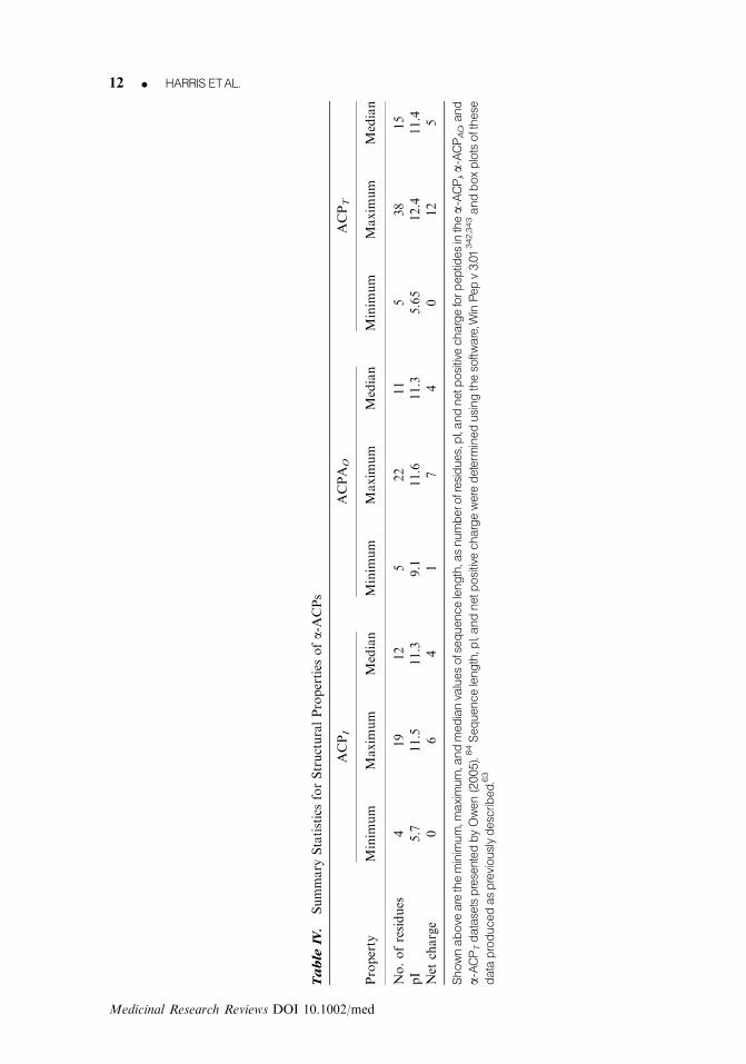
Ta
ble
IV.
Summary
StatisticsforStructuralProperties
ofa-ACPs
ACPI
ACPA
OACPT
Property
Minim
um
Maxim
um
Median
Minim
um
Maxim
um
Median
Minim
um
Maxim
um
Median
No.ofresidues
419
12
522
11
538
15
pI
5.7
11.5
11.3
9.1
11.6
11.3
5.65
12.4
11.4
Net
charge
06
41
74
012
5
Shownabove
aretheminimum,m
aximum,andmedianvaluesofsequencelength,asnumberofresidues,pI,andnetpositivechargeforpeptidesinthea-ACPI,a-ACPAO,and
a-ACPTdatasetspresentedbyOwen(2005).84Sequencelength,pI,andnetpositivechargeweredeterm
inedusingthesoftware,W
inPepv3.01342,343andboxplotsofthese
dataproducedaspreviouslydescribed.63
12 K HARRIS ETAL.
Medicinal Research Reviews DOI 10.1002/med

these peptides. Indeed, previous studies have reported no clear correlation between the se-quence length and toxicity of defense peptides.63,101,202
The sequences of peptides in the a-ACPAO, a-ACPT, and a-ACPI datasets were furtheranalyzed to ascertain the relative frequencies of their component amino acid residuesand most residues were found to be represented at some level (Fig. 1). The only post-translational modification of residues observed in these datasets was C-terminal amidation,which is the most common modification observed in cationic defense peptides.198 It is no-ticeable that the residue distributions of the a-ACPAO, a-ACPT, and a-ACPI datasets (Fig. 1)are broadly similar to those reported for a-AMPs,101,202 which presumably reflects theirmany common structure/function relationships. However, for further comparisons, residuedistributions of the McCaldon and Argos dataset, which is a random sample of a-helicaloligopeptides,203 were included alongside those of the a-ACPAO, a-ACPT, and a-ACPI
datasets (Fig. 1). It can be clearly seen from Figure 1 that the residue profiles of the datasetsformed by a-ACPs are vastly different to that of the McCaldon and Argos dataset, generallyreflecting the specialized structure/function relationships of these ACPs.
6. HISTIDINE-RELATED EFFECTS AND a-ACPs
Histidine is often described as an aromatic residue due to its possession of an imidazole sidechain. At physiological pH, the pKa of this side chain, which is circa 6, means that the residueis uncharged.204 However, at low pH, histidine becomes fully charged and may be consideredto be a moderately hydrophilic basic residue.205 The propensity of histidine for a-helixformation is also affected by pH and is moderate at neutral pH but enhanced at low pH.206
Based on the fact that the surface of membranes from both cancer and microbial cellsgenerally exhibit a pH lower than the pH of 7.2 to 7.4 found in the bulk phase of normaltissues/organs,207,208 it has previously been suggested that pH-dependent properties of his-tidine may feature in the anticancer action of some ACPs.209 However, the occurrence of theresidue in ACPs is generally low (circa 2% of residues)83 and it can be seen from Figure 1 thathistidine was found to be present at only very low levels in the a-ACPAO, a-ACPT, anda-ACPI datasets (relative frequencies o0.05). These results suggest that histidine does notgenerally play a major role in the mechanisms that underpin the selectivity of toxicity of thea-ACPs analyzed to cancer cells. Nonetheless, a number of histidine-rich defense peptideshave been recently reported, which has prompted investigation into the role of the residue inthe anticancer and antimicrobial actions of these peptides.210–213 These latter studies foundthat low pH increased the positive charge carried by these peptides via the protonation oftheir histidine residues, resulting in enhanced affinity for cancer and microbial cells alongwith potent toxicity to these cells.209,214–217 Based on these results, it was shown that byvarying their histidine content, the pH-dependent antimicrobial properties of defense pep-tides could be attenuated, which led to the design of peptides that were inactive at neutralpH, but at lower pH showed selective toxicity to microbial cells over normal eukaryoticcells.215,218–221 Using a similar approach, histidine-containing peptides have been designedthat function as pH-dependent a-ACPAO and a-ACPT peptides against both cancer cell linesand in vivo tumors.209,222,223 It has also been shown that substituting histidine residues intonaturally occurring a-ACPAO peptides can enhance their anticancer activity at low pH, asrecently demonstrated for citropin 1.1,199,222 an amphibian a-ACPAO peptide.201 Mostrecently, evidence has been presented which suggests that the protonation of histidine underthe acidic pH conditions of the cancer cell membrane promotes the ability of the ACPAO
peptide, cationic antimicrobial protein, to form amyloid structure, thereby contributing tothe peptide’s cancer cell toxicity by enabling it to induce inactivation of these cells via
ONTHESELECTIVITYANDEFFICACYOFDEFENSE K 13
Medicinal Research Reviews DOI 10.1002/med

permeabilization of their membranes.166,167 Interestingly, a number of close homologs ofcitropin 1.1 are a-ACPAO peptides, whose sequences include conserved histidine re-sidues224,225 withunknown function. It has been predicted that a number of these homologs have the potentialto form amyloid structure.226 There is also evidence to suggest that histidine-mediatedamyloid formation may be involved in the action of enodogenous human ACPs177,227 such asthe histidine-rich fragment, which is cleaved from the histidine-rich glycoprotein (HRGP)and inhibits angiogenic and tumor activity.228,229 Based on a propensity to form b-sheetstructures, it has been predicted that this peptide may be amyloidogenic177 and that proto-nation of its histidine residues in the low pH of the TAVE cell membrane may facilitate itsinteraction with these membranes, and thereby its antiangiogenic activity.228–230 Interest-ingly, HRGP has been recently shown to possess antibacterial231 and antifungal activity,232
and therefore may constitute a novel example of a host defense peptide encrypted within alarger protein.233,234 Most recently, several other encrypted peptides with the potential toserve as amyloidogenic host defense peptides have also been reported.235–238
Another property of histidine residues known to feature in the biological action of manypeptides is the ability of these residues to chelate metal ions,239 and a histidine-mediatedability to bind zinc seems to be required for both the antimicrobial and anticancer activity ofHRGP.231,232,240 In relation to established ACPs, the only major peptides reported to exhibita histidine-mediated ability to bind metal ions seems to be insect alloferons, which are lineardefense peptides isolated from larvae of the blow fly Calliphora vicina.241 These peptides wereshown to form complexes with copper ions242 and exhibit anticancer properties via theirability to activate immunity mechanisms against tumors. However, whether direct toxicity tocancer cells involving copper ion chelation is a feature of the anticancer action of alloferonsis, as yet, an open question.241,243 Indeed, the only other major ACP known to bind metals isthe mammalian milk protein, lactoferrin, and the role of iron in the anticancer activity of thisACPAO is a subject of great unresolved debate.244,245 It would, therefore, seem that currentlythere are no established instances of ACPs that require the presence of metal ions to facilitatetheir anticancer action, although such a requirement has been demonstrated for the anti-microbial activity of a number of defense peptides.233,244 The best characterized examples ofthese latter peptides are those rich in histidine residues,246 such as histatins, which area-AMPs found in the saliva of humans and other primates.247,248 The ability of histatins to
Figure 1. Amino acid distributions of peptides. Shown above are the overall amino acid residue compositions of peptides in
the a-ACPI (light grey), a-ACPAO (white), and a-ACPT (dark grey) datasets presented by Owen.84Also shown for comparative pur-
poses are the overall residue compositions of peptides in the McCaldon and Argos dataset (black), which is a random sample of
oligopeptides.203
Relative residue frequencies were determined, as previously described.63
14 K HARRIS ETAL.
Medicinal Research Reviews DOI 10.1002/med
form complexes with copper, zinc, and other metal ions249,250 seems to directly facilitateoxidative mechanisms that contribute to the antimicrobial activity of these peptides.251–253
Histatins are potent antifungal agents and there is evidence to suggest that these peptidesassociate with fungal mitochondria, thereby inhibiting respiration and inducing the forma-tion of reactive oxygen species to produce oxidative stress.247
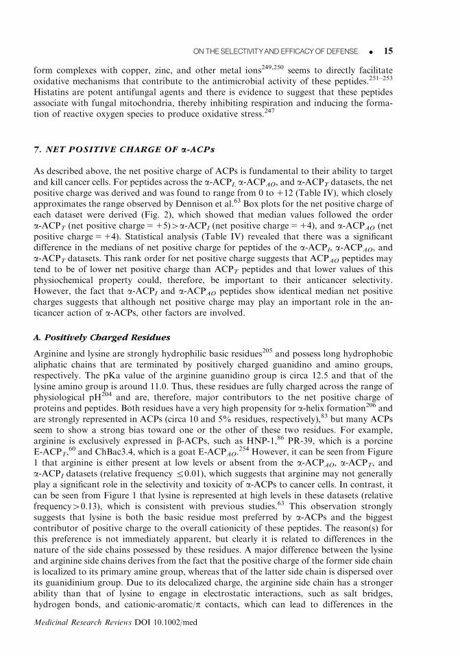
7. NET POSITIVE CHARGE OF a-ACPs
As described above, the net positive charge of ACPs is fundamental to their ability to targetand kill cancer cells. For peptides across the a-ACPI, a-ACPAO, and a-ACPT datasets, the netpositive charge was derived and was found to range from 0 to 112 (Table IV), which closelyapproximates the range observed by Dennison et al.63 Box plots for the net positive charge ofeach dataset were derived (Fig. 2), which showed that median values followed the ordera-ACPT (net positive charge5 15)4a-ACPI (net positive charge5 14), and a-ACPAO (netpositive charge5 14). Statistical analysis (Table IV) revealed that there was a significantdifference in the medians of net positive charge for peptides of the a-ACPI, a-ACPAO, anda-ACPT datasets. This rank order for net positive charge suggests that ACPAO peptides maytend to be of lower net positive charge than ACPT peptides and that lower values of thisphysiochemical property could, therefore, be important to their anticancer selectivity.However, the fact that a-ACPI and a-ACPAO peptides show identical median net positivecharges suggests that although net positive charge may play an important role in the an-ticancer action of a-ACPs, other factors are involved.
A. Positively Charged Residues
Arginine and lysine are strongly hydrophilic basic residues205 and possess long hydrophobicaliphatic chains that are terminated by positively charged guanidino and amino groups,respectively. The pKa value of the arginine guanidino group is circa 12.5 and that of thelysine amino group is around 11.0. Thus, these residues are fully charged across the range ofphysiological pH204 and are, therefore, major contributors to the net positive charge ofproteins and peptides. Both residues have a very high propensity for a-helix formation206 andare strongly represented in ACPs (circa 10 and 5% residues, respectively),83 but many ACPsseem to show a strong bias toward one or the other of these two residues. For example,arginine is exclusively expressed in b-ACPs, such as HNP-1,86 PR-39, which is a porcineE-ACPT,
60 and ChBac3.4, which is a goat E-ACPAO.254 However, it can be seen from Figure
1 that arginine is either present at low levels or absent from the a-ACPAO, a-ACPT, anda-ACPI datasets (relative frequency r0.01), which suggests that arginine may not generallyplay a significant role in the selectivity and toxicity of a-ACPs to cancer cells. In contrast, itcan be seen from Figure 1 that lysine is represented at high levels in these datasets (relativefrequency40.13), which is consistent with previous studies.63 This observation stronglysuggests that lysine is both the basic residue most preferred by a-ACPs and the biggestcontributor of positive charge to the overall cationicity of these peptides. The reason(s) forthis preference is not immediately apparent, but clearly it is related to differences in thenature of the side chains possessed by these residues. A major difference between the lysineand arginine side chains derives from the fact that the positive charge of the former side chainis localized to its primary amine group, whereas that of the latter side chain is dispersed overits guanidinium group. Due to its delocalized charge, the arginine side chain has a strongerability than that of lysine to engage in electrostatic interactions, such as salt bridges,hydrogen bonds, and cationic-aromatic/p contacts, which can lead to differences in the
ONTHESELECTIVITYANDEFFICACYOFDEFENSE K 15
Medicinal Research Reviews DOI 10.1002/med

ability of these residues to interact with membranes and their components.255–257 For ex-ample, the positively charged moieties of both the lysine and arginine side chains are able tointeract with the negatively charged centers of p ring systems found in the side chains ofaromatic residues. However, arginine engages in these cation–p interactions with a higherfrequency than lysine and contrasts with the latter residue, in that when so associated with anaromatic residue, the side chain of arginine is able to concomitantly form hydrogen bondswith the surrounding molecules. This ability allows the arginine side chain to promotemembrane interaction and use the shielding effect of its association with aromatic residues toreduce the energy cost of locating its guanidinium cation in the hydrophobic environment ofthe bilayer.258,259 It has also been suggested that the greater ability of the arginine side chainto engage in hydrogen bonding and electrostatic interactions is primarily responsible for theobservation that this latter side chain has a higher affinity for some anionic membranecomponents than that of lysine.260 Indeed, it is generally recognized that the guanidiniumgroup of the arginine side chain gives the residue a high affinity for both anionic andzwitterionic membranes. However, although the primary amine group of the lysine side chaingives the residue high affinity for anionic membranes, it exhibits a decreased affinity forzwitterionic membranes as compared to the guanidinium group of arginine.261 A clear im-plication from this latter observation is that lysine residues may play a role in the cancer cellselectivity of ACPs with a preference for this residue, given the anionic nature of cancer cellmembranes. In contrast, examination of Figure 1 shows that comparable levels of lysineoccur in the a-ACPAO and a-ACPT datasets, which indicates that factors other than thepresence of the residue are involved in the selectivity of the former peptides for cancer cellsover healthy eukaryotic cells. Another major difference between the lysine and arginine sidechains lies in the fact that the former side chain is significantly more hydrophobic than thelatter side chain,262 and taken with the differences in their charge characteristics, theseobservations clearly suggest that differences in the hydrophobicity and/or amphiphilicity ofthe lysine and arginine side chains could be a factor in the preference of ACPs for a givencationic residue. As an example, the positive charge, hydrophobicity, and amphiphilicity of
Figure 2. Boxplot analysis of net positive charge for a-ACPs. Shownabove is aboxplot analysis for thenet positive charge ofpeptides within the ACPI, ACPAO, and ACPT datasets.The plot shows the median (dark band) along with the minimum andmax-
imum of this measure.The box represents the lower (Q1525%) and upper (Q3575%) quartile range. Net positive charge was
determined andbox plots of these data produced, as previously described.63
16 K HARRIS ETAL.
Medicinal Research Reviews DOI 10.1002/med

these side chains are primarily responsible for the ability of both lysine and arginine tointeract with membranes via the snorkeling mechanism. According to this mechanism, thecharged moieties of these side chains maintain electrostatic interactions with the membranelipid head-group region, while the long apolar regions of these side-chains extend or snorkelinto the hydrophobic core of the bilayer, thereby enabling the parent peptide to penetratemore deeply into the membrane.263,264 However, the more hydrophobic lysine side-chain isknown to enhance the snorkeling ability of this residue compared to arginine.265,266 It haspreviously been suggested that snorkeling by lysine may play a role in the toxicity of a-ACPsto cancer cells.63,163 A recent study has also suggested that differences in the surface topo-graphy of defense peptides due to lysine and arginine may contribute to the preferences ofthese peptides for one or the other of these residues. This study, which focused on theantimicrobial activity of HNP-1 and other defensins, showed that differences betweenthe structural and chemical properties of lysine and arginine side chains were able to affect theaccessibility of these residues on the surface of the parent peptides, and thereby the ability ofthese molecules to interact with bacterial membranes and kill bacteria.267 Given that HNP-1inactivates cancer cells via membrane interaction,80,268,269 it seems possible that such topo-graphical differences may also contribute to the preferences of ACPs for lysine or arginine.
A number of a-AMPs are C-terminally amidated and it has previously been proposedthat this structural moiety may serve a structural function in the a-helices of these peptides.A C-terminal amide is able to provide an additional hydrogen bond to stabilize these a-helical architectures, and thereby strongly favor the propensity of a-AMPs to adopt a-helicalstructure.270 However, a C-terminal amide group also has the effect of increasing the positivecharge of the parent peptide; recent studies on a-AMPs showed that removing theseC-terminal moieties could influence the antimicrobial activity on a number of these pep-tides.270,271 Included in these a-AMPs were modelin-5, a synthetic peptide,270 PGLa fromXenopus laevis59 and aurein 2.3,272 which are also known to function as a-ACPAO pep-tides.59,84,201 This observation led to the suggestion that C-terminal amide groups may play arole in the anticancer action of some a-ACPAO peptides. Inspection of the APD2 databaseshows that, in addition to a-ACPAO peptides, a number of a-ACPT peptides are alsoC-terminally amidated.83 In response, a recent study investigated the role of this structuralmodification in the selectivity and toxicity of these a-ACPs to cancer cells. Using a range oftheoretical techniques, this study showed that the mutation of nonamidated a-ACPAO anda-ACPT peptides to their C-terminally amidated isoforms had no apparent effect on theirability to discriminate between healthy eukaryotic cells and cancer cells from carcinoma,adenocarcinoma, and melanoma. The same study also showed that the C-terminal amidationof a-ACPAO and a-ACPT peptides had a variable effect on the levels of toxicity exhibited bythese peptides to cancer cells, with these levels either remaining unaffected or showing up toten-fold increases or decreases in magnitude.198
B. Negatively Charged Residues
Glutamic acid and aspartic acid are strongly hydrophilic negatively charged residues205 withcarboxylated side chains that have pKa values in the region of 4, and therefore usually remainfully charged across the physiological range of pH.204 These residues differ in their propensityfor a-helix formation with glutamic acid showing a high tendency to adopt such structure,whereas the tendency of aspartic acid is low.206 These residues are found at low levels inACPs (o2.5% of residues)83 and it can be seen from Figure 1 that they are either present atlow levels or absent from the datasets studied (relative frequency r0.01). These resultssuggest that glutamic and aspartic acid do not generally play a major role in the mechanismsthat underpin the selectivity of toxicity of a-ACPs to cancer cells. In addition, these results
ONTHESELECTIVITYANDEFFICACYOFDEFENSE K 17
Medicinal Research Reviews DOI 10.1002/med

suggest that these residues make only a minor contribution to the overall charge of thea-ACPs examined, presumably to maximize the cationicity of these peptides. However, it hasbeen shown that a glutamic acid residue is key to the anticancer activity of cycloviolacin 02,which is a plant defensin or cyclotide. It was suggested that this residue played a structuralrole in the ability of the b-ACP to disrupt cancer cell membranes by facilitating efficientaggregation of the peptide in the bilayer, thereby initiating cytoxicity.273,274 This glutamicacid residue is highly conserved across other b-ACPs of the cyclotide family and it may bethat the residue plays a similar role in their anticancer mechanisms.275–278 In another study,Tytler et al.279 showed that a centrally placed glutamic acid residue in the hydrophobic faceof some a-AMPs contributed to the selectivity of these peptides for microbes by engaging ininteractions with the cholesterol of eukaryotic membranes, thereby inhibiting their lyticaction.279 Interestingly, FLAK50 T4, which is a synthetic peptide in the a-ACPI database,includes a glutamic residue in the center of its hydrophobic face (Fig. 3), and as discussedabove, cholesterol is known to inhibit the anticancer action of some ACPs.60,78 Studies on therole of aspartic acid residues in polybia-MP1, a wasp (Polybia paulista) a-AMP showed thatthe presence and position of these residues optimized the charge density of the peptide forinteraction with anionic membranes as opposed to zwitterionic membranes, thereby con-tributing to the selectivity and toxicity of polybia-MP1 for microbial membranes.280 Anionicresidues may serve a similar function in the anticancer action of the peptide, as was recentlyreported when it was found that polybia-MP1 showed selective toxicity to prostate andbladder cancer cells over nonmalignant cells.280,281 Nonetheless, currently, the general opi-nion appears to be that primary roles for glutamic and aspartic acid in cationic a-helicaldefense peptides may be to serve structural functions rather than aid selectivity. For example,several studies have observed that when the latter residues are present in a-AMPs, theytended to occupy positions in the a-helix that were i73 or i74 relative to basic residues. Ithas been suggested that this structural positioning may promote helix formation via saltbridging and may be a strategy for improving the rigidity of a-helical residue arrangements,and hence changing efficacy.101,202
It is worthy of note that currently, in addition to the multitude of cationic defensepeptides known, circa 100 anionic defense peptides have been reported. In these peptides,glutamic and aspartic acid are the predominantly charged residues, where both residues havebeen shown to play important roles in facilitating the selectivity and toxicity of a number ofthese peptides to microbial cells.233,234 As an example, aspartic acid residues in a number ofovine anionic a-AMPs bind zinc ions to form a cationic salt bridge between the parentpeptide and the negatively charged bacterial membrane, thereby facilitating the bacterialtargeting and initiating the antibacterial action of these a-AMPs.282 Based on the work ofTytler et al.279 there is a possibility that interactions between the glutamic and asparticresidues of some anionic defense peptides and the membrane cholesterol of cancer cellmembranes may inhibit the potential of these peptides for anticancer action. Nonetheless,given that many defense peptides possess these residues and exhibit selective anticanceraction, it may be that anionic defense peptides represent a source of peptides for investigationas ACPs.
8. HYDROPHOBICITY
Hydrophobicity can be taken as a measure of the affinity of a peptide for the apolar core regionof the bilayer195 and is generally accepted as a key driver in the ability of a-ACPs to partitioninto the membranes of cancer cells.60,78 However, there have been few comparative studiesbetween a-ACPAO and a-ACPT peptides to elucidate the potential role of hydophobicity in the
18 K HARRIS ETAL.
Medicinal Research Reviews DOI 10.1002/med

selectivity and toxicity of these peptides for cancer cells. It has been postulated that the dis-tribution of hydrophobicity along the peptide long axis may have a key role in the toxicity ofACPs. An asymmetric distribution of hydrophobicity is a structural characteristic of obliquelyorientated a-helices or tilted peptide structure, which causes these a-helices to insert intomembranes at a shallow angle of between 30 and 601, thereby destabilizing membrane structureand promoting a range of membrane-related processes, such as lysis or pore formation.283,284
Several techniques have been developed to detect hydrophobicity gradients within a-ACPs, suchas the inspection of hydrophobicity profiles produced by hydropathy plot analysis.193 Use ofthis analysis and experimental studies have confirmed the potential for a-ACPAO peptides, suchas aurein 1.2 and citropin 1.1 to utilize tilted peptide structure in their membrane interac-tions;63,97,98 it was predicted that such structure may feature in the anticancer action of manyother a-ACPs.63 Most recently, it was shown that peptides in both the a-ACPAO and a-ACPT
Figure 3. Two-dimensional axial projections of a-ACPs. Shown above are peptides from the a-ACPI, a-ACPAO, and a-ACPTdatasets when represented as a-helical wheels, which are two-dimensional axial projections of these peptides, assuming an
angular periodicity of1001.344
The examples shown includemagainin 2 and FLAK50 Z5, which are endogenous and synthetic of
the a-ACPAO dataset, respectively; melittin and FLAK98, which are endogenous and synthetic members, of the a-ACPT dataset,respectively; andFLAK50T4, which isasyntheticmemberof thea-ACPIdataset.Theamphiphilicityof these a-ACPs canbe clearlyseen by the segregation of hydrophilic residues (dark grey) andhydrophobic residues (black) about the a-helical longaxis. It canalso be seen that centrally located within the hydrophobic residues of FLAK50 T4 is a glutamic acid residue. The sequences of
these examples were obtained from Owen84andwere represented as a-helical wheels using the software, AntheProt v 5.0.345
ON THESELECTIVITYANDEFFICACYOFDEFENSE K 19
Medicinal Research Reviews DOI 10.1002/med

datasets showed the potential to form tilted peptide structure, although this potential was farmore pronounced in a-ACPT peptides.199 These authors suggested that the use of this structuredid not play a major role in the selectivity of a-ACPAO peptides for cancer cells over non-malignant cells. Rather, it was suggested that the use of tilted structure by a-ACPT peptides todestabilize target cell membranes may be more associated with their broader spectrum of targetspecificity shown by these latter peptides as compared to a-ACPAO peptides.199 Nonetheless,there seems to be no specific structural characteristics of tilted segments that can be associatedwith the general toxicity of a-ACPT peptides. Model tilted defense peptides were found to bestrongly hemolytic and reversing the hydrophobicity gradients of these peptides had no sig-nificant effect on their levels of hemolysis.285,286 More recently, it was reported that no apparentrelationship existed between the cancer cell toxicity of peptides in the a-ACPT dataset and thedirection, magnitude, length, and residue composition of their hydrophobicity gradients.199
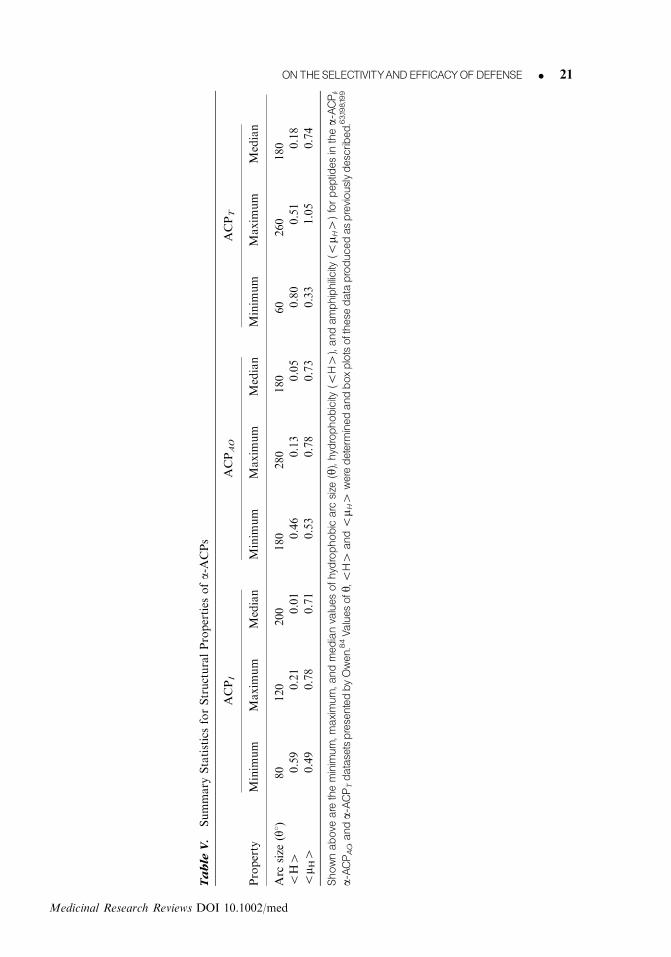
A number of studies on a-AMPs have investigated the role of hydrophobicity in the anti-microbial action of these peptides and established that both their hydrophobic arc size (y) andmean hydrophobicity (oH4) are key factors in their ability to partition into microbial mem-branes and kill microbial cells.286,287 These hydrophobicity measures were quantified as describedin Table V for the ACPI, ACPAO, and ACPT datasets, and it was found that across the datasetshydrophobic arc sizes ranged between y5601 and y52601, whilst oH4 ranged between �0.8and 0.51 (Table V). These ranges in y andoH4 are comparable to those previously reported fora-ACPs63 and suggest that there is a wide variation in the affinity of these peptides for themembrane lipidic region. Box plots (Fig. 4) showed that the medians of y for thesepeptides followed the order ACPI (y52001) 4ACPAO (y51801) and a-ACPT (y51801), whiletheir medians for oH4 followed the rank order: ACPI (oH45�0.01) 4a-ACPAO(oH45�0.05)4ACPT (oH45�0.18). Statistical analysis (Table V) revealed that there wasno significant difference in the medians of either y or oH4 for peptides of the datasets. Thisresult clearly suggests that hydrophobicity does not play a major role in the selectivity of the a-ACPs studied for cancer cells over untransformed cells. However, this result could indicate that athreshold value of this structural property may be necessary to drive the interaction of thesepeptides with the hydrophobic core of target cell membranes, thereby affecting their toxicity toeukaryotic cells. Strongly supporting this suggestion, a number of studies have suggested thatthreshold hydrophobicites are required to drive the membrane interactions of a-AMPs and othermembrane interactive a-helices.288–290
As a measure of peptide hydrophobicity, oH4 is probably the most widely usedparameter and clearly it is a function of the residue composition of the parent ACP. Residuesthat make positive contributions to oH4 vary widely in their individual levels of hydro-phobicity, and in a number of cases, it seems that some of these residues may also play otherroles in the anticancer mechanisms of both a-ACPs and other ACPs.
Cysteine is weakly hydrophobic205 and occurs in ACPs at high levels forming circa 6% ofthe residues in these peptides.83 In a vast majority of cases, these cysteine residues are foundin b-ACPs,60,78 where they participate in cystine bonds or disulphide bridges due to theability of their thiol side chains to form covalent bonds with other cysteine side chains.204
This ability is fundamental in stabilizing the secondary/tertiary structures of ACPs, such asthe b-hairpin molecule of gomesin,79 the b-sheet architectures of defensins,61,86 and thecysteine knot structures of some plant cyclotides.275–278 The occurrence of cysteine as a freeresidue in ACPs is very rare with only one such peptide, OEP3121, from the earthworm,Eisenia foetida, reported in the 105 ACPs recorded in the APD2 database.83 Taken with thefact that cysteine has only a low propensity for a-helix formation,206 these observationswould seem to explain the results of our analyses, which showed that the residue was absentfrom the three datasets of a-ACPs studied here. Proline is also weakly hydrophobic195 andhas been reported as being in ACPs at high levels (circa 5% of residues),83 but was found to
20 K HARRIS ETAL.
Medicinal Research Reviews DOI 10.1002/med
Ta
ble
V.
Summary
StatisticsforStructuralProperties
ofa-ACPs
ACPI
ACPAO
ACPT
Property
Minim
um
Maxim
um
Median
Minim
um
Maxim
um
Median
Minim
um
Maxim
um
Median
Arc
size
(y1)
80
120
200
180
280
180
60
260
180
oH4
�0.59
0.21
�0.01
�0.46
0.13
�0.05
�0.80
0.51
�0.18
om H
40.49
0.78
0.71
0.53
0.78
0.73
0.33
1.05
0.74
Shownabove
aretheminimum,maximum,andmedianvaluesofhydrophobicarc
size
(y),hydrophobicity
(oH4),andamphiphilicity
(om H
4)forpeptidesinthea-ACPI,
a-ACPAO,anda-ACPTdatasetspresentedbyOwen.84Valuesofy,oH4
andom H
4weredeterm
inedandboxplotsofthesedataproducedaspreviouslydescribed.63,198,199
ON THESELECTIVITYANDEFFICACYOFDEFENSE K 21
Medicinal Research Reviews DOI 10.1002/med

be either present at very low levels or absent from the a-ACPAO, a-ACPT, and a-ACPI
datasets (relative frequency o0.005), which suggests that the residue does not generally playa role in the selectivity or toxicity of these ACPs for cancer cells. These results would seem toreflect the fact that the residue has a very low propensity to adopt a-helical structure206 andcan interrupt such structure when located within an a-helix-forming sequence. Proline isstrictly an imino acid, and either breaks or kinks a helix due to the inability of its side chainto donate an amide hydrogen bond and the steric interference caused to a-helix formation bythis side chain.291 Internal prolines are observed in a number of a-ACPAO peptides,83 such asBMAP28, BMAP-27,60 and buforin IIb,132 and a-ACPT peptides, such as melittin.80 In thecase of melittin, it has been shown that the flexibility given to the backbone of the peptide bythe presence of proline is important for efficient membrane interaction by the peptide, im-plying that for some structures the residue has a key role to play.114,292 Proline is also overlyrepresented in some ACPs and seems to contribute to the conformational flexibility of the E-ACPAO peptide, ChBac3.4,254 and the E-ACPT peptides, prophenin PF2293 and PR-39.60 Inthe case of PR-39, there is some evidence to suggest that its anticancer action may involve thebinding of proline residues to SH3 domains in intracellular proteins, thereby affecting thecellular signaling of host cancer cells.294,295
Methioinine is moderately hydrophobic195 and exhibits a high propensity for a-helixformation.206 However, the residue is present in ACPs at very low levels (0.5% of residues,respectively)83 and was found to be absent from the datasets studied here. Taken together,these results suggest that, in general, methionine plays no major role in the anticancer actionof ACPs. Glycine is also moderately hydrophobic205 and is strongly represented in ACPs(circa 10% of residues),83 but was found to be present in the datasets studied here at lowlevels (relative frequency o0.1). This result could be related to the fact that, due to the lackof a side chain,204 glycine possesses high conformational flexibility and a very low propensityto adopt a-helical structure.206 This conformational flexibility results in the residue actingsimilarly to proline in that it can function as a breaker of a-helical structure when locatedwithin an a-helix-forming sequence.291 However, glycine was found to be twice as abundantin the ACPI dataset (relative frequency5 0.075) than the a-ACPAO and a-ACPT datasets(relative frequency o0.025), which implies that the inactivity of peptides in the a-ACPI
dataset may, in some cases, be related to a reduced propensity for a-helix formation.
Figure 4. Boxplotanalysis ofhydrophobicitymeasures fora-ACPs.Shownaboveareboxplotanalysesofhydrophobicityas y(A) andoH4 (B) for peptideswithin the ACPI, ACPAO, and ACPT datasets.Theplots show themedian eØ andoH4 (dark bands)
along with the minimum and maximum values of these measures. The boxes represent the lower (Q1525%) and upper
(Q3575%) quartile ranges. Values of y and oH4 were determined and box plots of these data produced, as previously de-
scribed.63,198,199
22 K HARRIS ETAL.
Medicinal Research Reviews DOI 10.1002/med

A number of AMPs191,296 and ACPs with multiple glycine residues are known.80 Mostrecently, it was reported that the conformational flexibility of the glycine-rich N-terminaldomain of SK84, an insect ACPAO, may facilitate the anticancer action of the peptide byforming an elastic structure in the membrane of target cancer cells, which results in bilayerdisruption.297 In contrast, glycine is also known to be an efficient N-capping agent, which isimportant to the propensity of peptides to form a-helical structure, and it has previously beensuggested that the residue might serve such a purpose in a-ACPs.63 Inspection of the APD2database shows that glycine is the N-terminal residue in a number of a-ACPs, includingaureins, BMAP-27, and BMAP-28.83 In addition, glycine is also known to play a role inmaintaining the balance between hydrophobicity and amphiphilicity necessary for themembrane action of obliquely orientated a-helical structure,187,298 which as described above,seems to feature in the anticancer action of some a-ACPs.63,97,98
Phenylalinine, tyrosine, and tryptophan are generally grouped together, for, in additionto being considered hydrophobic residues due to the fact that their side chains195 areuncharged, they all share aromatic characteristics due to the presence of p ring systemsin their side chains.204 These ring systems exhibit a significant quadropole moment, whichgives rise to negatively charged regions that allow these aromatic residues to engage incation–p contacts with other positively charged species, such as ions and the basic residuesdescribed above.258 In addition to cation–p contacts, tyrosine and tryptophan are alsoable to engage in hydrogen bonding and other electrostatic interactions with components ofthe lipid headgroup region, which gives these residues a strong preference for an interfaciallocation.299–302
Tyrosine is weakly hydrophobic205 and is represented in ACPs at low levels (2.2% ofresidues).83 The residue has a moderate propensity for a-helix formation,206 but as seen inFigure 1, tyrosine is either absent or present at very low levels in the three data sets analyzedhere (relative frequency 0.001). These results are consistent with previous data63 and suggestthat tyrosine does not generally play a major role in the mechanisms that underpinthe selectivity of toxicity of a-ACPs to cancer cells. Tryptophan has a significantly higherhydrophobicity than tyrosine205 and a propensity for a-helix formation that is comparable tothe latter residue.206 Tryptophan is present at low levels in ACPs (1.3% of residues)83 andwas found to have a low frequency of occurrence in the datasets studied here (relativefrequencyo0.025). Nonetheless, as seen from Figure 1, the residue is present in the a-ACPAO
and a-ACPT datasets but is absent from the a-ACPI data set, which suggests that the residuemay play a role in the toxicity of some of these peptides to cancer cells. In support of thissuggestion, it has been suggested that the membrane interactions of some a-ACPAO anda-ACPT peptides may be mediated by the ability of their tryptophan residues to form a stablecomplex with cholesterol.114 Several studies have suggested that there may be a preferentialinteraction between cholesterol and tryptophan residues located near the membrane/waterinterface, although this has been disputed.303 Tryptophan is overly represented in someACPs, such as the bovine E-ACPT and indolicidin.84 Studies on the b-ACP and LfB havesuggested that the residue is important for the membrane interactions of the peptide andhence its toxicity to cancer cells,304 which may also be the case for a-ACPs. Phenylalinine isstrongly hydrophobic of all naturally occurring residues205 and is present in ACPs at higherlevels than both tyrosine and tryptophan, (4.3% of residues).83 The residue has a propensityfor a-helix formation that is comparable to the other aromatic residues206 and was found tobe strongly represented in the a-ACPAO, a-ACPT, and a-ACPI datasets with relative fre-quencies between 0.075 and 0.125 (Fig. 1). The high levels of the residue in the a-ACPAO anda-ACPT datasets suggest functionality, and it is generally accepted that phenylalinine plays amajor role in the anticancer action of a-ACPs by enhancing the affinity of these peptidesfor target cancer cell membranes.63 It is well established that in contrast to tyrosine and
ONTHESELECTIVITYANDEFFICACYOFDEFENSE K 23
Medicinal Research Reviews DOI 10.1002/med

tryptophan, phenylalinine does not possess a hydrogen bond donor and has a weaker ca-pacity for cation–p contacts, which taken with hydrophobic and lipophilic effects leads theresidue to possess a strong preference for location in the hydrophobic core of the bilayer.301
In addition, studies on defense peptides have suggested that phenylalinine may play otherroles in the anticancer action of a-ACPs. In aureins, which function as both a-AMPs and a-ACPs, the residue seems to play a role in maintaining the surface topography of the pep-tide,305 whereas in the case of clavanins, which are a-AMPs, the residue seems to play a rolein maintaining the conformational flexibility of these peptides.306 Phenylalanine is overlyrepresented in E-ACPT peptides, such as prophenin PF2293 and PR-39,60 and it may be thatthe residue contributes to the conformational flexibility of the extended structures possessedby these peptides.
Alanine, leucine, isoleucine, and valine are strongly hydrophobic residues205 and arerepresented at high levels in ACPs at comparable levels, ranging between 7.2 and 9.6% ofresidues.83 These residues possess moderate-to-high propensities to adopt a-helical struc-ture,206 and were well represented in the datasets studied here with levels that, in the case ofeach residue, were comparable across a-ACPAO, a-ACPT, and a-ACPI datasets. It is gen-erally accepted that, similar to phenylalinine, the strong hydrophobicity of these residues is amajor factor in the affility of a-ACPs for cancer cell membranes and the anticancer action ofthese peptides.63 The occurrences of alanine, leucine (relative frequencies5 0.21 and 0.26,respectively) in the a-ACPAO, a-ACPT, and a-ACPI datasets are much greater than those ofvaline and isoleucine (relative frequencies o0.06). A bias for the former two residues overother hydrophobic residues has previously been reported for a-ACPs63 and a-AMPs.101 Itwould seem that alanine and leucine are the major hydrophobic residues found in a-ACPs,which may be related to the fact that leucine and alanine have the highest a-helix-formingpropensities of the naturally occurring hydrophobic residues.206 Nonetheless, it is generallybelieved that the mutation of alanine and leucine for valine, isoleucine, and phenylalinine areconservative substitutions. For example, studies on aureins have shown that homologs thatdiffer solely by the mutation of leucine to phenylalinine exhibit similar general profiles ofactivity when directed against the same range of cancer cells.225 In addition, the mutation ofleucine and alanine to isoleucine, valine, and phenylalinine has been identified in a number ofhomologous a-ACPs, such as BPAP-27 and BMAP-28, and gaegurin 5 and gaegurin 6.60
At present, the reasons(s) underlying a preference for leucine and alanine by a-ACPs areunclear.
9. AMPHIPHILICITY
Amphiphilicity is the ordered spatial segregation of hydrophilic and hydrophobic residueswithin the structure of a protein or peptide, and is recognized as a major determinant in theability of these molecules to partition into the amphiphilic environment of the bilayer.196,307
Some ACPs may be classified as exhibiting tertiary amphiphilicity and these peptides areexemplified by the cysteine knot structures of plant cyclotides.308 In such ACPs, residues thatare distal in the primary structure of the peptide are brought together in its final three-dimensional to form amphiphilic sites. However, most known ACPs demonstrate secondaryamphiphilicity, which arises when the folding of these peptides leads to residue segregationand secondary structures with opposing polar and apolar faces.196 Typical examples of thisform of amphiphilicity are provided by b-ACPs, such as gomesin,79,174 and a-ACPs, such asaureins,225 and peptides in the a-ACPI, a-ACPAO, and a-ACPT datasets, which arerepresented as helical wheels in Figure 3. Secondary amphiphilicity can be quantified by anumber of Fourier transform-based techniques, but the hydrophobic moment (omH4) is the
24 K HARRIS ETAL.
Medicinal Research Reviews DOI 10.1002/med

most commonly used.195,196,309 Developed by Eisenberg et al.310 the hydrophobic momentquantifies amphiphilicity by treating the hydrophobicity of successive amino acids ina-helical sequences as vectors. These vectors are summed in two dimensions, assuming anamino acid side chain periodicity of 1001, and the resultant of this summation leads to themean hydrophobic moment,omH4 which is the generally used form of the parameter. Here,values of omH4 were computed for peptides of the a- ACPI, a-ACPAO, and a-ACPT
datasets, as described in Table IV, and it was found that these values ranged between 0.33and 1.05. This range is in close agreement with previous work and consistent with the viewthat a-ACPs are active at the interface, generally accepted as a common step in the anticancermechanisms of all known ACPs.63 To investigate the potential role of amphiphilicity in theselectivity and toxicity of peptides in the a-ACPI, a-ACPAO, and a-ACPT datasets for cancercells, box plots for theomH4 values of these datasets were derived (Fig. 5). Inspection of themedians for omH4 of peptides in these datasets shows that they follow the rank order:a-ACPT (omH45 0.74)4a-ACPAO (omH45 0.73)4a-ACPI (omH45 0.71). Statisticalanalysis (Table V) revealed that there was no significant difference in the medians of omH4for peptides of the a-ACPI, a-ACPAO, and a-ACPT datasets, although the comparison ofomH4 for the a-ACPI dataset to that of the ACPAO dataset suggests that there may be athreshold in omH4 values for the anticancer-only activity of the latter peptides. Similarly,comparison of omH4 for the a-ACPT dataset to that of the a-ACPAO dataset suggests thatthere may be a ceiling in the omH4 values for the anticancer-only activity of the latterpeptides. Thus, an optimal range in omH4 values for the anticancer action of ACPAO
peptides seems most probably to be important to the ability of these peptide to kill targetcancer cells, although it is possible that optimisation of omH4 may also play a role in theselectivity of these peptides for cancer cells. Nonetheless, our analysis clearly shows thatalthough amphiphilicity clearly plays an important role in the anticancer action of a-ACPs,other factors are also involved.
Figure 5. Box plot analysis of amphiphilicity for a-ACPs. Shown above is a box plot analysis of amphiphilicity as omH> forpeptides within the ACPI, ACPAO, and ACPT datasets.The plot shows the median (dark band) along with the minimum andmax-
imum values of this measure.The box represents the lower (Q1525%) and upper (Q3575%) quartile range.Values ofomH4were determined and box plots of these data producedas previously described.
63,198,199
ON THESELECTIVITYANDEFFICACYOFDEFENSE K 25
Medicinal Research Reviews DOI 10.1002/med

10. CONCLUSIONS
This review has considered the evidence that a number of membrane-based factors contributeto the anticancer action of both a-ACPs and other ACPs. The elevated levels of microvilliand higher transmembrane potentials associated with cancer cells seem to contribute to theselectivity of ACPAO peptides for malignant cells over nonmalignant cells. It seems possiblethat these differences in membrane properties could also contribute to the toxicity of boththese latter peptides and ACPT peptides for target eukaryotic cells, although this possibilitydoes not appear to have been extensively investigated. The net negative surface charge carriedby cancer cells, as compared to the overall neutral charge carried by untransformed cells, islikely to be a key factor in the anticancer action of ACPs. This negative surface chargefacilitates the targeting and electrostatic binding of cancer cells by cationic ACPs, and ingeneral these binding events seem to be a major determinant in the selectivity and toxicity ofACPAO peptides for cancer cells and the toxicity of ACPT peptides for these latter cells.Consistent with these observations, this review has shown that in relation to nonmalignantcells, a variety of anionic membrane-based moieties, including sialic acid residues, sulfatedGAG and PS, are overrepresented on the surface of cancer cells. Nonetheless, this review hasalso shown that these anionic moieties can exhibit widely differing levels of expressionbetween cancer cell types and vary greatly in their structural characteristics, which undersome circumstances can reduce both the selectivity of toxicity of ACPs for cancer cells.Indeed, it seems that the structural characteristics of some anionic moieties on the surface ofnonmalignant cells, which carry no net positive charge, enable these moieties to bind ACPsand inhibit their lytic action, thereby effectively diminishing the selectivity and toxicity ofACPs for cancer cells. In contrast, the overrepresentation of these anionic moieties on thesurface of endothelial cells associated with tumor vasculature enables these moieties to bindACPs and induce angiogenesis, thereby effectively enhancing the selectivity and toxicity ofACPs for cancer cells.
Cholesterol is present in the membranes of both maliganant and nonmalignant cells; thispresence seems to have a variable effect on the selectivity and toxicity of ACPs for these cells.The presence of the sterol in the membranes of nonmalignant cells seems able to inhibit thelytic action of some a-ACPAO peptides against these cells via changes in the fluidity of thesemembranes. Clearly, this ability can contribute to the lack of toxicity shown by these peptidesfor untransformed cells and effectively enhance their selectivity for cancer cells. However, inthe membranes of cancer cells, the sterol seems to interact directly with a number of ACPAO
peptides, thereby reducing their toxicity to these cells and effectively reducing the selectivityof these peptides for untransformed cells.
This review has also considered the differences in a number of peptide-based factors thatcould contribute to the anticancer action of ACPs. There is evidence that some a-ACPs andACPs may possess the ability to recognize membrane-based receptors, which could con-tribute to both the selectivity and toxicity of these peptides for cancer cells. Our own analyseshave suggested that differences between a-ACPAO and a-ACPT peptides in relation to theirresidue composition, sequence length, net positive charge, and molecular architecture, suchas those inherent in amphiphilicity, may each have some influence on the selectivity of theformer peptides for cancer cells over untransformed cells. Indeed, a recent study demon-strated that the manipulation of peptide-based characteristics, such as hydrophobicity, viaamino acid substitutions in the apolar face of a-ACPs allowed the interconversion of a-ACPT
peptides and a-ACPAO peptides.311 However, taken overall, this review strongly suggests thatno single membrane- or peptide-based factor alone is responsible for the anticancer action ofACPs. Consistent with this suggestion, a recent statistical analysis could find no combinationof the peptide-based factors—net positive charge, hydrophobicity, and amphiphilicity—that
26 K HARRIS ETAL.
Medicinal Research Reviews DOI 10.1002/med

was optimal for the efficacy of this action, suggesting that it is the synergistic action of thediffering physiochemical properties and their distribution within the peptide that is importantto this action.199 Moreover, interplay between these peptide-based factors and membrane-based factors associated with cancer cells can also have synergistic effects on the interactionsof ACPs with cancer cell membranes. For example, the positive charge of an ACPapproaching a cancer cell membrane can be enhanced by local conditions, such as pH, thesalt concentration, and the presence of charged and polar moieties on membrane proteinsand lipid. The negative charge carried by these membrane-based moieties can be reciprocallyenhanced by the cationic charge of the incoming peptide and these mutual charge regulationeffects are able to significantly enhance the affinity of ACPs for cancer cells, thereby influ-encing their selectivity and toxicity for these cells.312,313 It would seem that the selectivity andtoxicity shown by ACPAO peptides for cancer cells along with the general toxicity shown toeukaryotic cells by ACPT peptides depends upon the varying contributions from each of anumber of peptide- and membrane-based factors. In a sense, it may be that the anticanceraction of ACPs parallels the ‘‘lock and key’’ model postulated for enzyme activity,314 wherethe molecular architecture of the peptide has to support its binding and insertion into amembrane of a given composition. Such binding and insertion is not only clearly dependenton the shape and physiochemical properties of the peptide but also on the membranepacking. For example, as described above, net positive charge acts as a major component ofthe ‘‘key’’ in the anticancer action of many ACPs by engaging in electrostatic interactionsthat enable the peptide to target and bind anionic moieties on the cancer cell surface. In somecases, it may be that when these two factors are not balanced, such as when the anionicmoieties on the cancer cell surface bind a peptide too tightly or in insufficient quantities foranticancer action, the peptide ‘‘key’’ cannot fully engage or efficiently fit the ‘‘lock’’ formedfrom the lipid packing arrangement and anticancer action is not initiated.
Currently, many studies are attempting to produce a second generation of ACPs withimproved selectivity and toxicity for cancer cells that show the potential for therapeuticdevelopment. In many cases, whilst the first generation of these peptides or native ACPsseemed to show such potential in vitro and ex vivo, it was found that in vivo toxicity could bea problem due to their reduced selectivity and unwanted toxicity profiles.86,315 These findingshave limited both the systemic use and therapeutic potential of first generation ACPs, andpresently the only major candidate in this class of peptides with promise for medical use is theneutraceutical protein, lactoferrin (Table III), which has been patented as an anticanceragent.316 In previous human clinical trials, orally administered lactoferrin was shown toreduce the risk of colon carcinogenesis,317 and currently the protein is in an advanced humanclinical trials as an orally administered treatment for nonsmall lung cell cancer (TableIII).318–320 The strategy adopted by most efforts to design a therapeutically useful secondgeneration ACPs seems to be primarily focused on optimizing the structural and physio-chemical parameters of these peptides for this use.321–323 Based on the findings of this review,we suggest that the most efficient strategies for the design of therapeutically useful ACPs willbe those which take into account the characteristics of not only these peptides, but also thoseof their target cancer cell membranes. Indeed, there is a wide variation in the characteristicsof cancer cell membranes and it is our view that the design of ACPs should be guided by acancer cell-specific approach rather than the generic approach, which seems to be currently inuse. Supporting this view, it would seem that a similar ethos is emerging in the researcharena, as evidenced by the number of very recent reports. One study investigated the abilityof HS expressed on the surface of some tumor cells to inhibit the anticancer action of LfB, asdescribed above, and showed that structural modification of the peptide-generated analogswere able to select and kill these tumor cells with this ability unaffected by the presence of HSon their surface.324 Using a different approach, some studies have attempted to design cancer
ONTHESELECTIVITYANDEFFICACYOFDEFENSE K 27
Medicinal Research Reviews DOI 10.1002/med

cell-specific ACPs by mimicking the ability of peptides, such as HNP-1, which, as describedabove, seems to target some cancer cells via specific membrane receptors. Based on thediscovery that certain tumor types overexpress specific receptors on their membrane surface,novel ACPs that include homing domains specific for such receptors have been designed andtheir anticancer efficacy demonstrated in preclinical trials.55,60,78,175,325–329
ABBREVIATIONS
ACPs Anticancer peptidesACPAO Peptides with toxicity to cancer cells onlyACPT Peptides with toxicity to cancer and non cancer cellsACPI Peptides with no toxicity to cancer cellsa-ACPs a-Helical anticancer peptidesa-ACPAO a-Helical peptides with toxicity to cancer cells onlya-ACPT a-Helical peptides with toxicity to cancer and non cancer cellsa-ACPI a-helical Peptides with no toxicity to cancer cellsa-AMPs a-Helical antimicrobial peptidesb-ACPs b-Sheet anticancer peptidesb-ACPAO b-Sheet peptides with toxicity to cancer cells onlyb-ACPT b-Sheet peptides with toxicity to cancer and non cancer cellsE-ACPs Extended structure anticancer peptidesE-ACPAO Extended structure peptides with toxicity to cancer cells onlyE-ACPT Extended structure peptides with toxicity to cancer and non
cancer cellsCS Chondroitin sulfateCT ChemotherapyGAG glycosaminoglycanoH4 HydrophobicityHRGP Histidine-rich glycoproetinHS Heparin sulfateLfB Lactoferrin BMDR Muliti-drug resistancePG ProteoglycansPS PhosphatidylserineRT Radiation therapyTAVE Tumor-associated vascular endothelialomH4 Mean hydrophobic moment
REFERENCES
1. Thun MJ, DeLancey JO, Center MM, Jemal A, Ward EM. The global burden of cancer: Priorities
for prevention. Carcinogenesis 2010;31:100–110.
2. Kreeger PK, Lauffenburger DA. Cancer systems biology: A network modeling perspective.
Carcinogenesis 2010;31:2–8.
3. Marusyk A, Polyak K. Tumor heterogeneity: Causes and consequences. Biochim Biophys Acta
Rev Cancer 2009;1805:105–117.
4. Brooks SA, Lomax-Browne HJ, Carter TM, Kinch CE, Hall DM. Molecular interactions in
cancer cell metastasis. Acta Histochem 2010;112:3–25.
28 K HARRIS ETAL.
Medicinal Research Reviews DOI 10.1002/med

5. Sung SY, Johnstone PAS. Tumor microenvironment promotes cancer progression, metastasis,
and therapeutic resistance. Curr Probl Cancer 2007;31:36–100.
6. Urruticoechea A, Alemany R, Balart J, Villanueva A, Vinals F, Capella G. Recent advances in
cancer therapy: An overview. Curr Pharm Des 2009;16:3–10.
7. Lee LJ, Harris JR. Innovations in radiation therapy (RT) for breast cancer. Breast 2009;18:S103–S111.
8. Mallick I, Waldron JN. Radiation therapy for head and neck cancers. Semin Oncol Nurs
2009;25:193–202.
9. Aebi S. Chemotherapy—Overview and future perspectives. Breast 2006;15:S11–S14.
10. Verweij J, de Jonge MJA. Achievements and future of chemotherapy. Eur J Cancer 2000;36:1479–1487.
11. Schiff D, Wen PY, van den Bent MJ. Neurological adverse effects caused by cytotoxic and
targeted therapies. Nat Rev Clin Oncol 2009;6:596–603.
12. Soussain C, Ricard D, Fike JR, Mazeron JJ, Psimaras D, Delattre JY. CNS complications of
radiotherapy and chemotherapy. Lancet 2009;374:1639–1651.
13. Ralhan R, Kaur J. Alkylating agents and cancer therapy. Expert Opin Ther Pat
2007;17:1061–1075.
14. Fu Y, Li S, Zu Y, Yang G, Yang Z, Luo M, Jiang S, Wink M, Efferth T. Medicinal chemistry of
paclitaxel and its analogues. Curr Med Chem 2009;16:3966–3985.
15. Ruan K, Song G, Ouyang G. Role of hypoxia in the hallmarks of human cancer. J Cell Biochem
2009;107:1053–1062.
16. Livesey KM, Tang D, Zeh HJ, Lotze MT. Autophagy inhibition in combination cancer treatment.
Curr Opin Invest Drugs 2009;10:1269–1279.
17. Gillet J-P, Gottesman MM. Mechanisms of multidrug resistance in cancer. MDR Cancer
2010:47–76.
18. Baguley BC. Multidrug resistance in cancer. MDR Cancer 2009:1–14.
19. Liu FS. Mechanisms of chemotherapeutic drug resistance in cancer therapy—A quick review.
Taiwan J Obstet Gynecol 2009;48:239–244.
20. Goda K, Bacso Z, Szabo G. Multidrug resistance through the spectacle of P-glycoprotein. Curr
Cancer Drug Targets 2009;9:281–297.
21. Ozben T. Mechanisms and strategies to overcome multiple drug resistance in cancer. FEBS Lett
2006;580:2903–2909.
22. Jorritsma A, Schumacher TNM, Haanen J. Immunotherapeutic strategies: The melanoma
example. Immunotherapy 2009;1:679–690.
23. Mocellin S, Pilati P, Nitti D. Peptide-based anticancer vaccines: Recent advances and future
perspectives. Curr Med Chem 2009;16:4779–4796.
24. Mishra S, Sinha S. Immunoinformatics and modeling perspective of T cell epitope-based cancer
immunotherapy: A holistic picture. J Biomol Struct Dyn 2009;27:293–305.
25. Kanduc D. Epitopic peptides with low similarity to the host proteome: Towards biological
therapies without side effects. Expert Opin Biol Ther 2009;9:45–53.
26. Bodles-Brakhop AM, Draghia-Akli R. DNA vaccination and gene therapy: Optimization and
delivery for cancer therapy. Expert Rev Vaccines 2008;7:1085–1101.
27. Copier J, Dalgleish AG, Britten CM, Finke LH, Gaudernack G, Gnjatic S, Kallen K, Kiessling R,
Schuessler-Lenz M, Singh H, Talmadge J, Zwierzina H, Hakansson L. Improving the efficacy of
cancer immunotherapy. Eur J Cancer 2009;45:1424–1431.
28. Mathew M, Verma RS. Humanized immunotoxins: a new generation of immunotoxins for
targeted cancer therapy. Cancer Science 2009;100:1359–1365.
29. Ilett EJ, Prestwich RJD, Melcher AA. The evolving role of dendritic cells in cancer therapy.
Expert Opin Biol Ther 2010;10:369–379.
30. Collins SA, Guinn BA, Harrison PT, Scallan MF, O’Sullivan GC, Tangney M. Viral vectors in
cancer immunotherapy: Which vector for which strategy? Curr Gene Ther 2008;8:66–78.
ONTHESELECTIVITYANDEFFICACYOFDEFENSE K 29
Medicinal Research Reviews DOI 10.1002/med

31. Huang ZQ, Buchsbaum DJ. Monoclonal antibodies in the treatment of pancreatic cancer.
Immunotherapy 2009;1:223–239.
32. Eshhar Z. Adoptive cancer immunotherapy using genetically engineered designer T-cells: First
steps into the clinic. Curr Opin Mol Therap 2010;12:55–63.
33. Elkord E, Hawkins RE, Stern PL. Immunotherapy for gastrointestinal cancer: current status and
strategies for improving efficacy. Expert Opin Biol Ther 2008;8:385–395.
34. Spitz MR, Bondy ML. The evolving discipline of molecular epidemiology of cancer.
Carcinogenesis 2010;31:127–134.
35. Fuchs H, Bachran C. Targeted Tumor Therapies at a Glance. Curr Drug Targets 2009;10:89–93.
36. Sarkar FH, Li Y. Harnessing the fruits of nature for the development of multi-targeted cancer
therapeutics. Cancer Treat Rev 2009;35:597–607.
37. Pallis AG, Serfass L, Dziadziusko R, van Meerbeeck JP, Fennell D, Lacombe D, Welch J,
Gridelli C. Targeted therapies in the treatment of advanced/metastatic NSCLC. Eur J Cancer
2009;45:2473–2487.
38. Minniti G, Muni R, Lanzetta G, Marchetti P, Enrici RM. Chemotherapy for glioblastoma:
current treatment and future perspectives for cytotoxic and targeted agents. Anticancer Res
2009;29:5171–5184.
39. Pathania D, Millard M, Neamati N. Opportunities in discovery and delivery of anticancer drugs
targeting mitochondria and cancer cell metabolism. Adv Drug Deliv Rev 2009;61:1250–1275.
40. Breen EC, Walsh JJ. Tubulin-targeting agents in hybrid drugs. Curr Med Chem 2010;17:609–639.
41. Li Y, Cozzi PJ. Angiogenesis as a strategic target for prostate cancer therapy. Med Res Rev
2009;30:23–66.
42. Rodon J, Perez J, Kurzrock R. Combining targeted therapies: Practical issues to consider at the
bench and bedside. Oncologist 2010;15:37–50.
43. Cao S, Cripps A, Wei MQ. New strategies for cancer gene therapy: Progress and opportunities.
Clin Exp Pharmacol Physiol 2010;37:108–114.
44. Gough MJ, Crittenden MR. Combination approaches to immunotherapy: The radiotherapy
example. Immunotherapy 2009;1:1025–1037.
45. Zahorowska B, Crowe PJ, Yang J-L. Combined therapies for cancer: A review of EGFR-targeted
monotherapy and combination treatment with other drugs. J Cancer Res Clin Oncol
2009;135:1137–1148.
46. Seufferlein T, Ahn J, Krndija D, Lother U, Adler G, von Wichert G. Tumor biology and cancer
therapy—An evolving relationship. Cell Commun Signal 2009;7:1–10.
47. Cirstea D, Vallet S, Raje N. Future novel single agent and combination therapies. Cancer J
2009;15:511–518.
48. Kamrava M, Bernstein MB, Camphausen K, Hodge JW. Combining radiation, immunotherapy,
and antiangiogenesis agents in the management of cancer: The Three Musketeers or just another
quixotic combination? Mol BioSyst 2009;5:1262–1270.
49. Sakamoto J, Matsui T, Kodera Y. Paclitaxel chemotherapy for the treatment of gastric cancer.
Gastric Cancer 2009;12:69–78.
50. Klimm B, Engert A. Combined modality treatment of Hodgkin’s lymphoma. Cancer J
2009;15:143–149.
51. Magne N, Deutsch E, Haie-Meder C. Current data on radiochemotherapy and potential of
targeted therapies for cervical cancers. Cancer Radiother 2008;12:31–36.
52. Shannon AM, Williams KJ. Antiangiogenics and radiotherapy. J Pharm Pharmacol
2008;60:1029–1036.
53. Raucher D, Moktan S, Massodi I, Bidwell GL. Therapeutic peptides for cancer therapy. Part II—Cell
cycle inhibitory peptides and apoptosis-inducing peptides. Expert Opin Drug Deliv 2009;6:1049–1064.
54. Bidwell GL, Raucher D. Therapeutic peptides for cancer therapy. Part I—Peptide inhibitors of
signal transduction cascades. Expert Opin Drug Deliv 2009;6:1033–1047.
30 K HARRIS ETAL.
Medicinal Research Reviews DOI 10.1002/med

55. Bhutia SK, Maiti TK. Targeting tumors with peptides from natural sources. Trends Biotechnol
2008;26:210–217.
56. Mendoza FJ, Espino PS, Cann KL, Bristow N, McCrea K, Los M. Anti-tumor chemotherapy
utilizing peptide-based approaches—Apoptotic pathways, kinases, and proteasome as targets.
Arch Immunol Ther Exp 2005;53:47–60.
57. Gomes A, Bhattacharjee P, Mishra R, Biswas AK, Dasgupta SC, Giri B, Debnath A, Das Gupta S,
Das T, Gomes A. Anticancer potential of animal venoms and toxins. Indian J Exp Biol
2010;48:93–103.
58. Smolarczyk R, Cichon T, Szala S. Peptides: A new class of anticancer drugs. Postepy Hig Med
Dosw 2009;63:360–368.
59. Giuliani A, Pirri G, Nicoletto SF. Antimicrobial peptides: An overview of a promising class of
therapeutics. Cent Eur J Biol 2007;2:1–33.
60. Hoskin DW, Ramamoorthy A. Studies on anticancer activities of antimicrobial peptides. Biochim
Biophys Acta Biomembr 2008;1778:357–375.
61. Slocinska M, Marciniak P, Rosinski G. Insects antiviral and anticancer peptides: New leads for
the future? Protein Pept Lett 2008;15:578–585.
62. Mader JS, Smyth D, Marshall J, Hoskin DW. Bovine lactoferricin inhibits basic fibroblast growth
factor- and vascular endothelial growth factor (165)-induced angiogenesis by competing for
heparin-like binding sites on endothelial cells. Am J Pathol 2006;169:1753–1766.
63. Dennison SR, Whittaker M, Harris F, Phoenix DA. Anticancer alpha-helical peptides and
structure/function relationships underpinning their interactions with tumour cell membranes.
Curr Protein Pept Sci 2006;7:487–499.
64. Zhang L, Falla TJ. Potential therapeutic application of host defense peptides. Antimicrobial
Peptides. Methods Mol Biol 2010;618:303–327.
65. Li G-H, Zhang J-H, Qu M-R, You J-M. Lactoferricin: A lactoferrin-derived multifunctional
antimicrobial peptide. Zhongguo Shengwu Huaxue yu Fenzi Shengwu Xuebao 2009;25:796–804.
66. Diamond G, Beckloff N, Weinberg A, Kisich KO. The roles of antimicrobial peptides in innate
host defense. Curr Pharma Des 2009;15:2377–2392.
67. Dawson RM, Liu CQ. Cathelicidin peptide SMAP-29: Comprehensive review of its properties and
potential as a novel class of antibiotics. Drug Dev Res 2009;70:481–498.
68. Zairi A, Tangy F, Bouassida K, Hani K. Dermaseptins and magainins: Antimicrobial peptides
from frogs’ skin—New sources for a promising spermicides microbicides: A mini review. J Biomed
Biotechnol 2009;2009:452–567.
69. Steinstraesser L, Kraneburg UM, Hirsch T, Kesting M, Steinau HU, Jacobsen F, Al-Benna S.
Host defense peptides as effector molecules of the innate immune response: A sledgehammer for
drug resistance? Int J Mol Sci 2009;10:3951–3970.
70. Sang YM, Blecha F. Porcine host defense peptides: Expanding repertoire and functions. Dev
Comp Immunol 2009;33:334–343.
71. Harrison CJ. Innate immunity as a key element in host defense against methicillin resistant
Staphylococcus aureus. Minerva Pediatr 2009;61:503–514.
72. Zhang LJ, Falla TJ. Host defense peptides for use as potential therapeutics. Curr Opin Invest
Drugs 2009;10:164–171.
73. Estrela AB, Heck MG, Abraham WR. Novel approaches to control biofilm infections. Curr Med
Chem 2009;16:1512–1530.
74. Ding J, Chou YY, Chang TL. Defensins in viral infections. J Innate Immun 2009;1:413–420.
75. Falco A, Ortega-Villaizan M, Chico V, Brocal I, Perez L, Coll JM, Estepa A. Antimicrobial
peptides as model molecules for the development of novel antiviral agents in aquaculture. Mini
Rev Med Chem 2009;9:1159–1164.
76. Rivas L, Luque-Ortega JR, Andreu D. Amphibian antimicrobial peptides and protozoa: Lessons
from parasites. Biochim Biophys Acta Biomembr 2009;1788:1570–1581.
ONTHESELECTIVITYANDEFFICACYOFDEFENSE K 31
Medicinal Research Reviews DOI 10.1002/med

77. Ajesh K, Sreejith K. Peptide antibiotics: An alternative and effective antimicrobial strategy to
circumvent fungal infections. Peptides 2009;30:999–1006.
78. Schweizer F. Cationic amphiphilic peptides with cancer-selective toxicity. Eur J Pharmacol
2009;625:190–194.
79. Rodrigues EG, Dobroff AS, Taborda CP, Travassos LR. Antifungal and antitumor models of
bioactive protective peptides. Anais Da Academia Brasileira De Ciencias 2009;81:503–520.
80. Mader JS, Hoskin DW. Cationic antimicrobial peptides as novel cytotoxic agents for cancer
treatment. Expert Opin Invest Drugs 2006;15:933–946.
81. Papo N, Shai Y. Host defense peptides as new weapons in cancer treatment. Cell Mol Life Sci
2005;62:784–790.
82. Leuschner C, Hansel W. Membrane disrupting lytic peptides for cancer treatments. Curr Pharma
Des 2004;10:2299–2310.
83. Wang GS, Li X, Wang Z. APD2: The updated antimicrobial peptide database and its application
in peptide design. Nucleic Acids Res 2009;37:D933–D937.
84. Owen DR, Helix BioMedix, Inc., assignee. Short bioactive peptides patent US 06875744. 2005.
85. Haammami R, Fliss I. Current trends in antimicrobial research: Chemo and bioinformatics
approaches. Drug Discov Today 2010;15:540–546.
86. Droin N, Hendra JB, Ducoroy P, Solary E. Human defensins as cancer biomarkers and
antitumour molecules. J Proteomics 2009;72:918–927.
87. Cerovsky V, Budesinsky M, Hovorka O, Cvacka J, Voburka Z, Slaninova J, Borovickova L,
Fucik V, Bednarova L, Votruba I, Straka J. Lasioglossins: Three novel antimicrobial peptides
from the venom of the eusocial bee Lasioglossum laticeps (Hymenoptera: Halictidae).
Chembiochem 2009;10:2089–2099.
88. Lin WJ, Chien YL, Pan CY, Lin TL, Chen JY, Chiu SJ, Hui CF. Epinecidin-1, an antimicrobial
peptide from fish (Epinephelus coioides) which has an antitumor effect like lytic peptides in human
fibrosarcoma cells. Peptides 2009;30:283–290.
89. Chen JY, Lin WJ, Wu JL, Her GM, Hui CF. Epinecidin-1 peptide induces apoptosis which
enhances antitumor effects in human leukemia U937 cells. Peptides 2009;30:2365–2373.
90. Chen JY, Lin WJ, Lin TL. A fish antimicrobial peptide, tilapia hepcidin TH2-3, shows potent
antitumor activity against human fibrosarcoma cells. Peptides 2009;30:1636–1642.
91. Ehrenstein G, Lecar H. Electrically gated ionic channels in lipid bilayers. Q Rev Biophys
1977;10:1–34.
92. Huang HW. Action of antimicrobial peptides: Two-state model. Biochemistry 2000;39:
8347–8352.
93. Heller WT, Waring AJ, Lehrer RI, Huang HW. Multiple states of beta-sheet peptide protegrin in
lipid bilayers. Biochemistry 1998;37:17331–17338.
94. Ludtke SJ, He K, Heller WT, Harroun TA, Yang L, Huang HW. Membrane pores induced by
magainin. Biochemistry 1996;35:13723–13728.
95. Matsuzaki K, Murase O, Miyajima K. Kinetics of pore formation by an antimicrobial peptide,
magainin-2, in phospholipid-bilayers. Biochemistry 1995;34:12553–12559.
96. Pouny Y, Shai Y. Interaction of D-amino-acid incorporated analogs of paradoxin with
membranes. Biochemistry 1992;31:9482–9490.
97. Dennison SR, Harris F, Phoenix DA. Are oblique orientated alpha-helices used by antimicrobial
peptides for membrane invasion? Protein Pept Lett 2005;12:27–29.
98. Marcotte I, Wegener KL, Lam YH, Chia BCS, de Planque MRR, Bowie JH, Auger M, Separovic
F. Interaction of antimicrobial peptides from Australian amphibians with lipid membranes. Chem
Phys Lipids 2003;122:107–120.
99. Zasloff M. Antimicrobial peptides of multicellular organisms. Nature 2002;415:389–395.
100. Bechinger B, Lohner K. Detergent-like actions of linear amphipathic cationic antimicrobial
peptides. Biochim Biophys Acta Biomembr 2006;1758:1529–1539.
32 K HARRIS ETAL.
Medicinal Research Reviews DOI 10.1002/med

101. Dennison SR, Wallace J, Harris F, Phoenix DA. Amphiphilic alpha-helical antimicrobial peptides
and their structure/function relationships. Protein Pept Lett 2005;12:31–39.
102. Brogden KA. Antimicrobial peptides: Pore formers or metabolic inhibitors in bacteria? Nat Rev
Microbiol 2005;3:238–250.
103. Simons K, Ikonen E. Cell biology—How cells handle cholesterol. Science 2000;290:1721–1726.
104. Steiner H, Andreu D, Merrifield RB. Binding and action of cecropin and cecropin analogs—
Antibacterial peptides from insects. Biochim Biophys Acta 1988;939:260–266.
105. Silvestro L, Weiser JN, Axelsen PH. Structure-function studies of cecropin A activity in lipid
membranes. Biophys J 1997;72:TU332–TU332.
106. Wojcik C, Sawicki W, Marianowski P, Benchaib M, Czyba JC, Guerin JF. Cyclodextrin enhances
spermicidal effects of magainin-2-amide. Contraception 2000;62:99–103.
107. Matsuzaki K, Sugishita K, Fujii N, Miyajima K. Molecular-basis for membrane selectivity of an
antimicrobial peptide, magainin-2. Biochemistry 1995;34:3423–3429.
108. Raghuraman H, Chattopadhyay A. Cholesterol inhibits the lytic activity of melittin in
erythrocytes. Chem Phys Lipids 2005;134:183–189.
109. Sood R, Kinnunen PKJ. Cholesterol, lanosterol, and ergosterol attenuate the membrane
association of LL-37(W27F) and temporin L. Biochim Biophys Acta Biomembr
2008;1778:1460–1466.
110. Maher S, McClean S. Melittin exhibits necrotic cytotoxicity in gastrointestinal cells which is
attenuated by cholesterol. Biochem Pharmacol 2008;75:1104–1114.
111. Raghuraman H, Chattopadhyay A. Interaction of melittin with membrane cholesterol:
A fluorescence approach. Biophys J 2004;87:2419–2432.
112. Rinaldi AC, Mangoni ML, Rufo A, Luzi C, Barra D, Zhao HX, Kinnunen PKJ,
Bozzi A, Di Giulio A, Simmaco M. Temporin L: Antimicrobial, haemolytic and
ytotoxic activities, and effects on membrane permeabilization in lipid vesicles. Biochem J
2002;368:91–100.
113. Wu WK, Sung JJ, To KF, Yu L, Li HT, Li ZJ, Chu KM, Yu J, Cho CH. The host defense peptide
LL-37 activates the tumor-suppressing bone morphogenetic protein signaling via inhibition of
proteasome in gastric cancer cells. J Cell Physiol 2010;223:178–186.
114. Raghuraman H, Chattopadhyay A. Melittin: A membrane-active peptide with diverse functions.
Biosci Rep 2007;27:189–223.
115. De Kruijff B. Cholesterol as a target for toxins. Biosci Rep 1990;10:127–130.
116. Li YC, Park MJ, Ye SK, Kim CW, Kim YN. Elevated levels of cholesterol-rich lipid rafts in
cancer cells are correlated with apoptosis sensitivity induced by cholesterol-depleting agents. Am J
Pathol 2006;168:1107–1118.
117. Utsugi T, Schroit AJ, Connor J, Bucana CD, Fidler IJ. Elevated expression of phosphatidylserine
in the outer-membrane leaflet of human tumor-cells and recognition by activated human blood
monocytes. Cancer Res 1991;51:3062–3066.
118. Kozlowska K, Nowak J, Kwiatkowski B, Cichorek M. ESR study of plasmatic membrane of the
transplantable melanoma cells in relation to their biological properties. Exp Toxicol Pathol
1999;51:89–92.
119. Sok M, Sentjurc M, Schara M. Membrane fluidity characteristics of human lung cancer. Cancer
Lett 1999;139:215–220.
120. Zwaal RFA, Schroit AJ. Pathophysiologic implications of membrane phospholipid asymmetry in
blood cells. Blood 1997;89:1121–1132.
121. Chan SC, Hui L, Chen HM. Enhancement of the cytolytic effect of anti-bacterial cecropin by the
microvilli of cancer cells. Anticancer Res 1998;18:4467–4474.
122. Chan SC, Yau WL, Wang W, Smith DK, Sheu FS, Chen HM. Microscopic observations of the
different morphological changes caused by anti-bacterial peptides on Klebsiella pneumoniae and
HL-60 leukemia cells. J Pept Sci 1998;4:413–425.
ONTHESELECTIVITYANDEFFICACYOFDEFENSE K 33
Medicinal Research Reviews DOI 10.1002/med

123. Lohner K, Prenner EJ. Differential scanning calorimetry and X-ray diffraction studies of the
specificity of the interaction of antimicrobial peptides with membrane-mimetic systems. Biochim
Biophys Acta Biomembr 1999;1462:141–156.
124. Zachowski A. Phospholipids in animal eukaryotic membranes—transverse asymmetry and
movement. Biochem J 1993;294:1–14.
125. Yeaman MR, Yount NY. Mechanisms of antimicrobial peptide action and resistance. Pharma
Rev 2003;55:27–55.
126. Miyagi T, Yamaguchi K. Biochemistry of glycans: sialic acids. In: Kamerling JP, Boons G-J,
Lee YC, Suzuki A, Taniguchi N, Voragen AGJ, editors. Comprehensive glycoscience—From
chemistry to systems biology. Volume 1. Oxford: Elsevier BV; 2007. pp 297–322.
127. Lopez PHH, Schnaar RL. Gangliosides in cell recognition and membrane protein regulation. Curr
Opin Struct Biol 2009;19:549–557.
128. Kobata A. Glycoprotein glycan structures. In: Kamerling JP, Boons G-J, Lee YC, Suzuki A,
Taniguchi N, Voragen AGJ, editors. Comprehensive glycoscience—From chemistry to systems
biology. Volume 1. Oxford: Elsevier BV; 2007. pp 39–72.
129. Yu RK, Yanagisawa M, Ariga T. Glycosphingolipid structures. In: Kamerling JP, Boons G-J,
Lee YC, Suzuki A, Taniguchi N, Voragen AGJ, editors. Comprehensive glycoscience—From
chemistry to systems biology. Volume 1. Oxford: Elsevier BV; 2007. pp 73–122.
130. Varki A. Sialic acids in human health and disease. Trends Mol Med 2008;14:351–360.
131. Wang P-H. Altered glycosylation in cancer: Sialic acids and sialyltransferases. J Cancer Mol
2005;1:73–81.
132. Lee HS, Park CB, Kim JM, Jang SA, Park IY, Kim MS, Cho JH, Kim SC. Mechanism of
anticancer activity of buforin IIb, a histone H2A-derived peptide. Cancer Lett 2008;271:47–55.
133. Risso A, Zanetti M, Gennaro R. Cytotoxicity and apoptosis mediated by two peptides of innate
immunity. Cell Immunol 1998;189:107–115.
134. Cho JH, Sung BH, Kim SC. Buforins: Histone H2A-derived antimicrobial peptides from toad
stomach. Biochim Biophys Acta Biomembr 2009;1788:1564–1569.
135. Ohyama C. Glycosylation in bladder cancer. Int J Clin Oncol 2008;13:308–313.
136. Fredman P, Hedberg K, Brezicka T. Gangliosides as therapeutic targets for cancer. Biodrugs
2003;17:155–167.
137. Bucki R, Namiot DB, Namiot Z, Savage PB, Janmey PA. Salivary mucins inhibit antibacterial
activity of the cathelicidin-derived LL-37 peptide but not the cationic steroid CSA-13.
J Antimicrob Chemother 2008;62:329–335.
138. Lohner K, Prenner EJ. Differential scanning calorimetry and X-ray diffraction studies of the
specificity of the interaction of antimicrobial peptides with membrane-mimetic systems. Biochim
Biophys Acta 1999;1462:141–156.
139. Nieuwdorp M, Meuwese MC, Mooij HL, Ince C, Broekhuizen LN, Kastelein JJP, Stroes ESG,
Vink H. Measuring endothelial glycocalyx dimensions in humans: A potential novel tool to
monitor vascular vulnerability. J Appl Physiol 2008;104:845–852.
140. Schaefer L, Schaefer RM. Proteoglycans: From structural compounds to signaling molecules. Cell
Tissue Res 2010;339:237–246.
141. Taylor KR, Gallo RL. Glycosaminoglycans and their proteoglycans: Host-associated molecular
patterns for initiation and modulation of inflammation. FASEB J 2006;20:9–22.
142. Koo CY, Sen YP, Bay BH, Yip GW. Targeting heparan sulfate proteoglycans in breast cancer
treatment. Recent Pat Anticancer Drug Discov 2008;3:151–158.
143. Asimakopoulou AP, Theocharis AD, Tzanakakis GN, Karamanos NK. The biological
role of chondroitin sulfatein cancer and chondroitin-based anticancer agents. In Vivo
2008;22:385–389.
144. Poon GMK, Gariepy J. Cell-surface proteoglycans as molecular portals for cationic peptide and
polymer entry into cells. Biochem Soc Trans 2007;35:788–793.
34 K HARRIS ETAL.
Medicinal Research Reviews DOI 10.1002/med

145. Lehmann J, Retz M, Sidhu SS, Suttmann H, Sell M, Paulsen F, Harder J, Unteregger G,
Stockle M. Antitumor activity of the antimicrobial peptide magainin II against bladder cancer cell
lines. Eur Urol 2006;50:141–147.
146. Baranska-Rybak W, Sonesson A, Nowicki R, Schmidtchen A. Glycosaminoglycans inhibit the
antibacterial activity of LL-37 in biological fluids. J Antimicrob Chemother 2006;57:260–265.
147. Klocek G, Seelig J. Melittin interaction with sulfated cell surface sugars. Biochemistry
2008;47:2841–2849.
148. Goncalves E, Kitas E, Seelig J. Structural and thermodynamic aspects of the interaction between
heparan sulfate and analogues of melittin. Biochemistry 2006;45:3086–3094.
149. Wu JM, Jan PS, Yu HC, Haung HY, Fang HJ, Chang YI, Cheng JW, Chen HM. Structure and
function of a custom anticancer peptide, CB1a. Peptides 2009;30:839–848.
150. Gifford JL, Hunter HN, Vogel HJ. Lactoferricin: A lactoferrin-derived peptide with
antimicrobial, antiviral, antitumor and immunological properties. Cell Mol Life Sci
2005;62:2588–2598.
151. Fadnes B, Rekdal O, Uhlin-Hansen L. The anticancer activity of lytic peptides is inhibited by
heparan sulfateon the surface of the tumor cells. BMC Cancer 2009;9:183.
152. Iwasaki T, Ishibashi J, Tanaka H, Sato M, Asaoka A, Taylor D, Yamakawa M. Selective cancer
cell cytotoxicity of enantiomeric 9-mer peptides derived from beetle defensins depends on
negatively charged phosphatidylserine on the cell surface. Peptides 2009;30:660–668.
153. Schroder-Borm H, Bakalova R, Andra J. The NK-lysin derived peptide NK-2 preferentially kills
cancer cells with increased surface levels of negatively charged phosphatidylserine. FEBS Lett
2005;579:6128–6134.
154. Itano N, Kimata K. Altered hyaluronan biosynthesis in cancer progression. Semin Cancer Biol
2008;18:268–274.
155. Stern R. Hyaluronan metabolism: A major paradox in cancer biology. Pathol Biol
2005;53:372–382.
156. Chen J, Xu X-M, Underhill CB, Yang S, Wang L, Chen Y, Hong S, Creswell K, Zhang L.
Tachyplesin activates the classic complement pathway to kill tumor cells. Cancer Res
2005;65:4614–4622.
157. Zwaal RFA, Comfurius P, Bevers EM. Surface exposure of phosphatidylserine in pathological
cells. Cell Mol Life Sci 2005;62:971–988.
158. Dobrzynska I, Szachowicz-Petelska B, Sulkowski S, Figaszewski Z. Changes in electric charge and
phospholipids composition in human colorectal cancer cells. Mol Cell Biochem 2005;276:113–119.
159. Woehlecke H, Pohl A, Alder-Baerens N, Lage H, Herrmann A. Enhanced exposure of
phosphatidylserine in human gastric carcinoma cells overexpressing the half-size ABC transporter
BCRP (ABCG2). Biochem J 2003;376:489–495.
160. Sugimura M, Donato R, Kakkar VV, Scully MF. Annexin-V as a probe of the contribution of
anionic phospholipids to the procoagulant activity of tumor-cell surfaces. Blood Coagul Fibrin
1994;5:365–373.
161. Rao LVM, Tait JF, Hoang AD. Binding of annexin-V to a human ovarian-carcinoma cell-line
(Oc-2008)—Contrasting effects on cell-surface factor-Viia/tissue factor activity and prothrombi-
nase activity. Thromb Res 1992;67:517–531.
162. Papo N, Seger D, Makovitzki A, Kalchenko V, Eshhar Z, Degani H, Shai Y. Inhibition of tumor
growth and elimination of multiple metastases in human prostate and breast xenografts by
systemic inoculation of a host defense-like lytic peptide. Cancer Res 2006;66:5371–5378.
163. Dennison SR, Harris F, Phoenix DA. The interactions of aurein 1.2 with cancer cell membranes.
Biophys Chem 2007;127:78–83.
164. Gehman JD, Luc F, Hall K, Lee TH, Boland MP, Pukala TL, Bowie JH, Aguilar MI,
Separovic F. Effect of antimicrobial peptides from Australian tree frogs on anionic phospholipid
membranes. Biochemistry 2008;47:8557–8565.
ONTHESELECTIVITYANDEFFICACYOFDEFENSE K 35
Medicinal Research Reviews DOI 10.1002/med

165. Kinnunen PKJ. Amyloid formation on lipid membrane surfaces. Open Biol J 2009;2:163–175.
166. Torrent M, Odorizzi F, Nogues MV, Boix E. Eosinophil cationic protein aggregation:
Identification of an N-terminus amyloid prone region. Biomacromolecules 2010;11:1983–1990.
167. Navarro S, Aleu J, Jimenez M, Boix E, Cuchillo CM, Nogues MV. The cytotoxicity of eosinophil
cationic protein/ribonuclease 3 on eukaryotic cell lines takes place through its aggregation on the
cell membrane. Cell Mol Life Sci 2008;65:324–337.
168. Bystrom J, Amin K, Bishop-Bailey D. Analysing the eosinophil cationic protein—A clue to the
function of the eosinophil granulocyte. Respir Res 2011;12:10.
169. Zhao H, Sood R, Jutila A, Bose S, Fimland G, Nissen-Meyer J, Kinnunen PKJ. Interaction of the
antimicrobial peptide pheromone Plantaricin A with model membranes: Implications for a novel
mechanism of action. Biochim Biophys Acta Biomembr 2006;1758:1461–1474.
170. Sand SL, Oppegard C, Ohara S, Iijima T, Naderi S, Blomhoff HK, Nissen-Meyer J, Sand O.
Plantaricin A, a peptide pheromone produced by Lactobacillus plantarum, permeabilizes the cell
membrane of both normal and cancerous lymphocytes and neuronal cells. Peptides 2010;3:1237–1244.
171. Zhao HX, Jutila A, Nurminen T, Wickstrom SA, Keski-Oja J, Kinnunen PKJ. Binding of
endostatin to phosphatidylserine-containing membranes and formation of amyloid-like fibers.
Biochemistry 2005;44:2857–2863.
172. Ran S, Downes A, Thorpe PE. Increased exposure of anionic phospholipids on the surface of
tumor blood vessels. Cancer Res 2002;62:6132–6140.
173. Papett M, Herman IM. Mechanisms of normal and tumor-derived angiogenesis. Am J Physiol
Cell Physiol 2002;282:C947–C970.
174. Rodrigues EG, Dobroff ASS, Cavarsan CF, Paschoalin T, Nimrichter L, Mortara RA, Santos
EL, Fazio MA, Miranda A, Daffre S, Travassos LR. Effective topical treatment of subcutaneous
murine B16F10-Nex2 melanoma by the antimicrobial peptide gomesin. Neoplasia 2008;10:61–68.
175. Chen YX, Xu XM, Hong SG, Chen JG, Liu NF, Underhill CB, Creswell K, Zhang LR. RGD-
tachyplesin inhibits tumor growth. Cancer Res 2001;61:2434–2438.
176. Fu Y, Tang HD, Huang YJ, Song N, Luo YZ. Unraveling the mysteries of endostatin. IUBMB
Life 2009;61:613–626.
177. Gebbink MFBG, Voest EE, Reijerkerk A. Do antiangiogenic protein fragments have amyloid
properties? Blood 2004;104:1601–1605.
178. Doyle J, Brinkworth CS, Wegener KL, Carver JA, Llewellyn LE, Olver IN, Bowie JH, Wabnitz
PA, Tyler MJ. nNOS inhibition, antimicrobial and anticancer activity of the amphibian
skin peptide, citropin 1.1 and synthetic modifications—The solution structure of a modified
citropin 1.1. Eur J Biochem 2003;270:1141–1153.
179. Chia BCS, Carver JA, Mulhern TD, Bowie JH. Maculatin 1.1, an anti-microbial peptide from the
Australian tree frog, Litoria genimaculata: Solution structure and biological activity. Eur J
Biochem 2000;267:1894–1908.
180. Hetru C, Letellier L, Oren Z, Hoffmann JA, Shai Y. Androctonin, a hydrophilic disulphide-
bridged non-haemolytic anti-microbial peptide: A plausible mode of action. Biochem J
2000;345:653–664.
181. Vunnam S, Juvvadi P, Rotondi KS, Merrifield RB. Synthesis and study of normal, enantio, retro,
and retroenantio isomers of cecropin A-melittin hybrids, their end group effects and selective
enzyme inactivation. J Pept Res 1998;51:38–44.
182. Merrifield RB, Juvvadi P, Andreu D, Ubach J, Boman A, Boman HG. Retro and retroenantio
analogs of cecropin-melittin hybrids. Proc Natl Acad Sci USA 1995;92:3449–3453.
183. Maloy WL, Kari UP. Structure-activity studies on magainins and other host-defense peptides.
Biopolymers 1995;37:105–122.
184. Wade D, Boman A, Wahlin B, Drain CM, Andreu D, Boman HG, Merrifield RB. All-D amino
acid-containing channel-forming antibiotic peptides. Proc Natl Acad Sci USA 1990;87:
4761–4765.
36 K HARRIS ETAL.
Medicinal Research Reviews DOI 10.1002/med

185. Bessalle R, Kapitkovsky A, Gorea A, Shalit I, Fridkin M. All-D-magainin: Chirality,
antimicrobial activity and proteolytic resistance. FEBS Lett 1990;274:151–155.
186. Oudhoff MJ, Bolscher JGM, Nazmi K, Kalay H, van’t Hof W, Amerongen AVN, Veerman ECI.
Histatins are the major wound-closure stimulating factors in human saliva as identified in a cell
culture assay. FASEB J 2008;22:3805–3812.
187. Fehlbaum P, Bulet P, Chernysh S, Briand JP, Roussel JP, Letellier L, Hetru C,
Hoffmann JA. Structure-activity analysis of thanatin, a 21-residue inducible insect defense
peptide with sequence homology to frog skin antimicrobial peptides. Proc Natl Acad Sci USA
1996;93:1221–1225.
188. Wei G, de Leeuw E, Pazgier M, Yuan WR, Zou GZ, Wang JF, Ericksen B, Lu WY, Lehrer RI,
Lu WY. Through the looking glass, mechanistic insights from enantiomeric human defensins.
J Biol Chem 2009;284:29180–29192.
189. Ulvatne H, Vorland LH. Bactericidal kinetics of 3 lactoferricins against Staphylococcus aureus and
Escherichia coli. Scand J Infect Dis 2001;33:507–511.
190. Vorland LH, Ulvatne H, Andersen J, Haukland HH, Rekdal O, Svendsen JS, Gutteberg TJ.
Antibacterial effects of lactoferricin B. Scand J Infect Dis 1999;31:179–184.
191. Nicolas P. Multifunctional host defense peptides: Intracellular-targeting antimicrobial peptides.
FEBS J 2009;276:6483–6496.
192. Han X, Kang WJ. Sequence analysis and membrane partitioning energies of alpha-helical
antimicrobial peptides. Bioinformatics 2004;20:970–973.
193. Harris F, Daman A, Wallace J, Dennison SR, Phoenix DA. Oblique orientated alpha-helices and
their prediction. Curr Protein Pept Sci 2006;7:529–537.
194. Harris F, Dennison S, Phoenix DA. The prediction of hydrophobicity gradients within membrane
interactive protein alpha-helices using a novel graphical technique. Protein Pept Lett
2006;13:595–600.
195. Phoenix DA, Harris F. The hydrophobic moment and its use in the classification of amphiphilic
structures (review). Mol Membr Biol 2002;19:1–10.
196. Phoenix DA, Harris F, Daman OA, Wallace J. The prediction of amphiphilic alpha-helices. Curr
Protein Pept Sci 2002;3:201–221.
197. Harris F, Wallace J, Phoenix DA. Use of hydrophobic moment plot methodology to aid the
identification of oblique orientated alpha-helices. Mol Membr Biol 2000;17:201–207.
198. Dennison SR, Harris F, Bhatt T, Singh J, Phoenix DA. The effect of C-terminal amidation on the
efficacy and selectivity of antimicrobial and anticancer peptides. Mol Cell Biochem
2009;332:43–50.
199. Dennison SR, Harris F, Bhatt T, Singh J, Phoenix DA. A theoretical analysis of secondary
structural characteristics of anticancer peptides. Mol Cell Biochem 2010;333:129–135.
200. Fernandez DI, Gehman JD, Separovic F. Membrane interactions of antimicrobial peptides from
Australian frogs. Biochim Biophys Acta Biomembr 2009;1788:1630–1638.
201. Apponyi MA, Pukala TL, Brinkworth CS, Maselli VA, Bowie JH, Tyler MJ, Booker GW,
Wallace JC, John A, Separovic F, Doyle J, Llewellyn LE. Host-defense peptides of Australian
anurans: Structure, mechanism of action and evolutionary significance. Peptides
2004;25:1035–1054.
202. Tossi A, Sandri L, Giangaspero A. Amphipathic, alpha-helical antimicrobial peptides.
Biopolymers 2000;55:4–30.
203. McCaldon P, Argos P. Oligopeptide biases in protein sequences and their use in predicting protein
coding regions in nucleotide-sequences. Protein Struct Funct Genet 1988;4:99–122.
204. Nozaki Y, Tanford C, Hirs CHW. Examination of titration behavior. Methods in enzymology.
Volume 11. Academic Press; 1967. pp 715–734.
205. Eisenberg D, Schwarz E, Komaromy M, Wall R. Analysis of membrane and surface protein
sequences with the hydrophobic moment plot. J Mol Biol 1984;179:125–142.
ONTHESELECTIVITYANDEFFICACYOFDEFENSE K 37
Medicinal Research Reviews DOI 10.1002/med

206. Pace CN, Scholtz JM. A helix propensity scale based on experimental studies of peptides and
proteins. Biophys J 1998;75:422–427.
207. Raghunand N, He X, van Sluis R, Mahoney B, Baggett B, Taylor CW, Paine-Murrieta G, Roe D,
Bhujwalla ZM, Gillies RJ. Enhancement of chemotherapy by manipulation of tumour pH. Br J
Cancer 1999;80:1005–1011.
208. Engin K, Leeper DB, Cater JR, Thistlethwaite AJ, Tupchong L, McFarlane JD. Extracellular pH
distribution in human tumors. Int J Hyperther 1995;11:211–216.
209. Tu ZG, Volk M, Shah K, Clerkin K, Liang JF. Constructing bioactive peptides with pH-
dependent activities. Peptides 2009;30:1523–1528.
210. Mak P. Hemocidins in a functional and structural context of human antimicrobial peptides. Front
Biosci 2008;13:6859–6871.
211. Mason AJ, Bertani P, Moulay G, Marquette A, Perrone B, Drake AF, Kichler A, Bechinger B.
Membrane interaction of chrysophsin-1, a histidine-rich antimicrobial peptide from red sea
bream. Biochemistry 2007;46:15175–15187.
212. Lai R, Takeuchi H, Lomas LO, Jonczy J, Rigden DJ, Rees HH, Turner PC. A new type of
antimicrobial protein with multiple histidines from the hard tick, Amblyomma hebraeum. FASEB J
2004;18:1447–1449.
213. Lee IH, Cho Y, Lehrer RI. Effects of pH and salinity on the antimicrobial properties of clavanins.
Infect Immun 1997;65:2898–2903.
214. Mak P, Siwek M, Pohl J, Dubin A. Menstrual hemocidin HbB115-146 is an acidophilic
antibacterial peptide potentiating the activity of human defensins, cathelicidin and lysozyme. Am J
Reprod Immunol 2007;57:81–91.
215. Kacprzyk L, Rydengard V, Morgelin M, Davoudi M, Pasupuleti M, Malmsten M,
Schmidtchen A. Antimicrobial activity of histidine-rich peptides is dependent on acidic conditions.
Biochim Biophys Acta Biomembr 2007;1768:2667–2680.
216. Mason AJ, Gasnier C, Kichler A, Prevost G, Aunis D, Metz-Boutigue MH, Bechinger B.
Enhanced membrane disruption and antibiotic action against pathogenic bacteria by designed
histidine-rich peptides at acidic pH. Antimicrob Agents Chemother 2006;50:3305–3311.
217. van Kan EJM, Demel RA, Breukink E, van der Bent A, de Kruijff B. Clavanin permeabilizes target
membranes via two distinctly different pH-dependent mechanisms. Biochemistry 2002;41:7529–7539.
218. Li L, He J, Eckert R, Yarbrough D, Lux R, Anderson M, Shi WY. Design and characterization of
an acid-activated antimicrobial peptide. Chem Biol Drug Des 2010;75:127–132.
219. Mason AJ, Moussaoui W, Abdelrahman T, Boukhari A, Bertani P, Marquette A, Shooshtar-
izaheh P, Moulay G, Boehm N, Guerold B, Sawers RJH, Kichler A, Metz-Boutigue MH, Candolfi E,
Prevost G, Bechinger B. Structural determinants of antimicrobial and antiplasmodial activity and
selectivity in histidine-rich amphipathic cationic peptides. J Biol Chem 2009;284:119–133.
220. Makovitzki A, Shai Y. pH-Dependent antifungal lipopeptides and their plausible mode of action.
Biochemistry 2005;44:9775–9784.
221. Vogt TCB, Bechinger B. The interactions of histidine-containing amphipathic helical peptide
antibiotics with lipid bilayers: The effects of charges and pH. J Biol Chem 1999;274:29115–29121.
222. Tu ZG, Young A, Murphy C, Liang JF. The pH sensitivity of histidine-containing lytic peptides.
J Pept Sci 2009;15:790–795.
223. Shai Y, Arik M; Yeda Research and Development Co. Ltd., assignee. Histidine-containing
diasteromeric peptides and uses thereof patent WO/2007/074457. 2009.
224. Rozek T, Bowie JH, Wallace JC, Tyler MJ. The antibiotic and anticancer active aurein peptides
from the Australian bell frogs Litoria aurea and Litoria raniformis. Part. 2. Sequence determination
using electrospray mass spectrometry. Rapid Commun Mass Spectrom 2000;14:2002–2011.
225. Rozek T, Wegener KL, Bowie JH, Olver IN, Carver JA, Wallace JC, Tyler MJ. The antibiotic and
anticancer active aurein peptides from the Australian bell frogs Litoria aurea and Litoria
raniformis: The solution structure of aurein 1.2. Eur J Biochem 2000;267:5330–5341.
38 K HARRIS ETAL.
Medicinal Research Reviews DOI 10.1002/med

226. Mahalka AK, Kinnunen PKJ. Binding of amphipathic alpha-helical antimicrobial peptides to
lipid membranes: Lessons from temporins B and L. Biochim Biophys Acta Biomembr
2009;1788:1600–1609.
227. Dings RP, Nesmelova I, Griffioen AW, Mayo KH. Discovery and development of anti-angiogenic
peptides: A structural link. Angiogenesis 2003;6:83–91.
228. Olsson AK, Larsson H, Dixelius J, Johansson I, Lee C, Oellig C, Bjork I, Claesson-Welsh L.
A fragment of histidine-rich glycoprotein is a potent inhibitor of tumor vascularization. Cancer
Res 2004;64:599–605.
229. Karrlander M, Lindberg N, Olofsson T, Kastemar M, Olsson A-K. Histidine-rich
glycoprotein can prevent development of mouse experimental glioblastoma. PLoS ONE 2009;4:12.
230. Poon IKH, Patel KK, Davis DS, Parish CR, Hulett MD. Histidine-rich glycoprotein: The Swiss
army knife of mammalian plasma. Blood 2011;117:2093–2101.
231. Rydengard V, Olsson AK, Morgelin M, Schmidtchen A. Histidine-rich glycoprotein exerts
antibacterial activity. FEBS J 2007;274:377–389.
232. Rydengard V, Shannon O, Lundqvist K, Kacprzyk L, Chalupka A, Olsson A-K, Morgelin M,
Jahnen-Dechent W, Malmsten M, Schmidtchen A. Histidine-rich glycoprotein protects from
systemic Candida infection. PLoS Pathog 2008;4:e1000116.
233. Harris F, Dennison SR, Phoenix DA. Anionic antimicrobial peptides from eukaryotic organisms.
Curr Protein Pept Sci 2009;10:585–606.
234. Harris F, Dennison SR, Phoenix DA. Anionic antimicrobial peptides from eukaryotic organisms
and their mechanisms of action. Curr Chem Biol 2011;5:142–153.
235. Shanmugam G, Phambu N, Polavarapu PL. Unusual structural transition of antimicrobial VP1
peptide. Biophys Chem 2011;155:104–108.
236. Soscia SJ, Kirby JE, Washicosky KJ, Tucker SM, Ingelsson M, Hyman B, Burton MA, Goldstein
LE, Duong S, Tanzi RE, Moir RD. The Alzheimer’s disease-associated amyloid beta-protein is an
antimicrobial peptide. PLoS 2010;5:e9505.
237. Dennison SR, Morton LHG, Harris F, Phoenix DA. The impact of membrane lipid
composition on antimicrobial function of an alpha-helical peptide. Chem Phys Lipids 2008;151:
92–102.
238. Dennison SR, Morton LHG, Harris F, Phoenix DA. Antimicrobial properties of a lipid
interactive alpha-helical peptide VP1 against Staphylococcus aureus bacteria. Biophys Chem
2007;129:279–283.
239. Sovago I, Osz K. Metal ion selectivity of oligopeptides. Dalton Trans 2006;32:3841–3854.
240. Vanwildemeersch M, Olsson AK, Gottfridsson E, Claesson-Welsh L, Lindahl U, Spillmann D.
The anti-angiogenic His/pro-rich fragment of histidine-rich glycoprotein binds to endothelial cell
heparan sulfate in a Zn21-dependent manner. J Biol Chem 2006;281:10298–10304.
241. Chernysh S, Kim SI, Bekker G, Pleskach VA, Filatova NA, Anikin VB, Platonov VG,
Bulet P. Antiviral and antitumor peptides from insects. Proc Natl Acad Sci USA
2002;99:12628–12632.
242. Kowalik-Jankowska T, Biega L, Kuczer M, Konopinska D. Mononuclear copper(II) complexes
of alloferons 1 and 2: A combined potentiometric and spectroscopic studies. J Inorg Biochem
2009;103:135–142.
243. Ryu MJ, Anikin V, Hong SH, Jeon H, Yu YG, Yu MH, Chernysh S, Lee C. Activation of NF-
kappa B by alloferon through down-regulation of antioxidant proteins and I kappa B alpha. Mol
Cell Biochem 2008;313:91–102.
244. Adlerova L, Bartoskova A, Faldyn M. Lactoferrin: A review. Vet Med 2008;53:457–468.
245. Rodrigues L, Teixeira J, Schmitt F, Paulsson M, Mansson HL. Lactoferrin and cancer disease
prevention. Crit Rev Food Sci Nutr 2009;49:203–217.
246. Rydengard V, Nordahl EA, Schmidtchen A. Zinc potentiates the antibacterial effects of histidine-
rich peptides against Enterococcus faecalis. FEBS J 2006;273:2399–2406.
ONTHESELECTIVITYANDEFFICACYOFDEFENSE K 39
Medicinal Research Reviews DOI 10.1002/med

247. Signarvic RS, DeGrado WF. Metal-binding dependent disruption of membranes by designed
helices. J Am Chem Soc 2009;131:2277–3384.
248. Calderon-Santiago M, de Castro MDL. The dual trend in histatins research. Trends Anal Chem
2009;28:1011–1018.
249. Sun XL, Salih E, Oppenheim FG, Helmerhorst EJ. Kinetics of histatin proteolysis in whole saliva
and the effect on bioactive domains with metal-binding, antifungal, and wound-healing properties.
FASEB J 2009;23:2691–2701.
250. Grogan J, McKnight CJ, Troxler RF, Oppenheim FG. Zinc and copper bind to unique sites of
histatin 5. FEBS Lett 2001;491:76–80.
251. Tay WM, Hanafy AI, Angerhofer A, Ming LJ. A plausible role of salivary copper in antimicrobial
activity of histatin-5-metal binding and oxidative activity of its copper complex. Bioorg Med
Chem Lett 2009;19:6709–6712.
252. Houghton EA, Nicholas KM. In vitro reactive oxygen species production by histatins and
copper(I,II). J Biol Inorg Chem 2009;14:243–251.
253. Cabras T, Patamia M, Melino S, Inzitari R, Messana I, Castagnola M, Petruzzelli R. Pro-oxidant
activity of histatin 5 related Cu(II)-model peptide probed by mass spectrometry. Biochem Biophys
Res Commun 2007;358:277–284.
254. Shamova O, Orlov D, Stegemann C, Czihal P, Hoffmann R, Brogden K, Kolodkin N, Sakuta G,
Tossi A, Sahl HG, Kokryakov V, Lehrer RI. ChBac3.4: A novel proline-rich antimicrobial peptide
from goat leukocytes. Int J Pept Res Ther 2009;15:31–42.
255. Baud F, Karlin S. Measures of residue density in protein structures. Proc Natl Acad Sci USA
1999;96:12494–12499.
256. Dougherty DA. Cation-pi interactions in chemistry and biology: A new view of benzene, Phe, Tyr,
and Trp. Science 1996;271:163–168.
257. Gallivan JP, Dougherty DA. Cation-pi interactions in structural biology. P Natl Acad Sci USA
1999;96:9459–9464.
258. Dougherty DA. Cation-pi interactions involving aromatic amino acids. J Nutr
2007;137:1504S–1508S.
259. Chan DI, Prenner EJ, Vogel HJ. Tryptophan- and arginine-rich antimicrobial peptides: Structures
and mechanisms of action. Biochim Biophys Acta Biomembr 2006;1758:1184–1202.
260. Fromm JR, Hileman RE, Caldwell EEO, Weiler JM, Linhardt RJ. Differences in the
interaction of heparin with arginine and lysine and the importance of these basic-amino-acids
in the binding of heparin to acidic fibroblast growth-factor. Arch Biochem Biophys
1995;323:279–287.
261. Yang ST, Shin SY, Lee CW, Kim YC, Hahm KS, Kim JI. Selective cytotoxicity following Arg-to-
Lys substitution in tritrpticin adopting a unique amphipathic turn structure. FEBS Lett
2003;540:229–233.
262. Mant CT, Kovacs JM, Kim HM, Pollock DD, Hodges RS. Intrinsic amino acid side-chain
hydrophilicity/hydrophobicity coefficients determined by reversed-phase high-performance liquid
chromatography of model peptides: Comparison with other hydrophilicity/hydrophobicity scales.
Biopolymers 2009;92:573–595.
263. Sengupta D, Smith JC, Ullmann GM. Partitioning of amino-acid analogues in a five-slab
membrane model. Biochim Biophys Acta Biomembr 2008;1778:2234–2243.
264. Nyholm TKM, Ozdirekcan S, Killian JA. How protein transmembrane segments sense the lipid
environment. Biochemistry 2007;46:1457–1465.
265. Johansson ACV, Lindahl E. Position-resolved free energy of solvation for amino acids in lipid
membranes from molecular dynamics simulations. Protein Struct Funct Bioinform
2008;70:1332–1344.
266. Dennison SR, Dante S, Hauss T, Brandenburg K, Harris F, Phoenix DA. Investigations into the
membrane interactions of m-calpain domain V. Biophys J 2005;88:3008–3017.
40 K HARRIS ETAL.
Medicinal Research Reviews DOI 10.1002/med

267. Zou GZ, de Leeuw E, Li C, Pazgier M, Li CQ, Zeng PY, Lu WY, Lubkowski J, Lu WY. Toward
understanding the cationicity of defensins: Arg and Lys versus their noncoded analogs. J Biol
Chem 2007;282:19653–19665.
268. McKeown STW, Lundy FT, Nelson J, Lockhart D, Irwin CR, Cowan CG, Marley JJ. The
cytotoxic effects of human neutrophil peptide-1 (HNP1) and lactoferrin on oral squamous cell
carcinoma (OSCC) in vitro. Oral Oncol 2006;42:685–690.
269. Barker E, Reisfeld RA. A mechanism for neutrophil-mediated lysis of human neuroblastoma-
cells. Cancer Res 1993;53:362–367.
270. Dennison SR, Phoenix DA. Influence of C-terminal amidation on the efficacy of modelin-5.
Biochemistry 2011;50:1514–1523.
271. Strandberg E, Tiltak D, Ieronimo M, Kanithasen N, Wadhwani P, Ulrich AS. Influence of
C-terminal amidation on the antimicrobial and hemolytic activities of cationic alpha-helical
peptides. Pure Appl Chem 2007;79:717–728.
272. Pan YL, Cheng JT, Hale J, Pan J, Hancock RE, Straus SK. Characterization of the structure and
membrane interaction of the antimicrobial peptides aurein 2.2 and 2.3 from Australian southern
bell frogs. Biophys J 2007;92:2854–2864.
273. Goransson U, Herrmann A, Burman R, Haugaard-Jonsson LM, Rosengren KJ. The
conserved Glu in the cyclotide cycloviolacin O2 has a key structural role. Chembiochem
2009;10:2354–2360.
274. Herrmann A, Svangard E, Claeson P, Gullbo J, Bohlin L, Goransson U. Key role of glutamic acid
for the cytotoxic activity of the cyclotide cycloviolacin O2. Cell Mol Life Sci 2006;63:235–245.
275. Chan LY, Wang CKL, Major JM, Greenwood KP, Lewis RJ, Craik DJ, Daly NL. Isolation and
characterization of peptides from Momordica cochinchinensis seeds. J Nat Prod
2009;72:1453–1458.
276. Svangard E, Burman R, Gunasekera S, Lovborg H, Gullbo J, Goransson U. Mechanism of
action of cytotoxic cyclotides: Cycloviolacin O2 disrupts lipid membranes. J Nat Prod
2007;70:643–647.
277. Svangard E, Goransson U, Hocaoglu Z, Gullbo J, Larsson R, Claeson P, Bohlin L. Cytotoxic
cyclotides from Viola tricolor. J Nat Prod 2004;67:144–147.
278. Lindholm P, Goransson U, Johansson S, Claeson P, Gullbo J, Larsson R, Bohlin L, Backlund A.
Cyclotides: A novel type of cytotoxic agents. Mol Cancer Ther 2002;1:365–369.
279. Tytler EM, Anantharamaiah GM, Walker DE, Mishra VK, Palgunachari MN, Segrest JP.
Molecular-basis for prokaryotic specificity of magainin-induced lysis. Biochemistry
1995;34:4393–4401.
280. dos Santos Cabrera MP, Arcisio-Miranda M, Gorjao R, Leite NB, de Souza BM, Palma MS,
Cury R, Neto JR, Procopio J. Influence of the bilayer composition on the membrane-disruption
effect of polybia-MP1: A mastoparan peptide with antimicrobial and leukemic cell selectivity.
Biophys J 2009;96:156a–156a.
281. Wang KR, Zhang BZ, Zhang W, Yan JX, Li J, Wang R. Antitumor effects, cell selectivity and
structure-activity relationship of a novel antimicrobial peptide polybia-MPI. Peptides
2008;29:963–968.
282. Brogden KA, Ackermann M, McCray PB, Tack BF. Antimicrobial peptides in animals and their
role in host defenses. Int J Antimicrob Agents 2003;22:465–478.
283. Charloteaux B, Lorin A, Brasseur R, Lins L. The ‘‘Tilted Peptide Theory’’ links
membrane insertion properties and fusogenicity of viral fusion peptides. Protein Pept Lett
2009;16:718–725.
284. Lins L, Brasseur R. Tilted peptides: A structural motif involved in protein membrane insertion.
J Pept Sci 2008;14:416–422.
285. Zelezetsky I, Pag U, Sahl HG, Tossi A. Tuning the biological properties of amphipathic at-helical
antimicrobial peptides: Rational use of minimal amino acid substitutions. Peptides
2005;26:2368–2376.
ONTHESELECTIVITYANDEFFICACYOFDEFENSE K 41
Medicinal Research Reviews DOI 10.1002/med

286. Zelezetsky I, Tossi A. Alpha-helical antimicrobial peptides: Using a sequence template to
guide structure-activity relationship studies. Biochim Biophys Acta Biomembr
2006;1758:1436–1449.
287. Dathe M, Wieprecht T. Structural features of helical antimicrobial peptides: Their potential to
modulate activity on model membranes and biological cells. Biochim Biophys Acta Biomembr
1999;1462:71–87.
288. Matsuzaki K. Control of cell selectivity of antimicrobial peptides. Biochim Biophys Acta
Biomembr 2009;1788:1687–1692.
289. Glukhov E, Burrows LL, Deber CM. Membrane interactions of designed cationic antimicrobial
peptides: The two thresholds. Biopolymers 2008;89:360–371.
290. Deber CM, Liu LP, Wang C, Goto NK, Reithmeier RAF. The hydrophobicity threshold for
peptide insertion into membranes: Peptide-lipid interactions. Volume 52, current topics in
membranes. San Diego: Academic Press Inc; 2002. pp 465–479.
291. Richardson JS. The anatomy and taxonomy of protein structure. Adv Protein Chem
1981;34:167–339.
292. Dempsey CE, Bazzo R, Harvey TS, Syperek I, Boheim G, Campbell ID. Contribution of proline-
14 to the structure and actions of melittin. FEBS Lett 1991;281:240–244.
293. Pleskach VA, Aleshina GM, Artsybasheva IV, Shamova OV, Kozhukharova IV, Goilo TA,
Kokriakov VN. Cytotoxic and mitogenic effect of antimicrobial peptides from neutrophils on
cultured cells. Tsitologiia 2000;42:228–234.
294. Tanaka K, Fujimoto Y, Suzuki M, Suzuki Y, Ohtake T, Saito H, Kohgo Y. PI3-kinase p85 alpha
is a target molecule of proline-rich antimicrobial peptide to suppress proliferation of ras-
transformed cells. Japan J Cancer Res 2001;92:959–967.
295. Ohtake T, Fujimoto Y, Ikuta K, Saito H, Ohhira M, Ono M, Kohgo Y. Proline-rich antimicrobial
peptide, PR-39 gene transduction altered invasive activity and actin structure in human
hepatocellular carcinoma cells. Br J Cancer 1999;81:393–403.
296. Conlon JM, Abdel-Wahab YHA, Flatt PR, Leprince J, Vaudry H, Jouenne T, Condamine E.
A glycine-leucine-rich peptide structurally related to the plasticins from skin secretions of the frog
Leptodactylus laticeps (Leptodactylidae). Peptides 2009;30:888–892.
297. Lu J, Chen ZW. Isolation, characterization and anti-cancer activity of SK84, a novel glycine-rich
antimicrobial peptide from Drosophila virilis. Peptides 2010;31:44–50.
298. Fujii G. To fuse or not to fuse: the effects of electrostatic interactions, hydrophobic forces, and
structural amphiphilicity on protein-mediated membrane destabilization. Adv Drug Deliv Rev
1999;38:257–277.
299. Blaser G, Sanderson JM, Wilson MR. Free-energy relationships for the interactions of tryptophan
with phosphocholines. OBC 2009;7:5119–5128.
300. Stopar D, Spruijt RB, Hemminga MA. Anchoring mechanisms of membrane-associated M13
major coat protein. Chem Phys Lipids 2006;141:83–93.
301. Norman KE, Nymeyer H. Indole localization in lipid membranes revealed by molecular
simulation. Biophys J 2006;91:2046–2054.
302. Petersen FNR, Jensen MO, Nielsen CH. Interfacial tryptophan residues: A role for the cation-pi
effect? Biophys J 2005;89:3985–3996.
303. Holt A, de Almeida RFM, Nyholm TKM, Loura LMS, Daily AE, Staffhorst R, Rijkers DTS,
Koeppe RE, Prieto M, Killian JA. Is there a preferential interaction between cholesterol and
tryptophan residues in membrane proteins? Biochemistry 2008;47:2638–2649.
304. Vogel HJ, Schibli DJ, Jing WG, Lohmeier-Vogel EM, Epand RF, Epand RM. Towards a
structure-function analysis of bovine lactoferricin and related tryptophan- and arginine-contain-
ing peptides. Biochem Cell Biol Biochim Biol Cell 2002;80:49–63.
305. Dennison SR, Harris F, Phoenix DA. A Study on the importance of phenylalanine for aurein
functionality. Protein Pept Lett 2009;16:1455–1458.
42 K HARRIS ETAL.
Medicinal Research Reviews DOI 10.1002/med

306. van Kan EJM, Demel RA, van der Bent A, de Kruijff B. The role of the abundant phenylalanines
in the mode of action of the antimicrobial peptide clavanin. Biochim Biophys Acta Biomembr
2003;1615:84–92.
307. Pewsey AR, Phoenix DA, Roberts MG. Monte Carlo analysis of potential C-terminal membrane
interactive alpha-helices. Protein Pept Lett 1996;3:185–192.
308. Daly NL, Rosengren KJ, Craik DJ. Discovery, structure and biological activities of cyclotides.
Adv Drug Deliv Rev 2009;61:918–930.
309. Phoenix D, Harris F. The multi-purpose amphiphilic alpha-helix: A historical perspective. Curr
Protein Pept Sci 2006;7:471–472.
310. Eisenberg D, Weiss RM, Terwilliger TC. The helical hydrophobic moment: A measure of the
amphiphilicity of a helix. Nature 1982;299:371–374.
311. Huang YB, Wang XF, Wang HY, Liu Y, Chen YX. Studies on mechanism of action of anticancer
peptides by modulation of hydrophobicity within a defined structural framework. Mol Cancer
Ther 2011;10:416–426.
312. Teixeira AAR, Lund M, da Silva FLB. Fast proton titration scheme for multiscale modeling of
protein solutions. J Chem Theory Comput 2010;6:3259–3266.
313. Lund M, Akesson T, Jonsson B. Enhanced protein adsorption due to charge regulation. Langmuir
2005;21:8385–8388.
314. Fischer E. Einfluss der Configuration auf die Wirkung der Enzyme. Berichte der deutschen
chemischen Gesellschaft 1894;27:2985–2993.
315. Wu WKK, Wang GS, Coffelt SB, Betancourt AM, Lee CW, Fan DM, Wu KC, Yu J, Sung JJY,
Cho CH. Emerging roles of the host defense peptide LL-37 in human cancer and its potential
therapeutic applications. Int J Cancer 2010;127:11741–11747.
316. Varadhachary A, Barsky R, Pericle F, Petrak K, Wang Y. Agennix Incorporated, assignee.
Lactoferrin in the treatment of malignant neoplasms and other hyperproliferative diseases patent
US 07901879. 2011.
317. Tsuda H, Kozu T, Iinuma G, Ohashi Y, Saito Y, Saito D, Akasu T, Alexander DB, Futakuchi M,
Mitsuru F, Katsumi X, Kakizoe T, Iigo M. Cancer prevention by bovine lactoferrin: From animal
studies to human trial. Biometals 2010;23:399–409.
318. Digumarti R, Wang YY, Raman G, Doval DC, Advani SH, Julka PK, Parikh PM, Patil S, Nag S,
Madhavan J, Bapna A, Ranade AA, Varadhachary A, Malik RA. Randomized, double-blind,
placebo-controlled, phase II study of oral talactoferrin in combination with carboplatin and
paclitaxel in previously untreated locally advanced or metastatic non-small cell lung cancer.
J Thorac Oncol 2011;6:1098–1103.
319. Kelly RJ, Giaccone G. The role of talactoferrin alfa in the treatment of non-small cell lung cancer.
Expert Opin Biol Ther 2010;10:1379–1386.
320. de Mejia EG, Dia VP. The role of nutraceutical proteins and peptides in apoptosis, angiogenesis,
and metastasis of cancer cells. Cancer Metast Rev 2010;29:511–528.
321. Held-Kuznetsov V, Rotem S, Assaraf YG, Mor A. Host-defense peptide mimicry for novel
antitumor agents. FASEB J 2009;23:4299–4307.
322. Koszalka P, Kamysz E, Wejda M, Kamysz W, Bigda J. Antitumor activity of antimicrobial
peptides against U937 histiocytic cell line. Acta Biochim Pol 2011;58:111–117.
323. Steinstraesser L, Schubert C, Hauk J, Becerikli M, Stricker I, Koeller M, Hatt H, von Duering M,
Shai Y, Steinau H-U, Jacobsen F. Oncolytic designer host defense peptide suppresses growth of
human liposarcoma. Int J Cancer 2011;128:2994–3004.
324. Fadnes B, Uhlin-Hansen L, Lindin I, Rekdal O. Small lytic peptides escape the inhibitory effect of
heparan sulfate on the surface of cancer cells. BMC Cancer 2011;11:116.
325. Shadidi M, Sioud M. Selective targeting of cancer cells using synthetic peptides. Drug Resist
Update 2003;6:363–371.
ONTHESELECTIVITYANDEFFICACYOFDEFENSE K 43
Medicinal Research Reviews DOI 10.1002/med

326. Soman NR, Baldwin SL, Hu G, Marsh JN, Lanza GM, Heuser JE, Arbeit JM,
Wickline SA, Schlesinger PH. Molecularly targeted nanocarriers deliver the cytolytic peptide
melittin specifically to tumor cells in mice, reducing tumor growth. J Clin Invest 2009;119:
2380–2842.
327. Liu S, Yang H, Wan L, Cai HW, Li SF, Li YP, Cheng JQ, Lu XF. Enhancement of cytotoxicity of
antimicrobial peptide magainin II in tumor cells by bombesin-targeted delivery. Acta Pharmacol
Sinica 2011;32:79–88.
328. Cai HW, Yang H, Xiang B, Li SF, Liu S, Wan L, Zhang J, Li YP, Cheng JQ, Lu XF. Selective
apoptotic killing of solid and hematologic tumor cells by bombesin-targeted delivery of
mitochondria-disrupting peptides. Mol Pharma 2010;7:586–596.
329. Yang H, Liu S, Cai HW, Wan L, Li SF, Li YP, Cheng JQ, Lu XF. Chondroitin sulfate as a
molecular portal that preferentially mediates the apoptotic killing of tumor cells by penetratin-
directed mitochondria-disrupting peptides. J Biol Chem 2010;285:25666–25676.
330. Jin XB, Mei HF, Li XB, Ma Y, Zeng AH, Wang Y, Lu XM, Chu FJ, Wu Q, Zhu JY. Apoptosis-
inducing activity of the antimicrobial peptide cecropin of Musca domestica in human
hepatocellular carcinoma cell line BEL-7402 and the possible mechanism. Acta Biochim Biophys
Sinica 2010;42:259–265.
331. Rajanbabu V, Chen JY. Applications of antimicrobial peptides from fish and perspectives for the
future. Peptides 2011;32:415–420.
332. Wang KR, Yan JX, Zhang BZ, Song JJ, Jia PF, Wang R. Novel mode of action of polybia-MPI, a
novel antimicrobial peptide, in multi-drug resistant leukemic cells. Cancer Lett 2009;278:65–72.
333. Heinen TE, da Veiga ABG. Arthropod venoms and cancer. Toxicon 2011;57:497–511.
334. Herrmann A, Burman R, Mylne JS, Karlsson G, Gullbo J, Craik DJ, Clark RJ, Goransson U.
The alpine violet, Viola biflora, is a rich source of cyclotides with potent cytotoxicity.
Phytochemistry 2008;69:939–952.
335. Gerlach SL, Goransson U, Mondal D. Monitoring the anti-cancer effects and chemosensitizing
abilities of novel cyclotides from Psychotria leptothyrsa. FASEB J 2009;23:756.
336. Lu CX, Nan KJ, Lei Y. Agents from amphibians with anticancer properties. Anticancer Drug
2008;19:931–939.
337. Kim SK, Kim SS, Bang YJ, Kim SJ, Lee BJ. In vitro activities of native and designed peptide
antibiotics against drug sensitive and resistant tumor cell lines. Peptides 2003;24:945–953.
338. Lizzi AR, Carnicelli V, Clarkson MM, Di Giulio A, Oratore A. Lactoferrin-derived peptides:
Mechanisms of action and their perspectives as antimicrobial and antitumoral agents. Mini Rev
Med Chem 2009;9:687–695.
339. Henriques ST, Craik DJ. Cyclotides as templates in drug design. Drug Discov Today
2010;15:57–64.
340. Catrina SB, Refai E, Andersson M. The cytotoxic effects of the anti-bacterial peptides on
leukocytes. J Pept Sci 2009;15:842–848.
341. Johnstone SA, Gelmon K, Mayer LD, Hancock RE, Bally MB. In vitro characterization of the
anticancer activity of membrane-active cationic peptides. I. Peptide-mediated cytotoxicity and
peptide-enhanced cytotoxic activity of doxorubicin against wild-type and p-glycoprotein over-
expressing tumor cell lines. Anti-Cancer Drug Des 2000;15:151–160.
342. Hennig L. WinGene/WinPep: User-friendly software for the analysis of amino acid sequences.
Biotechniques 1999;26:1170–1172.
343. Hennig L. WinPep 2.11: Novel software for PC-based analyses of amino acid sequences. Prep
Biochem Biotechnol 2001;31:201–207.
344. Schiffer M, Edmundso Ab. Use of helical wheels to represent structures of proteins and to identify
segments with helical potential. Biophys J 1967;7:121–135.
345. Deleage G, Combet C, Blanchet C, Geourjon C. ANTHEPROT: An integrated protein sequence
analysis software with client/server capabilities. Comp Biol Med 2001;31:259–267.
44 K HARRIS ETAL.
Medicinal Research Reviews DOI 10.1002/med

346. Burman R, Svedlund E, Felth J, Hassan S, Herrmann A, Clark RJ, Craik DJ, Bohlin L,
Claeson P, Goransson U, Gullbo J. Evaluation of toxicity and antitumor activity of cycloviolacin
O2 in mice. Biopolymers 2010;94:626–634.
Dr. Frederick Harris is currently a Research Fellow in the School of Forensic and InvestigativeSciences at the University of Central Lancashire. Fred graduated from this University in 1993with a degree in Biochemistry with Microbiology and remained there to undertake postgraduatestudies on the penicillin-binding proteins of Escherichia coli, which led to the award of hisdoctorate in 1998. Fred then went on to post-doctorial research at the University of Utrecht,Holland, and the Leibniz-Centre for Medicine and Biosciences, Forschungszentrum Borstel,Germany, before returning to England to take up his present post in 2000. Since then, Fred hasundertaken research in a number of areas, including the structure/function relationships ofantimicrobial and anticancer peptides, the role of calpains in cataractogenesis, bacterial nickel-binding proteins, and photodynamic antimicrobial chemotherapy.
Dr. Sarah R. Dennison became a chartered member in the Society of Biology after graduatingfrom the University of Wales, Bangor, in 1999 with a degree in Environmental Biology.Subsequently, she undertook postgraduate research in Biochemistry/Biophysics which led to adoctorate entitled: ’Investigation into the structure/function relationship of the membraneinteraction of amphiphilic alpha helical antimicrobial peptides’ being awarded to her by theUniversity of Central Lancashire, in 2004. Sarah continues her research at this University as aResearch Associate in the School of Pharmacy and Biomedical Sciences where she is currentlyinvestigating the biological activity of antimicrobial and anticancer peptides using a number oftechniques such as Langmuir Blodgett monolayer analysis and CD spectroscopy.
Professor Jaipaul Singh obtained his doctorate in Physiology from the University ofSt. Andrews in 1978. He worked in several institutions including St. Andrews, Dundee, andLiverpool before joining the University of Central Lancashire in 1984. He has published widelyon diabetes-induced cardiomyopathy and exocrine gland insufficiencies, magnesium biology, andthe ageing process. His contribution to these areas of Physiology was recognized in 2011 whenhe was awarded a Doctor of Science by the University of Central Lancashire. Professor Singhhas supervised more than 45 postgraduate research students successfully and moreover, he iswell known nationally and internationally for his research.
Professor David A. Phoenix obtained his doctorate in Biochemistry from the University ofLiverpool. He has published widely on the structure/function relationship of amphiphilicbiomolecules and his contribution to the field of Biochemistry was recognized in 2009 when hewas awarded a Doctor of Science by the University of Liverpool. David is a fellow in the Societyof Biology (FSB), the Royal Society of Chemistry (FRSC), the Institute of Mathematics andIts Applications (FIMA), and the Royal Society of Medicine. He was made an Officer of theOrder of the British Empire in 2010 for Services to Science and Higher education.
ONTHESELECTIVITYANDEFFICACYOFDEFENSE K 45
Medicinal Research Reviews DOI 10.1002/med