Embed Size (px)
Citation preview
University of Pennsylvania University of Pennsylvania
ScholarlyCommons ScholarlyCommons
Departmental Papers (Dental) Penn Dental Medicine
2-2013
Onset of Mandible and Tibia Osteoradionecrosis – a Comparative Onset of Mandible and Tibia Osteoradionecrosis – a Comparative
Pilot Study in the rat Pilot Study in the rat
Monika Damek-Poprawa University of Pennsylvania
Stefan Booth University of Pennsylvania
Alexander C. Wright University of Pennsylvania
Amit Maity University of Pennsylvania
Sunday O. Akintoye University of Pennsylvania
Follow this and additional works at: https://repository.upenn.edu/dental_papers
Part of the Dentistry Commons
Recommended Citation Recommended Citation Damek-Poprawa, M., Booth, S., Wright, A. C., Maity, A., & Akintoye, S. O. (2013). Onset of Mandible and Tibia Osteoradionecrosis – a Comparative Pilot Study in the rat. Oral Surgery, Oral Medicine, Oral Pathology and Oral Radiology, 115 (2), 201-211. http://dx.doi.org/10.1016/j.oooo.2012.09.008
This paper is posted at ScholarlyCommons. https://repository.upenn.edu/dental_papers/86 For more information, please contact [email protected].
Onset of Mandible and Tibia Osteoradionecrosis – a Comparative Pilot Study in Onset of Mandible and Tibia Osteoradionecrosis – a Comparative Pilot Study in the rat the rat
Abstract Abstract
Abstract Abstract Objectives
Osteoradionecrosis (ORN) is common in the jaws following radiotherapy. We hypothesized that mandible is more susceptible to ORN than tibia based on site-disparity in hypoxic-hypocellular-hypovascular tissue breakdown.
Study Design
Twelve rats received 50 Gy irradiation to mandible or tibia; 4 of 12 rats further received minor surgical trauma to the irradiated sites. Structural and cellular skeletal changes were assessed with computer tomography, histology and immunostaining.
Results
Mandible developed ORN with 70% mean bone loss 10 weeks post-irradiation (p < 0.05) while tibia was structurally and radiological intact for 20 weeks post-irradiation. Hypocellularity, hypoxia and oxidative stress were higher in irradiated mandible (p < 0.001) than tibia (p < 0.01) but vascular damage was similar at both skeletal sites. Combined effects of radiation and minor trauma promoted mandibular alveolar bone loss and tibial fracture
Conclusion Conclusion ORN has a more rapid onset in mandible relative to tibia in the rat
Keywords Keywords Osteoradionecrosis, Hypovascular, Hypocellular, Radiotherapy, Oxidative stress, Animal model
Disciplines Disciplines Dentistry
This journal article is available at ScholarlyCommons: https://repository.upenn.edu/dental_papers/86
Onset of mandible and tibia osteoradionecrosis – a comparativepilot study in the rat
Monika Damek-Poprawa, PhD1, Stefan Both, PhD2, Alexander C. Wright, PhD3, Amit Maity,MD,PhD4, and Sunday O. Akintoye, BDS,DDS,MS5,*
1Department of Microbiology, School of Dental Medicine, University of Pennsylvania,Philadelphia, PA 19104, USA2Division of Medical Physics, Department of Radiation Oncology, School of Medicine, Universityof Pennsylvania, Philadelphia PA 19104, USA3Department of Radiology, School of Medicine, University of Pennsylvania, Philadelphia, PA191044Division of Radiation Biology, Department of Radiation Oncology, School of Medicine, Universityof Pennsylvania, Philadelphia PA 19104, USA5Department of Oral Medicine, School of Dental Medicine, University of Pennsylvania,Philadelphia, PA 19104, USA
AbstractObjectives—Osteoradionecrosis (ORN) is common in the jaws following radiotherapy. Wehypothesized that mandible is more susceptible to ORN than tibia based on site-disparity inhypoxic-hypocellular-hypovascular tissue breakdown.
Study Design—Twelve rats received 50 Gy irradiation to mandible or tibia; 4 of 12 rats furtherreceived minor surgical trauma to the irradiated sites. Structural and cellular skeletal changes wereassessed with computer tomography, histology and immunostaining.
Results—Mandible developed ORN with 70% mean bone loss 10 weeks post-irradiation (p <0.05) while tibia was structurally and radiological intact for 20 weeks post-irradiation.Hypocellularity, hypoxia and oxidative stress were higher in irradiated mandible (p < 0.001) thantibia (p < 0.01) but vascular damage was similar at both skeletal sites. Combined effects ofradiation and minor trauma promoted mandibular alveolar bone loss and tibial fracture
Conclusion—ORN has a more rapid onset in mandible relative to tibia in the rat
KeywordsOsteoradionecrosis; Hypovascular; Hypocellular; Radiotherapy; Oxidative stress; Animal model
© 2012 Mosby, Inc. All rights reserved.*Corresponding Author: Sunday O. Akintoye BDS, DDS, MS, University of Pennsylvania School of Dental Medicine, Department ofOral Medicine, Robert Schattner Room 211, 240 S. 40th Street, Philadelphia PA 19104, Office: 215-898-9932: Fax: [email protected].
Publisher's Disclaimer: This is a PDF file of an unedited manuscript that has been accepted for publication. As a service to ourcustomers we are providing this early version of the manuscript. The manuscript will undergo copyediting, typesetting, and review ofthe resulting proof before it is published in its final citable form. Please note that during the production process errors may bediscovered which could affect the content, and all legal disclaimers that apply to the journal pertain.
DISCLOSURESThe authors declare that they have no conflict of interest.
NIH Public AccessAuthor ManuscriptOral Surg Oral Med Oral Pathol Oral Radiol. Author manuscript; available in PMC 2014February 01.
Published in final edited form as:Oral Surg Oral Med Oral Pathol Oral Radiol. 2013 February ; 115(2): 201–211. doi:10.1016/j.oooo.2012.09.008.
$waterm
ark-text$w
atermark-text
$waterm
ark-text
INTRODUCTIONIonizing radiation is commonly used to treat head and neck cancers; however,osteoradionecrosis (ORN) is a major complication. Incidence of ORN can be as high as 10%or higher depending on dose of radiation and comorbid factors 1–3. ORN is characterized bytissue dehiscence, chronic bone devitalization, hypocellularity and osteolysis. Histologicalchanges in irradiated bone include decreased osteocyte count, empty osteocyte lacunae aswell as increased osteoclast number and activity 1, 4
While ORN is a common complication of cancer radiotherapy in the head and neck, post-irradiation skeletal fractures and delayed healing are more common in axial andappendicular bones. The events associated with this clinical disparity have not been directlyassessed in a small animal model 5. Additionally, clinical reports suggest that radiation-induced bone damage progresses much more rapidly to necrosis in orofacial bones, but site-specific radiation-induced cellular and structural changes that potentially promote jawsusceptibility to radiation damage are yet to be fully elucidated 5–7. Currently ORN isassociated with radiation-induced ‘hypoxic-hypocellular-hypovascular’ tissue followed bytissue breakdown and chronic non-healing wound in which metabolic and structuralprecursor demands exceed supply 8. This hypothesis has also not been conclusivelyclarified.
Bone marrow stromal cells (BMSCs) supply progenitor cells vital for bone healing, butskeletal site disparity in radio-responsiveness of BMSCs suggest that oral and long bonesmay be differentially susceptible to ORN 9. Developing a small animal model to studyskeletal site-specific pathophysiological mechanisms of ORN is often hindered by technicaldifficulties associated with targeting radiation to specific sites in the jaw of small animals.This is because external beam radiation of rodent jaws is often complicated by collateraldamage to other craniofacial structures and animals do not survive long enough for long-term follow-up 10. When radiation is delivered via a catheter attached to a remote afterloadermicroSelectron high dose rate (HDR) machine, it is possible to irradiate defined skeletalsites in small animals with minimal collateral damage to adjacent structures. Since orofacialand long bones have different embryological origins and BMSCs that reside in them areskeletal site specific in terms of their responsiveness to irradiation 9, we used a rat ORNmodel to test the hypothesis that mandible (oral bone) is more susceptible to ORN than tibia(appendicular bone).
MATERIALS AND METHODSAnimal irradiation and surgical procedures
Localized high dose rate (HDR) radiation was delivered by brachytherapy to female NIH-RNU 6 week-old rats [(n = 8), Charles Rivers Laboratories (Wilmington, MA, USA)] underanesthesia induced with ketamine (37 mg/kg body weight) and medetomidine (0.5 mg/kgbody weight). Animal protocol was approved by University of Pennsylvania Animal Careand Use Committee. Due to previous reports of specie and skeletal site differences at lowradiation doses, 9, 11, 12 we chose NIH-RNU rats so that graft therapy for ORN will befeasible in follow-up studies; we also performed pre-study survival dose response with 30 –60 Gy (data not shown) to ensure that animals will survive, and spontaneous ORN willdevelop10, 13. Hence, 50 Gy, a clinically relevant dose, was delivered to either right body ofthe mandible or right proximal tibia through a 150 mm long by 2 mm close-tipped lumencatheter (5F, Nucleotron BV, Veenendaal, The Netherlands) positioned cutaneously. Thecatheter was attached to a remote afterloader microSelectron HDR machine having a 10CiIr192 source (Nucleotron BV, Veenendaal, The Netherlands). Pretreatment planninginvolved preliminary in vivo microscopic computed tomography (micro-CT) to calculate
Damek-Poprawa et al. Page 2
Oral Surg Oral Med Oral Pathol Oral Radiol. Author manuscript; available in PMC 2014 February 01.
$waterm
ark-text$w
atermark-text
$waterm
ark-text
dosimetry and depth of radiation penetration (Figs. 1A – D) using computer assistedstandard dose-calculation planning system (PLATO-Brachytherapy Version 14.2.6Nucleotron BV, Veenendaal, The Netherlands). Each animal’s non-irradiated left mandibleor left tibia served as respective control sites. Post irradiation, anesthesia was reversed withatipamezole (1 mg/kg body weight). Animals were maintained on liquid diet (PMI® microstabilized rodent liquid diet Cat # LD101, TestDiet, Richmond IN) and evaluated twiceweekly for signs of post-radiation complications; these included swelling, redness, moistdesquamation and ulceration. When animals showed any sign of distress, a staff veterinariangave analgesic medication. Four of the 12 animals were further challenged with minorsurgical trauma to the irradiated sites 8 weeks post-irradiation as follows (Fig. 1E): Underanesthesia as above, a tungsten carbide dental bur (#556) attached to a portable dentalhandpiece unit was used to create 0.1 mm diameter cortical hole in the right mandibularbuccal plate adjacent to the first molar (n=2) or in the proximal lateral cortical plate of theright tibia (n=2). The animals were monitored twice weekly for post-irradiationcomplications.
In vivo microscopic computed tomography (micro-CT)Longitudinal skeletal changes were monitored with in vivo micro-CT at 4-week intervals for20 weeks (Fig. 1E). Animals were anesthetized as described above for micro-CT scanningusing MicroCAT II (ImTek Inc. Knoxville TN) and the following parameters: 80 kVp, 500µA, 375 ms exposure and 360 projections. Gantry was a clear plastic supplied bymanufacturer and images were reconstructed with manufacturer-supplied software(MicroCAT: Image Reconstruction, Visualization, & Analysis) using Feldkamp algorithmwith a Shepp-Logan Filter 14. Voxel size was 103 µm × 103 µm × 103 µm cubic voxels.Radiation damage based on relative voxel intensity of complimentary anatomic regions ofinterest (ROI) in irradiated versus non-irradiated mandibles were assessed with AMIDE (AMedical Image Data Examiner, version 0.8.19) 15. Similarly, bone loss based on percentageof regional bone volume relative to total volume was also assessed in complimentary ROI inirradiated versus non-irradiated sites 16.
Ex vivo high-resolution micro-CT of skeletal samplesAfter rats were euthanized with carbon dioxide asphyxiation, the mandible and tibia of eachanimal were carefully dissected free of soft tissues, fixed with 4 % paraformaldehyde (PFA)in PBS (pH 7.4). Samples were immersed in water in a plastic sample tube for ex vivomicro-CT scanning using an eXplore Locus SP specimen scanner (GE Healthcare,Waukesha, WI, USA): 80 kVp, 80 µA, 250-µm Al filter, 760 views, 0.5° steps, 1.7 secexposure, 2 × 2 detector bin mode, 4 frame averages, and 2 hour scan time. Raw data werereconstructed at 16 µm or 29.5 µm isotropic resolution via a modified Feldkampalgorithm 14. Reconstructed images were viewed with OsiriX software (www.osirix-viewer.com), which allowed multi-planar reformatting at arbitrary oblique slices, maximumintensity projection and volume rendering. For volume rendering, a bone/muscle colorpalette was chosen together with a logarithmic inverse opacity function to enhance subtleintensity differences in soft tissue, enamel, dentin and bone.
Histology and immunostainingAfter high resolution micro-CT, the PFA-fixed skeletal parts were decalcified with 10 %EDTA in PBS (pH 8.0), embedded in paraffin and 5 µm tissue sections were stained withhematoxylin/eosin (H&E) for histological evaluation. To determine severity ofosteonecrosis, we quantified dead osteocytes in histological sections based on number ofempty lacunae (without stained nucleus) per unit area and evaluated adipocyte density usingImageJ version 1.45b (National Institutes of Health, Bethesda MD) as previouslydescribed 17, 18. Furthermore, tissue sections were deparaffinized and immunostained with
Damek-Poprawa et al. Page 3
Oral Surg Oral Med Oral Pathol Oral Radiol. Author manuscript; available in PMC 2014 February 01.
$waterm
ark-text$w
atermark-text
$waterm
ark-text
the following primary antibodies: rabbit polyclonal anti-human vascular endothelial factor(VEGF, 1:1000 dilution, catalogue # PC315, EMD4Biosciences, Gibbstown, NJ), anti-human fibroblast growth factor-basic (bFGF, 1:100 dilution, catalogue # PC16,EMD4Biosciences, Gibbstown, NJ), anti-human malondialdehyde (MDA, 1:5000 dilution,catalogue # ab6463, Abcam, Cambridge MA), and mouse monoclonal anti-human hypoxiainducible factor 1α (HIF1α, 1:1000 dilution, catalogue # ab463, Abcam, Cambridge, MA).Immunoreactivity of primary antibodies was visualized with DakoCytomation EnVision+DualLinksystem-HRP (DAB+) kit (catalogue # K4065, Dako, North America, Carpinteria,CA) following manufacturer’s recommendations. Negative controls omitted primaryantibodies substituted with non-immune serum for rabbit antibodies and IgG isotype controlfor monoclonal antibody. Quantitative angiographic assessment of immunostained bloodvessels was performed with ImageJ version 1.45b (National Institutes of Health, BethesdaMD).
Statistical analysisData variables from irradiated and non-irradiated sites were expressed as mean ± standarddeviation. Results were analyzed and compared by two-way analysis of variance (ANOVA)followed by post hoc comparisons with Holm–Sidak test using SigmaStat 3.1 statisticalpackage (Systat Software, Inc., Chicago, IL). Statistical significance was set at p < 0.05.
RESULTSTwelve animals were involved in this pilot study of which n = 4 (33.3%) were furtherchallenged with minor surgical trauma. The HDR afterloader delivered a dose of 50 Gy thatpenetrated the full diameter of mandible and tibia (Figs. 1A – D); it also confined radiation-induced erythema and dermatitis to the irradiated sites without extensive collateral damageto other structures (Figs. 2A and B). In the right mandible, erythema became associated withedema and pronounced unilateral swelling around the irradiated region (Figs. 2C and D).Soft tissue ulceration and osteonecrosis with bone sequestrum developed in the mandiblewithin 10 weeks post-irradiation (Figs. 2E and F). Rats irradiated in the mandible alsodisplayed slower and variable increase in body weight (Fig. 2G) and were thereforeeuthanized at 10 weeks. Rats irradiated in the tibia did not display clinical signs ofosteonecrosis or distress, so they were followed up for 20 weeks post-irradiation.
In vivo micro-CT revealed a necrotic region characteristic of ORN in right mandibleincluding opacification of pulp chambers of right lower incisor (Figs. 3A and B), but nodistinct radiological features of ORN were present in the tibia up to 20 weeks beforeeuthanasia (Fig. 3C). Quantitative analysis of radiologically observable bone in irradiatedmandible indicated up to 70% reduction in bone tissue compared with non-irradiated controlside (p < 0.05, Figs.3D and E). Three-dimensional volume rendering of micro-CT imagesdemonstrated advanced lingual, periodontal, trabecular and cortical bone loss associatedwith pulp chamber opacification of incisor and molar teeth in the irradiated region (Figs. 3F,G, H and I). There was a mean difference of 50% alveolar bone loss on the lingual (p <0.001) and buccal (p< 0.01) surfaces of the first molar in irradiated versus non-irradiatedmandible. Irradiation also caused ankylosis of right incisor with consequent delayederuption (Fig. 3J) that apparently impacted feeding and erratic weight gain (Fig. 2G).Histological features of ORN in mandible included necrotic acellular bone with loss ofosteocytes, formation of sequestrum, ruptured vasculature with extravasated erythrocytesand eosinophilic amorphous ground substance infiltrated by basophilic cells (Figs. 4A – 4E).Irradiated right mandibular incisor became sclerotic with warped dentin and loss of pulpaltissues consistent with pulp necrosis (Fig. 4C). Histology of irradiated tibia showed distortedgrowth plate architecture, trabecular micro-fractures and altered marrow elements consistingof marked adipogenesis, reduced hematopoietic and stromal cells, coalesced adipocytes and
Damek-Poprawa et al. Page 4
Oral Surg Oral Med Oral Pathol Oral Radiol. Author manuscript; available in PMC 2014 February 01.
$waterm
ark-text$w
atermark-text
$waterm
ark-text
fatty infiltration of inter-trabecular marrow spaces (Figs. 4F – 4J). Both irradiated mandible(p < 0.001) and tibia (p < 0.01) ORN displayed empty osteocyte lacunae characteristic ofdead osteocytes (Fig. 4K) but there was a discordant effect on adipocytic differentiation.While adipocytes were suppressed in irradiated mandible, the irradiated tibia wasdistinctively marked by adipocytic infiltration (p < 0.001) (Fig. 4L). These site-dependenthistological features of ORN were consistent when irradiated mandible and tibia werefurther challenged with minor surgical trauma (Figs. 5A and B); however, micro -CTshowed advanced alveolar and periodontal bone loss in irradiated mandible relative to non-irradiated site (Figs. 5C and E). Along this line, minor surgery to irradiated tibia increasedthe risk of fracture to both irradiated tibia and associated fibula that was otherwise sparedfrom irradiation (Figs. 5D and F).
We used immunostaining to assess skeletal site dependent effects of ionizing radiation.Mechanistically, bone vasculature is vital for maintaining bone marrow stromal cell ‘niche’needed to repopulate the pool of osteoprogenitor cells involved in bone healing 5, 19, 20.Also, VEGF promotes angiogenesis, and bFGF promotes collagen synthesis. Along this line,immunoreactivity of irradiated mandible and tibia with VEGF (Figs. 6A, B, E and F) andbFGF (Figs. 6C, D, G and H) confirmed a reduction in number of immunostained bloodvessels relative to non-irradiated left side although this was not significantly differentbetween mandible and tibia (Fig. 6I). Hypoxia and production of MDA have been associatedwith oxidative stress in radiation damage 21, 22. Lipid hydroperoxides that accumulateduring cellular oxidative stress decompose to form MDA, a thiobarbituric acid reactivesubstance (TBARS) used to evaluate levels of lipid peroxidation. Therefore, increased levelsof MDA epitopes usually correlate with higher lipid peroxidation and oxidative stress.Immunostaining of irradiated mandible relative to control showed higher levels of HIF1α (amarker of hypoxia, Figs. 7A and B) and MDA (an indirect assessment of lipid peroxidation,Figs. 7E and F). Although HIF1α immunoreactivity was also upregulated in irradiated tibia,no differences were seen in degree of MDA immunoreactivity (Figs. 7D, E, G and H).
DISCUSSIONThe goal of head and neck cancer radiotherapy is to induce maximum permanent damage tocancer tissues with minimal collateral damage to normal tissues 1, 2, 23. Despiteimprovements in irradiation protocols, the jaw bone and salivary glands often receivecollateral radiation damage with consequent development of jaw ORN and salivaryhypofunction respectively 1–3. A small animal model of jaw ORN is essential to fullyunderstand cellular events in ORN and develop preventive measures that will minimizemorbidity while enhancing cancer survivorship 5. Since jaw ORN can develop eitherspontaneously or following dental extraction, 10, 13, previous ORN animal models that wereinduced by tooth extractions in the irradiated site have not fully illuminated the early cellularevents that potentially enhance jaw susceptibility to ORN 1, 10, 16, 17, 24. In this study, 50 Gy,a clinically-relevant dose delivered using HDR afterloader Unit induced spontaneous jawORN independent of surgical trauma 9, 11, 12 and animals that received radiation in the tibiasurvived long enough for evaluation of radiation outcomes. The added challenge of minortrauma however increased alveolar bone loss and risk of tibial fracture so animals had to beeuthanized, further limiting longitudinal follow up. A major reason for using female NIH-RNU rats for the ORN models in this pilot study is because their ability to minimize graftrejection will enable us test graft therapy for ORN in future studies10, 13. Female rats alsoattain skeletal maturity faster than males25 just like humans26. As ossification of rat cranialand appendicular bones occurs about 7–8 days after birth, using 6-week old female rats is anappropriate model to evaluate pathophysiological events of ORN in mature bones 22
Damek-Poprawa et al. Page 5
Oral Surg Oral Med Oral Pathol Oral Radiol. Author manuscript; available in PMC 2014 February 01.
$waterm
ark-text$w
atermark-text
$waterm
ark-text
Clinically, ORN is a chronic disorder with a slow onset 1–3. It begins with an acute phaseresponse to ionizing radiation that consists of erythema due to vascular hyperemia anddermatitis caused by tissue inflammation. ORN gradually progresses into the chronic phasethat includes vascular damage leading to thrombosis and tissue fibrosis caused by acombination of cell death and deposition of extracellular collagen 27. Although theradiation-induced macroscopic tissue changes in the tibia were latent and non-observable bymicro-CT, histological analysis revealed trabecular bone breakdown, altered marrow cellularcomponents and adipocytic trans-differentiation typical of bone marrow necrosis 28, 29.These pathological cellular changes remained subclinical much longer in the tibia but werepossibly accelerated in the mandible. The observed tibial response is in accord withenhanced adipocytic differentiation often seen in the long bones of patients with alcohol-induced osteonecrosis 30. Taken together, these disparate responses between mandible andtibia indicate that onset and course of ORN are apparently skeletal site-dependent.
Radiation promotes release of free radicals like superoxide (O−) and hydroxyl (OH−) thatplay important roles in radiation-induced cell death and delayed healing by inhibitingproliferation of osteoprogenitor cells while stimulating osteoclast proliferation 5, 30, 31.Radiation damage to vasculature also causes thrombosis and damage to the bone marrowstromal cell (BMSC) ‘niche’ that provide progenitor cells needed to repair damaged bone.These mechanistic changes of increased hypoxia, hypocellularity and oxidative stress weremore demonstrable in irradiated mandible than tibia7
Healing of irradiated bone is initiated by BMSCs mobilized from the marrow to the site ofinjury to provide osteoprogenitor cells 30, 32; thus delayed or abnormal healing pattern in themandible relative to tibia may have been precipitated by higher radiation-induced loss ofmandibular (orofacial) BMSCs (OFMSCs) as a result of the combined effects of hypoxia,hypovascularity and oxidative stress. This is consistent with earlier reports in humans androdents that BMSCs are phenotypically and functionally different depending on theirskeletal site of origin 12, 33–35. In same individuals, OFMSCs are highly proliferative withlong population doubling times; when irradiated they become slow-cycling cells and remainquiescent much longer than iliac crest BMSCs (ICMSCs) 5, 9. It is possible that altered cellcycle properties of OFMSCs could translate to stromal cell suppression and dysregulatedhealing in irradiated jaw making the jaw more susceptible to ORN along a sequence ofevents that include: 1) hypoxic-hypocellular marrow; 2) hypovascularity; 3) increasedadipogenesis and fatty infiltration; 4) delayed healing; 5) trabecular bone breakdown; 6)cortical bone breakdown and finally, 7) accelerated bone breakdown with formation ofsequestrum.
Furthermore, minor trauma, high microbial load, damage to teeth and supporting structuresare other factors that could complicate jaw ORN and cancer survivorship 36. A combinationof radiation and minor trauma induced further alveolar bone loss in the mandible andincreased the risk of tibial fracture. These are consistent with clinical features of ORN oftenseen in humans. Additionally, mandibular right incisor and first molar of irradiated rats inthis study developed necrotic pulp, sclerotic dentin, hypercalcification and ankylosis thatmay have impacted mastication and erratic weight gain. These sequence of events in our ratmodel will be logistically impracticable to demonstrate in humans due to ethical reasons, butthey are consistent with human clinical conditions where tooth devitalization, delayederuption patterns and periodontal disease after head and neck radiotherapy make ORN moredebilitating 36. Furthermore, our data affirm that underlying damage to cellular and vascularstructures following head and neck cancer radiotherapy apparently work in concert withdental surgery and periodontal disease to advance ORN. Although severity of osteonecrosiscan be assessed in histological sections based on loss of viable osteocytes 11, 17, the earliesthistological criteria for definitive diagnosis of osteonecrosis are still debatable due to its
Damek-Poprawa et al. Page 6
Oral Surg Oral Med Oral Pathol Oral Radiol. Author manuscript; available in PMC 2014 February 01.
$waterm
ark-text$w
atermark-text
$waterm
ark-text
multifactorial etiology from radiation, drugs, alcohol, infection and trauma 6, 24, 27, 37. Thispilot study involves a limited number of experimental animals but it is consistent with aprevious ORN study that showed that limited sample size can still be used to define skeletaloutcomes of radiation 38. Despite the limited sample size, the outcomes suggest apparentsite-disparity in the onset and cellular features of ORN in the jaws relative to long boneswith mandible being more susceptible to ORN than tibia. This report also underscore theneed for more studies to illuminate skeletal site-dependent effects of radiation-inducedhypoxia, hypocellularity, hypovascularity, oxidative stress and trauma in the pathogenesis ofORN.
AcknowledgmentsThe authors thank Drs. Liu Qing, Faizan Alawi and Muralidhar Mupparapu for their useful comments and Mr. ErikBlankmeyer for technical assistance. This project was supported in part by United Stated Department of Health andHuman Services/National Institutes of Health/National Cancer Institute (NIH/NCI) grant 5K08CA120875 and PennCenter for Musculoskeletal Disorders funded by NIH/National Institute of Arthritis, Musculoskeletal and SkinDisorders (NIH/NIAMS) research grant AR050950.
REFERENCES1. Center for Disease Control. , editor. CDC. National Oral Health Surveillance System-Complete
Tooth Loss. 2009 Jun 15. 2010.
2. Fischer DJ, Epstein JB. Management of patients who have undergone head and neck cancer therapy.Dent Clin North Am. 2008 Jan; 52(1):39–60. viii. [PubMed: 18154864]
3. Vissink A, Jansma J, Spijkervet FK, Burlage FR, Coppes RP. Oral sequelae of head and neckradiotherapy. Crit Rev Oral Biol Med. 2003; 14(3):199–212. [PubMed: 12799323]
4. Schou S, Holmstrup P, Worthington HV, Esposito M. Outcome of implant therapy in patients withprevious tooth loss due to periodontitis. Clin Oral Implants Res. 2006 Oct; 17(Suppl 2):104–123.[PubMed: 16968387]
5. Cao X, Wu X, Frassica D, Yu B, Pang L, Xian L, et al. Irradiation induces bone injury by damagingbone marrow microenvironment for stem cells. Proceedings of the National Academy of Sciences ofthe United States of America. 2011 Jan 25; 108(4):1609–1614. [PubMed: 21220327]
6. Marx RE. Osteoradionecrosis: a new concept of its pathophysiology. Journal of oral andmaxillofacial surgery : official journal of the American Association of Oral and MaxillofacialSurgeons. 1983 May; 41(5):283–288. [PubMed: 6572704]
7. Yu J, Piao BK, Pei YX, Qi X, Hua BJ. Protective effects of tetrahydropalmatine against gamma-radiation induced damage to human endothelial cells. Life sciences. 2010 Jul 3; 87(1–2):55–63.[PubMed: 20562023]
8. Marx RE. A new concept in the treatment of osteoradionecrosis. J Oral Maxillofac Surg. 1983;41(6):351–357. [PubMed: 6574217]
9. Damek-Poprawa M, Stefanik D, Levin LM, Akintoye SO. Human bone marrow stromal cellsdisplay variable anatomic site-dependent response and recovery from irradiation. Archives of OralBiology. 2010 May; 55(5):358–364. [PubMed: 20378097]
10. Lerouxel E, Moreau A, Bouler JM, Giumelli B, Daculsi G, Weiss P, et al. Effects of high doses ofionising radiation on bone in rats: a new model for evaluation of bone engineering. The Britishjournal of oral & maxillofacial surgery. 2009 Dec; 47(8):602–607.
11. Kanis JA, Melton LJ 3rd, Christiansen C, Johnston CC, Khaltaev N. The diagnosis of osteoporosis.J Bone Miner Res. 1994 Aug; 9(8):1137–1141. [PubMed: 7976495]
12. Akintoye SO, Lam T, Shi S, Brahim J, Collins MT, Robey PG. Skeletal site-specificcharacterization of orofacial and iliac crest human bone marrow stromal cells in same individuals.Bone. 2006 Jun; 38(6):758–768. [PubMed: 16403496]
13. Liddelow G, Klineberg I. Patient-related risk factors for implant therapy. A critique of pertinentliterature. Aust Dent J. 2011 Dec; 56(4):417–426. quiz 41. [PubMed: 22126353]
Damek-Poprawa et al. Page 7
Oral Surg Oral Med Oral Pathol Oral Radiol. Author manuscript; available in PMC 2014 February 01.
$waterm
ark-text$w
atermark-text
$waterm
ark-text
14. Feldkamp LA, Davis LC, Kress JW. Practical cone beam algorithm. Journal of the Optical Societyof America A: Optics, Image Science, and Vision 1984 June. 1984; 1(6):612–619.
15. Loening AM, Gambhir SS. AMIDE: a free software tool for multimodality medical image analysis.Mol Imaging. 2003 Jul; 2(3):131–137. [PubMed: 14649056]
16. Bone HG, Hosking D, Devogelaer JP, Tucci JR, Emkey RD, Tonino RP, et al. Ten years'experience with alendronate for osteoporosis in postmenopausal women. N Engl J Med. 2004 Mar18; 350(12):1189–1199. [PubMed: 15028823]
17. Pazianas M, Miller P, Blumentals WA, Bernal M, Kothawala P. A review of the literature onosteonecrosis of the jaw in patients with osteoporosis treated with oral bisphosphonates:prevalence, risk factors, and clinical characteristics. Clin Ther. 2007 Aug; 29(8):1548–1558.[PubMed: 17919538]
18. Tezal M, Wactawski-Wende J, Grossi SG, Ho AW, Dunford R, Genco RJ. The relationshipbetween bone mineral density and periodontitis in postmenopausal women. J Periodontol. 2000Sep; 71(9):1492–1498. [PubMed: 11022780]
19. Crisan M, Yap S, Casteilla L, Chen CW, Corselli M, Park TS, et al. A perivascular origin formesenchymal stem cells in multiple human organs. Cell Stem Cell. 2008 Sep 11; 3(3):301–313.[PubMed: 18786417]
20. Shi S, Gronthos S. Perivascular niche of postnatal mesenchymal stem cells in human bone marrowand dental pulp. Journal of bone and mineral research : the official journal of the AmericanSociety for Bone and Mineral Research. 2003 Apr; 18(4):696–704. [PubMed: 12674330]
21. Bornstein MM, Cionca N, Mombelli A. Systemic conditions and treatments as risks for implanttherapy. Int J Oral Maxillofac Implants. 2009; 24(Suppl):12–27. [PubMed: 19885432]
22. Hughes PC, Tanner JM. The assessment of skeletal maturity in the growing rat. J Anat. 1970 Mar;106(Pt 2):371–402. [PubMed: 4315144]
23. NIH. The Surgeon General’s Report on Bone Health and Osteoporosis: What It Means to You NIHPublication No. 12-7827. Bethesda MD: National Institutes of Health; 2012.
24. Ruggiero SL, Dodson TB, Assael LA, Landesberg R, Marx RE, Mehrotra B. AmericanAssociation of Oral and Maxillofacial Surgeons position paper on bisphosphonate-relatedosteonecrosis of the jaws--2009 update. J Oral Maxillofac Surg. 2009 May; 67(5 Suppl):2–12.[PubMed: 19371809]
25. Hughes PC, Tanner JM. A longitudinal study of the growth of the black-hooded rat: methods ofmeasurement and rates of growth for skull, limbs, pelvis, nose-rump and tail lengths. J Anat. 1970Mar; 106(Pt 2):349–370. [PubMed: 5442228]
26. Tanner, JM.; Whitehouse, RH.; Healy, MJR. A new system for estimating skeletal maturity fromthe hand and wrist, with standards derived from a study of 2600 healthy British children.Part II.the Scoring System. Paris: International Childrens Center; 1962.
27. Marx RE, Johnson RP. Studies in the radiobiology of osteoradionecrosis and their clinicalsignificance. Oral surgery, oral medicine, and oral pathology. 1987 Oct; 64(4):379–390.
28. Colella G, Campisi G, Fusco V. American Association of Oral and Maxillofacial Surgeons positionpaper: Bisphosphonate-Related Osteonecrosis of the Jaws-2009 update: the need to refine theBRONJ definition. J Oral Maxillofac Surg. 2009 Dec; 67(12):2698–2699. [PubMed: 19925998]
29. Miyanishi K, Yamamoto T, Irisa T, Yamashita A, Jingushi S, Noguchi Y, et al. Bone marrow fatcell enlargement and a rise in intraosseous pressure in steroid-treated rabbits with osteonecrosis.Bone. 2002 Jan; 30(1):185–190. [PubMed: 11792583]
30. Yeh CH, Chang JK, Ho ML, Chen CH, Wang GJ. Different differentiation of stroma cells frompatients with osteonecrosis: a pilot study. Clinical orthopaedics and related research. 2009 Aug;467(8):2159–2167. [PubMed: 19330390]
31. Garrett IR, Boyce BF, Oreffo RO, Bonewald L, Poser J, Mundy GR. Oxygen-derived free radicalsstimulate osteoclastic bone resorption in rodent bone in vitro and in vivo. The Journal of clinicalinvestigation. 1990 Mar; 85(3):632–639. [PubMed: 2312718]
32. FitzGerald TJ, Santucci MA, Harigaya K, Woda B, McKenna M, Sakakeeny MA, et al.Radiosensitivity of permanent human bone marrow stromal cell lines: effect of dose rate. Int JRadiat Oncol Biol Phys. 1988 Nov; 15(5):1153–1159. [PubMed: 3182348]
Damek-Poprawa et al. Page 8
Oral Surg Oral Med Oral Pathol Oral Radiol. Author manuscript; available in PMC 2014 February 01.
$waterm
ark-text$w
atermark-text
$waterm
ark-text
33. Aghaloo TL, Chaichanasakul T, Bezouglaia O, Kang B, Franco R, Dry SM, et al. Osteogenicpotential of mandibular vs. long-bone marrow stromal cells. Journal of Dental Research. 2010Nov; 89(11):1293–1298. [PubMed: 20811069]
34. Matsubara T, Suardita K, Ishii M, Sugiyama M, Igarashi A, Oda R, et al. Alveolar bone marrow asa cell source for regenerative medicine: differences between alveolar and iliac bone marrowstromal cells. Journal of bone and mineral research : the official journal of the American Societyfor Bone and Mineral Research. 2005 Mar; 20(3):399–409. [PubMed: 15746984]
35. Yamaza T, Ren G, Akiyama K, Chen C, Shi Y, Shi S. Mouse mandible contains distinctivemesenchymal stem cells. Journal of Dental Research. 2011 Mar; 90(3):317–324. [PubMed:21076121]
36. Kielbassa AM, Hinkelbein W, Hellwig E, Meyer-Luckel H. Radiation-related damage to dentition.Lancet Oncol. 2006 Apr; 7(4):326–335. [PubMed: 16574548]
37. Sarin J, Derossi SS, Akintoye SO. Updates on bisphosphonates and potential pathobiology ofbisphosphonate-induced jaw osteonecrosis. Oral Dis. 2008 May; 14(3):277–285. [PubMed:18336375]
38. Armin BB, Hokugo A, Nishimura I, Tamplen M, Beumer J 3rd, Steinberg ML, et al.Brachytherapy-mediated bone damage in a rat model investigating maxillary osteoradionecrosis.Arch Otolaryngol Head Neck Surg. 2012 Feb; 138(2):167–171. [PubMed: 22351863]
Damek-Poprawa et al. Page 9
Oral Surg Oral Med Oral Pathol Oral Radiol. Author manuscript; available in PMC 2014 February 01.
$waterm
ark-text$w
atermark-text
$waterm
ark-text
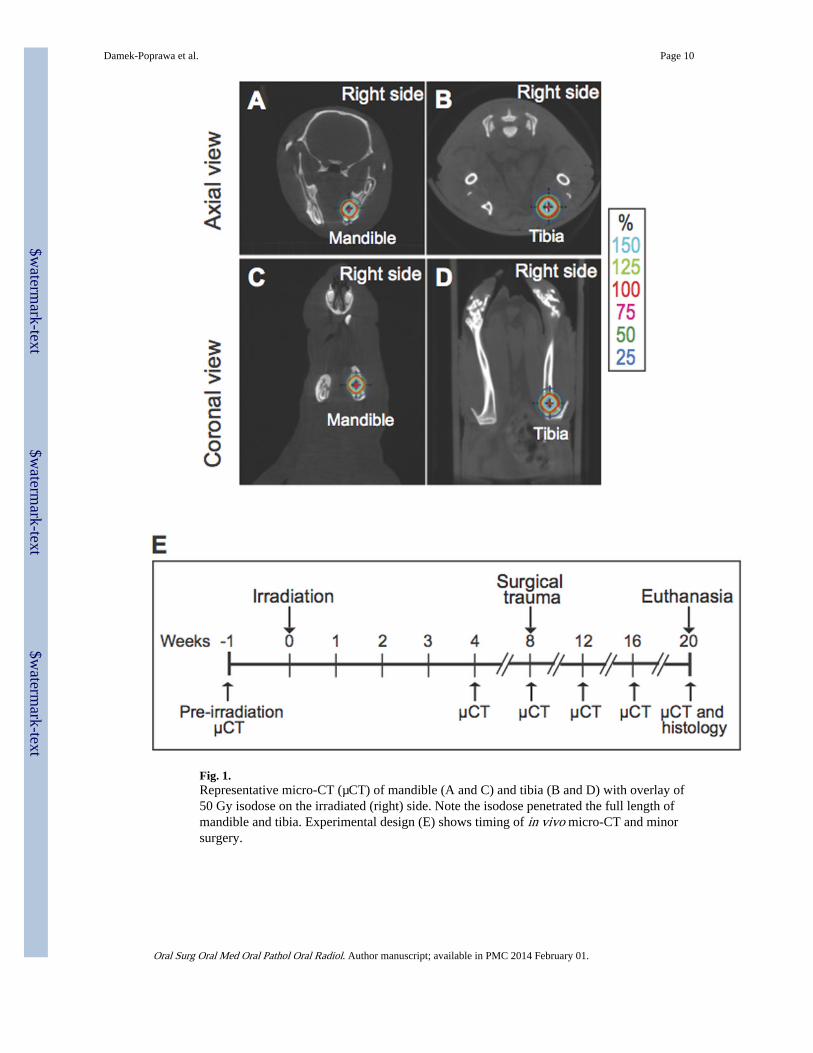
Fig. 1.Representative micro-CT (µCT) of mandible (A and C) and tibia (B and D) with overlay of50 Gy isodose on the irradiated (right) side. Note the isodose penetrated the full length ofmandible and tibia. Experimental design (E) shows timing of in vivo micro-CT and minorsurgery.
Damek-Poprawa et al. Page 10
Oral Surg Oral Med Oral Pathol Oral Radiol. Author manuscript; available in PMC 2014 February 01.
$waterm
ark-text$w
atermark-text
$waterm
ark-text
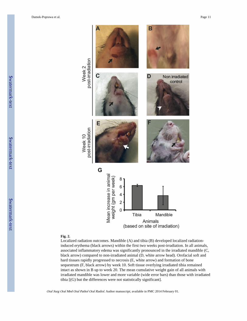
Fig. 2.Localized radiation outcomes. Mandible (A) and tibia (B) developed localized radiation-induced erythema (black arrows) within the first two weeks post-irradiation. In all animals,associated inflammatory edema was significantly pronounced in the irradiated mandible (C,black arrow) compared to non-irradiated animal (D, white arrow head). Orofacial soft andhard tissues rapidly progressed to necrosis (E, white arrow) and formation of bonesequestrum (F, black arrow) by week 10. Soft tissue overlying irradiated tibia remainedintact as shown in B up to week 20. The mean cumulative weight gain of all animals withirradiated mandible was lower and more variable (wide error bars) than those with irradiatedtibia [(G) but the differences were not statistically significant].
Damek-Poprawa et al. Page 11
Oral Surg Oral Med Oral Pathol Oral Radiol. Author manuscript; available in PMC 2014 February 01.
$waterm
ark-text$w
atermark-text
$waterm
ark-text
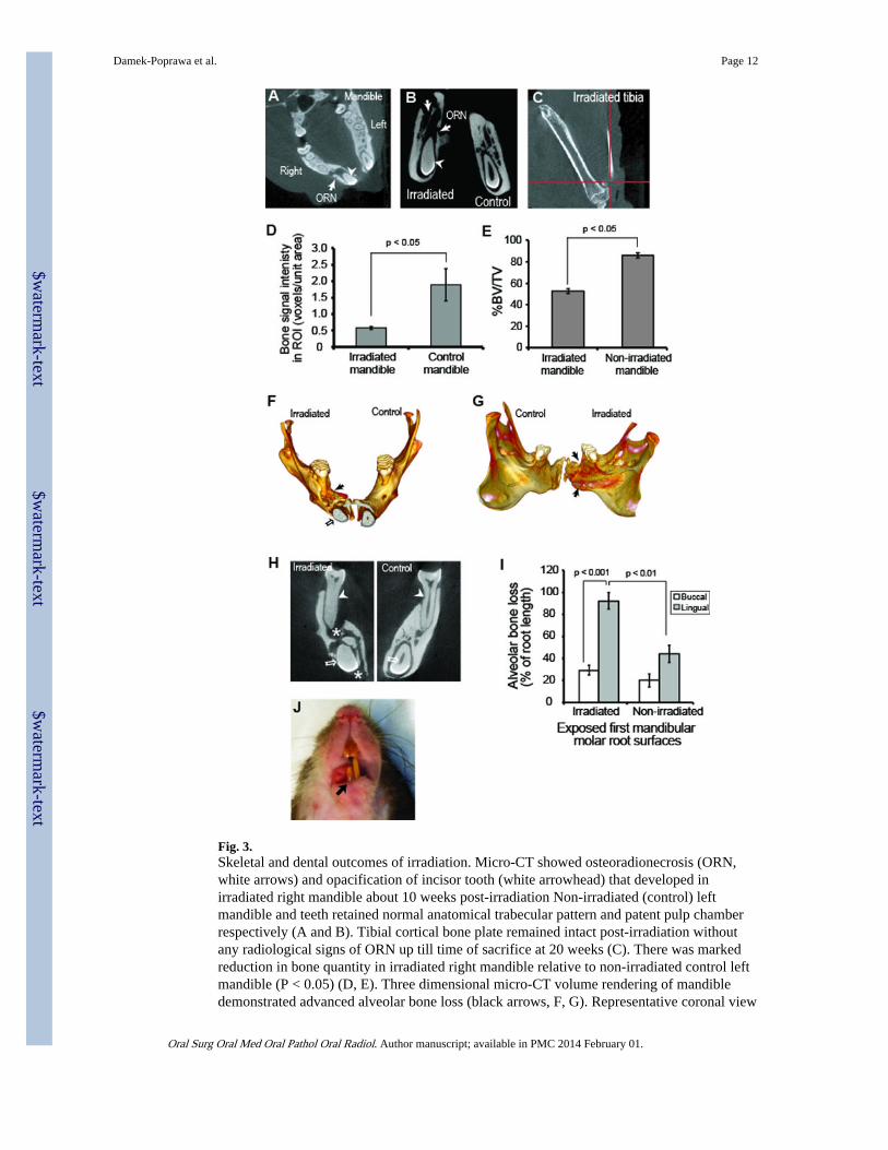
Fig. 3.Skeletal and dental outcomes of irradiation. Micro-CT showed osteoradionecrosis (ORN,white arrows) and opacification of incisor tooth (white arrowhead) that developed inirradiated right mandible about 10 weeks post-irradiation Non-irradiated (control) leftmandible and teeth retained normal anatomical trabecular pattern and patent pulp chamberrespectively (A and B). Tibial cortical bone plate remained intact post-irradiation withoutany radiological signs of ORN up till time of sacrifice at 20 weeks (C). There was markedreduction in bone quantity in irradiated right mandible relative to non-irradiated control leftmandible (P < 0.05) (D, E). Three dimensional micro-CT volume rendering of mandibledemonstrated advanced alveolar bone loss (black arrows, F, G). Representative coronal view
Damek-Poprawa et al. Page 12
Oral Surg Oral Med Oral Pathol Oral Radiol. Author manuscript; available in PMC 2014 February 01.
$waterm
ark-text$w
atermark-text
$waterm
ark-text
of mandibular ORN at the level of the first molar (H) displayed severe periodontal bone loss(white arrowheads), trabecular bone loss (white star) and pulpal calcification of both molarand long incisor teeth (white arrow and arrowheads). Periodontal bone loss was notablymore severe on the lingual than the buccal side in both irradiated (p < 0.001) and non-irradiated mandible (p < 0.01) (I). There was also delayed eruption of right mandibularincisor relative to the left (J).
Damek-Poprawa et al. Page 13
Oral Surg Oral Med Oral Pathol Oral Radiol. Author manuscript; available in PMC 2014 February 01.
$waterm
ark-text$w
atermark-text
$waterm
ark-text
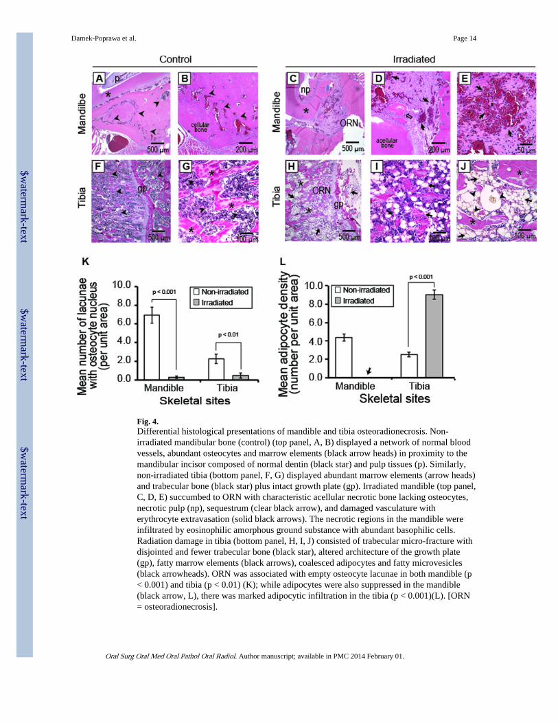
Fig. 4.Differential histological presentations of mandible and tibia osteoradionecrosis. Non-irradiated mandibular bone (control) (top panel, A, B) displayed a network of normal bloodvessels, abundant osteocytes and marrow elements (black arrow heads) in proximity to themandibular incisor composed of normal dentin (black star) and pulp tissues (p). Similarly,non-irradiated tibia (bottom panel, F, G) displayed abundant marrow elements (arrow heads)and trabecular bone (black star) plus intact growth plate (gp). Irradiated mandible (top panel,C, D, E) succumbed to ORN with characteristic acellular necrotic bone lacking osteocytes,necrotic pulp (np), sequestrum (clear black arrow), and damaged vasculature witherythrocyte extravasation (solid black arrows). The necrotic regions in the mandible wereinfiltrated by eosinophilic amorphous ground substance with abundant basophilic cells.Radiation damage in tibia (bottom panel, H, I, J) consisted of trabecular micro-fracture withdisjointed and fewer trabecular bone (black star), altered architecture of the growth plate(gp), fatty marrow elements (black arrows), coalesced adipocytes and fatty microvesicles(black arrowheads). ORN was associated with empty osteocyte lacunae in both mandible (p< 0.001) and tibia (p < 0.01) (K); while adipocytes were also suppressed in the mandible(black arrow, L), there was marked adipocytic infiltration in the tibia (p < 0.001)(L). [ORN= osteoradionecrosis].
Damek-Poprawa et al. Page 14
Oral Surg Oral Med Oral Pathol Oral Radiol. Author manuscript; available in PMC 2014 February 01.
$waterm
ark-text$w
atermark-text
$waterm
ark-text
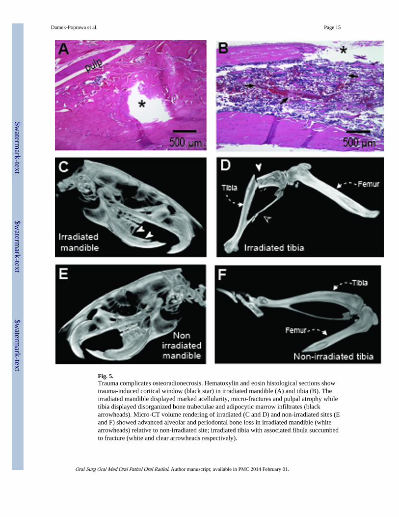
Fig. 5.Trauma complicates osteoradionecrosis. Hematoxylin and eosin histological sections showtrauma-induced cortical window (black star) in irradiated mandible (A) and tibia (B). Theirradiated mandible displayed marked acellularity, micro-fractures and pulpal atrophy whiletibia displayed disorganized bone trabeculae and adipocytic marrow infiltrates (blackarrowheads). Micro-CT volume rendering of irradiated (C and D) and non-irradiated sites (Eand F) showed advanced alveolar and periodontal bone loss in irradiated mandible (whitearrowheads) relative to non-irradiated site; irradiated tibia with associated fibula succumbedto fracture (white and clear arrowheads respectively).
Damek-Poprawa et al. Page 15
Oral Surg Oral Med Oral Pathol Oral Radiol. Author manuscript; available in PMC 2014 February 01.
$waterm
ark-text$w
atermark-text
$waterm
ark-text
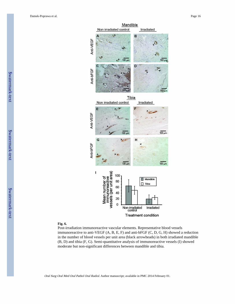
Fig. 6.Post-irradiation immunoreactive vascular elements. Representative blood vesselsimmunoreactive to anti-VEGF (A, B, E, F) and anti-bFGF (C, D, G, H) showed a reductionin the number of blood vessels per unit area (black arrowheads) in both irradiated mandible(B, D) and tibia (F, G). Semi-quantitative analysis of immunoreactive vessels (I) showedmoderate but non-significant differences between mandible and tibia.
Damek-Poprawa et al. Page 16
Oral Surg Oral Med Oral Pathol Oral Radiol. Author manuscript; available in PMC 2014 February 01.
$waterm
ark-text$w
atermark-text
$waterm
ark-text
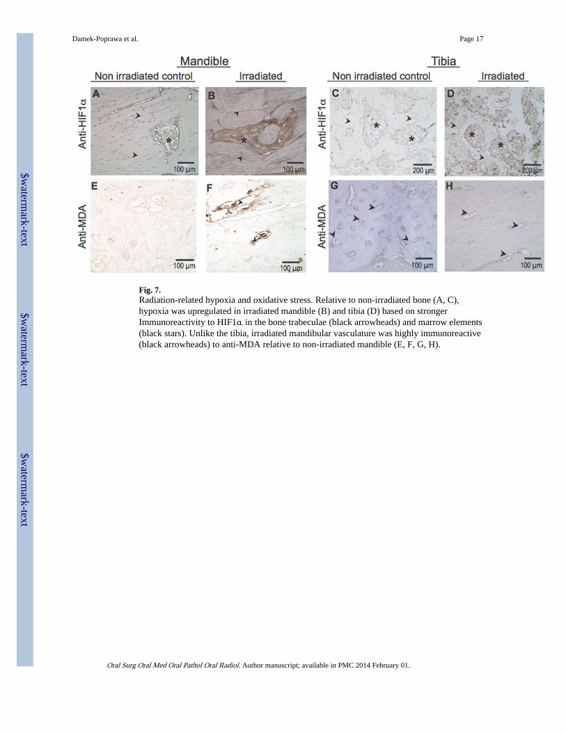
Fig. 7.Radiation-related hypoxia and oxidative stress. Relative to non-irradiated bone (A, C),hypoxia was upregulated in irradiated mandible (B) and tibia (D) based on strongerImmunoreactivity to HIF1α in the bone trabeculae (black arrowheads) and marrow elements(black stars). Unlike the tibia, irradiated mandibular vasculature was highly immunoreactive(black arrowheads) to anti-MDA relative to non-irradiated mandible (E, F, G, H).
Damek-Poprawa et al. Page 17
Oral Surg Oral Med Oral Pathol Oral Radiol. Author manuscript; available in PMC 2014 February 01.
$waterm
ark-text$w
atermark-text
$waterm
ark-text