Embed Size (px)
Citation preview

Citation: Chowdhury I, Thomas K and Thompson WE. Prohibitins in Reproduction- A Timeline. Austin J Reprod Med Infertil. 2016; 3(2): 1042.
Austin J Reprod Med Infertil - Volume 3 Issue 2 - 2016ISSN : 2471-0393 | www.austinpublishinggroup.com Chowdhury et al. © All rights are reserved
Austin Journal of Reproductive Medicine & Infertility
Open Access
Abstract
Prohibitins are ubiquitous and an evolutionary conserved protein family that is present in multiple cellular organelles including mitochondria in addition to the nucleus. The Prohibitins are involved in multiple cellular functions such as cellular differentiation, anti-proliferation, morphogenesis and play a major role in maintaining the functional integrity of the mitochondria. Our laboratory and other groups have performed experimental studies on the expression and distribution pattern of prohibitins in various reproductive tissues of different species, which are include mice, rats, pigs, humans and few lower vertebrates and invertebrates. Moreover, recent studies have shown that prohibitins are strongly associated with spermatogenesis, folliculogenesis and the functions of the accessory reproductive organs. In this brief review, we highlight experimental evidence that supports the conserved roles that the prohibitins play in reproductive physiology.
Keywords: Prohibitin (PHB); Repressor of estrogen activity (REA); Reproduction
present across all organisms. Currently our understanding of the complex biology of the prohibitins in reproductive physiology is limited. However, our laboratory and other groups have performed a number of experimental studies on the expression and distribution pattern of PHB and REA in various reproductive tissues of different species. These studies are performed in mice, rats, pigs, humans and few lower vertebrates and invertebrates including the red crayfish Cherax quadricarinatus, Octopus tankahkeei, Chinese mitten crab Eriocheir sinensis, Danio rerio, Gallus gallus, Salmo salar, and Bos taurus. Moreover, recent studies have shown that prohibitins are strongly associated with spermatogenesis, folliculogenesis and functions of the accessory reproductive organ. In this brief review, we highlight some of the experimental evidence supporting a conserved role for the prohibitins in reproductive physiology.
ProhibitinsIn humans, the PHB gene (hPHB) spans ~11 kb with 7 exons
and map to chromosome locus 17q21 [3]. The first exon and a small portion of the second exon comprise the 5’ untranslated region, whereas the largest exon, exon 7 contains ~700 bp of 3’ untranslated RNA. Several transcripts of the PHB gene are transcribed with varying lengths of 3’ untranslated region [4]. The longer transcripts are present at higher levels in proliferating tissues and cells [5]. The abundance of PHB mRNA is inversely related to markers of cellular proliferation in different cells and tissues [6-10]. Comparative genomic alignment studies have shown that the human and rat PHB genes are similar except for intron 2 and 3, which are ~1 kb larger in the rat gene [11]. The hPHB gene encodes ~30 kDa protein, also known as B-cell receptor associated protein-32 (BAP32) gene. PHB contains four highly conserved domains, namely, an N-terminal hydrophobic domain; a PHB domain (amino acid residues 26-187) encoded by exon 3, 4 and 5, and which is conserved from protozoa to mammals; a coiled-coil (CC) alpha helices domain (amino acids residues 177–211) present at the C-terminal end of the protein, and encoded largely by exon 6; and a putative nuclear export sequence (amino acid residues 257 to 270) which present at the C-terminal.
Introduction Sexual reproduction is a complex multistep hormonal dependent
process where a male gamete, the spermatozoa, fertilizes a female gamete, the ova to form a zygote. In vertebrates, the formation of a mature ova and a sperm are through the process of ovarian folliculogenesis and spermetogenesis respectively under the control of endocrine factors including gonadotropins (follicle stimulating hormone, FSH and luteinizing hormone, LH). During these processes, multiple autocrine and paracrine factors, and steroid hormones play important roles as regulators of folliculogenesis and spermatogensis. The coordinated biosynthesis of steroids in the ovary and the testis are critical for progression of the reproductive cycle, successful ovulation and release of spermatozoa, and eventually fertilization and pregnancy. The binding of gonadotropins to specific membrane G-protein coupled receptors (GPCRs), leads to the activation of multiple signal transduction pathways, including the adenylate cyclase-/cAMP-dependent protein kinase A (PKA) pathway, mitogen-activated protein kinase (MAP kinase) signaling and calcium-/calmodul independent pathways that are known to be involved in the regulation of steroidogenesis and gametogenesis in vertebrates. Furthermore, multiple cross-talk among these signal transduction systems has been well documented. Interestingly, several other proteins are involved in the process of gametogenesis and interacting regulatory pathways.
Prohibitins are ubiquitous and evolutionary conserved protein family that belongs to the SPFH family which is characterized by the presence of the stomatin/prohibitin/flotillin/HflK/C (SPFH) domain (also known as the PHB domain). Members of this protein family include prohibitin (PHB/PHB1), repressor of estrogen activity (REA/PHB2), stomatins, plasma membrane proteins of Escherichia coli (HflKC), flotillins, the human insulin receptor (HIR) proteins, the stomatin-like-proteins (SLPs), podocin and the erlins and plant defense proteins [1,2]. Based on extensive database analysis approximately 1800 PHB domain-containing proteins exist which includes 340 animal proteins and 142 mammalian proteins that are
Review Article
Prohibitins in Reproduction- A TimelineChowdhury I1*, Thomas K2 and Thompson WE1,3
1Department of Obstetrics and Gynecology, Morehouse School of Medicine, USA2Department of Neurobiology, Morehouse School of Medicine, USA3Department of Physiology, Morehouse School of Medicine, USA
*Corresponding author: Indrajit Chowdhury, Department of Obstetrics and Gynecology, Reproductive Science Research Program, Morehouse School of Medicine, 720 Westview Drive Southwest, Atlanta, GA 30310, USA
Received: September 02, 2016; Accepted: October 15, 2016; Published: October 18, 2016

Austin J Reprod Med Infertil 3(2): id1042 (2016) - Page - 02
Chowdhury I Austin Publishing Group
Submit your Manuscript | www.austinpublishinggroup.com
The N-terminal hydrophobic domain is critical for its attachment to the mitochondrial inner membrane, whereas the CC-domain is important for protein–protein interactions.
The human PHB2 (REA/hPHB2) [12], also referred as prohibitone [13]/B-cell receptor associated protein-37 (BAP37) [14] gene (PHB2) is located at chromosome 12p13 [15]. This gene has 10 exons, with smaller introns than PHB and spans ~5.3 kb. The REA/PHB2/BAP37 gene encodes a protein of ~37 kDa. Similar to PHB structure, REA has a PHB domain, which is encoded by residues 39–201; a CC-domain present at the C terminal end of the protein; a putative signal peptide (residues 1–36), and a putative nuclear localization signal peptide (residues 86–89). In eukaryotes PHB and PHB2 have highly conserved PHB domains. The PHB protein is 54% homologous with PHB2 [16,17] and has a single amino acid difference between rodents and humans [11]. Orthologues of the PHB gene have been identified in several organisms including bacteria [18,19], plants [20,21],
Trypanosoma brucei [22], Saccharomyces cerevisiae (yeast) [23,24], Caenorhabditis elegans [25] and Drosophila [26].
Prohibitins and Reproduction Distinct differences in PHB and PHB2 (REA) levels have
been observed during ovarian folliculogenesis, spermatogenesis, in mammary gland development, and in uterine tissue. Immunolocalization, Western blotting and immunogold staining of ovaries and testis had shown that both PHB and PHB2 (REA) are present in multiple cellular locations including mitochondria and nucleus, suggesting that they play additional roles in those cellular compartments. Due to complex post-translational modification and changes in cellular localization occurring during different physiological states, it has been difficult to identify the distinct roles that these prohibitins isoforms play in reproductive organs. Currently ours and other research groups are trying to understand the functional roles that the prohibitins play in reproduction by
Year and References Critical findings
[27] • Identifiedandcharacterizedprohibitin(PHB)asanintracellularprotein(p28kDa)duringdifferentiationofratgranulosacells(GCs)isolated from preantral and early antral follicles.
[28]• PHBwaslocalizedwithinratGCsofinfantandjuvenileovaries.• A strong expression pattern of PHB in rat oocyte at all stages of follicular development, in rat corpus luteum and in follicles
undergoing atresia. [29] • PHBroleswereconfirmedinmitochondrialstructureandfunctionduringgrowthanddifferentiationofGCs.
[30] • PHBgeneexpressionprofileswereanalyzedinnormalandincarcinoma-inducedfemaleratmammarygland.
[25] • Demonstratedtherolesofprohibitin(PHBandREA)complexforCaenorhabditiselegans (C. elegans) embryonic development, and necessary for normal mitochondrial morphology and respiration.
[31] • GnRHdependentPHBexpressionisimportantformaturationofTlymphocytesandratthymicgrowth.
[32]
• PHBwasimmunolocalizedinratGCs,theca-interstitialcells,andtheoocyte;andinporcineoocytes,zygotes,andblastocyst.• GonadotropindependentPHBexpressionwasinverselycorrelatedtoPCNAexpressionduringfollicularmaturationandpositively
co-localizedwithP450scc.• Inatreticfollicles,germinalvesicle-stageoocytes,zygotes,andblastocystsPHBwastranslocatedfromthecytoplasmtothe
nucleus.
[33] • In human fundus, the expressions of repressor of estrogen receptor activity (REA) is reduced during labor, and suggest that REA gene is involved in regulatory pathways of estrogen receptor alpha (ERA) activity.
[34] • REA is required for the maintenance of estrogen receptor (ER) activity and normal mammary gland development. The reduction or lossofREAfunctionpromotesover-activationofERandincreasebreastcancerriskinhumans.
[35-37]• InratGCs,mitochondrialPHBactthroughphospho-PHB(pPHB)-MEK-pERKpathwayandregulatestheBcl/Bcl-xLwhichinhibit
Bax-Bakexpression.Theseeventsdirectlyinhibitthereleaseofcytochromecfromtheinter-mitochondrialspaceandinhibitdownstream activation of cleaved caspase 3.
[38] • In rainbow trout (Oncorhynchusmykiss),ahigherREAmRNAabundanceineggsinhibitsdevelopmentofembryo.
[39]• Demonstratedanovelmechanismforcontrolofestrogen-inducedluminalepithelialproliferationinvolvinguterineendometrial
stromal(ST)Krüppel-likefactor9(KLF9)regulationsofparacrinefactor(s)torepressepithelialexpressionofco-repressorREA/PHB2 in mice.
[40] • PHB was found to be an estrogen-regulated gene essential for uterine development and function in mice, and selectively required for estrogen-regulated gene expression in uterus.
[41] • Studies demonstrated that REA is essential for mammary gland development and has a gene dosage-dependent role in the regulationofstage-specificphysiologicalfunctionsofthemammaryglandduringpregnancyandlactationstages.
[42]
• In cloned placenta of domestic cat (Feliscatus),theexpressionofPHBandcathepsinD(CD)werecorrelatedwiththegenerationofreactiveoxygenspecies(ROS),leadingtodecreasedmitochondrialmembranepotentialandtelomericDNA,whichareassociatedwith cellular senescence and apoptosis. The abnormal PHB protein patterns were associated with impaired development, and hence decreased fetal viability.
[43] • REAexpressionwasintenseinboththeperinuclearcytoplasmandthenucleusinGCsfromfolliclesatallstagesofdevelopmentinbovine. However, REA expression was less intense in thecal tissue.
[44,45] • PHBwasregulatedbyfolliclestimulatinghormone(FSH)inafollicularstage-dependentmannerin vitroinpre-antralGCsisolatedfromdiethylstilbestrol(DES)treatedratsandantralGCsisolatedfromequinechorionicgonadotropin(eCG)-treatedrats.
[46]• REAphysiologicallyrestrainedhumanendometrialstromalcelldecidualization,controllingthetimingandmagnitudeof
decidualizationtoenablepropercoordinationofuterinedifferentiationwithconcurrentembryodevelopmentthatisessentialforimplantation and optimal fertility.
[47] • Theaberrantexpressionofglycolysis-relatedenzymesinhumanendometriomatissuewasassociatedwithenhancedglycolyticmetabolism.Themalignant-likefeaturemaybepartiallycausedbylow-expressionofPHBgeneinendometrioticstromalcells.
[48] • Inhumans,REAmodulatedcrosstalkamongmultiplecelltypesintheuterinetissueandhostbackground,servingasabrakeontheestradiol-ER axis and restraining multiple aspects that contribute to the pathologic progression of endometriosis.
[49] • Foliclestimulatinghormone(FSH)-dependentPHB/pPHBupregulationinGCsisrequiredtosustainthedifferentiatedstateofGCs.
Table 1: Prohibitins role in female reproduction.
Austin J Reprod Med Infertil 3(2): id1042 (2016) - Page - 03
Chowdhury I Austin Publishing Group
Submit your Manuscript | www.austinpublishinggroup.com
utilizing conditional knock-out mice. We have highlighted a few important findings on the functional roles that PHB and REA play in reproductive physiology in Table 1 and 2.
ConclusionAt present, we are just beginning to understand the critical roles
that PHB and PHB2 (REA) play in ovarian, uterine, mammary gland, testis and accessory reproductive organ functions. Although current knowledge indicates that PHB and REA are involved in regulating the fate of folliculogenesis, spermatogenesis and other reproductive processes. A number of basic questions still remain to be answered. These include physiological role that PHB and PHB2 (REA) play in regulating gene expression and signaling in spermatogenesis, folliculogenesis, and the functions of the accessory reproductive organs.
AcknowledgementThis study was supported in part by National Institutes of Health
Grants 1SC3GM113751, 1RO1HD057235 and G12-RR03034. This investigation was conducted in a facility constructed with support from Research Facilities Improvement Grant #C06 RR18386 from NIH/NCRR.
References1. Nadimpalli R, Yalpani N, Johal GS, Simmons, CR. Prohibitins, stomatins
and plant disease response genes compose a super family that controls cell proliferation, ion channels regulation and death. J Biol Chem. 2000; 275:29579-29586.
2. Nijtmans LG, Artal SM, Grivell LA, Coates PJ. The mitochondrial PHBcomplex: roles in mitochondrial respiratory complex assembly, aging and degenerativedisease.CellMolLifeSci.2002;59:143-155.
3. SatoT,SaitoH,SwensenJ,OlifantA,WoodC,DannerD,etal.Thehumanprohibitin gene located on chromosome 17q21 is mutated in sporadic breast cancer. Cancer Res. 1992: 52: 1643-1646.
4. Jupe ER, Liu XT, Kiehlbauch JL, McClung JK, Dell’Orco RT. The 3’untranslated region of prohibitin and cellular immortalization. Exp CellRes.1996;224:128-135.
5. Choongkittaworn NM, Kim KH, Danner DB, Griswold MD. Expression of
prohibitin in rat seminiferous epithelium. Biol Reprod.1993; 49: 300-310.
6. Hussain-Hakimjee EA, Peng X, Mehta RR, Mehta RG. Growth inhibitionof carcinogen-transformed MCF-12F breast epithelial cells and hormone-sensitive BT-474 breast cancer cells by 1alpha-hydroxyvitamin D5.Carcinogenesis. 2006; 27: 551-559.
7. Jupe ER, Liu XT, Kiehlbauch JL, McClung JK, Dell’Orco RT. Prohibitinantiproliferativeactivityandlackofheterozygosityinimmortalizedcelllines.ExpCellRes.1995;218:577-580.
8. LumpkinCKJr,MooreTL,TarpleyMD,TaylorJM,BadgerTM,McClungJK.Acute ethanol and selected growth suppressor transcripts in regenerating rat liver. Alcohol. 1995; 12: 357-362.
9. Miyamoto S,Qin J, Safer B.Detection of early gene expression changesduring activation of human primary lymphocytes by in vitro synthesis of proteinsfrompolysomeassociatedmRNAs.ProteinSci.2001;10:423-433.
10. TannoS,FukudaI,SaitoY,OgawaK.Prohibitinexpressionisdecreasedinthe regenerating liver but not in chemically induced hepatic tumors in rats. JpnJCancerRes.1997;88:1155-1164.
11. Altus MS,Wood CM, Stewart DA, Roskams AJ, Friedman V, HendersonT, et al. Regions of evolutionary conservation between the rat and human prohibitin-encodinggenes.Gene.1995;158:291-294.
12. Montano MM, Ekena K, Delage-Mourroux R, Chang W, Martini P,Katzenellenbogen BS. An estrogen receptor-selective coregulator thatpotentiates the effectiveness of antiestrogens and represses the activity of estrogens.ProcNatlAcadSciUSA.1999;96:6947-6952.
13. LamersMC,BacherS.Prohibitinandprohibitone,ubiquitousandabundantproteins that are reluctant to reveal their real identity. Int Arch Allergy Immunol. 1997; 113: 146-149.
14. TerashimaM,KimKM,AdachiT,NielsenPJ,RethM,KohlerG,etal.TheIgM antigen receptor of B lymphocytes is associated with prohibitin and a prohibitinrelatedprotein.EMBOJ.1994;13:3782-3792.
15. Ansari-Lari MA, Shen Y, Muzny DM, Lee W, Gibbs RA. Large-scalesequencing in human chromosome 12p13: experimental and computational genestructuredetermination.GenomeRes.1997;7:268-280.
16. ChowdhuryI,Garcia-BarrioM,HarpD,ThomasK,MatthewsR,ThompsonWE.Theemergingrolesofprohibitinsinfolliculogenesis.Front.Biosci.(EliteEd). 2012; 4: 690-699.
17. GambleSC,ChotaiD,OdontiadisM,DartDA,BrookeGN,PowellSM,etal. Prohibitin, a protein downregulated by androgens, represses androgen receptoractivity.Oncogene.2007;26:1757-1768.
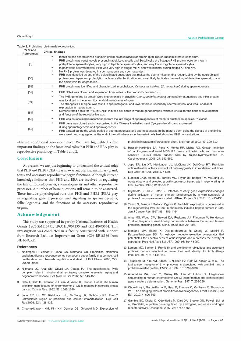
Year and References Critical findings
[5]
• Identifiedandcharacterizedprohibitin(PHB)asanintracellularprotein(p30kDa)inratseminiferousepithelium.• PHBproteinwasconstitutivelypresentinadultLeydigcellsandSertolicellsatallstages.PHBproteinwereverylowin
preleptotenespermatocytes,veryhighinleptotenespermatocytes,andverylowinzygotenespermatocytes.• Inpachytenespermatocytes,PHBwasveryhighinstagesVII-XIandwasminimalduringstagesXIIandXIV.• NoPHBproteinwasdetectedinspermatogoniaandspermatocytes.
[50]• PHBwasidentifiedasoneoftheubiquitinatedsubstratesthatmakesthespermmitochondriarecognizablebytheegg'subiquitin-
proteasomedependentproteolyticmachineryafterfertilizationandmostlikelyfacilitatesthemarkingofdefectivespermatozoainthe epididymis for degradation.
[51] • PHBproteinwasidentifiedandcharacterizedincephalopodOctopus tankahkeei (O. tankahkeei) during spermiogenesis.
[52] • PHBcDNAwasclonedandsequencedfromtestesofthecrabEriocheirsinensis.
[53]
• ThePHBgeneanditsproteinwerecharacterizedincrayfish(Cherazquadricarinatus) during spermatogenesis and PHB protein waslocalizedintheinnermitochondrialmembranesofsperm
• ThestrongestPHBsignalwasfoundinspermatogonia,andlowerlevelsinsecondaryspermatocytes,andweakorabsentexpression in mature sperm.
[54] • DemonstratedaroleforPHBinGnRH-inducedcelldeathinmaturegonadotropes,whichiscrucialforthenormaldevelopmentand function of the reproductive axis.
[55] • PHBwasco-localizedinmitochondriafromthelatestageofspermiogenesisofmacruracrustaceanspecies,P. clarkia.
[56]
• PHBgenewasclonedandcharacterizedintheChinesefire-belliednewtCynopsorientalis, and expressed during spermatogenesis and spermiogenesis.
• PHB existed during the whole period of spermatogenesis and spermiogenesis. In the mature germ cells, the signals of prohibitins wereweakandaggregatedattheendofthecell,whereasinthesertolicellshadabundantPHBconcentrations.
Table 2: Prohibitins role in male reproduction.
Austin J Reprod Med Infertil 3(2): id1042 (2016) - Page - 04
Chowdhury I Austin Publishing Group
Submit your Manuscript | www.austinpublishinggroup.com
18.de Monbrison F, Picot S. Introducing antisense oligonucleotides intoPneumocystiscarinii.JMicrobiolMethods.2002;50:211-213.
19. NarasimhanS,ArmstrongM,McClungJK,RichardsFF,SpicerEK.Prohibitin,a putative negative control element present in Pneumocystis carinii. Infect Immun.1997; 65: 5125-5130.
20. SneddenWA,FrommH.Characterizationoftheplanthomologueofprohibitin,a gene associated with antiproliferative activity in mammalian cells. Plant Mol Biol. 1997; 33: 753-756.
21. TakahashiA,KawasakiT,WongHL,SuharsonoU,HiranoH,ShimamotoK. Hyperphosphorylation of a mitochondrial protein, prohibitin, is induced by calyculinAinaricelesion-mimicmutantcdr1.PlantPhysiol.2003;132:1861-1869.
22. WelburnSC,MurphyNB.ProhibitinandRACKhomologuesareup-regulatedin trypanosomes induced to undergo apoptosis and in naturally occurring terminallydifferentiatedforms.CellDeathDiffer.1998;5:615-622.
23. KirchmanPA,MiceliMV,WestRL,JiangJC,KimS,JazwinskiSM.Prohibitinsand Ras2 protein cooperate in the maintenance of mitochondrial function during yeast aging. Acta Biochim Pol. 2003; 50: 1039-1056.
24. TatsutaT,ModelK,LangerT.Formationofmembraneboundringcomplexesbyprohibitininmitochondria.MolBiolCell.2005;16:248-259.
25. Artal-SanzM,TsangWY,WillemsEM,GrivellLA,LemireBD,vanderSpekH, et al. The mitochondrial prohibitin complex is essential for embryonic viabilityandgermlinefunctioninCaenorhabditiselegans.JBiolChem2003;278:32091-32099.
26. EvelethDDJr,MarshJL.SequenceandexpressionoftheCcgene,amemberofthedopadecarboxylasegeneclusterofDrosophila:possibletranslationalregulation.NucleicAcidRes.1986;14:6169-6183.
27. ThompsonWE,SanbuisshoA, LeeGY,AndersonE.Steroidogenic acuteregulatory(StAR)protein(p25)andprohibitin(p28)fromculturedratovariangranulosacells.J.ReprodFertil.1997;109:337-348.
28.Thompson WE, Powell JM, Whittaker JA, Sridaran R, Thomas KH.Immunolocalizationandexpressionofprohibitin,amitochondrialassociatedproteinwithintheratovaries.AnatRec.1999;256:40-48.
29. ThompsonWE,BranchA,WhittakerJA,LynD,ZilbersteinM,MayoKE,etal.Characterizationofprohibitininanewlyestablishedratovariangranulosecellline.Endocrinology.2001;142:4076-4085.
30. Shan L, He M, Yu M, Qiu C, Lee NH, Liu ET, et al. cDNA microarrayprofiling of rat mammary gland carcinomas induced by 2-amino-1-methyl-6-phenylimidazo[4,5-b]pyridine and 7,12-dimethylbenz[a]anthracene.Carcinogenesis.2002;23:1561-1568.
31. DixitVD,SridaranR,EdmonsondMA,TaubD,ThompsonWE.Gonadotropinreleasing hormone attenuates pregnancy-associated thymic involution and modulates the expression of antiproliferative gene product prohibitin. Endocrinology. 2003; 144: 1496-1505.
32. ThompsonWE, Asselin E, Branch A, Stiles JK, Sutovsky P, Lai L, et al.Regulation of prohibitin expression during follicular development and atresia inthemammalianovary.BiolReprod.2004;71:282-290.
33. Bukowski R, Hankins GD, Saade GR, Anderson GD, Thornton S. Labor-associated gene expression in the human uterine fundus, lower segment, andcervix.PLoSMed.2006;3:e169.
34. MussiP,LiaoL,ParkSE,CianaP,MaggiA,KatzenellenbogenBS,etal.Haploinsufficiency of the corepressor of estrogen receptor activity (REA)enhancesestrogenreceptorfunctioninthemammarygland.ProcNatlAcadSciUSA.2006;103:16716-16721.
35. ChowdhuryI,XuW,StilesJK,ZeleznikA,YaoX,MatthewsR,etal.Apoptosisof rat granulosa cells after staurosporine and serum withdrawal is suppressed by adenovirus-directed over expression of prohibitin. Endocrinology. 2007; 148:206-217.
36. Chowdhury I, Branch A, Olatinwo M, Thomas K, Matthews R, Thompson WE. Prohibitin (PHB) acts as a potent survival factor against ceramide induced apoptosisinratgranulosacells.LifeSci.2011;89:295-303.
37. Chowdhury I, Thompson WE, Welch C, Thomas K, Matthews R. Prohibitin (PHB)inhibitsapoptosisinratgranulosacells(GCs)throughtheextracellularsignal-regulatedkinase1/2(ERK1/2)andtheBclfamilyofproteins.Apoptosis.2013;18:1513-1525.
38.BonnetE,FostierA,BobeJ.Microarray-basedanalysisoffisheggqualityafternaturalorcontrolledovulation.BMCGenomics.2007;8:55-71.
39. PabonaJM,VelardeMC,ZengZ,SimmenFA,SimmenRC.Nuclearreceptorco-regulator Krüppel-like factor 9 and prohibitin 2 expression in estrogen-inducedepithelialcellproliferationinthemouseuterus.JEndocrinol.2009;200: 63-73.
40. HeB,KimTH,KommaganiR,FengQ,LanzRB,JeongJW,etal.Estrogen-regulated prohibitin is required for mouse uterine development and adult function. Endocrinology. 2011; 152: 1047-1056.
41. ParkS,ZhaoY,YoonS,XuJ,LiaoL,LydonJ,etal.Repressorofestrogenreceptor activity (REA) is essential for mammary gland morphogenesis and functional activities: studies in conditional knockout mice. Endocrinology.2011; 152: 4336-4349.
42. Bang JI, BaeDW, LeeHS, DebGK, KimMO, Sohn SH, et al. Proteomicanalysisofplacentasfromclonedcatembryosidentifiesasetofdifferentiallyexpressed proteins related to oxidative damage, senescence and apoptosis. Proteomics. 2011; 11: 4454-4467.
43. SalvettiNR,AlfaroNS,VelázquezMM,AmwegAN,MatillerV,DíazPU,etal.Alteration in localizationof steroidhormone receptorsandcoregulatoryproteins in follicles from cows with induced ovarian follicular cysts. Reproduction. 2012; 144: 723-735.
44. WangQ,LeaderA,TsangBK.InhibitoryrolesofprohibitinandchemerininFSH-inducedratgranulosacellsteroidogenesis.Endocrinology.2013;154:956-967.
45. Wang Q, Leader A, Tsang BK. Follicular stage-dependent regulation ofapoptosisandsteroidogenesisbyprohibitininratgranulosacells.JOvarianRes. 2013; 6: 6-23.
46. ZhaoY,ParkS,BagchiMK,TaylorRN,KatzenellenbogenBS.Thecoregulator,repressor of estrogen receptor activity (REA), is a crucial regulator of the timingandmagnitudeofuterinedecidualization.Endocrinology.2013;154:1349-1360.
47. QiX,ZhangY,JiH,WuX,WangF,XieM,etal.Knockdownofprohibitinexpression promotes glucose metabolism in eutopic endometrial stromal cells from women with endometriosis. Reprod Biomed Online. 2014; 29: 761-770.
48.ZhaoY,ChenY,KuangY,BagchiMK,TaylorRN,Katzenellenbogen JA,etal.MultipleBeneficialRolesofRepressorofEstrogenReceptorActivity(REA) in Suppressing the Progression of Endometriosis. Endocrinology. 2015; 157: 900-912.
49. ChowdhuryI,ThomasK,ZeleznikA,ThompsonWE.ProhibitinregulatestheFSHsignalingpathwayinratgranulosacelldifferentiation.JMolEndocrinol.2016; 56: 325-336.
50. ThompsonWE,Ramalho-SantosJ,SutovskyP.Ubiquitinationofprohibitinin mammalian sperm mitochondria: possible roles in the regulation of mitochondrial inheritance and sperm quality control. Biol Reprod. 2003; 69: 254-260.
51. MaoHT,WangDH,LanZ,ZhouH,YangWX.GeneexpressionprofilesofprohibitinintestesofOctopustankahkeei(ot-phb)revealingitspossibleroleduringspermiogenesis.MolBiolRep.2012;39:5519-5528.
52. Mao H, Wang DH, Zhou H, Yang WX. Characterization and expressionanalysis of prohibitin in the testis of Chinese mitten crab Eriocheirsinensis. Mol Biol Rep. 2012; 39: 7031-7039.
53. Fang DA, Wang Y, Wang J, Liu LH, Wang Q. Characterization ofCheraxquadricarinatus prohibitin and its potential role in spermatogenesis. Gene.2013;519:318-325.
54. SavulescuD,FengJ,PingYS,MaiO,BoehmU,HeB,etal.Gonadotropin-releasing hormone-regulated prohibitin mediates apoptosis of the gonadotropecells.MolEndocrinol.2013;27:1856-1870.

Austin J Reprod Med Infertil 3(2): id1042 (2016) - Page - 05
Chowdhury I Austin Publishing Group
Submit your Manuscript | www.austinpublishinggroup.com
55. DongWL,HouCC,YangWX.Mitochondrialprohibitinanditsubiquitinationduring crayfishProcambarusclarkiispermiogenesis.Cell TissueRes. 2015;359: 679-692.
56. JinJM,HouCC,TanFQ,YangWX.Thepotentialfunctionofprohibitinduringspermatogenesis inChinesefire-belliednewtCynopsorientalis.CellTissueRes.2016;363:805-22.
Citation: Chowdhury I, Thomas K and Thompson WE. Prohibitins in Reproduction- A Timeline. Austin J Reprod Med Infertil. 2016; 3(2): 1042.
Austin J Reprod Med Infertil - Volume 3 Issue 2 - 2016ISSN : 2471-0393 | www.austinpublishinggroup.com Chowdhury et al. © All rights are reserved