Embed Size (px)
Citation preview

Universal Journal of Environmental Research and Technology
All Rights Reserved Euresian Publication © 2012 eISSN 2249 0256
Available Online at: www.environmentaljournal.org
Volume 2, Issue 3: 135-142
Open Access Research Article
135
S. Arumugam
Toxic Effects of Flucloxacillin on the Early Development of the Polychaete
Hydroides elegans
S. Arumugam
Department of Zoology, Presidency College, Chennai, Tamil Nadu – 600 005
Corresponding author: [email protected]
Abstract: Hydroides elegans is a gregarious tube building polychaete which occurs in tropical and sub-tropical waters.
Adults were found to have good tolerance in laboratory assays using. The effect of flucloxacillin on
fertilization and early development was explained through three experiments. Flucloxacillin is a antibiotics
to treat bacterial infections. In normal fertilization the percentage of successful development at FM stage
was 100±0.00 upto larval release stage was 80.11±0.68. In Expt. I higher concentration 50% of the embryos
showed abnormal development and deformities. Expt. II in lower concentrations upto 800ppm the embryo
showed high deformities. Expt. III, in 400ppm nearly 60% of embryos showed abnormal development in
Blastula stage. Finally, more than 100ppm concentrations did not reach the release stage. The percentage
of successful development was decreased in Expt. I to Expt. III. The present paper deals with the toxic
effects of flucloxacillin on fertilization and early development of H. elegans and the percentage of successful
development of embryos were studied.
Keywords: developmental stages, flucloxacillin, Hydroides elegans, polychaete, toxic effects
1.0 Introduction: Hydroides elegans (Haswell) is a tube-building
polychaete species conspicuous in tropical and
subtropical coastal fouling communities. In most
places, settlement of H. elegans peaks in summer
or autumn (Qiu and Qian, 1997; Gopalakrishnan et
al., 2005). H. elegans is a sedentary fouling
tubiculous polychaete available in plenty on the
hulls of the ships, fishing boats and floating
material on the sea water (Udhayakumar and
Karande, 1996). The H. elegans on fishing boat
reduces the speed of the boat and increasing the
fuel coast (Raja, 1999; Gopalakrishnan et al., 2005;
Rani, 2005). H. elegans a sedentary polychaete
common in temperate intertidal waters produces
viable gametes throughout the year. The
organism is widespread forming dense layers
within the intertidal zone (Marsden and Anderson,
1981; Gopalakrishnan et al., 2005).
In the past several decades, laboratory studies on
Hydroides sp. have mainly focused on systematic
(Fauchald, 1977), oogenesis and fertilization
(Nordbaek, 1956), maturation (Leone, 1970),
salinity tolerance in adults (Mohan and Aruna,
1994), larval settlement (Hurlbut, 1991) and
description of early life history (Miura and
Kajihara, 1981; Gopalakrishnan et al., 2005).
Marine invertebrate larvae are usually more
sensitive to stress than adults and juveniles of the
same species and such sensitivity may help us
explain seasonal or annual variations in
recruitment success in the field (Qiu and Qian,
1997). H. elegans adults would retain their
gametes where external salinity dropped to levels
too to support fertilization and development.
Whether failure of fertilization or a failure of
fertilized eggs to cleave (Pechanic et al., 2007).
Toxic effects on aquatic and sediment dwelling
organisms caused by antibiotics uptake inhibitor
were recently summarized (Brooks, 2003).
Antibiotic is a chemical substance produced by a
micro organism that suppresses the growth or
directly kills another micro organism (Levine,
1978). Antibiotics are used to assist the immune
system and inhibits the growth of pathogenic
microorganisms (Timmreck, 1998). Flucloxacillin is
an antibiotic used for the treatment of infections
caused by susceptible gram-positive bacteria. It is
commonly used when a patient is suffering from
various infections such as bone and joint, skin and
wound, organs involved in breathing and the
inflammation of the lining of the heart cavity and
valves due to the bacteria Staphylococcus
(Harrison, 2005). Flucloxacillin is a white or almost
white powder. Slightly soluble in water and in

Universal Journal of Environmental Research and Technology
136
S. Arumugam
chloroform; freely soluble in methyl alcohol
(Sweetman, 2005). The toxic effects of antibiotics
on fertilization and early development of H.
elegans were studied by Rani (2005), Hemalatha
(2006) and Senthil (2006) and suitability of H.
elegans to assess the toxicity of pollutants were
established. Flucloxacillin may not cause any
damage to fertilization in human beings at normal
concentration. Increased concentrations of
flucloxacillin may cause damage to fertilization in
human beings and other animals. The present
study is designed to study the toxic effect of
flucloxacillin on fertilization and early
development of H. elegans.
2.0 Materials and Methods:
2.1. Source of Animals: Adult Hydroides elegans were collected from the
hulls of fishing boats berthed at Rayapuram fish
landing center, Chennai, India. Other sedentary
organisms like lepas, bernacles, neries, mytilus,
ascidians, algae, few crustacean, arthropods and
some mollusk were also seen. Other fouling
organisms were carefully removed from the
collection before H. elegans were placed in the
collection chambers. The specimens were
transported to the laboratory (Department of
Zoology, Presidency college, Chennai), within two
or three hours after collection. Adults were found
to have good tolerance in laboratory assays using.
2.2. Obtaining gametes for fertilization
studies: Gametes were obtained using standard
procedures (Raja and Sellappan, 1993;
Gopalakrishnan et al., 2005). H. elegans has
separated sexes. Tubes of adult H. elegans were
gently broken and observed for the release of
gametes, which occurred after few minutes. Eggs
were pink in colour and sperm milky white.
Gametes were then generally released by mature
individuals within several minutes and were
collected by pipette. Eggs were immediately
transferred into about 30 ml of seawater at a
salinity of 35‰ while sperm were kept undiluted
in small beakers until use. Sperm were then
diluted with about 10-20 ml of filtered seawater.
For each experiment, eggs were obtained from 5-8
females, and sperm were obtained from 4-6 males
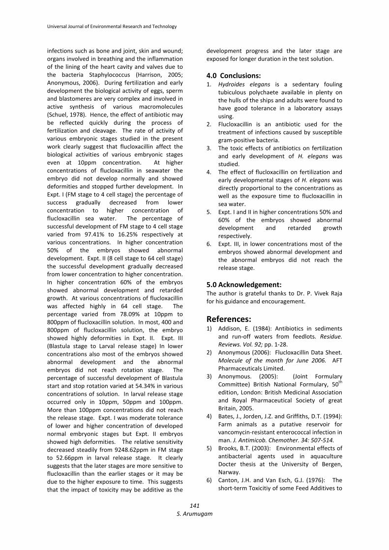
(Fig.1 and 2).
Fig. 1: Alive Hydroides elegans inside the tube and removed from the tube
Fig. 2: After mixing of gametes

Universal Journal of Environmental Research and Technology
137
S. Arumugam
2.3. Properties of Flucloxacillin: Flucloxacillin is a white or almost white, crystalline
hygroscopic, powder. Freely soluble in water and
in methyl alcohol; soluble in alcohol. A 10%
solution in water has a pH of 5.0 to 7.0. store at a
temperature non exceeding 25°C in air tight
containers (Sweetman, 2005). A systematic
(IUPAC) name 6-((s)-3-(2-chloro-6-fluourophenyl)-5-
methylisoxazole-4-carboxamido)-3, 3-dimthyl-7-oxo-4-
thia-l azabicyclo [3.2.0] heptane-2-carboxylic acid.
The molecular formula of flucloxacillin
C19H17ClFN3O5S with a molecular weight 453.87
(Anonymous, 2005). Fluxloxacillin is an isoxazolyl
penicillin used primarily for the treatment of
infections due to staphylococci resistant to
benzylpenicillin.
2.4. Preparation of Working Stock
Solution: 200 mg of flucloxacillin was dissolved and made up
to 250 ml of filtered sea water in a volumetric flask
to prepare 800ppm flucloxacillin solution. This
stock solution was stored in amber colored bottle.
From the stock solution the following
concentrations of flucloxacillin were prepared as
10ppm, 50ppm, 100ppm, 200ppm, 400ppm and
800ppm (Table 1).
Structure of Flucloxacillin
Table 1: Various concentrations of flucloxacillin prepared from stock solution
200 mg of flucloxacillin + 250 ml of filtered sea water = 800 ppm of stock solution
Sr.
No.
Stock solution
(ml)
Sea water
(ml)
Concentrations of flucloxacillin
solution (ppm)
1. 100 ml --- 800ppm
2. 50 ml 50 ml 400ppm
3. 25 ml 75 ml 200ppm
4. 12.5 ml 87.5 ml 100ppm
5. 6.25 ml 93.75 ml 50ppm
6. 1.25 ml 98.75 ml 10ppm
2.5. Experiment: The present work, the time of mixing gametes was
taken as time of fertilization and the percentage of
successful development of eggs in each
developmental stage were recorded. The
developmental stages were divided into three
experiments as Expt. I (FM stage to 4 cell stage),
Expt. II (8 cell stage to 64 cell stage) and Expt. III
(Blastula stage to Larval release stage).
Expt. I: Fertilization membrane stage to 4
cell stage Expt. I was designed to examine flucloxacillin
affect in developmental stages of FM stage to 4
cell stage. The experiment conducted on various
concentrations (10ppm, 50ppm, 100ppm, 200ppm,
400ppm and 800ppm) of flucloxacillin. These
concentrations are prepared from fresh stock
solution. Developmental stages were checked
often, and the cumulative time and the percentage
of successful egg development was recorded.
Expt. II: 8 cell stage to 64 cell stage Expt. II was designed to examine the effects of
flucloxacillin on survival and duration of
developmental stages of 8 cell stage to 64 cell
stage. The experiment was the same as in Expt. I.
Expt. III: Blastula stage to Larval release
stage Expt.III was designed to examine the effects of
fluxloxacillin on survival and duration of
development of Blastula stage to Larval release
stage. The experiment was the same as in Expt.I.

Universal Journal of Environmental Research and Technology
138
S. Arumugam
2.6. Data analysis: ANOVA was done by transforming values to ranks
and then applying parametric statistics on the
data, as described in Zar (1984). A 2-way ANOVA
(analysis of variance) was used to detect
flucloxacillin effects on duration of developmental
stages in Expt. I, II and III. The difference in the
mean was statistically significant between
different concentrations of flucloxacillin for all
developmental stages at 0.05% level. EC50 or
median effective concentration value of
flucloxacillin for different early embryonic stages
of H. elegans were calculated by probit analysis
followed by Finney (1971). EC50 values were
calculated for each experiment and all
developmental stages.
3.0 Results and Discussion:
3.1. Normal Fertilization and Early
Development: After fertilization, the negatively buoyant eggs sink
to the bottom where they undergo cleavage upto
the larval release stage. The first cleavage occurs
after 30 minutes of fertilization, at optimal
conditions such as 27˚C, pH 8.1 and salinity 35‰.
The elevation of fertilization membrane was
initiated 3 to 5 minutes after fertilization. The
meiotic division is not completed before spawning.
The first cleavage plane was meridional and
complete, both the blastomeres were almost of
equal in size. Completion of first cleavage
occurred at 30 minutes. The percentage of
successful development at FM stage was
100±0.00. The second cleavage plane was also
meridional but right angle to the first one. The
completion of 4 cell stage occurred 52 minutes
after fertilization. The successful development of
this stage was 94.54±1.41. The third plane of
cleavage was horizontal, dividing the four
blastomeres into 8 cells. The time required for the
completion of 8 cell stage was 1hour 2 minutes
and the percentage of successful development of
this stage was 93.05±1.88. The embryo reached
64 cell stage at 1 hour 39 minutes after
fertilization. The successful egg development at
this stage was 88.06±2.83. The blastula stage
reached at 1 hour 58 minutes after fertilization.
The percentage of successful egg development
was 86.09±1.78. Finally the larval release stage
occurred 4 hours 50 minutes after fertilization. At
the end of the stage 80.11±0.68 percentage egg
was successfully developed.
3.2. Toxic Effect of Flucloxacillin on
Fertilization and Early Development:
Expt. I: Fertilization membrane stage to 4
cell stage Expt. I was explained FM stage to 4 cell stage (FM
stage, 2 cell stage, 3 cell stage and 4 cell stage) at
various concentrations of flucloxacillin. In FM
stage, after fertilization the time showed a steady
increase from 6 minutes at 10ppm concentration
to 7.5 minutes at 800ppm concentrations of
flucloxacillin. The percentage of successful
development of FM stage showed variation at
various concentrations of flucloxacillin. The
percentage of success was 97.41±0.41 up to
10ppm and then declined steadily to 82.16±0.05 in
800ppm. After formation of fertilization
membrane reached 4 cell stage with a steady
increase from 54 minutes at 10ppm to 1 hour 15
minutes at 800ppm of solution. The percentage of
successful development of 4 cell stage declined
steadily from 84.18±5.92 at 10ppm to 16.25±0.40
at 800ppm (Table 2) of flucloxacillin solution. In
higher concentration 50% of the embryos showed
abnormal development, retarded growth and
deformities (Fig.3).
Fig. 3: Expt. I: Fertilization membrane stage
to 4 cell stage
Fig.4: Expt. II: 8 cell stage to 64 cell stage

Universal Journal of Environmental Research and Technology
139
S. Arumugam
Table 2: Percentage of success * of various embryonic stages of H. elegans in normal and in different
concentrations of flucloxacillinin sea water
Devpt.
Stages
Control
Percentage of successful development
Concentrations of Flucloxacillin expressed in ppm
10ppm 50ppm 100ppm 200ppm 400ppm 800ppm
Expt. I
FM-
Stage
100
±0.00
97.41
±0.41
96.35
±0.48
93.73
±1.58
92.41
±0.91
86.36
±0.84
82.16
±0.05
2- Cell
Stage
97.52
±1.45
92.85
±1.55
88.52
±1.16
85.06
±2.11
78.81
±0.06
53.19
±13.95
28.86
±4.47
3- Cell
Stage
95.03
±0.92
89.09
±3.20
81.23
±1.23
75.39
±1.53
68.33
±1.67
42.79
±8.62
20.86
±1.35
4- Cell
Stage
94.54
±1.41
84.18
±5.92
74.97
±2.34
67.71
±4.08
57.43
±6.32
33.60
±1.96
16.25
±0.40
Expt. II
8- Cell
Stage
93.05
±1.88
79.57
±8.92
69.2
±6.05
60.67
±7.27
48.26
±10.49
26.86
±0.28
12.26
±1.15
16- Cell
Stage
91.52
±1.39
72.69
±12.9
62.39
±9.76
55.98
±9.39
43.05
±11.94
20.83
±0.58
9.92
±1.04
32- Cell
Stage
90.55
±2.36
66.35
±16.35
54.55
±12.45
48.87
±11.37
38.47
±14.03
14.1
±0.18
6.98
±0.32
64- Cell
Stage
88.06
±2.83
62.13
±18.66
48.28
±14.6
41.05
±11.51
30.59
±12.41
10.29
±0.29
4.04
±0.39
Expt. III
Blastula
Stage
86.09
±1.78
56.44
±21.66
44.15
±14.64
35.07
±12.35
22.98
±10.76
8.00
±0.86
2.96
±0.63
Blastula
Start
Rotation
85.09
±2.78
55.49
±20.71
41.51
±14.15
31.37
±10.92
16.94
±8.06
6.01
±0.30
2.86
±0.64
Blastula
Stop
Rotation
83.11
±1.72
52.86
±22.43
36.33
±12.12
23.48
±9.85
11.52
±5.97
3.32
±0.47
1.15
±0.05
Release
Stage
80.11
±0.68
50.83
±23.66
23.41
±2.36
12.73
±1.37
ND ND ND
ND= No Development, n= number of experiments, ± = Standard Deviation, * = number of eggs observed in
each concentration = 10
Expt. II: 8 cell stage to 64 cell stage Various concentrations of flucloxacillin affected
embryos during development from 8 cell stage to
64 cell stage (8 cell stage, 16 cell stage, 32 cell
stage and 64 cell stage). Flucloxacillin strongly
affected embryos and duration of development at
10ppm to 800ppm concentrations of flucloxacillin
solution. The cumulative time of 8 cell stage
increased steadily from 1 hour 7 minutes at 10ppm
to 1 hour 33 minutes at 800ppm concentrations.
The successful development of 8 cell stage
declined from 79.57±8.92 at 10ppm to 12.26±1.15
at 800ppm. In 400ppm nearly 60% of the embryos
showed abnormal development and deformities
where as abnormal development increased in 16
cell stage. At higher concentration to reach 64 cell
stage time was taken from 2 hours 22 minutes.
The percentage of successful development of 64
cell stage decreased 4.04±0.39 at 800ppm (Table
2). In lower concentrations upto 800ppm the
embryo showed high deformities (Fig.4).
Expt. III: Blastula stage to Larval release
stage Cleavage of embryo during development from
Blastula to Larval release stage (Blastula stage,
Blastula start rotation, Blastula stop rotation and
Larval release stage) was affected by various
concentrations of flucloxacillin solution. However,
duration of embryo development was highly
affected with increasing flucloxacillin solution. The
cumulative time of Blastula stage increased

Universal Journal of Environmental Research and Technology
140
S. Arumugam
steadily from 1 hour 27 minutes at 10ppm to 3
hours 12 minutes at 800ppm. In 400ppm nearly
60% of embryos showed abnormal development in
this stage. In lower concentrations also most of
the embryos showed abnormal development and
deformed embryos did not reach rotation stage.
The percentage of successful development of
Blastula start rotation and stop rotation stage in
2.86±0.64 and 1.15±0.05 at 800ppm respectively.
It has taken long period reach release stage after
fertilization from 5 hours 17 minutes at 10ppm to
5 hours 37 minutes at 100ppm of flucloxacillin
solution. More than 100ppm concentrations did
not reach the release stage (Table 2 and Fig.5).
BL - Blastula stage BR - Blastula Rotation stage TL - Trochopore larva
Fig.5. Expt. III: Blastula stage to larval release stage
3.3. Relative Sensitivity of Flucloxacillin: A careful perusal of the embryonic stages were more sensitive than the FM stage and the sensitivity decreased
steadily as the developing embryo enter into next stage of development. The FM stage (9248.62ppm) was the
least sensitive stage of flucloxacillin. The larval release stage (52.66ppm) was the highest sensitive stage of
flucloxacillin solution (Table 3).
Table 3: Relative sensitivity (EC50 values) of
Flucloxacillin for different
embryonic stages of H. elegans
(EC50 values are expressed in ppm)
Antibiotics are drugs of natural or synthetic origin
that have the capacity to kill or to inhibit the
growth of microorganisms. Antibiotics that are
sufficiently non-toxic to the host are used as
chemotherapeutic agents in the treatment of
infectious diseases of human, animals and plants.
They have long been present in the environment
and have played a crucial role in the battle
between man and animals (Serrano, 2005). The
wide use of antibiotics in animal nutrition and
disease has resulted in the sensitization of a
relatively large number of the susceptible people,
many of whom react violent in contact with these
drugs (Harvey, 1975). In recent years
environmental researchers have drawn attention
to reports on measure contents of drugs in waste
water, surface water, ground water and drinking
water all over the world (Canton, 1976).
Concentrations of the antibiotic, not necessarily
pure are then examined for toxicity in mice. As
well as measured contents of drugs in the
environmental information have also begun to be
available in the literature on drugs toxic effects on
aquatic organisms (Addison, 1984). The discharge
of antibiotics and their metabolites in farm waste
could create a reservoir of resistant micro
organisms in the environment. Several antibiotics
enter aquatic and terrestrial ecosystems through
the discharge of effluents from farms (Bates et al.,
1994). Flucloxacillin is an antibiotic, commonly
used when a patient is suffering from various
DEVELOPMENTAL STAGES EC50 VALUES
FM-STAGE 9248.62
2-CELL STAGE 508.58
3-CELL STAGE 394.77
4-CELL STAGE 309.39
8-CELL STAGE 249.89
16-CELL STAGE 236.05
32-CELL STAGE 211.11
64-CELL STAGE 165.49
BLASTULA STAGE 139.69
BLASTULA START
ROTATION STAGE 109.22
BLASTULA STOP
ROTATION STAGE 73.71
RELEASE STAGE 52.66
Universal Journal of Environmental Research and Technology
141
S. Arumugam
infections such as bone and joint, skin and wound;
organs involved in breathing and the inflammation
of the lining of the heart cavity and valves due to
the bacteria Staphylococcus (Harrison, 2005;
Anonymous, 2006). During fertilization and early
development the biological activity of eggs, sperm
and blastomeres are very complex and involved in
active synthesis of various macromolecules
(Schuel, 1978). Hence, the effect of antibiotic may
be reflected quickly during the process of
fertilization and cleavage. The rate of activity of
various embryonic stages studied in the present
work clearly suggest that flucloxacillin affect the
biological activities of various embryonic stages
even at 10ppm concentration. At higher
concentrations of flucloxacillin in seawater the
embryo did not develop normally and showed
deformities and stopped further development. In
Expt. I (FM stage to 4 cell stage) the percentage of
success gradually decreased from lower
concentration to higher concentration of
flucloxacillin sea water. The percentage of
successful development of FM stage to 4 cell stage
varied from 97.41% to 16.25% respectively at
various concentrations. In higher concentration
50% of the embryos showed abnormal
development. Expt. II (8 cell stage to 64 cell stage)
the successful development gradually decreased
from lower concentration to higher concentration.
In higher concentration 60% of the embryos
showed abnormal development and retarded
growth. At various concentrations of flucloxacillin
was affected highly in 64 cell stage. The
percentage varied from 78.09% at 10ppm to
800ppm of flucloxacillin solution. In most, 400 and
800ppm of flucloxacillin solution, the embryo
showed highly deformities in Expt. II. Expt. III
(Blastula stage to Larval release stage) In lower
concentrations also most of the embryos showed
abnormal development and the abnormal
embryos did not reach rotation stage. The
percentage of successful development of Blastula
start and stop rotation varied at 54.34% in various
concentrations of solution. In larval release stage
occurred only in 10ppm, 50ppm and 100ppm.
More than 100ppm concentrations did not reach
the release stage. Expt. I was moderate tolerance
of lower and higher concentration of developed
normal embryonic stages but Expt. II embryos
showed high deformities. The relative sensitivity
decreased steadily from 9248.62ppm in FM stage
to 52.66ppm in larval release stage. It clearly
suggests that the later stages are more sensitive to
flucloxacillin than the earlier stages or it may be
due to the higher exposure to time. This suggests
that the impact of toxicity may be additive as the
development progress and the later stage are
exposed for longer duration in the test solution.
4.0 Conclusions: 1. Hydroides elegans is a sedentary fouling
tubiculous polychaete available in plenty on
the hulls of the ships and adults were found to
have good tolerance in a laboratory assays
using.
2. Flucloxacillin is an antibiotic used for the
treatment of infections caused by susceptible
gram-positive bacteria.
3. The toxic effects of antibiotics on fertilization
and early development of H. elegans was
studied.
4. The effect of flucloxacillin on fertilization and
early developmental stages of H. elegans was
directly proportional to the concentrations as
well as the exposure time to flucloxacillin in
sea water.
5. Expt. I and II in higher concentrations 50% and
60% of the embryos showed abnormal
development and retarded growth
respectively.
6. Expt. III, in lower concentrations most of the
embryos showed abnormal development and
the abnormal embryos did not reach the
release stage.
5.0 Acknowledgement: The author is grateful thanks to Dr. P. Vivek Raja
for his guidance and encouragement.
References: 1) Addison, E. (1984): Antibiotics in sediments
and run-off waters from feedlots. Residue.
Reviews. Vol. 92; pp. 1-28.
2) Anonymous (2006): Flucloxacillin Data Sheet.
Molecule of the month for June 2006. AFT
Pharmaceuticals Limited.
3) Anonymous. (2005): (Joint Formulary
Committee) British National Formulary, 50th
edition, London: British Medicinal Association
and Royal Pharmaceutical Society of great
Britain, 2005.
4) Bates, J., Jorden, J.Z. and Griffiths, D.T. (1994):
Farm animals as a putative reservoir for
vancomycin-resistant enterococcal infection in
man. J. Antimicob. Chemother. 34: 507-514.
5) Brooks, B.T. (2003): Environmental effects of
antibacterial agents used in aquaculture
Docter thesis at the University of Bergen,
Narway.
6) Canton, J.H. and Van Esch, G.J. (1976): The
short-term Toxicitiy of some Feed Additives to

Universal Journal of Environmental Research and Technology
142
S. Arumugam
Different Freshwater Organisms. Bull. Environ.
Contam. Toxicol.. 15: 720-725.
7) Fauchald, K. (1977): The polychaete worm
definitions and keys to orders, families and
genera. Nat. His. Mus. Los Angel Cty. Sci. Ser.,
28:1-190.
8) Finney, D.J. (1971): "Probit Analysis"
Cambridge University Press. London. p. 333.
9) Gopalakrishnan, S., Thilagam, H., Sellappan,
M. and Raja, P.V. (2005): Larval Development
and settlement of a marine Biofouling
Organisms. Internet. Confe. On Recent.
Advances in Marine Antifouling Tech. pp. 430-
440.
10) Harrison, K. (2005): Flucloxacillin chemistry
and structure. Molecule of the month for June
2005.
11) Harvey, S.C. (1975): Aantimicrobial Drugs.
Remingtons Pharmaceutical Sciences, pp.
1113-1116. Eds: Osol, John E, Hoover. Mack
Publishing Company, 15th
edition, Easton
Pennsylvania.
12) Hemalatha, P. (2006): Effect on cephalexin on
fertilization and early development of a
sedentary polychaete Hydroides elegans
(Haswell, 1883). M.Phil. Dissertation.
University of Madras.
13) Hurlbut, C.J. (1991): Community recruitment:
settlement and juvenile survival of seven co-
occuring species of sessile marine
invertebrates. Mar. Biol., 109:507-516.
14) Leone, D.E. (1970): The maturation of
Hydroides dianthus. Bio. Bull. (Woods Hole)
138:306-315.
15) Levine, R.R. (1978): Pharmacology Drug
Action and Reaction. Little, Brown and
Company 2nd
edition Boston, pp. 444.
16) Marsden, J.R. and Anderson, D.T. (1981):
Larval development and metamorphosis of
the serpulid polychaete Galeolaria caespitosa
Lamarck. Aus. J. Mar. Freshwater Res.,
32:667-680.
17) Miura, T. and Kajihara, T. (1981): The
development a serpulid worm Hydroides
ezoensis (Annelida, polychaete). Proc. Jap.
Soc. Sys. Zool., 20:7-12.
18) Mohan, P.C. and Aruna, C. (1994): The biology
of serpulid worms in relation to biofouling. In:
Thompson MF, Nagabhushanam, R., Sarojini,
R., Fingerman, M. (eds). Recent
developments in biofouling control. AA
Balkema, Rotterdam, p59-64.
19) Nordback, K. (1956): On the oogenesis and
fertilization of the serpulid Hydroides norvegia
(Gunnerus). Nytt. Mag. Zool., 4:121-123.
20) Pechenik, J.A., Pearse, J.S. and Qian, P.Y.
(2007): Effect of salinity on spawning and
early development of the tube building
polychaete Hydroides elegans in Hong Kong:
Not just the sperm’s fault? Biol. Bull.,
212:151-160.
21) Qiu, J.W. and Qian, P.Y. (1997): Combined
effects of salinity, temperature and food on
early development of the polychaete
Hydroides elegans. Mar. Ecol. Prog. Ser.,
152:79-88.
22) Raja, P.V. (1999): Hydroides elegans (Haswell)
an ideal organism to study pollution in
seawater XXVI International Ethological
Conference, 29th
August 1999. Bangalore.
Adva. Ethol. 34; 225.
23) Raja, P.V. and M. Sellappan (1993): Hydroides
elegans as a model specimen in quality
assessment of water for aquaculture. Effect of
mercury on fertilization and early
development. 11th
National Symposium on
Reproductive Biology and Comparative
Endocrinoloogy, Tirupathi. Abstract p. 54.
24) Rani, K.V.S.J. (2005): Effect of Ampicillin and
Amoxcillin on fertilization and early
development of a sedentary polychaete
Hydroids elegans (Haswell, 1883). M.Phil.
Dissertation. University of Madras.
25) Schuel, H. (1978): Secretary functions of egg
cortical granules in fertilization and
development. A critical review. Gamete
Research. 1 : 299-382.
26) Senthil, G.S. (2007): Effect of tetracycline on
fertilization and early development of a
sedentary polychaete Hydroides elegans
(Haswell, 1883). M.Phil. Dissertation,
University of Madras.
27) Serrano, P.H. (2005): Responsibe use of
antibiotics in Aquaculture. FAO Fisheries
Technical paper 469.
28) Sweetman, S.C. (2005): Martindale. The
complete drug reference. 34th
edition,
London, Chicago Pharmaceutical press, 2005.
29) Timmreck, T.K. (1998): An Introduction to
Epidemiology. Jones and Bartiett Publishers.
2nd
edition, Sudbury, Massachuetts. Pp. 49.
30) Udhayakumar and Karande (1996): Field
notes an a fouling serpulid Hydroides elegans
Haswell (polychaeta serpulids) present in
confined waters of Bombay. Indian J. Mar.
Scien. 25: 133-136.
31) Zar, J.H. (1974): Bio-Statistical Analysis.
Prentic Hall Inc. pp. 718.