Embed Size (px)
Citation preview
Case ReportOptic Neuropathy with Features Suggestive of Optic Neuritis inCerebrotendinous Xanthomatosis
Miyuki Miyamoto,1 Nobuyuki Ishii ,1 Hitoshi Mochizuki ,1 Kazutaka Shiomi,1
Tomoko Kaida,2 Hideki Chuman,3 andMasamitsu Nakazato1
1Division of Neurology, Respirology, Endocrinology and Metabolism, Department of Internal Medicine, University of Miyazaki,Miyazaki, Japan2Miyata Eye Hospital, Miyakonojo, Miyazaki, Japan3Department of Ophthalmology, University of Miyazaki, Miyazaki, Japan
Correspondence should be addressed to Nobuyuki Ishii; nobuyuki [email protected]
Received 20 September 2018; Revised 15 January 2019; Accepted 3 February 2019; Published 12 February 2019
Academic Editor: Peter Berlit
Copyright © 2019 Miyuki Miyamoto et al. This is an open access article distributed under the Creative Commons AttributionLicense, which permits unrestricted use, distribution, and reproduction in any medium, provided the original work is properlycited.
We describe our encounter with a 39-year-oldmanwho exhibited acute painless visual loss and progressive gait disturbance. He hadtendinous xanthoma and several neuroophthalmological findings indicative of optic neuropathy in the right eye, including afferentpupillary defect, cecocentral scotoma, and optic disc swelling. Neurological examination showed cerebellar ataxia and pyramidalweakness. Brain magnetic resonance imaging revealed bilateral swelling in the optic nerves with gadolinium-enhancementsuggesting optic neuritis, an enlarged fourth ventricle, atrophy of the cerebellum, and hyperintensities in the bilateral dentatenuclei. The patient was diagnosed with cerebrotendinous xanthomatosis (CTX) based on an elevated serum cholestanol level anda homozygous missense mutation in CYP27A1. CTX is a genetic lipid storage disease caused by dysfunction of the mitochondrialenzyme sterol 27-hydroxylase. With respect to ophthalmological manifestations, juvenile cataracts and optic neuropathy arecommonfindings in patients withCTX, but there have been no reports of optic neuropathywith features suggestive of optic neuritis.Thus, this case illustrates that clinicians should consider a diagnosis of CTX in patients with cardinal features of CTX even if thepatients show signs indicative of optic neuritis.
1. Introduction
Cerebrotendinous xanthomatosis (CTX), an autosomalrecessive lipid storage disease, is characterized by varioussystemic and neurological symptoms, including chronicdiarrhea, juvenile cataracts, tendon xanthomas, anddevelopmental delay [1]. With respect to ophthalmologicalmanifestations, apart from cataracts, optic neuropathy withoptic disc paleness is a common finding in patients withCTX [2]. However, cases of CTX with optic neuritis havenot been previously reported. Here, we present the firstcase of CTX with optic neuropathy and findings suggestiveof optic neuritis, including disc edema and contrast agentenhancement of the peripapillary retina and optic nerve.
2. Case Presentation
A 39-year-old man, born of a consanguineous marriage, hadepilepsy since the age of 12 years, as well as dyslipidemia;he was referred to our hospital because of acute painlessvisual loss and progressive gait disturbance. His visual acuitywas 20/16 bilaterally after bilateral cataract surgery at the ageof 31 years. His developmental milestones were normal. Hedenied a history of neonatal jaundice or infantile diarrhea,smoked half a pack of cigarettes per day, and drank socially.His family history was unremarkable. Medications includedcarbamazepine, phenytoin, clobazam, and bezafibrate.
On physical examination, swelling was observed in thetendons of Achilles, patella, and triceps, which indicated
HindawiCase Reports in Neurological MedicineVolume 2019, Article ID 2576826, 4 pageshttps://doi.org/10.1155/2019/2576826
2 Case Reports in Neurological Medicine
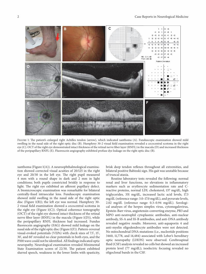
Figure 1: The patient’s enlarged right Achilles tendon (arrow), which indicated xanthoma (A). Funduscopic examination showed mildswelling in the nasal side of the right optic disc (B). Humphrey 30-2 visual field examination revealed a cecocentral scotoma in the righteye (C). OCT of the right eye demonstrated intact thickness of the retinal nerve fiber layer (RNFL) in the macula (D) and increased thicknessof the peripapillary RNFL (E). Fluorescein angiography exhibited profuse dye leakage on the right optic disc (B).
xanthoma (Figure 1(A)). A neuroophthalmological examina-tion showed corrected visual acuities of 20/125 in the righteye and 20/30 in the left eye. The right pupil measured4 mm with a round shape in dark and 2 mm in lightconditions; both pupils constricted briskly in response tolight. The right eye exhibited an afferent pupillary defect.A biomicroscopic examination was remarkable for bilateralcentrally-fixed intraocular lens. Funduscopic examinationshowed mild swelling in the nasal side of the right opticdisc (Figure 1(B)); the left eye was normal. Humphrey 30-2 visual field examination showed a cecocentral scotoma inthe right eye (Figure 1(C)). Optical coherence tomography(OCT) of the right eye showed intact thickness of the retinalnerve fiber layer (RNFL) in the macula (Figure 1(D)), whilethe peripapillary RNFL thickness had increased. Fundusfluorescein angiography (FAG) showed mild leakage on thenasal side of the right optic disc (Figure 1(F)). Pattern-reversalvisual-evoked potentials (VEPs) with check sizes of 7.5', 15',30', and 60' revealed no clear potentials in both eyes, and theP100wave could not be identified. All findings indicated opticneuropathy. Neurological examination revealed MinimentalState Examination scores of 22/30. The patient exhibitedslurred speech, weakness in the lower limbs with spasticity,
brisk deep tendon reflexes throughout all extremities, andbilateral positive Babinski sign. His gait was unstable becauseof truncal ataxia.
Routine laboratory tests revealed the following: normalrenal and liver functions, no elevations in inflammatorymarkers such as erythrocyte sedimentation rate and C-reactive proteins, normal LDL cholesterol, 137 mg/dL, hightriglycerides, 331 mg/dL, increased lactic acid levels, 17.3mg/dL (reference range: 3.0–17.0 mg/dL), and pyruvate levels,2.02 mg/dL (reference range: 0.3–0.94 mg/dL). Serologi-cal analyses of the herpes simplex virus, cytomegalovirus,Epstein-Barr virus, angiotensin-converting enzyme, PR3 andMPO anti-neutrophil cytoplasmic antibodies, anti-nuclearantibody, SS-A and SS-B antibodies, and anti-DNA antibodyrevealed negative results. Moreover, anti-aquaporin 4 andanti-myelin oligodendrocyte antibodies were not detected.No mitochondrial DNA mutations (i.e., nucleotide positions3460, 11,778, and 14,484) associated with Leber’s hereditaryoptic neuropathy (LHON) were observed. Cerebrospinalfluid (CSF) analysis revealed no cells but showed an increasedprotein level (77 mg/dL); isoelectric focusing revealed nooligoclonal bands in the CSF.
Case Reports in Neurological Medicine 3
(a) (b) (c)
(d) (e) (f)
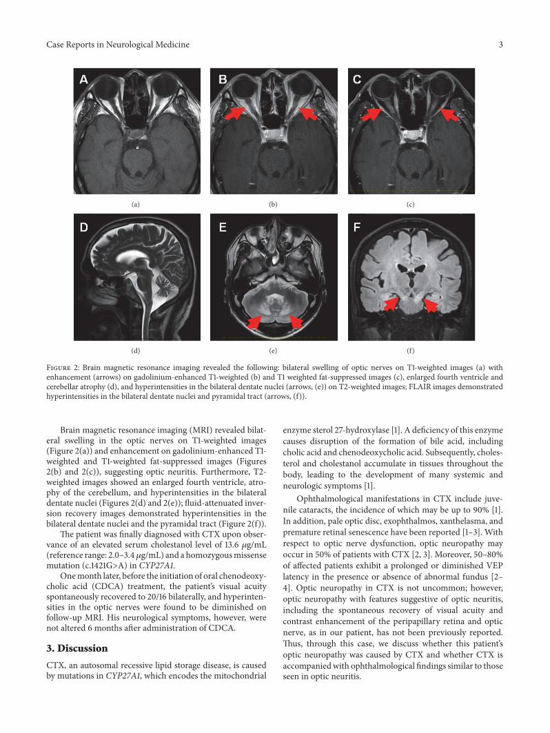
Figure 2: Brain magnetic resonance imaging revealed the following: bilateral swelling of optic nerves on T1-weighted images (a) withenhancement (arrows) on gadolinium-enhanced T1-weighted (b) and T1 weighted fat-suppressed images (c), enlarged fourth ventricle andcerebellar atrophy (d), and hyperintensities in the bilateral dentate nuclei (arrows, (e)) on T2-weighted images; FLAIR images demonstratedhyperintensities in the bilateral dentate nuclei and pyramidal tract (arrows, (f)).
Brain magnetic resonance imaging (MRI) revealed bilat-eral swelling in the optic nerves on T1-weighted images(Figure 2(a)) and enhancement on gadolinium-enhanced T1-weighted and T1-weighted fat-suppressed images (Figures2(b) and 2(c)), suggesting optic neuritis. Furthermore, T2-weighted images showed an enlarged fourth ventricle, atro-phy of the cerebellum, and hyperintensities in the bilateraldentate nuclei (Figures 2(d) and 2(e)); fluid-attenuated inver-sion recovery images demonstrated hyperintensities in thebilateral dentate nuclei and the pyramidal tract (Figure 2(f)).
The patient was finally diagnosed with CTX upon obser-vance of an elevated serum cholestanol level of 13.6 𝜇g/mL(reference range: 2.0–3.4𝜇g/mL) and a homozygousmissensemutation (c.1421G>A) in CYP27A1.
Onemonth later, before the initiation of oral chenodeoxy-cholic acid (CDCA) treatment, the patient’s visual acuityspontaneously recovered to 20/16 bilaterally, and hyperinten-sities in the optic nerves were found to be diminished onfollow-up MRI. His neurological symptoms, however, werenot altered 6 months after administration of CDCA.
3. Discussion
CTX, an autosomal recessive lipid storage disease, is causedby mutations in CYP27A1, which encodes the mitochondrial
enzyme sterol 27-hydroxylase [1]. A deficiency of this enzymecauses disruption of the formation of bile acid, includingcholic acid and chenodeoxycholic acid. Subsequently, choles-terol and cholestanol accumulate in tissues throughout thebody, leading to the development of many systemic andneurologic symptoms [1].
Ophthalmological manifestations in CTX include juve-nile cataracts, the incidence of which may be up to 90% [1].In addition, pale optic disc, exophthalmos, xanthelasma, andpremature retinal senescence have been reported [1–3]. Withrespect to optic nerve dysfunction, optic neuropathy mayoccur in 50% of patients with CTX [2, 3]. Moreover, 50–80%of affected patients exhibit a prolonged or diminished VEPlatency in the presence or absence of abnormal fundus [2–4]. Optic neuropathy in CTX is not uncommon; however,optic neuropathy with features suggestive of optic neuritis,including the spontaneous recovery of visual acuity andcontrast enhancement of the peripapillary retina and opticnerve, as in our patient, has not been previously reported.Thus, through this case, we discuss whether this patient’soptic neuropathy was caused by CTX and whether CTX isaccompaniedwith ophthalmological findings similar to thoseseen in optic neuritis.
4 Case Reports in Neurological Medicine
The etiology of optic neuropathy with acute visual lossincludes optic neuritis, arteritic and nonarteritic ischemicoptic neuropathy, infections, optic nerve compression,LHON, toxic and metabolic optic neuropathy, and traumaticoptic neuropathy [5]. Considering the pathophysiology ofCTX, which involves the accumulation of cholesterol andcholestanol in virtually all tissues, nonarteritic ischemicoptic neuropathy (NAION) is immediately included in thedifferential diagnosis of a patients with optic neuropathy.However, the presence of contrast-enhanced optic nerveson MRI and spontaneous recovery of visual acuity renderNAION a less likely diagnosis [6]. Except for idiopathicoptic neuritis, other differential diagnoses were also notcompatible with the patient’s history, neuroophthalmologicalexamination, and laboratory and imaging findings. Althoughwe could not exclude idiopathic optic neuritis, we speculatedthat the patient’s optic neuropathy was caused by CTXbecause optic neuropathy is commonly associated with CTX.
We hypothesized that optic neuropathy with findingssimilar to those seen in optic neuritis in CTX may involvemitochondrial dysfunction, although the exact mechanismremains unclear. Sterol 27-hydroxylase, a mitochondrialenzyme, is impaired in CTX, leading to abnormalities inmitochondrial function as well as lipid metabolism [1].Indeed, the following findings suggesting mitochondrialdysfunctionhave been revealed by previous studies: increasedlactic acid and pyruvate levels in the blood and CSF [7],a lactate peak on brain MR spectroscopy [8], decreasedactivities ofmitochondrial respiratory chain enzymes [7], andstructural abnormalities in the mitochondria [9].
Similarly, LHON is established as a mitochondrial disor-der. A typical clinical presentation of LHON includes acuteor subacute painless visual loss accompanied by disc swelling,which resembles our patient’s clinical course; however, leak-age in the fundus FAG and progressive optic nerve atrophyis not typical of LHON [10]. However, several studies havereported fundus edema, dye leakage in FAG, and gadolinium-enhancement of the optic nerve on MRI, masquerading asoptic neuritis [11, 12], as well as spontaneous improvementin visual acuity [13] in patients with LHON. The similar-ities between our case and LHON cases indicate that theophthalmological findings in our patient may have resultedfrom mitochondrial dysfunction in CTX. Furthermore, ourpatient’s clinical course might explain the mechanism under-lying the optic neuropathy commonly seen in CTX.
To the best of our knowledge, there have been no reportsof cases of optic neuropathy with features suggestive ofoptic neuritis in CTX. The exact underlying mechanismsremain unclear; however, we speculate that mitochondrialdysfunction caused by CTX may be involved. Thus, this caseillustrates that clinicians should consider a diagnosis of CTXin patients with cardinal features of CTX, such as xanthomasor hyperintensities of the dentate nuclei on brain MRI, evenin the presence of contrast enhancement of the optic discs andoptic nerves, indicating optic neuritis.
Consent
Written informed consent was obtained from the patient.
Conflicts of Interest
The authors declare that there are no conflicts of interestregarding the publication of this article.
Acknowledgments
This study did not receive any specific grants from fundingagencies in the public, commercial, or not-for-profit sectors.
References
[1] G. Salen and R. D. Steiner, “Epidemiology, diagnosis, andtreatment of cerebrotendinous xanthomatosis (CTX),” Journalof Inherited Metabolic Disease, vol. 40, no. 6, pp. 771–781, 2017.
[2] M. T. Dotti, A. Rufa, and A. Federico, “Cerebrotendinous xan-thomatosis: Heterogeneity of clinical phenotype with evidenceof previously undescribed ophthalmological findings,” Journalof Inherited Metabolic Disease, vol. 24, no. 7, pp. 696–706, 2001.
[3] J. R. M. Cruysberg, R. A. Wevers, B. G. M. Van Engelen, A.Pinckers, A. Van Spreeken, and J. J. M. Tolboom, “Ocular andsystemic manifestations of cerebrotendinous xanthomatosis,”American Journal of Ophthalmology, vol. 120, no. 5, pp. 597–604,1995.
[4] M.Mondelli, A. Rossi, C. Scarpini, M. T. Dotti, and A. Federico,“Evoked potentials in cerebrotendinous xanthomatosis andeffect induced by chenodeoxycholic acid,” JAMANeurology, vol.49, no. 5, pp. 469–475, 1992.
[5] A. T. Toosy, D. F. Mason, and D. H. Miller, “Optic neuritis,”TheLancet Neurology, vol. 13, no. 1, pp. 83–99, 2014.
[6] J. F. Rizzo III, C. M. Andreoli, and J. D. Rabinov, “Use ofmagnetic resonance imaging to differentiate optic neuritis andnonarteritic anterior ischemic optic neuropathy,” Ophthalmol-ogy, vol. 109, no. 9, pp. 1679–1684, 2002.
[7] M. T. Dotti, L. Manneschi, and A. Federico, “Mitochondrialenzyme deficiency in cerebrotendinous xanthomatosis,” Journalof the Neurological Sciences, vol. 129, no. 2, pp. 106–108, 1995.
[8] N. De Stefano, M. T. Dotti, M. Mortilla, and A. Federico,“Magnetic resonance imaging and spectroscopic changes inbrains of patients with cerebrotendinous xanthomatosis,” Brain,vol. 124, no. 1, pp. 121–131, 2001.
[9] A. Federico, M. T. Dotti, and N. Volpi, “Muscle mitochondrialchanges in cerebrotendinous xanthomatosis,” Annals of Neurol-ogy, vol. 30, no. 5, pp. 734-735, 1991.
[10] C. Meyerson, G. Van Stavern, and C. McClelland, “Leberhereditary optic neuropathy: Current perspectives,” ClinicalOphthalmology, vol. 9, pp. 1165–1176, 2015.
[11] A. Arianti, H. Chuman, N. Kajihara, N. Sakamoto, and N.Nao-I, “Atypical clinical and neuroimaging findings in leber’shereditary optic neuropathy:A case report,” JOJOphthalmology,vol. 6, no. 5, Article ID 555698, 2018.
[12] C. Lamirel, J. Cassereau, I. Cochereau et al., “Papilloedemaand MRI enhancement of the prechiasmal optic nerve at theacute stage of Leber hereditary optic neuropathy,” Journal ofNeurology, Neurosurgery & Psychiatry, vol. 81, no. 5, pp. 578–580, 2010.
[13] T.-K. Hsu, A.-G. Wang, M.-Y. Yen, and J.-H. Liu, “Leber’shereditary optic neuropathy masquerading as optic neuritiswith spontaneous visual recovery,” Clinical and ExperimentalOptometry, vol. 97, no. 1, pp. 84–86, 2014.

Stem Cells International
Hindawiwww.hindawi.com Volume 2018
Hindawiwww.hindawi.com Volume 2018
MEDIATORSINFLAMMATION
of
EndocrinologyInternational Journal of
Hindawiwww.hindawi.com Volume 2018
Hindawiwww.hindawi.com Volume 2018
Disease Markers
Hindawiwww.hindawi.com Volume 2018
BioMed Research International
OncologyJournal of
Hindawiwww.hindawi.com Volume 2013
Hindawiwww.hindawi.com Volume 2018
Oxidative Medicine and Cellular Longevity
Hindawiwww.hindawi.com Volume 2018
PPAR Research
Hindawi Publishing Corporation http://www.hindawi.com Volume 2013Hindawiwww.hindawi.com
The Scientific World Journal
Volume 2018
Immunology ResearchHindawiwww.hindawi.com Volume 2018
Journal of
ObesityJournal of
Hindawiwww.hindawi.com Volume 2018
Hindawiwww.hindawi.com Volume 2018
Computational and Mathematical Methods in Medicine
Hindawiwww.hindawi.com Volume 2018
Behavioural Neurology
OphthalmologyJournal of
Hindawiwww.hindawi.com Volume 2018
Diabetes ResearchJournal of
Hindawiwww.hindawi.com Volume 2018
Hindawiwww.hindawi.com Volume 2018
Research and TreatmentAIDS
Hindawiwww.hindawi.com Volume 2018
Gastroenterology Research and Practice
Hindawiwww.hindawi.com Volume 2018
Parkinson’s Disease
Evidence-Based Complementary andAlternative Medicine
Volume 2018Hindawiwww.hindawi.com
Submit your manuscripts atwww.hindawi.com