Embed Size (px)
Citation preview

Robert Babbel1
H. Ric Harnsberger Brent Nelson
Jerry Sonkens Steven Hunt
Received November 19, 1990; returned for revision February 25, 1991 ; revision received March 26, 1991 ; accepted April 22, 1991.
' All authors: Department of Radiology, University of Utah Medical Center, 50 N. Medical Dr. , Salt Lake City, VT 84132. Address reprint requests to H. R. Harnsberger.
0195-61 08/91 /1205-0849 © American Society of Neuroradiology
Optimization of Techniques in Screening CT of the Sinuses
849
The number of screening examinations of the sinuses performed with CT has markedly increased owing to the widespread and increasing use of endoscopic sinonasal surgery. We reviewed scans from 500 patients who had screening CT examinations of the sinuses for preendoscopic evaluation of inflammatory sinonasal disease to better define an optimal imaging protocol. Three aspects of direct coronal imaging of the paranasal sinuses were investigated: (1) preparation of the patient prior to the examination; (2) technical factors of the CT study, including positioning of the patient, optimal coronal angle, slice thickness, and CT exposure factors; and (3) data display. Our experience indicates that pretreatment of the patient with maximal medical therapy enables the best preendoscopic definition of anatomy, disease pattern, and nonreversible disease component for the treating surgeon. CT technical factors are optimized with scanning in the prone position with thin (3-mm) sections obtained through the anterior paranasal sinuses. This allows optimal visualization of the ostiomeatal unit. The remaining posterior portions of the sinuses are adequately imaged with thicker slices (5 mm). The coronal scan angle used is less critical. Exposure factors (mAs) can be reduced dramatically without image compromise. Data display is optimized when the bone algorithm is used to acquire the data and with image display at intermediate window center and width level.
Use of the techniques outlined in this article results in a cost-effective yet diagnostic scan of the sinuses with decreased radiation exposure to the patient.
AJNR 12:849-854, September/October 1991; AJR 157: November 1991
Functional endoscopic sinonasal surgery has resulted in a dramatic increase in the use of CT as a presurgical screening study of the sinuses [1-3]. In this report, we describe a limited preendoscopic CT examination of the sinuses performed in a single plane, without IV contrast material and at a reduced cost. Various techniques and approaches to the screening CT study have been described [1-9]. However, no well-documented studies defining optimal techniques were found in the literature. We examined the various aspects of the screening sinonasal CT examination to document optimal techniques for preendoscopic evaluation. Three general features of the screening examination were analyzed: preparation of the patient, CT technique, and display of the acquired data. This article describes our findings and summarizes our experience with a list of recommendations for the optimal preendoscopic CT screening examination of the sinuses.
Direct coronal imaging is especially well suited for radiologic evaluation of the sinonasal region prior to endoscopic surgery owing to the similar anatomic orientations of imaging and endoscopy [1-5 , 1 0]. Coronal imaging also affords an optimal view of the components of the ostiomeatal unit (OMU), including the maxillary ostium, infundibulum, uncinate process, ethmoid bullae, and middle meatus [2-5, 1 0). The OMU is a critical area in endoscopic sinonasal surgery and the most frequent site of disease in the paranasal sinuses [1 0-14]. Besides the OMU, the other bony and soft-tissue structures of the sinonasal region are

850 BABBEL ET AL. AJNR:12, September/October 1991
visualized superbly on coronal CT. Although the coronal imaging plane is assumed in this study to provide ideal visualization of the OMU for the surgeon, other facets of the CT examination were critically evaluated for this report.
Subjects and Methods
Investigational methods included analysis of two study groups. Group 1 consisted of routine screening CT scans of the sinuses in 500 consecutive patients scanned for preendoscopic evaluation of inflammatory sinonasal disease. Group 2 included an additional 44 patients whose scans were used in both a prospective and retrospective analysis. All scans were obtained with a General Electric 9800 Quickscan CT unit or a Picker 1200 SX CT unit by using bone algorithm with direct coronal scanning. Patients in group 1 were scanned with a slice width and interval of 3 mm anteriorly and 5 mm posteriorly. Patients in group 2 were scanned with a 5-mm slice width and interval throughout. All scans were reviewed by at least two of the authors.
To evaluate the relative merits of prone vs supine positioning, coronal scanning was attempted in both positions in each of 34 consecutive patients in group 2. The CT gantry was maximally angled in an attempt to obtain a true coronal plane of imaging in both positions. Factors assessed for each position were ease of patient positioning, diagnostic quality, demonstration of disease, and adequacy of visualization of the OMU.
In 10 group 1 patients, CT technical factors were varied to test the hypothesis that when the bone algorithm is used, high-quality CT scans can be obtained despite the use of significantly reduced CT exposure factors (mAs). In each case, one slice location was selected where repeated images were obtained with incremental decreases in mAs, while maintaining a constant kVp. The acquired images were photographed by using identical window levels and widths. These were compared for image quality and diagnostic utility.
The ability of patients to hyperextend their necks varied , often resulting in scans that were not truly coronal in angle. This resulted in scans with variable coronal angles, which allowed the retrospective analysis of the actual coronal angle used and the relationship, if any, of the scan angle to visualization of the OMU. The coronal scan angle was measured on each scan of the 40 patients in group 2, including 30 patients scanned in both supine and prone positions. The lateral scout localizer image, with annotated slices , was used to make the measurements in relationship to the hard palate. The angle of the
A B
scan was correlated with adequacy of visualization of the OMU. Visualization of the OMU was graded on a scale of 1-4. A grade of 1 was assigned when the OMU was not visualized, grade 2 was assigned when the OMU area could be identified but the OMU anatomy was poorly defined, grade 3 was assigned for good visualization, and grade 4 was assigned when visualization was excellent.
The importance of CT slice thickness and interval in visualizing the OMU was evaluated by determining the frequency with which these structures were seen optimally (good to excellent visualization) on 5-vs 3-mm contiguous scans. The 40 patients in group 2 were scanned with the 5-mm technique, and the 1-4 grading system was used to assess image quality on the coronal scans. This was compared with visualization of the OMU in the 500 patients in group 1 scanned with the 3-mm technique. Scans showing severely diseased OMUs were excluded to avoid introduction of uncontrollable factors.
The filming technique was appraised in five patients in group 1 with sinonasal disease to determine if multiple filming formats are necessary to evaluate the sinuses on CT. The bone algorithm was used in all cases [15] . The data were displayed at soft-tissue (450 H, 50 H), bone (4000 H, 1100 H), and intermediate (2500 H, 250 H) window widths and levels, respectively. The images were evaluated for ease of perception of soft-tissue and bone disease, and for ability to visualize normal structures.
Difficulty in study design precluded an adequate objective analysis of patient preparation before scanning. However, subjective evaluation of our experience with use of a sympathomimetic nasal spray, nose-blowing immediately prior to the scan, and use of a prescan course of antibiotics [2] was performed.
Results
Patient Position
Of 34 consecutive patients, 30 were able to assume both the supine and prone positions. Two were unable to assume either position, while two were unable to assume the prone position but were able to assume the supine position. In the 30 patients able to assume both positions, 21 (70%) were adequately evaluated in either position. In six patients (20%), the studies were complementary and allowed a more accurate assessment than did either study alone (Fig. 1). This was particularly true for determining if an air/fluid level was present
Fig. 1.-A and 8, Free migrating ball of debris within left maxillary sinus (arrows) is seen in different locations on direct coronal images with patient prone (A) and supine (8). On either im· age alone, debris has the appearance of a mucous retention cyst or polyp. In order to categorize abnormality correctly, both images are necessary to document that lesion is not attached to sinus wall.
AJNR:12, September/October 1991 SCREENING CT OF THE SINUS 851
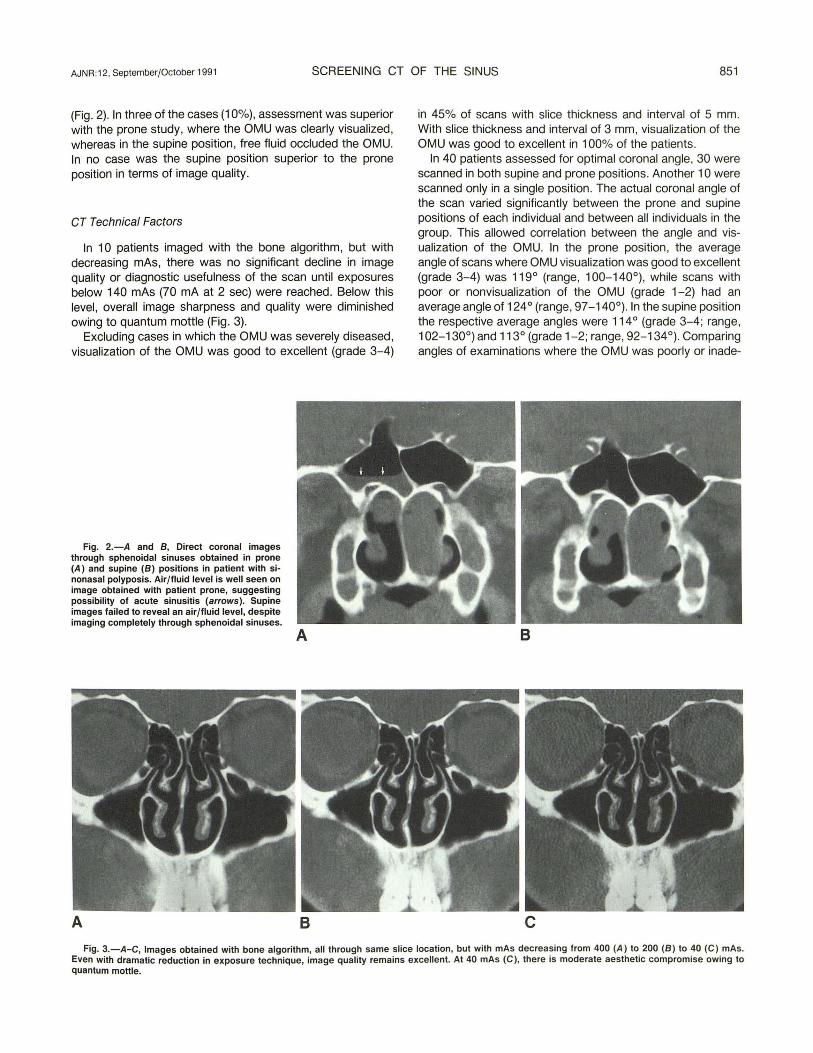
(Fig. 2). In three of the cases (1 0%), assessment was superior with the prone study, where the OMU was clearly visualized, whereas in the supine position, free fluid occluded the OMU. In no case was the supine position superior to the prone position in terms of image quality.
CT Technical Factors
In 10 patients imaged with the bone algorithm, but with decreasing mAs, there was no significant decline in image quality or diagnostic usefulness of the scan until exposures below 140 mAs (70 mA at 2 sec) were reached. Below this level, overall image sharpness and quality were diminished owing to quantum mottle (Fig. 3).
Excluding cases in which the OMU was severely diseased, visualization of the OMU was good to excellent (grade 3-4)
Fig. 2.-A and 8, Direct coronal images through sphenoidal sinuses obtained in prone (A) and supine (8) positions in patient with sinonasal polyposis. Air/fluid level is well seen on image obtained with patient prone, suggesting possibility of acute sinusitis (arrows). Supine images failed to reveal an air/fluid level, despite imaging completely through sphenoidal sinuses.
A
A
B
in 45% of scans with slice thickness and interval of 5 mm. With slice thickness and interval of 3 mm, visualization of the OMU was good to excellent in 1 00% of the patients.
In 40 patients assessed for optimal coronal angle, 30 were scanned in both supine and prone positions. Another 1 0 were scanned only in a single position. The actual coronal angle of the scan varied significantly between the prone and supine positions of each individual and between all individuals in the group. This allowed correlation between the angle and visualization of the OMU. In the prone position, the average angle of scans where OMU visualization was good to excellent (grade 3-4) was 119° (range, 100-140°), while scans with poor or nonvisualization of the OMU (grade 1-2) had an average angle of 124 o (range, 97 -140°). In the supine position the respective average angles were 114 o (grade 3-4; range, 1 02-130°) and 113° (grade 1-2; range, 92-134°). Comparing angles of examinations where the OMU was poorly or inade-
B
c Fig. 3.-A-C, Images obtained with bone algorithm, all through same slice location, but with mAs decreasing from 400 (A) to 200 (8) to 40 (C) mAs.
Even with dramatic reduction in exposure technique, image quality remains excellent. At 40 mAs (C), there is moderate aesthetic compromise owing to quantum mottle.

852 BABBEL ET AL. AJNR :12, September/October 1991
quately visualized with examinations where visualization was good to excellent, there was no statistically significant range of angles that resulted in consistently optimal visualization of the OMU.
Data Display
Of the five cases of noncomplicated inflammatory sinonasal disease filmed at three different window widths and levels (soft tissue, bone, and intermediate), none demonstrated significant disease on either bone or soft-tissue images that was not apparent on intermediate images. Image quality was excellent on the intermediate images, allowing superb visualization of bone and soft-tissue anatomy (Fig . 4).
Discussion
The results of our investigation enable delineation of a preendoscopic screening CT protocol with optimal techniques. The goals of a cost-effective, expeditious, low-radiation-dose preendoscopic examination are best attained when these techniques are used in performing the examination. Our recommendations and their justifications follow.
Patient Preparation
One of the goals of the screening CT examination of the sinuses is to define nonreversible disease; that is, the disease component that has not responded to conservative medical therapy and requires endoscopic sinonasal surgery [1-3]. Zinreich et al. [2] emphasized the importance of proper prescan patient preparation to eliminate as much reversible disease as possible. This can be accomplished by several measures, including administration of a course of antibiotics prior to the scan, thus eliminating any acute sinusitis component [2] ; use of a sympathomimetic nasal spray 15 min before the scan in association with vigorous nose blowing immediately
A 8
prior to scanning to eliminate reversible nasal congestion and mucous; continued use of previously prescribed oral antihistamines and decongestants to eliminate nasal congestion and mucous; and consideration of administration of a short course of steroids in cases of nasal polyposis or other suspected allergic sinonasal disease. Although objective data documenting the necessity of prescan preparation are difficult to obtain, our experience suggests that use of these techniques allows optimal delineation of the chronic, nonreversible disease component (Fig. 5).
CT Technique
It has been suggested that the total amount of radiation (mAs) used in screening CT of the sinuses could be significantly reduced if a bone algorithm were used (Akiya F, personal communication). Advantages of reducing exposure techniques include decreased radiation exposure to the patient, extended CT tube life, and reduced expense. On the basis of the results of our study, we have reduced our exposure factor from 420 mAs (140 mA at 3 sec) to 200 mAs (1 00 mA at 2 sec) without image compromise.
Delineation of the individual components of the OMU is critical to the correct interpretation of inflammatory disease of the nose and sinuses. Direct coronal CT imaging with the patient in the prone position affords an optimal view of these components, with dependent fluid layering away from the OMU. Supplemental coronal scans with the patient supine occasionally are helpful for complete pathologic characterization. Coronal imaging with the patient supine can also be used for the rare patient who is unable to assume the prone position.
Scan angle might at first seem important to the optimal definition of the structures of the OMU. However, the OMU structures are seen well with a variety of coronal and oblique coronal planes. An optimal scan angle for visualization of the
c Fig. 4.-A- C, Single slice, obtained with bone algorithm, photographed at three different window levels and widths, respectively: 450 and 50 H (A),
2000 and 250 H (8), and 4000 and 1100 H (C). Note how even subtle soft tissues remain clearly visible on intermediate images (arrow), yet bone detail is also v1suahzed accurately (8).
AJNR:12, September/October 1991 SCREENING CT OF THE SINUS 853
A B C
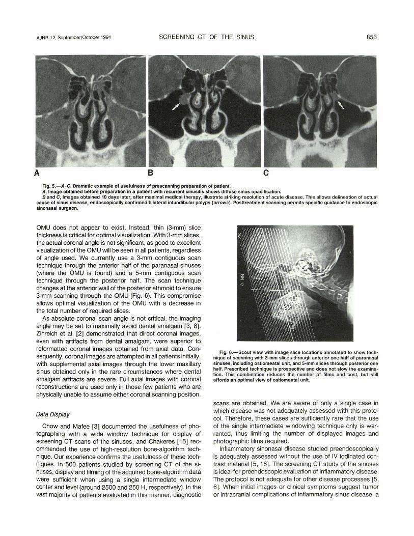
Fig. 5.-A-C, Dramatic example of usefulness of prescanning preparation of patient. A, Image obtained before preparation in a patient with recurrent sinusitis shows diffuse sinus opacification. 8 and C, Images obtained 10 days later, after maximal medical therapy, illustrate striking resolution of acute disease. This allows delineation of actual
cause of sinus disease, endoscopically confirmed bilateral infundibular polyps (arrows). Posttreatment scanning permits specific guidance to endoscopic sinonasal surgeon.
OMU does not appear to exist. Instead, thin (3-mm) slice thickness is critical for optimal visualization . With 3-mm slices, the actual coronal angle is not significant, as good to excellent visualization of the OMU will be seen in all patients, regardless of angle used. We currently use a 3-mm contiguous scan technique through the anterior half of the paranasal sinuses (where the OMU is found) and a 5-mm contiguous scan technique through the posterior half. The scan technique changes at the anterior wall of the posterior ethmoid to ensure 3-mm scanning through the OMU (Fig. 6). This compromise allows optimal visualization of the OMU with a decrease in the total number of required slices.
As absolute coronal scan angle is not critical, the imaging angle may be set to maximally avoid dental amalgam [3, 8] . Zinreich et al. [2] demonstrated that direct coronal images, even with artifacts from dental amalgam, were superior to reformatted coronal images obtained from axial data. Consequently, coronal images are attempted in all patients initially, with supplemental axial images through the lower maxillary sinus obtained only in the rare circumstances where dental amalgam artifacts are severe. Full axial images with coronal reconstructions are used only in those few patients who are physically unable to assume either coronal scanning position.
Data Display
Chow and Mafee [3] documented the usefulness of photographing with a wide window technique for display of screening CT scans of the sinuses, and Chakeres [15] recommended the use of high-resolution bone-algorithm technique. Our experience confirms the usefulness of these techniques. In 500 patients studied by screening CT of the sinuses, display and filming of the acquired bone-algorithm data were sufficient when using a single intermediate window center and level (around 2500 and 250 H, respectively). In the vast majority of patients evaluated in this manner, diagnostic
Fig. 6.-Scout view with image slice locations annotated to show technique of scanning with 3-mm slices through anterior one half of paranasal sinuses, including ostiomeatal unit, and 5-mm slices through posterior one half. Prescribed technique is prospective and does not slow the examination. This combination reduces the number of films and cost, but still affords an optimal view of ostiomeatal unit.
scans are obtained. We are aware of only a single case in which disease was not adequately assessed with this protocol. Therefore, these cases are sufficiently rare that the use of the single intermediate windowing technique only is warranted, thus limiting the number of displayed images and photographic films required.
Inflammatory sinonasal disease studied preendoscopically is adequately assessed without the use of IV iodinated contrast material [5 , 16]. The screening CT study of the sinuses is ideal for preendoscopic evaluation of inflammatory disease. The protocol is not adequate for other disease processes [5, 6]. When initial images or clinical symptoms suggest tumor or intracranial complications of inflammatory sinus disease, a

854 BABBEL ET AL. AJNR:12, September/October 1991
TABLE 1: Protocol and Technique for Screening CT Evaluation of the Sinuses
Protocol
Patient preparation
CT technique Patient position
Gantry angle
Scan extent
Section thickness
kVp mAs (dose per slice)
Imaging algorithm
Photography
Contrast material
Description
Key for eliminating reversible disease. Permits better delineation of anatomy and definition of chronic disease requiring endoscopic surgery. Preparation includes (1) completion of course of antibiotics; (2) continuation of oral antihistamines and decongestants; (3) sympathomimetic nasal spray 15 min before examination, followed by vigorous nose blowing; and (4) consideration of steroid use in allergic patients.
Coronal imaging with patient in prone position with head hyperextended, resting on chin (keeps free fluid out of infundibulum). In patients unable to maintain prone position or when images obtained in prone position are inconclusive, the supine position with head hyperextended over the edge of the table is used for coronal imaging .
Perpendicular to hard palate to obtain direct coronal images; actual coronal angle is not critical.
Posterior margin of sphenoid sinus to anterior margin of frontal sinus.
Posterior one half (posterior margin of sphenoid sinus to anterior margin of posterior ethmoid) scanned with 5-mm contiguous slices. Anterior one half (anterior margin of posterior ethmoid to anterior margin of frontal sinus) scanned with 3-mm contiguous slices; anterior thin sections are critical to ensure optimal visualization of ostiomeatal unit.
120 200 (1 00 mA x 2-sec scan time); low
exposure can be used because of bone-algorithm technique with filming at intermediate window.
Bone (maximum-edge-enhancement program).
Window width: 2000-2500 H; window center: 100-300 H. Both bone and soft tissues are visualized on the single set of images.
Not used.
Note.-These recommendations are for the screening CT examination of the sinuses in evaluating inflammatory disease. In cases of noninflammatory disease, or in cases of extensive or complicated inflammatory disease, a full CT examination of the sinuses should be considered. This includes IV contrast material with axial and coronal images and multiple filming techniques.
full sinonasal CT protocol is indicated. This includes use of IV contrast material, axial and coronal imaging of the sinuses and intracranial structures, and photography to emphasize both soft-tissue and bone detail.
In conclusion, our recommended approach to the screening CT examination of the sinuses is summarized in Table 1. With these guidelines, maximal diagnostic information is obtained with reduced cost and radiation exposure to the patient. In addition, a clear presurgical picture of the irreversible inflammatory changes of the sinonasal region is provided to the endoscopic surgeon.
REFERENCES
1. Kennedy DW, Zinreich SJ , Rosenbaum AE, Johns ME. Functional endoscopic sinus surgery. Arch Otolaryngol Head Neck Surg 1985;111 : 576-582
2. Zinreich SJ, Kennedy DW, Rosenbaum SE, et al. Paranasal sinuses: CT imaging requirements for endoscopic surgery. Radiology 1987;163: 769-775
3. Chow JM, Malee MF. Radiologic assessment preoperative to endoscopic sinus surgery. Otolaryngol Clin North Am 1989;22(4):691-701
4. Schatz CJ, Becker TS. Normal CT anatomy of the paranasal sinuses. Radio/ Clin North Am 1984;22(1):107-118
5. Pollei SR, Harnsberger HR. The radiologic evaluation of the sinonasal region. Postgrad Radio/1989;9:242-264
6. Scm PM. CT of the paranasal sinuses. Neuroradiology 1985;27:189-201 7. Unger JM, Shaffer K, Duncavage JA. Computed tomography in nasal and
paranasal sinus disease. Laryngoscope 1984;94:1319-1324 8. Sataloff RT, Grossman CB, Gonzales C, Naheedy MH. Computed tomog
raphy of the face and paranasal sinuses: Part I. Normal anatomy. Head Neck 1984;7: 110-122
9. McAlister WH, Lusk R, Muntz HR. Comparison of plain radiographs and coronal CT scans in infants and children with recurrent sinusitis. AJR 1989;153:1259-1264
10. Harnsberger HR. The sinuses and nose. In: Harnsberger HR , ed. Handbooks in radiology, head and neck imaging. Chicago: Year Book Medical, 1990:377-419
11. Drettner B. The obstructed maxillary ostium. Rhinology 1967;51 :100-104 12. Stammberger H. Endoscopic endonasal surgery-concepts in treatment
of recurring rhinosinusitis . I. Anatomic and pathophysiologic considerations. Otolaryngol Head Neck Surg 1986;94(2): 143-146
13. Stammberger H. Endoscopic endonasal surgery-concepts in treatment of recurring rhinosinusitis. II. Surgical technique. Otolaryngol Head Neck Surg 1986;94(2):147-156
14. Proctor DF. The mucociliary system. In: Proctor DF, Andersen IHP, eds. The nose: upper airway physiology and the atmospheric environment. New York: Elsevier, 1982:245-278
15. Chakeres DW. Computed tomography of the ethmoid sinuses. Otolaryngol C/in North Am 1985;18(1):29-42
16. Rice DH . Basic surgical techniques and variations of endoscopic sinus surgery. Otolaryngol Clin North Am 1989;22(4):713-726
The reader's attention is directed to the commentary on this article, which appears on the following pages.