Embed Size (px)
Citation preview
Review ArticleVolume 3 Issue 5 - November 2016DOI: 10.19080/ADOH.2016.02.555602
Adv Dent & Oral Health Copyright © All rights are reserved by Ahmed Abdelfattah
Oral Health, Diseases, Examination, Diagnosis, Treatment Plan
&Mouth Preparation
Ahmed Abdelfattah* Professor of Prosthodontics, Cairo University & Suez canal University, Saudi Arabia
Submission: : October 22, 2016; Published: November 30, 2016
*Corresponding author: Ahmed Abdelfattah, Professor of Prosthodontics, Professor of postgraduate research, consultant of prosthodontics, Cairo University & Suez canal University, King Fahd General Hospital, Dental Center; Jeddah, K.S.A., Saudi Arabia, Tel: ;
; Email:
Abstract
This literature review will organize dentist mind for ideal examination & diagnosis way to reach the best way for treatment plan & mouth preparation to achieve the goal of best treatment for the patient.
Adv Dent & Oral Health 3(5) : ADOH.MS.ID.555602 (2016)
IntroductionThe oral cavity composed of:
a. Hard tissues as: Bones of the upper jaw (maxilla ) & lower jaw ( mandible ), bone of the palate, and teeth.
b. Soft tissues as: Periodontium, soft palate, tongue, floor of the mouth, cheeks & lips.
The most common diseases always affect the teeth and the periodontium.
The Periodontium is composed of : Gingiva: The normal gingiva is firm and pink in color and
composed of :
a) Free gingival: It is the movable part of the gingiva that extent from the gingival crest to the bottom of the gingival sulcus (normal sulcus depth is 1-2 mm).
b) Attached gingival: It is the stippled part of the gingiva.
Periodontal ligaments: The periodontal ligaments are arranged in 5 bundles in different directions. It is responsible for the nutrition and attachment of the teeth in addition to act as foundation for the prosthesis.
Alveolar bone crest: The alveolar bone crest is 2 mm away from the gingival sulcus. The periodontium is a connective tissue structure attached to the periosteum of both the maxilla & the mandible that serves to anchor the teeth in the maxillary and the
mandibular alveolar processes. It provides attachment & support, nutrition, synthesis & resorption, and mechanoreception.
The main element of the periodontium is the periodontal ligament (PDL), which consists of collagenous fibers (Sharpey’s fibers), embedded in bone and cementum, and giving support to the tooth in function.
There are five principal fiber groups in the PDL that traverse the space between the tooth root & alveolar bone, providing attachment and support :-
a. Trans-septal fibers: extending interproximally between adjacent teeth; their ends embedded in cementum.
b. Alveolar crest fibers: beginning just apical to the epithelial attachment and extending from cementum to the alveolar crest.
c. Horizontal fibers: coursing at right angles from cementum to the alveolar bone.
d. Oblique fibers: extending in an oblique direction apically, attaching cementum to the alveolar bone. They are the most numerous fibers.
e. Apical fibers: radiating from cementum into the alveolar bone at the apex of the root.
There are also smaller irregularly arranged fibers interspersed between the principal fiber groups. Cellular elements found in PDL included - fibroblast [the main synthetic cell, producing collagen & other proteoglycans], cementoblasts
001
How to cite this article: Ahmed A. Oral Health, Diseases, Examination, Diagnosis, Treatment Plan & Mouth Preparation. Adv Dent & Oral Health. 2016; 3(5): 555602. DOI: 10.19080/ADOH.2016.02.555602002
Advances in Dentistry & Oral Health
& cementoclasts, osteoblasts & osteoclasts, and mast cells & epithelial rests [playing a role in pathologic conditions of the periodontium].
At the base of the gingival sulcus is the epithelium - tooth interface named dentogingival junction (DGJ). This structural relationship between hard and soft tissues is unique in the body, which anchor the epithelial cells to the enamel and cemental surfaces. The depth of the sulcus is variable in healthy individuals, averaging 1.8 mm. In general, the shallower it is, the more likely is the gingiva to be in a state of health.
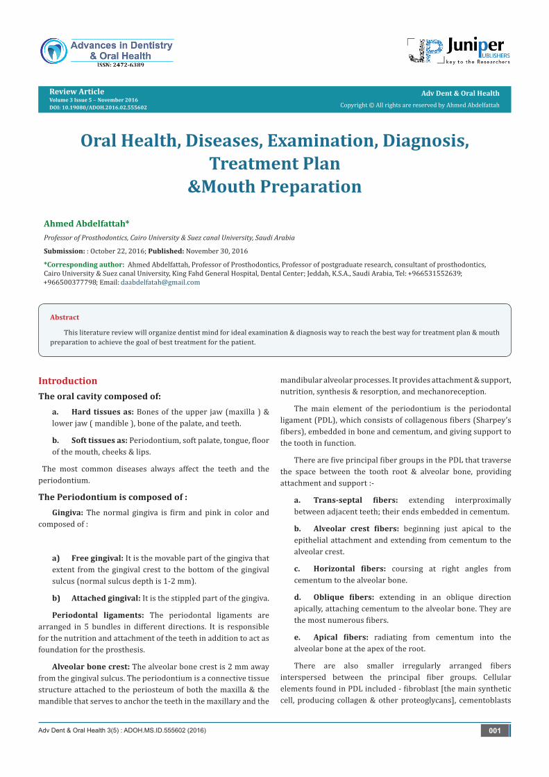
The Tooth is composed of : 1. Enamel: The hardest outer part of the tooth composed from enamel prisms embedded in a matrix which bonds the prisms together.
2. Dentin: The resilient part of the tooth which contains tiny tubes called dentinal tubules through which pass nerve filaments to collect at the junction between enamel & dentin ( dentino- enamel junction ) forming the nerve plexus.
3. Pulp: The soft connective tissues contain blood vessels and nerves, and fill the inner space of the tooth which called pulp chamber & root canal. It is responsible for nutrition of the tooth & sensation.
4. Cementum: The outer layer of the root covering the dentin. It is responsible for attaching the 5 bundles of PDL from one end to insure tooth anchor (Figure 1).
Figure 1: Normal tooth.
Common Oral Diseases Tooth decay
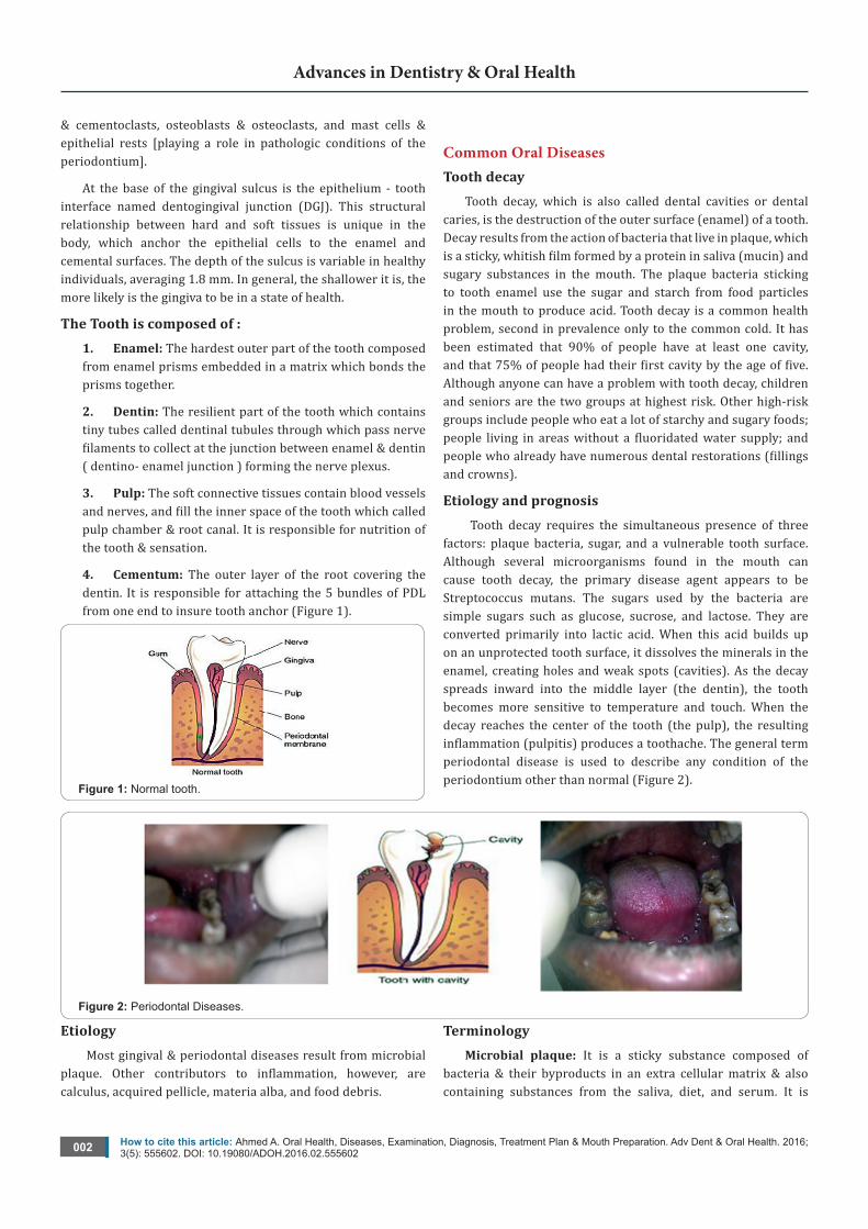
Tooth decay, which is also called dental cavities or dental caries, is the destruction of the outer surface (enamel) of a tooth. Decay results from the action of bacteria that live in plaque, which is a sticky, whitish film formed by a protein in saliva (mucin) and sugary substances in the mouth. The plaque bacteria sticking to tooth enamel use the sugar and starch from food particles in the mouth to produce acid. Tooth decay is a common health problem, second in prevalence only to the common cold. It has been estimated that 90% of people have at least one cavity, and that 75% of people had their first cavity by the age of five. Although anyone can have a problem with tooth decay, children and seniors are the two groups at highest risk. Other high-risk groups include people who eat a lot of starchy and sugary foods; people living in areas without a fluoridated water supply; and people who already have numerous dental restorations (fillings and crowns).
Etiology and prognosis Tooth decay requires the simultaneous presence of three
factors: plaque bacteria, sugar, and a vulnerable tooth surface. Although several microorganisms found in the mouth can cause tooth decay, the primary disease agent appears to be Streptococcus mutans. The sugars used by the bacteria are simple sugars such as glucose, sucrose, and lactose. They are converted primarily into lactic acid. When this acid builds up on an unprotected tooth surface, it dissolves the minerals in the enamel, creating holes and weak spots (cavities). As the decay spreads inward into the middle layer (the dentin), the tooth becomes more sensitive to temperature and touch. When the decay reaches the center of the tooth (the pulp), the resulting inflammation (pulpitis) produces a toothache. The general term periodontal disease is used to describe any condition of the periodontium other than normal (Figure 2).
Figure 2: Periodontal Diseases.
Etiology Most gingival & periodontal diseases result from microbial
plaque. Other contributors to inflammation, however, are calculus, acquired pellicle, materia alba, and food debris.
Terminology Microbial plaque: It is a sticky substance composed of
bacteria & their byproducts in an extra cellular matrix & also containing substances from the saliva, diet, and serum. It is
How to cite this article: Ahmed A. Oral Health, Diseases, Examination, Diagnosis, Treatment Plan & Mouth Preparation. Adv Dent & Oral Health. 2016; 3(5): 555602. DOI: 10.19080/ADOH.2016.02.555602003
Advances in Dentistry & Oral Health
basically a product of the growth of bacterial colonies & is the initiating factor in gingival & periodontal disease.
Calculus: It is a chalky or dark deposit attached to the tooth structure. It is essentially microbial plaque that has undergone mineralization with the passage of time.
Acquired pellicle: Pellicle is a thin brown or gray film of salivary proteins that develops on teeth after they have been cleaned. It frequently forms the interface between the tooth surface and dental deposits.
Materia alba: It is a white coating composed of microorganisms, dead epithelial cells, and leukocytes that is loosely adherent to the tooth. It can be removed from the tooth surface by water spray or by rinsing.
Pathogenesis: Sequence of events in the development of a gingivitis - periodontitis lesion is a very complex. It involves not only local phenomena in the gingiva, PDL, tooth surface, and alveolar bone, but also a number of complex host response mechanisms.
The chronic plaque induced lesion has been investigated & analyzed, so, divided into initial, early, established, and advanced stages.
Tooth Loss: Tooth lost may be due to:
A. Trauma: Trauma due to accident may cause:
1. Avulsion of tooth from its socket.
2. Fracture of the tooth crown with part of the root.
3. Internal fracture of the root with separation of the fractured part.
4. Alveolar bone fracture with gingival trauma and exposure of the root to oral cavity.
B. Unrestorability: Unrestorability of the tooth may be due to:
1. Mutilation from caries with complete destruction of the crown and part of the root.
2. Root caries with fraction involvement.
3. Failure to do R.C.T for the tooth.
C. Occlusion & path of insertion problems: Severe tilting of the tooth which causing problem with occlusion & fabrication of other prosthesis.
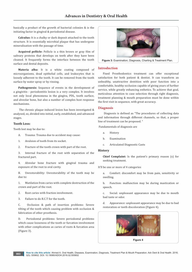
D. Periodontal problems: Severe periodontal problems which cause looseness of the tooth or furcation involvement with other complications as caries of roots & furcation area (Figure 3).
Figure 3: Examination, Diagnosis, Charting & Treatment Plan.
Introduction Fixed Prosthodontics treatment can offer exceptional
satisfaction for both patient & dentist. It can transform an unhealthy, unattractive dentition with poor function into a comfortable, healthy occlusion capable of giving years of further service, while greatly enhancing esthetics. To achieve that goal, meticulous attention to case selection through right diagnosis, treatment planning & mouth preparation must be done within the first visit in sequence, with great accuracy.
Diagnosis Diagnosis is defined as “The procedures of collecting data
and information through different channels, so that, a proper line of treatment can be proposed. “
Fundamentals of diagnosis are
a. History
b. Examination
c. Articulated Diagnostic Casts
History
Chief Complaint: Is the patient’s primary reason (s) for seeking treatment.
It’ll be one or more of 4 categories
a. Comfort: discomfort may be from pain, sensitivity or swelling.
b. Function: malfunction may be during mastication or speech.
c. Social: unpleasant appearance may be due to mouth bad taste or odor.
d. Appearance: unpleasant appearance may be due to bad restoration or teeth discoloration (Figure 4).
Figure 4
How to cite this article: Ahmed A. Oral Health, Diseases, Examination, Diagnosis, Treatment Plan & Mouth Preparation. Adv Dent & Oral Health. 2016; 3(5): 555602. DOI: 10.19080/ADOH.2016.02.555602004
Advances in Dentistry & Oral Health
Personal Details: Name, age, sex, address, education, occupation, marital status, children, and telephone number.
Medical History: Could be achieved under 4 categories :
A. Conditions affecting the treatment methodology, e.g., any disorders that necessitate the use of antibiotic premedication, any use of steroids or anticoagulants, & previous allergic response to medication or dental materials.
B. Conditions affecting the treatment plan, e.g., previous radiation therapy, hemorrhagic disorders, extremity of age, and terminal illness.
C. Systemic conditions with oral manifestations, e.g., + Periodontitis= diabetes, menopause, pregnancy, or the use of anticonvulsant drugs.
+ Erosion of teeth= stomach acid regurgitation in case of hiatal hernia, bulimia, or anorexia nervosa. + T.M.J. disorders or reduced salivary flow= as side effect of certain drugs.
D. Possible risk factors to dentist & auxiliary personnel, e.g., patients who are suspected or confirmed carriers of hepatitis, AIDs, or syphilis.
Dental History: Includes :1. Periodontal history and oral hygiene
2. Restorative history: filling and F.P.Ds
3. Endodontic history
4. Orthodontic history
5. Removable Prosthodontic history
6. Oral surgery history
7. Radiographic history
8. T.M.J. dysfunction history
Methods of Clinical Examination General examination
General appearance, skin color, and vital signs as respiration, pulse, temperature, and blood pressure.
Extra oral examination: special attention to facial asymmetry = may hint at serious conditions.
Cervical lymph nodes are palpated, as are the T.M.J., & the muscles of mastication. Lips position during smiling is critical in treatment planning.
Intra oral examination: Reveals considerable information concerning the condition of the soft tissues, teeth, and supporting structures. Lips tongue, floor of the mouth, vestibules, cheeks, and the hard & soft palates are examined; and any abnormalities are noted.
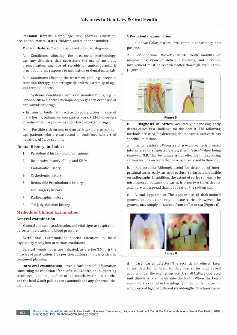
A.Periodontal examination: 1. Gingiva: Color, texture, size, contour, consistency, and position.
2. Periodontium: Pocket’s depth, tooth mobility or malpositions, open or deficient contacts, and furcation involvement must be recorded after thorough examination (Figure 5).
Figure 5 B. Diagnosis of caries: Accurately diagnosing early dental caries is a challenge for the dentist. The following methods are used for detecting dental caries, and each has specific limitations:-
a. Dental explorer: When a sharp explorer tip is pressed into an area of suspected caries, it will “stick” when being removed. N.B. This technique is not effective in diagnosing carious lesions on teeth that have been exposed to fluoride.
b. Radiographs: Although useful for detection of inter-proximal caries, early caries on occlusal surfaces is not visible on radiographs. In addition, the extent of caries can easily be misdiagnosed because the caries is often two times deeper and more widespread than it appear on the radiograph.
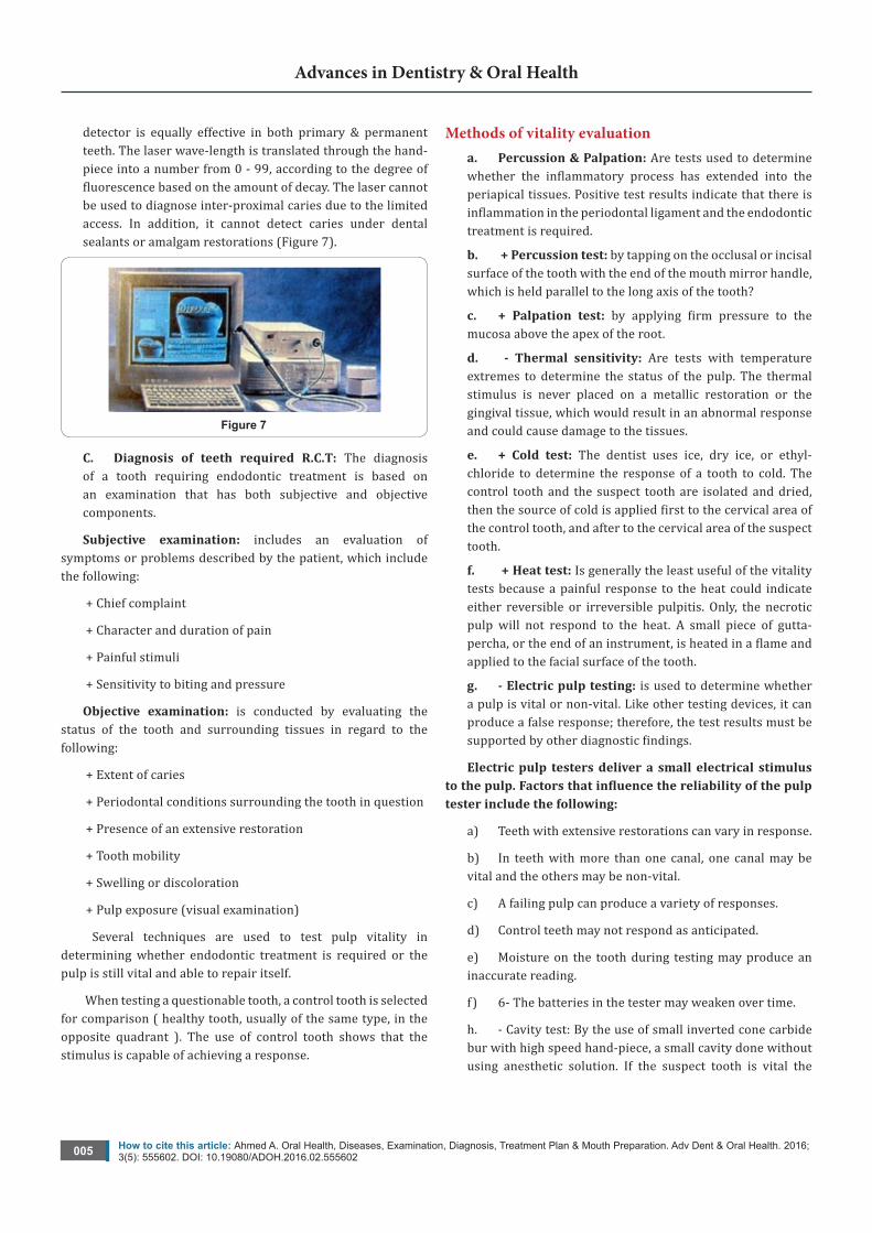
c. Visual appearance: The appearance of dark-stained grooves in the teeth may indicate caries. However, the grooves may simply be stained from coffee or tea (Figure 6).
Figure 6
d. Laser caries detector: The recently introduced laser caries detector is used to diagnose caries and reveal activity under the enamel surface. A small battery-operated unit directs a laser beam into the tooth. When the beam encounters a change in the integrity of the tooth, it gives off a fluorescent light of different wave-lengths. The laser caries
How to cite this article: Ahmed A. Oral Health, Diseases, Examination, Diagnosis, Treatment Plan & Mouth Preparation. Adv Dent & Oral Health. 2016; 3(5): 555602. DOI: 10.19080/ADOH.2016.02.555602005
Advances in Dentistry & Oral Health
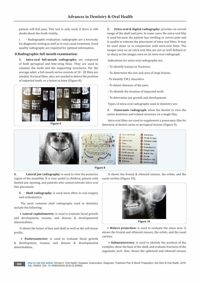
detector is equally effective in both primary & permanent teeth. The laser wave-length is translated through the hand-piece into a number from 0 - 99, according to the degree of fluorescence based on the amount of decay. The laser cannot be used to diagnose inter-proximal caries due to the limited access. In addition, it cannot detect caries under dental sealants or amalgam restorations (Figure 7).
Figure 7
C. Diagnosis of teeth required R.C.T: The diagnosis of a tooth requiring endodontic treatment is based on an examination that has both subjective and objective components.
Subjective examination: includes an evaluation of symptoms or problems described by the patient, which include the following:
+ Chief complaint
+ Character and duration of pain
+ Painful stimuli
+ Sensitivity to biting and pressure
Objective examination: is conducted by evaluating the status of the tooth and surrounding tissues in regard to the following:
+ Extent of caries
+ Periodontal conditions surrounding the tooth in question
+ Presence of an extensive restoration
+ Tooth mobility
+ Swelling or discoloration
+ Pulp exposure (visual examination)
Several techniques are used to test pulp vitality in determining whether endodontic treatment is required or the pulp is still vital and able to repair itself.
When testing a questionable tooth, a control tooth is selected for comparison ( healthy tooth, usually of the same type, in the opposite quadrant ). The use of control tooth shows that the stimulus is capable of achieving a response.
Methods of vitality evaluationa. Percussion & Palpation: Are tests used to determine whether the inflammatory process has extended into the periapical tissues. Positive test results indicate that there is inflammation in the periodontal ligament and the endodontic treatment is required.
b. + Percussion test: by tapping on the occlusal or incisal surface of the tooth with the end of the mouth mirror handle, which is held parallel to the long axis of the tooth?
c. + Palpation test: by applying firm pressure to the mucosa above the apex of the root.
d. - Thermal sensitivity: Are tests with temperature extremes to determine the status of the pulp. The thermal stimulus is never placed on a metallic restoration or the gingival tissue, which would result in an abnormal response and could cause damage to the tissues.
e. + Cold test: The dentist uses ice, dry ice, or ethyl-chloride to determine the response of a tooth to cold. The control tooth and the suspect tooth are isolated and dried, then the source of cold is applied first to the cervical area of the control tooth, and after to the cervical area of the suspect tooth.
f. + Heat test: Is generally the least useful of the vitality tests because a painful response to the heat could indicate either reversible or irreversible pulpitis. Only, the necrotic pulp will not respond to the heat. A small piece of gutta-percha, or the end of an instrument, is heated in a flame and applied to the facial surface of the tooth.
g. - Electric pulp testing: is used to determine whether a pulp is vital or non-vital. Like other testing devices, it can produce a false response; therefore, the test results must be supported by other diagnostic findings.
Electric pulp testers deliver a small electrical stimulus tothepulp.Factorsthatinfluencethereliabilityofthepulptester include the following:
a) Teeth with extensive restorations can vary in response.
b) In teeth with more than one canal, one canal may be vital and the others may be non-vital.
c) A failing pulp can produce a variety of responses.
d) Control teeth may not respond as anticipated.
e) Moisture on the tooth during testing may produce an inaccurate reading.
f) 6- The batteries in the tester may weaken over time.
h. - Cavity test: By the use of small inverted cone carbide bur with high speed hand-piece, a small cavity done without using anesthetic solution. If the suspect tooth is vital the
How to cite this article: Ahmed A. Oral Health, Diseases, Examination, Diagnosis, Treatment Plan & Mouth Preparation. Adv Dent & Oral Health. 2016; 3(5): 555602. DOI: 10.19080/ADOH.2016.02.555602006
Advances in Dentistry & Oral Health
patient will feel pain. This test is only used, if there is still doubt about the tooth vitality.
i. - Radiographic evaluation: radiographs are a necessity for diagnostic testing as well as in root canal treatment. Good quality radiographs are required for optimal information.
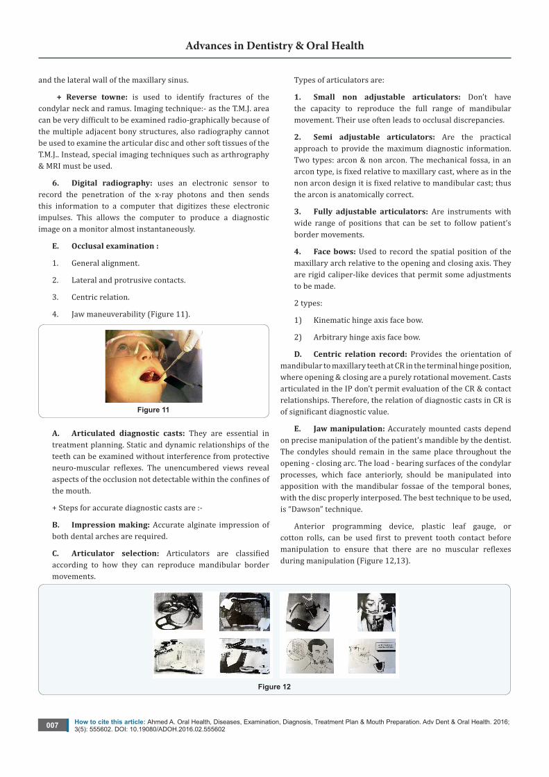
D.Radiographic full-mouth examination: 1. Intra-oral full-mouth radiographs: are composed of both periapical and bite-wing films. They are used to examine the teeth and the supporting structures. For the average adult, a full-mouth series consists of 18 - 20 films are needed. Occlusal films, also, are needed to detect the position of impacted teeth, or a lesion in bone (Figure 8).
Figure 8
2. Extra-oral & digital radiography: provides an overall image of the skull and jaws. In some cases the extra-oral film is used because the patient has swelling or severe pain and is unable to tolerate the placement of intra-oral films. It may be used alone or in conjunction with intra-oral films. The images seen on an extra-oral film are not as well defined or as sharp as the images seen on an intra-oral radiograph.
Indications for extra-oral radiographs are:
- To identify trauma or fractures.
- To determine the size and area of large lesions.
- To identify T.M.J. disorders.
- To detect diseases of the jaws.
- To identify the location of impacted teeth.
- To determine jaw growth and development.
Types of extra-oral radiographs used in dentistry are:
3. Panoramic radiograph: allow the dentist to view the entire dentition and related structure on a single film.
Intra-oral films are used to supplement a panoramic film for detection of dental caries or periapical lesions (Figure 9).
Figure 9
4. Lateral jaw radiography: is used to view the posterior region of the mandible. It is very useful in children, patient with limited jaw opening, and patients who cannot tolerate intra-oral film placement.
5. Skull radiography: is used most often in oral surgery and orthodontics.
The most common skull radiographs used in dentistry include the following:
+ Lateral cephalometric: is used to evaluate facial growth and development, trauma, and disease & developmental abnormalities.
It shows the bones of face and skull as well as the soft tissue profile.
+ Posteroanterior: is used to evaluate facial growth & development, trauma, and disease & developmental abnormalities.
It shows the frontal & ethmoid sinuses, the orbits, and the nasal cavities (Figure 10).
Figure 10
+ Waters projection: is used to evaluate the sinus area. It shows the frontal and ethmoid sinuses, the orbits, and the nasal cavities.
+ Submentovertex: is used to identify the position of the condyles, show the base of the skull, and evaluate fractures of the zygomatic arch. Also, shows the sphenoid and ethmoid sinuses
How to cite this article: Ahmed A. Oral Health, Diseases, Examination, Diagnosis, Treatment Plan & Mouth Preparation. Adv Dent & Oral Health. 2016; 3(5): 555602. DOI: 10.19080/ADOH.2016.02.555602007
Advances in Dentistry & Oral Health
and the lateral wall of the maxillary sinus.
+ Reverse towne: is used to identify fractures of the condylar neck and ramus. Imaging technique:- as the T.M.J. area can be very difficult to be examined radio-graphically because of the multiple adjacent bony structures, also radiography cannot be used to examine the articular disc and other soft tissues of the T.M.J.. Instead, special imaging techniques such as arthrography & MRI must be used.
6. Digital radiography: uses an electronic sensor to record the penetration of the x-ray photons and then sends this information to a computer that digitizes these electronic impulses. This allows the computer to produce a diagnostic image on a monitor almost instantaneously.
E. Occlusal examination :
1. General alignment.
2. Lateral and protrusive contacts.
3. Centric relation.
4. Jaw maneuverability (Figure 11).
Figure 11
A. Articulated diagnostic casts: They are essential in treatment planning. Static and dynamic relationships of the teeth can be examined without interference from protective neuro-muscular reflexes. The unencumbered views reveal aspects of the occlusion not detectable within the confines of the mouth.
+ Steps for accurate diagnostic casts are :-
B. Impression making: Accurate alginate impression of both dental arches are required.
C. Articulator selection: Articulators are classified according to how they can reproduce mandibular border movements.
Types of articulators are:
1. Small non adjustable articulators: Don’t have the capacity to reproduce the full range of mandibular movement. Their use often leads to occlusal discrepancies.
2. Semi adjustable articulators: Are the practical approach to provide the maximum diagnostic information. Two types: arcon & non arcon. The mechanical fossa, in an arcon type, is fixed relative to maxillary cast, where as in the non arcon design it is fixed relative to mandibular cast; thus the arcon is anatomically correct.
3. Fully adjustable articulators: Are instruments with wide range of positions that can be set to follow patient’s border movements.
4. Face bows: Used to record the spatial position of the maxillary arch relative to the opening and closing axis. They are rigid caliper-like devices that permit some adjustments to be made.
2 types:
1) Kinematic hinge axis face bow.
2) Arbitrary hinge axis face bow.
D. Centric relation record: Provides the orientation of mandibular to maxillary teeth at CR in the terminal hinge position, where opening & closing are a purely rotational movement. Casts articulated in the IP don’t permit evaluation of the CR & contact relationships. Therefore, the relation of diagnostic casts in CR is of significant diagnostic value.
E. Jaw manipulation: Accurately mounted casts depend on precise manipulation of the patient’s mandible by the dentist. The condyles should remain in the same place throughout the opening - closing arc. The load - bearing surfaces of the condylar processes, which face anteriorly, should be manipulated into apposition with the mandibular fossae of the temporal bones, with the disc properly interposed. The best technique to be used, is “Dawson” technique.
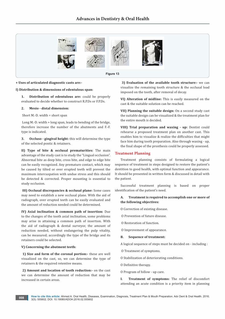
Anterior programming device, plastic leaf gauge, or cotton rolls, can be used first to prevent tooth contact before manipulation to ensure that there are no muscular reflexes during manipulation (Figure 12,13).
Figure 12
How to cite this article: Ahmed A. Oral Health, Diseases, Examination, Diagnosis, Treatment Plan & Mouth Preparation. Adv Dent & Oral Health. 2016; 3(5): 555602. DOI: 10.19080/ADOH.2016.02.555602008
Advances in Dentistry & Oral Health
Figure 13
+ Uses of articulated diagnostic casts are:-
I) Distribution & dimensions of edentulous span:
1. Distribution of edentulous are: could be properly evaluated to decide whether to construct R.P.Ds or F.P.Ds.
2. Mesio - distal dimension:
Short M.-D. width = short span
Long M.-D. width = long span, leads to bending of the bridge, therefore increase the number of the abutments and F.-F. type is indicated.
3. Occluso - gingival height: this will determine the type of the selected pontic & retainers.
II) Type of bite & occlusal prematurities: The main advantage of the study cast is to study the “Lingual occlusion”. Abnormal bite as deep bite, cross bite, and edge to edge bite can be easily recognized. Any premature contact, which may be caused by tilted or over erupted teeth will prevent the maximum intercuspation with undue stress and this should be detected & corrected. Proper mounting is essential to study occlusion.
III) Occlusal discrepancies & occlusal plane: Some cases may need to establish a new occlusal plane. With the aid of radiograph, over erupted teeth can be easily evaluated and the amount of reduction needed could be determined.
IV) Axial inclination & common path of insertion: Due to the changes of the tooth axial inclination, some problems may arise in attaining a common path of insertion. With the aid of radiograph & dental surveyor, the amount of reduction needed, without endangering the pulp vitality, can be measured; accordingly the type of the bridge and its retainers could be selected.
V) Concerning the abutment teeth:
1) Size and form of the coronal portion:- these are well visualized on the cast, so, we can determine the type of retainers & the required retentive means.
2) Amount and location of tooth reduction:- on the cast we can determine the amount of reduction that may be increased in certain areas.
3) Evaluation of the available tooth structure:- we can visualize the remaining tooth structure & the occlusal load imposed on the tooth, after removal of decay.
VI) Alteration of midline: This is easily measured on the cast & the suitable solution can be reached.
VII) Planning the suitable design: On a second study cast the suitable design can be visualized & the treatment plan for the entire mouth is decided.
VIII) Trial preparation and waxing - up: Dentist could rehearse a proposed treatment plan on another cast. This enables him to visualize & realize the difficulties that might face him during tooth preparation. Also through waxing - up, the final shape of the prosthesis could be properly assessed.
Treatment PlanningTreatment planning consists of formulating a logical
sequence of treatment in steps designed to restore the patient’s dentition to good health, with optimal function and appearance. It should be presented in written form & discussed in detail with the patient.
Successful treatment planning is based on proper identification of the patient’s need.
A. Treatment is required to accomplish one or more of the following objectives:
O Correction of existing disease.
O Prevention of future disease.
O Restoration of function.
O Improvement of appearance.
B. Sequence of treatment:
A logical sequence of steps must be decided on - including :
O Treatment of symptoms.
O Stabilization of deteriorating conditions.
O Definitive therapy.
O Program of follow - up care.
I. Treatment of symptoms: The relief of discomfort attending an acute condition is a priority item in planning
How to cite this article: Ahmed A. Oral Health, Diseases, Examination, Diagnosis, Treatment Plan & Mouth Preparation. Adv Dent & Oral Health. 2016; 3(5): 555602. DOI: 10.19080/ADOH.2016.02.555602009
Advances in Dentistry & Oral Health
treatment. A fully examination is neither desirable nor generally possible until the symptoms of the acute condition have been addressed. Also, urgent treatment of non acute problems such as a lost anterior crown, a broken porcelain veneer or fractured R.P.D., should receive priority attention.
II. Stabilization of deteriorating conditions: Such as dental caries or periodontal disease.
a. Replacement of defective restorations.
b. Removal of carious lesions.
c. Re contouring of over contoured prosthesis.
d. Removal of plaque & proper oral hygiene instructions.
III. Definitivetherapy: When the stabilization phase has been completed, successful elective long - term treatment aimed at promoting dental health, restoring function, and improving appearance can begin. Several therapeutic proposals may be applicable to a single patient. The advantages & disadvantages of each should be thoroughly explained to the patient, with a diagnostic casts and waxing - up used as guides. Usually oral surgical procedures are scheduled first, followed by periodontics, endodontics, orthodontics, fixed Prosthodontics, and finally removable Prosthodontics.
Oral surgery: The treatment plan should allow time for healing & ridge remodeling. All preprosthetic surgical procedures (e.g. ridge contouring) should be undertaken during the early phase of treatment.
Endodontics: Some endodontic treatment may have been accomplished as part of the relief of discomfort & stabilization of conditions. Elective endodontics may be needed to provide adequate space for a cast restoration or to provide retention for a badly damaged or worn tooth.
- Orthodontics: Minor orthodontic tooth movement is a common adjunct to fixed Prosthodontics, especially if tooth loss has been neglected & drifting has occurred.
- Fixed Prosthodontics: Is initiated only after the preceding modalities have been completed. This will permit modification of the original plan , as unforeseen difficulties should surface during treatment.
+ Occlusal adjustments: are often necessary before the initiation of fixed Prosthodontics. Where extensive F.P.D. is to be provided, an accurate & well-tolerated occlusal relationship may be obtainable only if a discrepancy between IP & CR is eliminated first.
+ Anterior restorations: are usually done first because they influence the border movements of the mandible & thus the shape of the occlusal surfaces of the posterior teeth.
+ Posterior restorations: it is often advantageous to restore opposing posterior segments at the same time. This permits
the development of an efficient occlusal scheme through the application of an additive wax technique.
+ Complex prosthetics: carefully planned treatment sequencing is particularly important when complex Prosthodontic treatments involving alteration of the vertical dimension or a combination of fixed & removable prosthesis are required. Two sets of diagnostic casts are accurately mounted, so, they can be precisely interchanged on the articulator. Definitive tooth preparation starts in one arch only, preserving the occlusal surfaces of the opposing arch to act as an essential reference for mounting the working cast. The definitive restorations are waxed against the diagnostically waxed cast, establishing optimal occlusion. When one arch has been completed, the opposing cast can be restored, achieving the predicted result.
- Removable Prosthodontics: Are the final procedures, but start to be planned for during fixed Prosthodontic treatment.
- Follow up: A specific program of follow up care and regular recall is an essential part of the treatment plan. The aim is to monitor dental health, identify the signs of disease early, and initiate prompt corrective measures as necessary.
IV.Clinical Tips:
Factors affecting the selection of prosthesis type:
(I) Biomechanical Considerations
1) The decision to remove a tooth: Is part of the treatment planning process & is made after the advantages & disadvantages associated with retention of the tooth have been assessed.
2) The edentulous span:
a- Distribution:
+ Cases with free end saddle will usually require a R.P.Ds.. Alternative treatment are:
- A distal fixture with implant.
- Cantilever bridge in selected cases.
+ Multiple edentulous spaces, though each of which may be restored with a fixed bridge, yet due to expenses & technical complexity, a R.P.Ds. May be used.
b- Length: All fixed bridges, long or short, possess a certain degree of bending when subjected to load; the longer the span, the greater the flexing.
Bending varies directly with the cube of the length & inversely with the cube of occluso-gingival thickness of the pontic considering other factors being equal.
+ Excessive flexing or bending under occlusal force may lead to:
- Fracture of porcelain veneer
- Connector breakage
How to cite this article: Ahmed A. Oral Health, Diseases, Examination, Diagnosis, Treatment Plan & Mouth Preparation. Adv Dent & Oral Health. 2016; 3(5): 555602. DOI: 10.19080/ADOH.2016.02.5556020010
Advances in Dentistry & Oral Health
- Retainer loosening
- An unfavorable soft tissue response
+ To minimize bending:
- Construct pontics & connectors of greater occluso-gingival dimensions
- Use an alloy of higher yield strength
+ Clinical considerations of the bridge flexing in treatment planning:
- The length of the edentulous area will affect the type of restoration to be suitable for replacing one or two missing teeth.
- Three posterior teeth is better to be replaced with R.P.D. as F.P.D. will be very questionable.
- Constructing a long span F.P.D. on short teeth is expected to have a very disappointing prognosis. + Alternative treatment modalities for long edentulous span:
- An implant supported F.P.D. {requires sufficient alveolar bone, broad flat ridge & favorable opposing occlusion}.
- Removable partial denture.
c- Arch form: The arch curvature affects the amount of stresses occurring in F.P.Ds constructed in the anterior segment, especially in the upper teeth.
In cases of pointed arch (V-shaped), pontics of F.P.D would lie far outside the inter abutment axis line, thus acting as lever arm producing torque movement on the supporting abutment. + Measures to be taken in bridge design: If the distance between the inter abutment axis & the pontic is increased = the force arm will be increased; this will need to increase the number of the abutments, to increase the resistance arm for this force.
(II) The Prospective Abutment: 1) The pulpal condition:
a- Vital sound tooth: the unrestored vital caries-free tooth is an ideal abutment as it facilitates conservative preparation for strong retentive restoration with good esthetics.
b- Carious tooth: after caries removal as well as all the undermined enamel, assessment of the remaining sound tooth structure & the pulpal condition should be performed. The existing situation would influence the line of treatment as following:
+ Selecting the most suitable type of restorative material & the necessary retentive means {e.g. pins, grooves or boxes}.
+ Doubtful pulpal condition or those teeth with pulpal capping should not be used as F.P.D abutments unless being endodontically treated.
c- Endodontically treated abutments: A perfectly
endodontically treated tooth (clinically & radio- graphically) can be used successfully as an abutment with a post & core for retention & strength.
+ Clinical consideration of endodontically treated abutments:
- As posts & cores are usually constructed to compensate for the lost coronal part, thus extreme care is needed to get sufficient retention from the post.
- Before deciding an endodontic treatment for a tooth to be used as abutment, it should be evaluated whether this tooth is restorable or not.
- In complex & expensive prosthesis whose success is dependent on an abutment that will require endodontic treatment, endodontic surgery or implants may be a better treatment choice.
2) Coronal variations and tooth alignment:
a- Over erupted teeth: to restore the dentition to complete function, free of interference, over- erupted teeth should be adjusted to the normal occlusal plane. In some situations intentional RCT may be necessary to permit enough shortening to correct the occlusal plane.
b- Short crowns: abutments with short clinical crown would create problems in constructing F.P.Ds. Careful selection of the bridge design, with suitable retentive retainers & types of pontics should be highly stressed on. Considerations in bridge design:
- F.-F. Bridge is the most indicated type.
- Full coverage retainer with additional retentive means are to be used.
- Establishing relative least convergence.
- Extend stump of preparation more cervically.
- To avoid excessive shortening = occlusal reduction to receive retainer with occlusal metal coverage. - Pontics & connectors should be of considerable occluso-gingival dimension to resist bending.
- Replacing missing 2 or 3 teeth in short occluso-gingival edentulous areas with R.P.D should be highly considered.
c- Mesially tilted 2nd molar: early loss of 1st mand. molar would create problems if the space is ignored, as mesial tilting of the 2nd molar. Constructing of F.P.D would face the problem of attaining a common path of insertion. Different treatment modalities:
1) Up-righting the tilted abutment orthodontically then normal F.-F bridge is considered the treatment of choice.
2) A proximal (mesial) ½ crown can be used as a retainer on the tilted abutment.
How to cite this article: Ahmed A. Oral Health, Diseases, Examination, Diagnosis, Treatment Plan & Mouth Preparation. Adv Dent & Oral Health. 2016; 3(5): 555602. DOI: 10.19080/ADOH.2016.02.5556020011
Advances in Dentistry & Oral Health
3) F.-S bridge with non rigid connector on the distal aspect of the premolar; so, the path of insertion of the bridge is parallel to the tilted molar.
4) F.F bridge with telescopic crown on the distal abutment.
+ Telescopic crown is consisted of:
- Thin thimble crown that prepared, constructed & cemented alone parallel to the long axis of the tilted abutment.
- The external surface is especially designed to be covered by the retainer of the bridge, which will not cover the distal surface as it will be inserted parallel to the normal path of insertion.
Mouth preparation
It has become clear that failures are often attributed to inadequate mouth preparation. In this case mouth preparation refers to the dental procedures that need to be accomplished before fixed prosthodontics can properly be undertaken. This is because the etiologic factors that lead to the need for F.P.Ds, also promote other pathologic conditions. F.P.Ds will be successful only if restorations are placed on well restored teeth in a healthy environment.
Comprehensive treatment planning will ensure that mouth preparation is undertaken in a logical & efficient sequence, and aimed at bringing the teeth & their supporting structures to optimum health. Equally important is the need to educate & motivate the patient to maintain long-term dental health through meticulous oral hygiene practices. As a general plan, the following sequence of treatment procedures in advance of F.P.D should be adhered to:-
(1) Relief of symptoms {C.C.}
(2) Removal of etiologic factors {caries & deposits}
(3) Repair of damage
(4) Maintenance of dental health
+ A typical sequence in the treatment of a patient presenting with extensive dental disease, could be as follows:
- Preliminary assessment
- Emergency treatment for symptoms {C.C.}
- Definitive data collection & assessment of needs
- Oral surgery
- Caries control & replacement of defective restorations
- Endodontic treatment
- Definitive periodontal treatment, possibly in conjunction with preliminary occlusal therapy
- Orthodontic treatment
- Definitive occlusal treatment
- F.P.D. treatment
- R.P.D. as immediate transient before implant treatment or permanent restoration
- Implant treatment
- Follow up care
Oral Surgery
A- Soft tissue procedures: Any abnormality that may require surgical intervention.
+ Elective soft tissue surgery may include:
- Alteration of muscle attachments
- Removal of soft tissue wedge distal to molars
- Increase of the vestibular depth
- Modification of edentulous ridges to accommodate F.P.Ds. or R.P.Ds.
B- Hard tissue procedures: It should be performed as early during treatment as possible to allow the maximum time of healing & osseous recon touring.
+ It may include:
- Simple tooth extraction or remaining roots removal
- Tuberosity reduction or, max. or mand., tori excision
- Impacted or unerupted supernumerary tooth or 3rd molar removal
C- Orthognathic surgery: Patients who are candidates for orthognathic surgery require3 careful restorative evaluation & attention before treatment. Otherwise, an expected improvement in the facial skeleton may be accompanied by unexpected occlusal dysfunction. After surgery, the connection between plaque control, caries prevention, and periodontal health should be stressed to the patient.
D-Implant-supportedfixedprosthesis: Successful implant dentistry necessitates that both, the patient be meticulously selected & the technique chosen be skillfully executed.
E- Caries and Existing Restorations Generally, when a crown is needed, the dentist should plan
to replace any existing restorations. Although, most teeth will require foundation restorations, & small defects resulting from less extensive lesions, can often be incorporated in the design of a cast restoration or be blocked out with cement. Assessment is more difficult when an existing crown or F.P.D is being replaced. Then the extent of damage can be seen only after the defective restoration has been removed.
+ Foundation restoration or core: Is used to build a damaged tooth to ideal anatomic form in advance of it’s being prepared for a crown. It should provide the patient with adequate function &
How to cite this article: Ahmed A. Oral Health, Diseases, Examination, Diagnosis, Treatment Plan & Mouth Preparation. Adv Dent & Oral Health. 2016; 3(5): 555602. DOI: 10.19080/ADOH.2016.02.5556020012
Advances in Dentistry & Oral Health
be contoured and finished to facilitate oral hygiene. Subsequent tooth preparation is greatly simplified if the tooth is built up to ideal contour.
+ Selection criteria of the foundation material depends on:
1- The extent of tooth destruction.
2- The overall treatment plan.
3- Operator preference.
It is important to consider the effect of subsequent tooth preparation for the cast restoration on the retention & resistance of the foundation. Retention features such as grooves or pins should be placed sufficiently pulpal to allow adequate room for the definitive restoration. Adhesive retention may be helpful in preventing lose of the foundation during tooth preparation.
F- Endodontics:
+ Assessment: the clinical examination should include:
- Vitality testing of all teeth in the dental arch.
- Tenderness to percussion should be noted.pulpal health.
- Any abnormal sensitivity, soft tissue swellings, fistulous tracts, or discolored teeth.
- Carefully examined radiographs for signs of periapical disease, if there is doubt concerning pulpal health.
+ Treatment: it should be a general rule to perform conventional rather than surgical endodontics, if possible; because apicoectomy adversely affects the crown/root ratio & thus the support of the planned prosthesis. When a post & core restoration is needed, 3-5 mm of apical seal should be retained. The post can usually be removed to access recurrent periapical lesion, using a “ Masserann Kit “. It may be desirable to perform elective endodontics in the following situations:-
1) When there are problems in obtaining a completely compatible line of withdrawal between multiple abutments.
2) When it is impossible to gain adequate retention in a badly worn tooth.
3) When the pulpal prognosis of an abutment tooth is compromised & additional preparation is likely to further jeopardize its longevity.
G- Definitive Periodontal Treatment: Certain specific periodontal procedures may be indicated to improve the prognosis of a restoration.
H- + Mucosal reparative therapy: It is recommended that a tooth to be treated with restoration extending into the gingival sulcus should have approximately 5 mm of keratinized gingiva, at least 3 mm of which is attached gingiva. Where less keratinized gingiva is present, or in areas of localized gingival recession, a
grafting procedure should be considered.
- Free autogenous gingival graft.
- Laterally positioned pedicle graft.
- Coronally positioned pedicle graft.
+ Crown lengthening procedure:- May be indicated:
1) To improve the appearance of an anterior tooth.
2) When the clinical crown is too short to provide adequate retention without the restoration’s impingement on the soft tissue { biologic width }.
3) In some patients with extensive sub gingival caries, sub gingival fracture, or root perforation resulting from endodontics.
When crown lengthening is the treatment of choice it may be accomplished either surgically or with combined orthodontic-periodontic techniques depending on the patient & dental situation.
I-Orthodontic Treatment: Minor orthodontic tooth movement can significantly
enhance the prognosis of subsequent restorative treatment. Up righting of malpositioned abutment tooth can improve axial alignment, create more favorable pontic space, and improve embrasure form in the fixed prosthesis. Additionally, it can direct occlusal forces along the long axis of the tooth & often lead to a substantial conservation of tooth structure.
J-DefinitiveOcclusalTreatment: Mouth preparation often involves reorganization of the
patient’s occlusion, typically to make IP coincide with CR & remove eccentric interferences. The coincidence of CR & IP greatly facilitates accurately transferring the patient’s casts to an articulator. When selective grinding of the natural dentition is being considered, it should be remembered that this is a purely subtractive procedure (tissue is removed) and is limited by the thickness of the enamel.
Obviously, before any irreversible changes are made in the dentition, a careful diagnosis must establish whether indeed restorations will be needed.
+ Diagnostic adjustment:-2 sets of articulated diagnostic casts are required. One set will serve as a reference; the other will be used to evaluate how much tooth structure has been removed and how much more must be removed to meet the objectives of the procedure. This will reveal the efficacy of the treatment plan before anything is done.
The primary objectives of selective occlusal grinding are:
1) To redistribute forces parallel to the long axes of the teeth by eliminating contacts an inclined planes & creating cusp-fossa occlusion.
How to cite this article: Ahmed A. Oral Health, Diseases, Examination, Diagnosis, Treatment Plan & Mouth Preparation. Adv Dent & Oral Health. 2016; 3(5): 555602. DOI: 10.19080/ADOH.2016.02.5556020013
Advances in Dentistry & Oral Health
2) To eliminate deflective occlusal contact; CR coincides with IP.
3) To improve worn occlusal anatomy, enhance cuspal shape, narrow occlusal tables, & reemphasize proper developmental & supplemental grooves in otherwise flat surfaces.
4) To correct marginal ridge discrepancies & extrusions, so oral hygiene will be easier.
5) To correct tooth malalignment through selective reshaping.
It will not always be possible to achieve every one of these goals.
If a choice must be made, corrective therapy should not be at the expense of functional surfaces & should not destroy any functional contact.
+ Clinical occlusal adjustment:
A- Patient selection: careful analysis of the diagnostic occlusal adjustment must be made to determine whether the patient is a good candidate for such irreversible subtractive treatment.
Precise reduction & close attention to the sequence are essential. A written record of each reduction is also recommended.
The following should be considered as contraindications to definitive occlusal adjustment:
i. A bruxer, whose habit cannot be controlled,
ii. When the diagnostic correction indicates that too much tooth structure will be removed.
iii. A complex spatial relationship [e.g. Angle class II & skeletal class III].
iv. When max. Palatal cusps contact mand. buccal cusps.
v. An open anterior occlusal relationship.
vi. Excessive wear.
vii. Before orthodontic or orthognathic treatment.
viii. Before physical or occlusal appliance therapy.
ix. A patient with T.M.J pain.
x. A patient whose jaw movements cannot be manipulated easily.
B- Occlusal adjustment: it needs to be undertaken in a logical sequence of steps, so, this will avoid repetition & improve the efficacy of treatment.
The steps are:
(1) Elimination of CR interferences:
As the mand. Rotates around the terminal hinge axis, each mandibular tooth follows its own arc of closure. If IP & CR positions don’t coincide, premature contacts will be unavoidable. Manipulate the mand. & mark the tooth, so that, the initial contact in CR & the extent and direction of jaw movement to IP are seen. This movement, or slide, can be in either an anterior or a lateral direction. Find any interference that because the condylar processes to be displaced & adjust it by selective grinding of the cuspal inclined planes.
(2) Elimination of lateral & protrusive interferences:
The goal of this phase of adjustment are to eliminate contacts between all posterior teeth during protrusive movements, mediotrusive [non working], & latero-trusive [working]. In certain patients, group function of the working side contacts should be considered rather than the more ideal mutually protected occlusion; especially when there is mobility, poor bone support, wear, or malpositioning of the canines. It is essential during this phase of adjustment, that no centric contacts be removed. In general, lateral & protrusive interferences are eliminated by creating a groove that permits escape of the centric cusp during eccentric movements [1-3].
References1. Contemporary Fixed Prosthodontics.
2. Fundamentals of fixed Prosthodontics.
3. Johnson’s Fixed Prosthodontics.
Your next submission with JuniperPublishers
will reach you the below assets
• Quality Editorial service
• Swift Peer Review
• Reprints availability
• E-prints Service
• Manuscript Podcast for convenient understanding
• Global attainment for your research
• Manuscript accessibility in different formats
( Pdf, E-pub, Full Text, Audio)
• Unceasing customer service
Track the below URL for one-step submission
http://juniperpublishers.com/online-submission.php