Embed Size (px)
Citation preview
Available online at www.sciencedirect.com
Organ printing: from bioprinter to organ biofabrication lineVladimir Mironov1, Vladimir Kasyanov2 and Roger R Markwald1
Organ printing, or the layer by layer additive robotic
biofabrication of functional three-dimensional tissue and organ
constructs using self-assembling tissue spheroid building
blocks, is a rapidly emerging technology that promises to
transform tissue engineering into a commercially successful
biomedical industry. It is increasingly obvious that similar well-
established industries implement automated robotic systems
on the path to commercial translation and economic success.
The use of robotic bioprinters alone however is not sufficient for
the development of large industrial scale organ biofabrication.
The design and development of a fully integrated organ
biofabrication line is imperative for the commercial translation
of organ printing technology. This paper presents recent
progress and challenges in the development of the essential
components of an organ biofabrication line.
Addresses1 Advanced Tissue Biofabrication Center, Department of Regenerative
Medicine and Cell Biology, Medical University of South Carolina, 173
Ashley Avenue, Charleston, SC 29425, USA2 Department of Rehabilitation, Riga Stradins University, 16 Dzirciema
Street, Riga LV-1007, Latvia
Corresponding authors: Mironov, Vladimir ([email protected]),
Markwald, Roger R ([email protected])
Current Opinion in Biotechnology 2011, 22:667–673
This review comes from a themed issue on
Tissue, cell and pathway engineering
Edited by Uwe T. Bornscheuer and Ali Khademhosseini
Available online 16th March 2011
0958-1669/$ – see front matter
# 2011 Elsevier Ltd. All rights reserved.
DOI 10.1016/j.copbio.2011.02.006
IntroductionSince its inception [1��] the concept of organ printing
using robotic bioprinters for the layer-by-layer additive
biofabrication of functional 3D tissues and organ con-
structs using self-assembling tissue spheroids has under-
gone progressive development [2,3,4��,5,6] and gradually
gained recognition as a reasonable bottom-up solid scaf-
fold-free alternative to the classic top-down or solid
scaffold-based approach to tissue engineering [7]. As
Dr. David Williams stated in recent influential review:
‘‘There is obviously some way to go before such a paradigm[. . .directed tissue self-assembly. . .] could be translated into apractical reality, but many steps have been taken’’ [8]. The
report on the 4th International Bioprinting and Biofabri-
cation Conference (2009) that took place in Bordeaux,
France, stated that ‘bioprinting is coming of age’ [9]. An
www.sciencedirect.com
increasing number of papers and reviews, publication of
the first books [10], the rapid development of new bio-
printing research centers around the world, creation of the
new Biofabrication journal and International Society for
Biofabrication (2010) and, most importantly, the devel-
opment of commercially available bioprinters are all
important progress milestones.
The potential competitive advantage with the use of self-
assembling tissue spheroids for organ printing has been
recently reviewed [3,4��,6]. It has been suggested that the
bottom-up solid scaffold-free approach can enhance the
development of tissue engineering technology by
enabling the automated and robotic industrial scale organ
biofabrication [4��]. History of the automobile industry
and the emergence of microelectronic industry have
taught us that an automated robotic approach is required
for the successful development of new commercially
profitable industries. The combination of computer-aided
robotics and tissue engineering will not only enable tissue
and organ bioassembly at large industrial scale, but will
also provide the necessary level of flexibility for patient
specific, customized organ biofabrication [2,4��,5,11–14].
It is become increasingly obvious that, from a systems
engineering point of view, it will take more then just
bioprinters to biofabricate complex human tissues and
organs. Indications suggest that the development of series
of integrated automated robotic tools, or an organ bio-
fabrication line (OBL) is required. Components of the
OBL must include a clinical cell sorter, stem cell propa-
gation bioreactor, cell differentiator, tissue spheroid bio-
fabricator (Figure 1), tissue spheroids encapsulator,
robotic bioprinter, and perfusion bioreactor. Certain com-
ponents of the OBL, such as clinical cell sorters (for
example, Celution, Cytori Therapeutics, USA), stem cells
propagation bioreactors (for example, Aastrom Bios-
ciences, USA), cell and tissue encapsulators (for example,
Nisco Engineering Inc., Switzerland), and robotic bio-
printers (for example, Envisiontech, Germany and BioAs-
sembly Tool, Sciperio/nScript, USA) (Figure 2) are
already commercially available whereas other com-
ponents are still under development. Ideally, all com-
ponents of an OBL must be compatible and able to
integrate. In this review we will present progress and
discuss the related challenges in the development of the
essential components of OBL, such as tissue spheroid
biofabricators, robotic bioprinters and bioreactors.
Design principles of organ biofabrication lineThe OBL must be developed using carefully formulated
engineering principles. For example, the engineering
Current Opinion in Biotechnology 2011, 22:667–673
668 Tissue, cell and pathway engineering
Figure 1
(a)
(b)
(d)
(f)
(g)
(h)
(c)
(e)
Current Opinion in Biotechnology
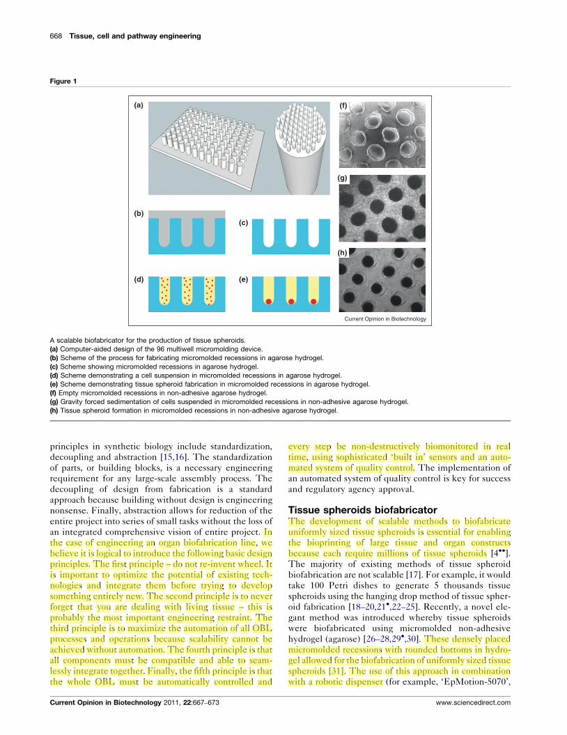
A scalable biofabricator for the production of tissue spheroids.
(a) Computer-aided design of the 96 multiwell micromolding device.
(b) Scheme of the process for fabricating micromolded recessions in agarose hydrogel.
(c) Scheme showing micromolded recessions in agarose hydrogel.
(d) Scheme demonstrating a cell suspension in micromolded recessions in agarose hydrogel.
(e) Scheme demonstrating tissue spheroid fabrication in micromolded recessions in agarose hydrogel.
(f) Empty micromolded recessions in non-adhesive agarose hydrogel.
(g) Gravity forced sedimentation of cells suspended in micromolded recessions in non-adhesive agarose hydrogel.
(h) Tissue spheroid formation in micromolded recessions in non-adhesive agarose hydrogel.
principles in synthetic biology include standardization,
decoupling and abstraction [15,16]. The standardization
of parts, or building blocks, is a necessary engineering
requirement for any large-scale assembly process. The
decoupling of design from fabrication is a standard
approach because building without design is engineering
nonsense. Finally, abstraction allows for reduction of the
entire project into series of small tasks without the loss of
an integrated comprehensive vision of entire project. In
the case of engineering an organ biofabrication line, we
believe it is logical to introduce the following basic design
principles. The first principle – do not re-invent wheel. It
is important to optimize the potential of existing tech-
nologies and integrate them before trying to develop
something entirely new. The second principle is to never
forget that you are dealing with living tissue – this is
probably the most important engineering restraint. The
third principle is to maximize the automation of all OBL
processes and operations because scalability cannot be
achieved without automation. The fourth principle is that
all components must be compatible and able to seam-
lessly integrate together. Finally, the fifth principle is that
the whole OBL must be automatically controlled and
Current Opinion in Biotechnology 2011, 22:667–673
every step be non-destructively biomonitored in real
time, using sophisticated ‘built in’ sensors and an auto-
mated system of quality control. The implementation of
an automated system of quality control is key for success
and regulatory agency approval.
Tissue spheroids biofabricatorThe development of scalable methods to biofabricate
uniformly sized tissue spheroids is essential for enabling
the bioprinting of large tissue and organ constructs
because each require millions of tissue spheroids [4��].The majority of existing methods of tissue spheroid
biofabrication are not scalable [17]. For example, it would
take 100 Petri dishes to generate 5 thousands tissue
spheroids using the hanging drop method of tissue spher-
oid fabrication [18–20,21�,22–25]. Recently, a novel ele-
gant method was introduced whereby tissue spheroids
were biofabricated using micromolded non-adhesive
hydrogel (agarose) [26–28,29�,30]. These densely placed
micromolded recessions with rounded bottoms in hydro-
gel allowed for the biofabrication of uniformly sized tissue
spheroids [31]. The use of this approach in combination
with a robotic dispenser (for example, ‘EpMotion-5070’,
www.sciencedirect.com
Organ printing: from bioprinter to organ biofabrication line Mironov, Kasyanov and Markwald 669
Figure 2
F NN
(a) (b) (c)
(d) (e)
Current Opinion in Biotechnology
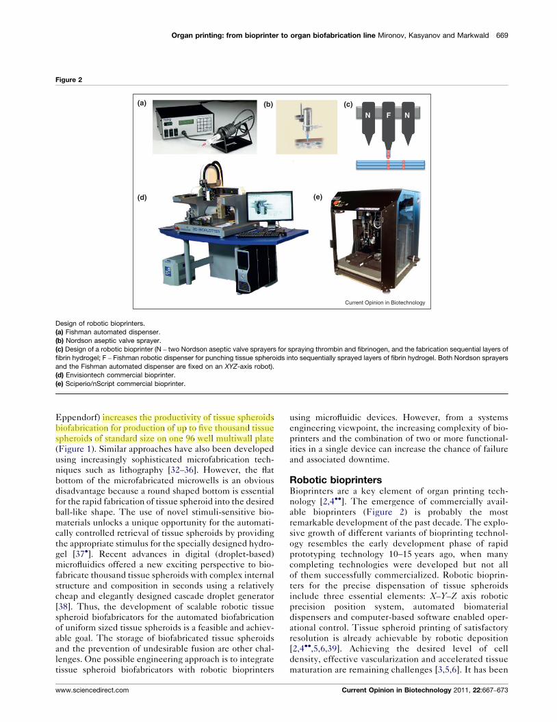
Design of robotic bioprinters.
(a) Fishman automated dispenser.
(b) Nordson aseptic valve sprayer.
(c) Design of a robotic bioprinter (N – two Nordson aseptic valve sprayers for spraying thrombin and fibrinogen, and the fabrication sequential layers of
fibrin hydrogel; F – Fishman robotic dispenser for punching tissue spheroids into sequentially sprayed layers of fibrin hydrogel. Both Nordson sprayers
and the Fishman automated dispenser are fixed on an XYZ-axis robot).
(d) Envisiontech commercial bioprinter.
(e) Sciperio/nScript commercial bioprinter.
Eppendorf) increases the productivity of tissue spheroids
biofabrication for production of up to five thousand tissue
spheroids of standard size on one 96 well multiwall plate
(Figure 1). Similar approaches have also been developed
using increasingly sophisticated microfabrication tech-
niques such as lithography [32–36]. However, the flat
bottom of the microfabricated microwells is an obvious
disadvantage because a round shaped bottom is essential
for the rapid fabrication of tissue spheroid into the desired
ball-like shape. The use of novel stimuli-sensitive bio-
materials unlocks a unique opportunity for the automati-
cally controlled retrieval of tissue spheroids by providing
the appropriate stimulus for the specially designed hydro-
gel [37�]. Recent advances in digital (droplet-based)
microfluidics offered a new exciting perspective to bio-
fabricate thousand tissue spheroids with complex internal
structure and composition in seconds using a relatively
cheap and elegantly designed cascade droplet generator
[38]. Thus, the development of scalable robotic tissue
spheroid biofabricators for the automated biofabrication
of uniform sized tissue spheroids is a feasible and achiev-
able goal. The storage of biofabricated tissue spheroids
and the prevention of undesirable fusion are other chal-
lenges. One possible engineering approach is to integrate
tissue spheroid biofabricators with robotic bioprinters
www.sciencedirect.com
using microfluidic devices. However, from a systems
engineering viewpoint, the increasing complexity of bio-
printers and the combination of two or more functional-
ities in a single device can increase the chance of failure
and associated downtime.
Robotic bioprintersBioprinters are a key element of organ printing tech-
nology [2,4��]. The emergence of commercially avail-
able bioprinters (Figure 2) is probably the most
remarkable development of the past decade. The explo-
sive growth of different variants of bioprinting technol-
ogy resembles the early development phase of rapid
prototyping technology 10–15 years ago, when many
completing technologies were developed but not all
of them successfully commercialized. Robotic bioprin-
ters for the precise dispensation of tissue spheroids
include three essential elements: X–Y–Z axis robotic
precision position system, automated biomaterial
dispensers and computer-based software enabled oper-
ational control. Tissue spheroid printing of satisfactory
resolution is already achievable by robotic deposition
[2,4��,5,6,39]. Achieving the desired level of cell
density, effective vascularization and accelerated tissue
maturation are remaining challenges [3,5,6]. It has been
Current Opinion in Biotechnology 2011, 22:667–673
670 Tissue, cell and pathway engineering
shown that adequate cell density can be achieved by
dielectrophoresis in combination with stereolithography
[40]. This investigation revealed that cell density
increased progressively, even in an initially low-density
printed cell-hydrogel construct, as the result of either
post-printed cell proliferation or the elimination of a
sacrificial biodegradable hydrogel [41–43]. The initial
high cell density can be achieved immediately after
bioprinting using self-assembling tissue spheroids
[4��,5,6,39,44��] or continuous self-assembling tissue
rods or longitudinal pellets [45]. There has also been
certain progress in solving the problem of vasculariza-
tion of thick 3D bioprinted tissue and organ constructs
[6]. Branched vascular tissue constructs have been bio-
printed using continuous rod dispensation [45��]. Vas-
cular tubes have also been bioassembled from vascular
tissue spheroids [2,4��,5,6,21�,39,46]. In other develop-
ments, new in vitro assays have been developed for the
systematic screening of potential chemical maturogenic
factors necessary for accelerated tissue maturation [47].
It has also been demonstrated that mechanical con-
ditioning can improve vascular tissue maturation
[21�]. Further progress in bioprinter development must
focus on improving nozzle and cartridge design, the
development of more flexible functionality and the
design of collectors or bioreactor for bioprinted con-
structs. In this context, recently reports have found
nozzle-free bioprinting to be an innovative engineering
concept [48�]. Another interesting development is
magnetic force driven tissue engineering with tissue
spheroids labeled with magnetic nanoparticles, which
could lead to the development of a magnetic bioprinter
Figure 3
(a) (b)
(d) (e)
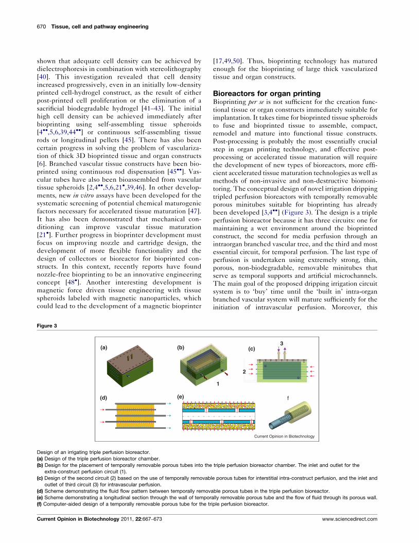
Design of an irrigating triple perfusion bioreactor.
(a) Design of the triple perfusion bioreactor chamber.
(b) Design for the placement of temporally removable porous tubes into the
extra-construct perfusion circuit (1).
(c) Design of the second circuit (2) based on the use of temporally removabl
outlet of third circuit (3) for intravascular perfusion.
(d) Scheme demonstrating the fluid flow pattern between temporally remova
(e) Scheme demonstrating a longitudinal section through the wall of tempor
(f) Computer-aided design of a temporally removable porous tube for the tr
Current Opinion in Biotechnology 2011, 22:667–673
[17,49,50]. Thus, bioprinting technology has matured
enough for the bioprinting of large thick vascularized
tissue and organ constructs.
Bioreactors for organ printingBioprinting per se is not sufficient for the creation func-
tional tissue or organ constructs immediately suitable for
implantation. It takes time for bioprinted tissue spheroids
to fuse and bioprinted tissue to assemble, compact,
remodel and mature into functional tissue constructs.
Post-processing is probably the most essentially crucial
step in organ printing technology, and effective post-
processing or accelerated tissue maturation will require
the development of new types of bioreactors, more effi-
cient accelerated tissue maturation technologies as well as
methods of non-invasive and non-destructive biomoni-
toring. The conceptual design of novel irrigation dripping
tripled perfusion bioreactors with temporally removable
porous minitubes suitable for bioprinting has already
been developed [3,4��] (Figure 3). The design is a triple
perfusion bioreactor because it has three circuits: one for
maintaining a wet environment around the bioprinted
construct, the second for media perfusion through an
intraorgan branched vascular tree, and the third and most
essential circuit, for temporal perfusion. The last type of
perfusion is undertaken using extremely strong, thin,
porous, non-biodegradable, removable minitubes that
serve as temporal supports and artificial microchannels.
The main goal of the proposed dripping irrigation circuit
system is to ‘buy’ time until the ‘built in’ intra-organ
branched vascular system will mature sufficiently for the
initiation of intravascular perfusion. Moreover, this
1
2
3(c)
f
Current Opinion in Biotechnology
triple perfusion bioreactor chamber. The inlet and outlet for the
e porous tubes for interstitial intra-construct perfusion, and the inlet and
ble porous tubes in the triple perfusion bioreactor.
ally removable porous tube and the flow of fluid through its porous wall.
iple perfusion bioreactor.
www.sciencedirect.com
Organ printing: from bioprinter to organ biofabrication line Mironov, Kasyanov and Markwald 671
Figure 4
Bioimaging
Pre-processing
Processing
Post-processing
Design
Biofabrication
Maturation
(a) (c)(b) (d) (e)
CAD
Blueprint
Bioink
Bioprinter
Biomonitoring
Bioreactor
Biopaper
Maturagenes
Current Opinion in Biotechnology
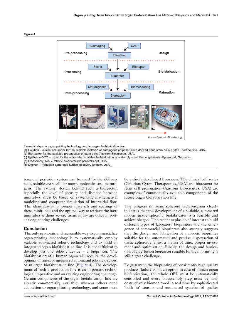
Essential steps in organ printing technology and an organ biofabrication line.
(a) Celution – clinical sell sorter for the scalable isolation of autologous adipose tissue derived adult stem cells (Cytori Therapeutics, USA).
(b) Bioreactor for the scalable propagation of stem cells (Aastrom Bioscience, USA).
(c) EpMotion-5070 – robot for the automated scalable biofabrication of uniformly sized tissue spheroids (Eppendorf, Germany).
(d) Bioasembly Tool – robotic bioprinter (Sciperio/nScript, USA).
(e) LifePort – Perfusion apparatus (Organ Recovery System, USA).
temporal perfusion system can be used for the delivery
cells, soluble extracellular matrix molecules and maturo-
gens. The rational design behind such a bioreactor,
especially the level of porosity and distance between
minitubes, must be based on systematic mathematical
modeling and computer simulation of interstitial flow.
The identification of proper materials and coatings of
these minitubes, and the optimal way to retrieve the inert
minitubes without severe tissue injury are other import-
ant engineering challenges.
ConclusionThe only economic and reasonable way to commercialize
organ-printing technology is to systematically employ
scalable automated robotic technology and to build an
integrated organ biofabrication line. It is not sufficient to
develop just one robotic device – a bioprinter. The
biofabrication of a human organ will require the devel-
opment of series of integrated automated robotic devices,
or an organ biofabrication line (Figure 4). The develop-
ment of such a production line is an important techno-
logical imperative and an exciting engineering challenge.
Certain components of the organ biofabrication line are
already commercially available, whereas others need
adaptation to organ printing technology, and some must
www.sciencedirect.com
be entirely developed from new. The clinical cell sorter
(Celution, Cytori Therapeutics, USA) and bioreactor for
stem cell propagation (Aastrom Biosciences, USA) are
examples of commercially available components of the
future organ biofabrication line.
The progress in tissue spheroid biofabrication clearly
indicates that the development of a scalable automated
robotic tissue spheroid biofabricator is a feasible and
achievable goal. The recent explosion of interest to build
different types of laboratory bioprinters and the emer-
gence of commercial bioprinters also strongly suggests
that the design and fabrication of a robotic bioprinter
suitable for the automated and precise dispensation of
tissue spheroids is just a matter of time, proper invest-
ment and optimization. Finally, the design and fabrica-
tion of a perfusion bioreactor suitable for organ printing is
still a great challenge.
To guarantee the bioprinting of consistently high quality
products (failure is not an option in case of human organ
biofabrication), the whole OBL must be automatically
controlled and every bioassembly step must be non-
destructively biomonitored in real time by sophisticated
‘built in’ sensors and automated systems of quality
Current Opinion in Biotechnology 2011, 22:667–673
672 Tissue, cell and pathway engineering
control. It is logical to assume that close collaboration
between biologists and engineers, and use of mathemat-
ical modeling and computer simulation to predict tissue
biofabrication processes, will significantly enhance, opti-
mize and accelerate the OBL design process.
AcknowledgementThis work was funded by NSF R-II grant ‘South Carolina Project for OrganBiofabrication’.
References and recommended readingPapers of particular interest, published within the annual period ofreview, have been highlighted as:
� of special interest�� of outstanding interest
1.��
Mironov V, Boland T, Trusk T, Forgacs G, Markwald RR: Organprinting: computer-aided jet-based 3D tissue engineering.Trends Biotechnol 2003, 21:157-161.
First introduction the concept of organ printing with using self-assemblingtissue spheroids as a ‘bioink’.
2. Jakab K, Norotte C, Marga F, Murphy K, Vunjak-Novakovic G,Forgacs G: Tissue engineering by self-assembly and bio-printing of living cells. Biofabrication 2010, 2:022001.
3. Mironov V, Kasyanov V, Drake C, Markwald RR: Organ printing:promises and challenges. Regen Med 2008, 3:93-103.
4.��
Mironov V, Visconti RP, Kasyanov V, Forgacs G, Drake CJ,Markwald RR: Organ printing: tissue spheroids as buildingblocks. Biomaterials 2009, 30:2164-2174.
Most comprehensive description of solid scaffold-free approach in tissueengineering using self-assembling tissue spheroids as building blocks.
5. Mironov V, Zhang J, Gentile C, Brakke K, Trusk T, Jakab K,Forgacs G, Kasyanov V, Visconti R, Markwald RR: Designer‘blueprint’ for vascular trees: morphology evolution of vasculartissue constructs. Virtual Phys Prototyping 2009, 4:63-74.
6. Visconti RP, Kasyanov V, Gentile C, Zhang J, Markwald RR,Mironov V: Towards organ printing: engineering an intra-organbranched vascular tree. Expert Opin Biol Ther 2010, 10:409-420.
7. Nichol JW, Khademhosseini A: Modular tissue engineering:engineering biological tissues from the bottom up.Soft Matter 2009, 5:1312-1319.
8. Williams DF: On the nature of biomaterials. Biomaterials 2009,30:5897-5909.
9. Guillemot F, Mironov V, Nakamura M: Bioprinting is coming ofage: report from the International Conference on Bioprintingand Biofabrication in Bordeaux (3B’09). Biofabrication 2010,2:010201.
10. Ringeisen BR, Spargo BJ, Wu PK (Eds): Cell and Organ Printing.Springer; 2010.
11. Boland T, Xu T, Damon B, Cui X: Application of inkjet printing totissue engineering. Biotechnol J 2006, 1:910-917.
12. Campbell PG, Weiss LE: Tissue engineering with the aid ofinkjet printers. Expert Opin Biol Ther 2007, 7:1123-1127.
13. Cohen DL, Malone E, Lipson H, Bonassar LJ: Direct freeformfabrication of seeded hydrogels in arbitrary geometries.Tissue Eng 2006, 12:1325-1335.
14. Fedorovich NE, Swennen I, Girones J, Moroni L, vanBlitterswijk CA, Schacht E, Alblas J, Dhert WJ: Evaluation ofphotocrosslinked lutrol hydrogel for tissue printingapplications. Biomacromolecules 2009.
15. Endy D: Foundations for engineering biology.Nature 2005, 438:449-453.
16. Purnick PE, Weiss R: The second wave of synthetic biology:from modules to systems. Nat Rev Mol Cell Biol 2009,10:410-422.
Current Opinion in Biotechnology 2011, 22:667–673
17. Lin RZ, Chang HY: Recent advances in three-dimensionalmulticellular spheroid culture for biomedical research.Biotechnol J 2008, 3:1172-1184.
18. Kelm JM, Djonov V, Hoerstrup SP, Guenter CI, Ittner LM,Greve F, Hierlemann A, Sanchez-Bustamante CD,Perriard JC, Ehler E et al.: Tissue-transplant fusion andvascularization of myocardial microtissues and macrotissuesimplanted into chicken embryos and rats. Tissue Eng 2006,12:2541-2553.
19. Kelm JM, Djonov V, Ittner LM, Fluri D, Born W, Hoerstrup SP,Fussenegger M: Design of custom-shaped vascularizedtissues using microtissue spheroids as minimal building units.Tissue Eng 2006, 12:2151-2160.
20. Kelm JM, Fussenegger M: Microscale tissue engineeringusing gravity-enforced cell assembly. Trends Biotechnol 2004,22:195-202.
21.�
Kelm JM, Lorber V, Snedeker JG, Schmidt D, Broggini-Tenzer A,Weisstanner M, Odermatt B, Mol A, Zund G, Hoerstrup SP: Anovel concept for scaffold-free vessel tissue engineering: self-assembly of microtissue building blocks. J Biotechnol 2010,148:46-55.
First demonstration of feasibility of biofabrication of tissue engineeredvascular graft from tissue spheroids using molding technique andmechanical conditioning.
22. Kelm JM, Timmins NE, Brown CJ, Fussenegger M, Nielsen LK:Method for generation of homogeneous multicellulartumor spheroids applicable to a wide variety of cell types.Biotechnol Bioeng 2003, 83:173-180.
23. Layer PG, Robitzki A, Rothermel A, Willbold E: Of layers andspheres: the reaggregate approach in tissue engineering.Trends Neurosci 2002, 25:131-134.
24. Mironov V, Markwald RR, Forgacs G: Organ printing:self-assembling cell aggregates as a ‘‘bioink’’.Sci Med 2003, 9:69-71.
25. Reininger-Mack A, Thielecke H, Robitzki AA: 3D-biohybridsystems: applications in drug screening. Trends Biotechnol2002, 20:56-61.
26. Dean DM, Morgan JR: Cytoskeletal-mediated tensionmodulates the directed self-assembly of microtissues.Tissue Eng Part A 2008, 14:1989-1997.
27. Dean DM, Napolitano AP, Youssef J, Morgan JR: Rods, tori, andhoneycombs: the directed self-assembly of microtissueswith prescribed microscale geometries. FASEB J 2007,21:4005-4012.
28. Napolitano AP, Chai P, Dean DM, Morgan JR: Dynamics of theself-assembly of complex cellular aggregates onmicromolded nonadhesive hydrogels. Tissue Eng 2007,13:2087-2094.
29.�
Napolitano AP, Dean DM, Man AJ, Youssef J, Ho DN, Rago AP,Lech MP, Morgan JR: Scaffold-free three-dimensional cellculture utilizing micromolded nonadhesive hydrogels.Biotechniques 2007, 43: 494, 496–500.
Novel approach for biofabrication of tissue spheroids of uniformed sizeusing micromolded non-adhesive hydrogel.
30. Rago AP, Dean DM, Morgan JR: Controlling cell position incomplex heterotypic 3D microtissues by tissue fusion.Biotechnol Bioeng 2009, 102:1231-1241.
31. Nagy-Mehesz A, Brown J, Beaver W, Gayk M, Hajdu Z, Visconti R,Norris RA, Trusk T, Markwald R, Mironov V: Scalablerobotic biofabrication of uniform size tissue spheroids.In 2010 International Conference on Biofabrication. Edited by SunW. Philadelphia, Pennsylvania, USA: 2010:71.
32. Fukuda J, Khademhosseini A, Yeo Y, Yang X, Yeh J, Eng G,Blumling J, Wang CF, Kohane DS, Langer R: Micromolding ofphotocrosslinkable chitosan hydrogel for spheroid microarrayand co-cultures. Biomaterials 2006, 27:5259-5267.
33. Fukuda J, Nakazawa K: Orderly arrangement of hepatocytespheroids on a microfabricated chip. Tissue Eng 2005,11:1254-1262.
www.sciencedirect.com
Organ printing: from bioprinter to organ biofabrication line Mironov, Kasyanov and Markwald 673
34. Nakazawa K, Izumi Y, Fukuda J, Yasuda T: Hepatocyte spheroidculture on a polydimethylsiloxane chip having microcavities.J Biomater Sci Polym Ed 2006, 17:859-873.
35. Okuyama T, Yamazoe H, Mochizuki N, Khademhosseini A,Suzuki H, Fukuda J: Preparation of arrays of cell spheroids andspheroid-monolayer cocultures within a microfluidic device.J Biosci Bioeng 2010, 110:572-576.
36. Otsuka H, Hirano A, Nagasaki Y, Okano T, Horiike Y, Kataoka K:Two-dimensional multiarray formation of hepatocytespheroids on a microfabricated PEG-brush surface.Chembiochem 2004, 5:850-855.
37.�
Tekin H, Anaya M, Brigham MD, Nauman C, Langer R,Khademhosseini A: Stimuli-responsive microwells forformation and retrieval of cell aggregates. Lab Chip 2010,10:2411-2418.
Innovative and potentially scalable approach for retrieval of tissue spher-oids from microfabricated microwells.
38. Shah RK, Shum HC, Rowat AC, Lee D, Agresti JJ, Utada AS,Chu LY, Kim JW, Fernandez-Nieves A, Martinez CJ et al.:Designer emulsions using microfluidics. Mater Today 2008,11:18-27.
39. Jakab K, Norotte C, Damon B, Marga F, Neagu A, Besch-Williford CL, Kachurin A, Church KH, Park H, Mironov V et al.: Tissueengineering by self-assembly of cells printed into topologicallydefined structures. Tissue Eng Part A 2008, 14:413-421.
40. Liu Tsang V, Chen AA, Cho LM, Jadin KD, Sah RL, DeLong S,West JL, Bhatia SN: Fabrication of 3D hepatic tissues byadditive photopatterning of cellular hydrogels. FASEB J 2007,21:790-801.
41. Wang X, Yan Y, Pan Y, Xiong Z, Liu H, Cheng J, Liu F, Lin F, Wu R,Zhang R et al.: Generation of three-dimensional hepatocyte/gelatin structures with rapid prototyping system.Tissue Eng 2006, 12:83-90.
42. Wang X, Yan Y, Zhang R: Recent trends and challenges incomplex organ manufacturing. Tissue Eng Part B Rev 2010,16:189-197.
www.sciencedirect.com
43. Yan Y, Wang X, Pan Y, Liu H, Cheng J, Xiong Z, Lin F, Wu R,Zhang R, Lu Q: Fabrication of viable tissue-engineeredconstructs with 3D cell-assembly technique. Biomaterials 2005,26:5864-5871.
44.��
Jakab K, Neagu A, Mironov V, Markwald RR, Forgacs G:Engineering biological structures of prescribed shape usingself-assembling multicellular systems. Proc Natl Acad Sci USA2004, 101:2864-2869.
Milestone paper describing self-assembling properties of tissue spher-oids in three-dimensional hydrogel.
45.��
Norotte C, Marga FS, Niklason LE, Forgacs G: Scaffold-freevascular tissue engineering using bioprinting. Biomaterials2009, 30:5910-5917.
First report about bioprinting of branched vascular segments usingcontinuous dispensing of longitudinal tissue rods.
46. Gentile C, Fleming PA, Mironov V, Argraves KM,Argraves WS, Drake CJ: VEGF-mediated fusion in thegeneration of uniluminal vascular spheroids. Dev Dyn 2008,237:2918-2925.
47. Hajdu Z, Mironov V, Mehesz AN, Norris RA, Markwald RR,Visconti RP: Tissue spheroid fusion-based in vitro screeningassays for analysis of tissue maturation. J Tissue Eng RegenMed 2010.
48.�
Moon S, Hasan SK, Song YS, Xu F, Keles HO, Manzur F,Mikkilineni S, Hong JW, Nagatomi J, Haeggstrom E et al.:Layer by layer three-dimensional tissue epitaxy bycell-laden hydrogel droplets. Tissue Eng Part C Methods 2010,16:157-166.
First report about nozzle-free bioprinting.
49. Ho VH, Muller KH, Barcza A, Chen R, Slater NK: Generation andmanipulation of magnetic multicellular spheroids. Biomaterials2010, 31:3095-3102.
50. Lin RZ, Chu WC, Chiang CC, Lai CH, Chang HY:Magnetic reconstruction of three-dimensional tissuesfrom multicellular spheroids. Tissue Eng Part C Methods 2008,14:197-205.
Current Opinion in Biotechnology 2011, 22:667–673