Embed Size (px)
Citation preview
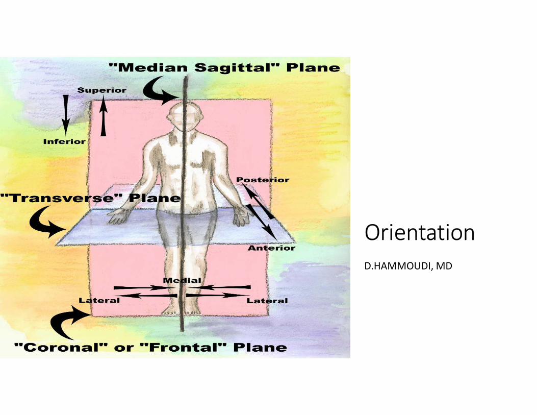
Orientation
D.HAMMOUDI, MD
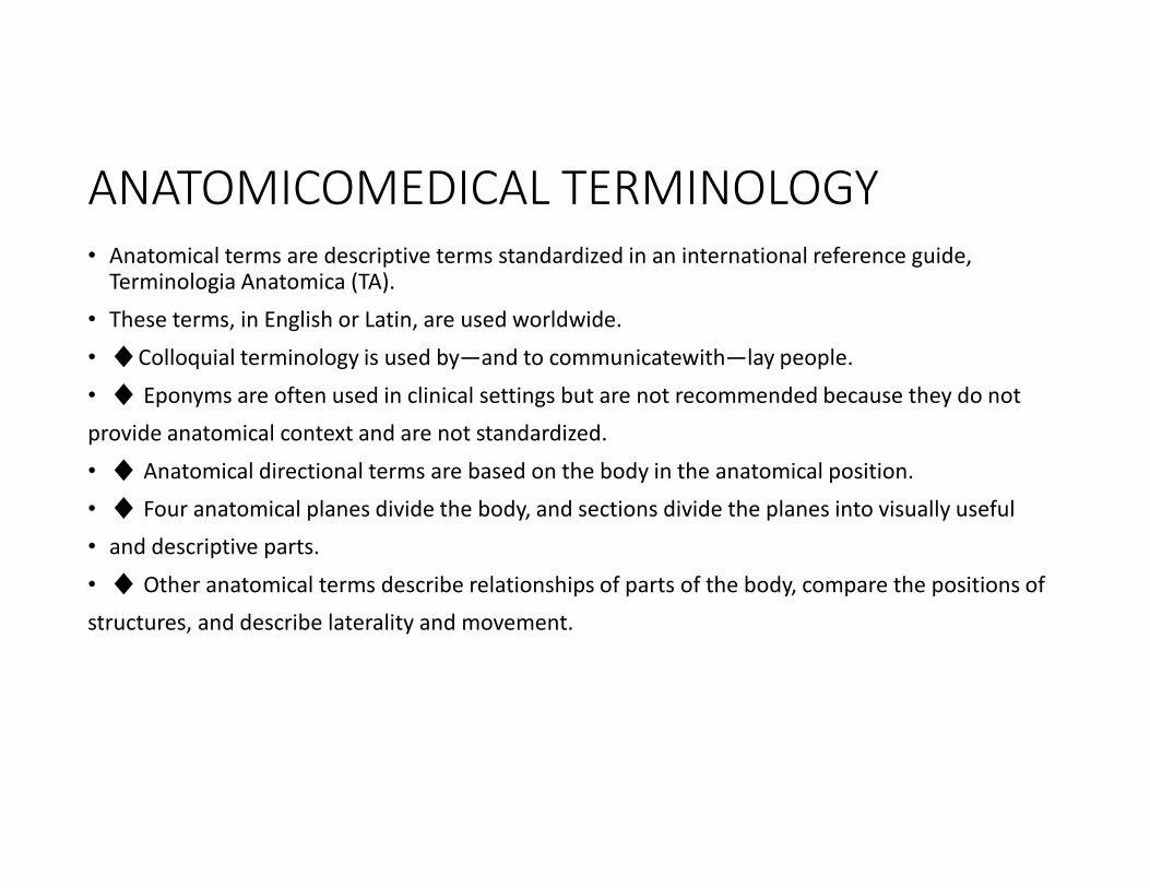
ANATOMICOMEDICAL TERMINOLOGY
• Anatomical terms are descriptive terms standardized in an international reference guide, Terminologia Anatomica (TA).
• These terms, in English or Latin, are used worldwide.
• ♦Colloquial terminology is used by—and to communicatewith—lay people.
• ♦ Eponyms are often used in clinical settings but are not recommended because they do not
provide anatomical context and are not standardized.
• ♦ Anatomical directional terms are based on the body in the anatomical position.
• ♦ Four anatomical planes divide the body, and sections divide the planes into visually useful
• and descriptive parts.
• ♦ Other anatomical terms describe relationships of parts of the body, compare the positions of
structures, and describe laterality and movement.
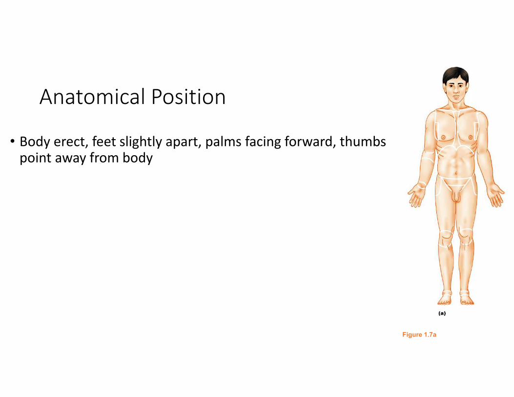
Anatomical Position
• Body erect, feet slightly apart, palms facing forward, thumbs point away from body
Figure 1.7a

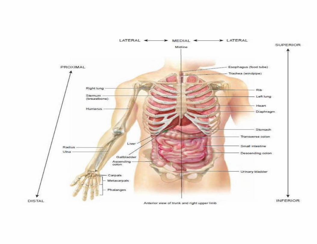
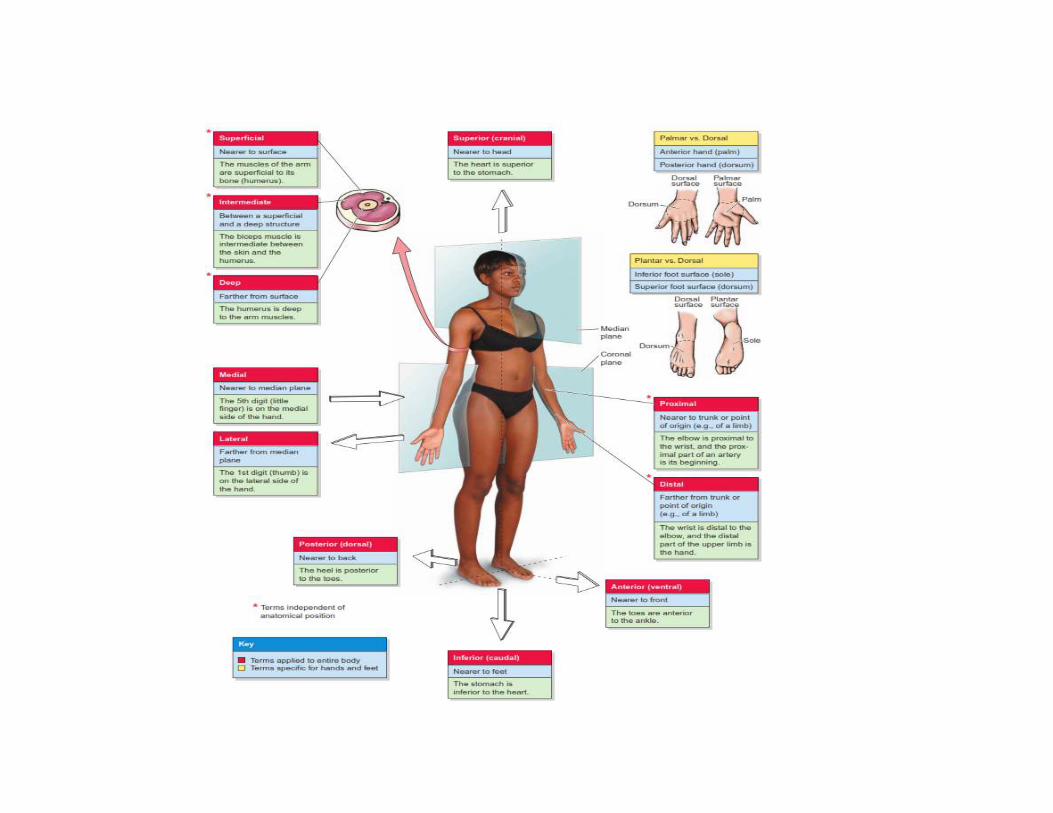
Directional Terms
• Superior and inferior – toward and away from the head, respectively
• Anterior and posterior – toward the front and back of the body
• Medial, lateral, and intermediate – toward the midline, away from the midline, and between a more medial and lateral structure
• Paired structures having right and left members (e.g., the kidneys)are bilateral, whereas those occurring
on one side only (e.g., the spleen) are unilateral.
• Designating whether you are referring specifically to the right or left member of bilateral structures can
be critical, and is a good habit to begin at the outset of one’s training to become a health professional.
• Something occurring on the same side of the body as another structure is ipsilateral; the right thumb and
right great (big) toe are ipsilateral, for example.
• Contralateral means occurring on the opposite side of the body relative to another structure;
• the right hand is contralateral to the left hand.
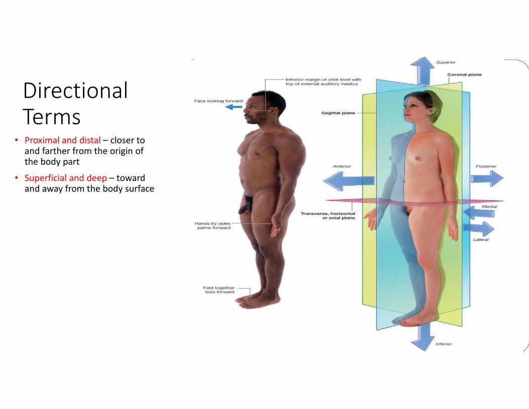
Directional
Terms• Proximal and distal – closer to
and farther from the origin of the body part
• Superficial and deep – toward and away from the body surface
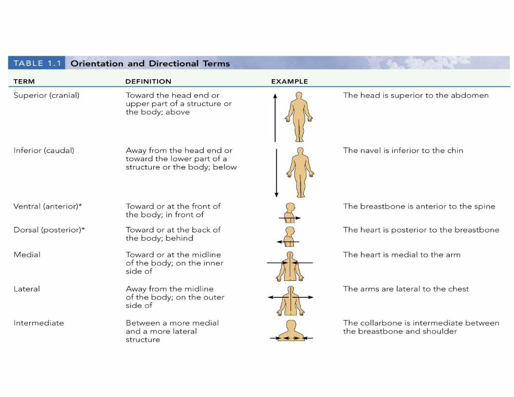
Directional Terms
Table 1.1a
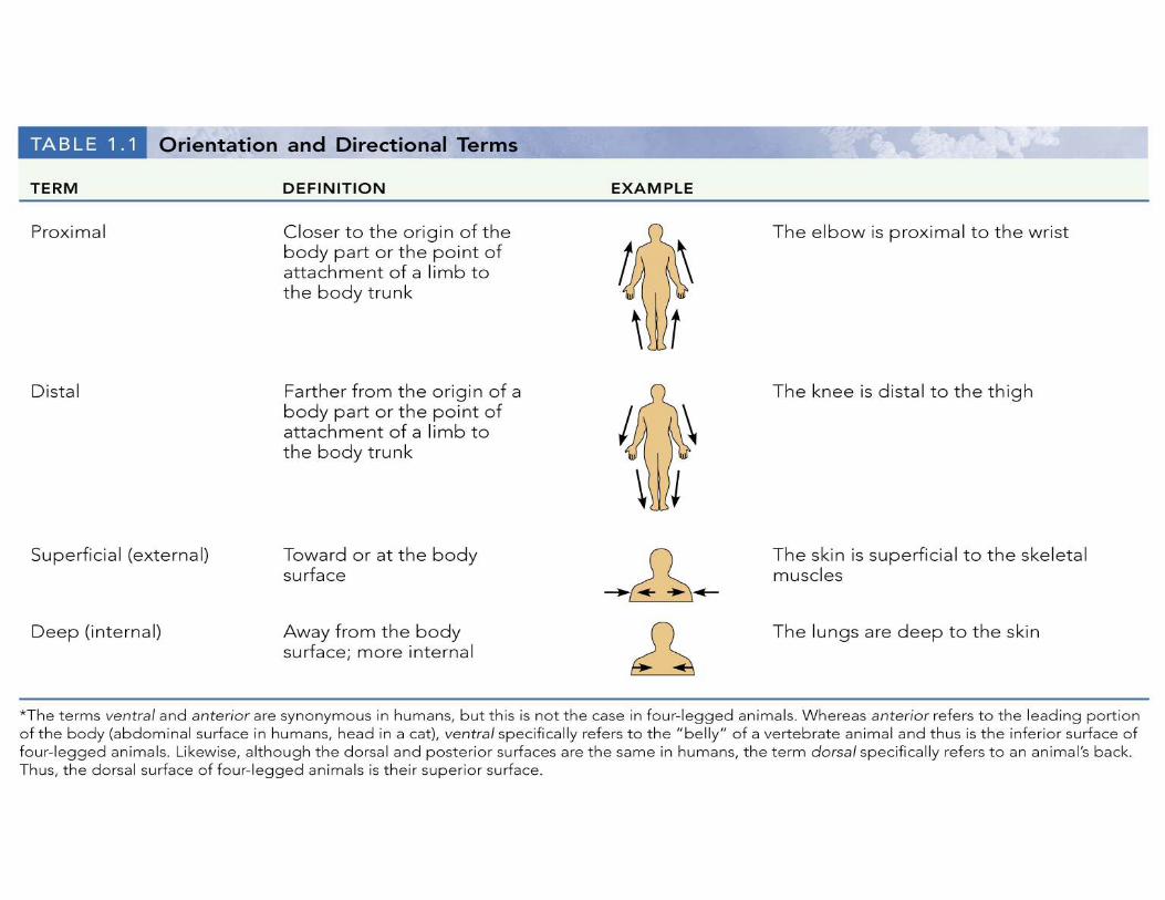
Directional Terms
Table 1.1b
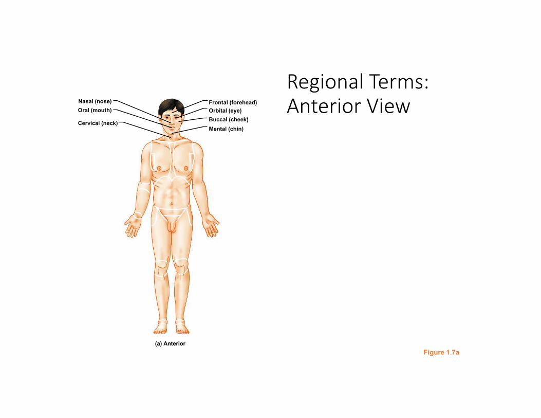
Regional Terms:
Anterior View
Figure 1.7a
Nasal (nose)
Oral (mouth)
Cervical (neck)
Frontal (forehead)
Orbital (eye)
Buccal (cheek)
Mental (chin)
(a) Anterior
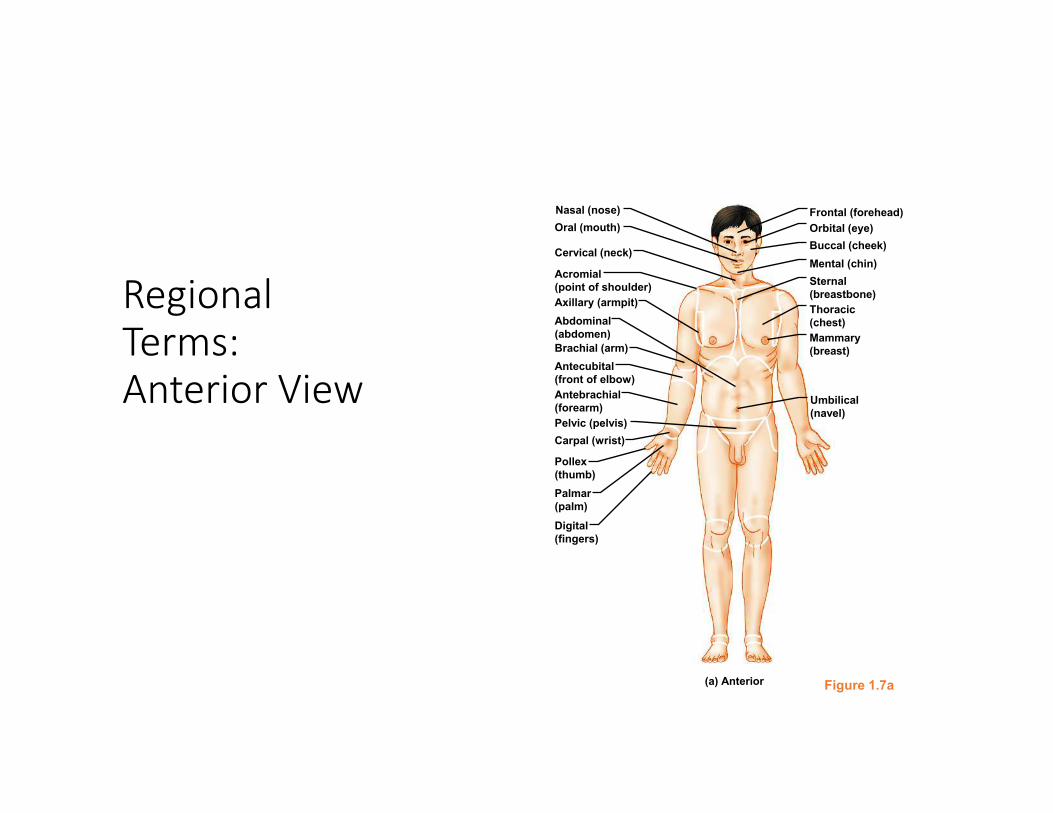
Regional
Terms:
Anterior View
Figure 1.7a
Nasal (nose)
Oral (mouth)
Cervical (neck)
Acromial
(point of shoulder)
Axillary (armpit)
Brachial (arm)
Antecubital
(front of elbow)
Abdominal
(abdomen)
Pelvic (pelvis)
Antebrachial
(forearm)
Carpal (wrist)
Palmar
(palm)
Pollex
(thumb)
Digital
(fingers)
Mammary
(breast)
Frontal (forehead)
Orbital (eye)
Buccal (cheek)
Sternal
(breastbone)
Thoracic
(chest)
Mental (chin)
Umbilical
(navel)
(a) Anterior
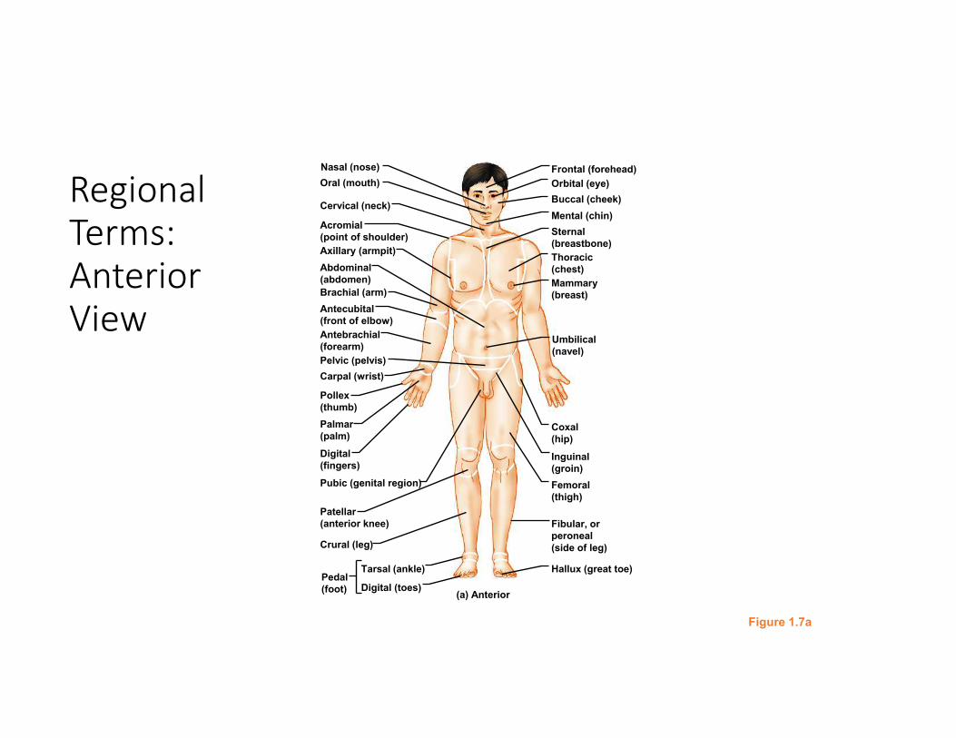
Regional
Terms:
Anterior
View
Figure 1.7a
Nasal (nose)
Oral (mouth)
Cervical (neck)
Acromial
(point of shoulder)
Axillary (armpit)
Brachial (arm)
Antecubital
(front of elbow)
Abdominal
(abdomen)
Pelvic (pelvis)
Antebrachial
(forearm)
Carpal (wrist)
Palmar
(palm)
Pollex
(thumb)
Digital
(fingers)
Pubic (genital region)
Patellar
(anterior knee)
Crural (leg)
Tarsal (ankle)Pedal
(foot) Digital (toes)
Inguinal
(groin)
Coxal
(hip)
Femoral
(thigh)
Fibular, or
peroneal
(side of leg)
Hallux (great toe)
Mammary
(breast)
Frontal (forehead)
Orbital (eye)
Buccal (cheek)
Sternal
(breastbone)
Thoracic
(chest)
Mental (chin)
Umbilical
(navel)
(a) Anterior
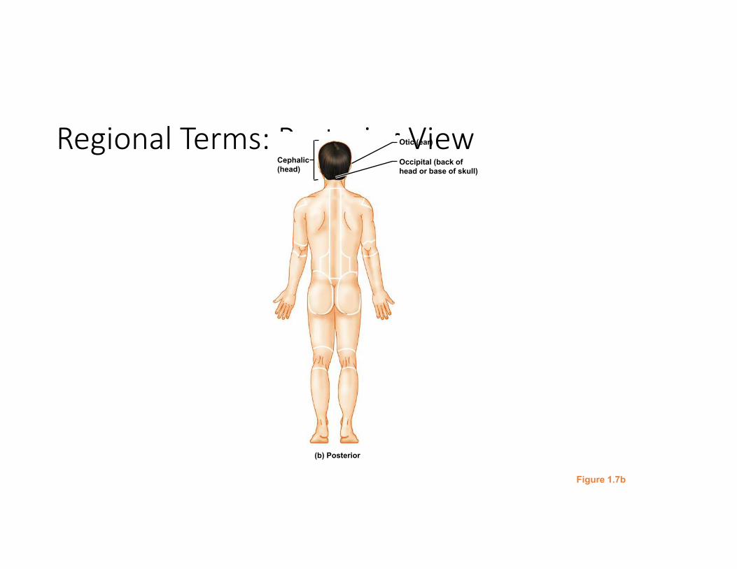
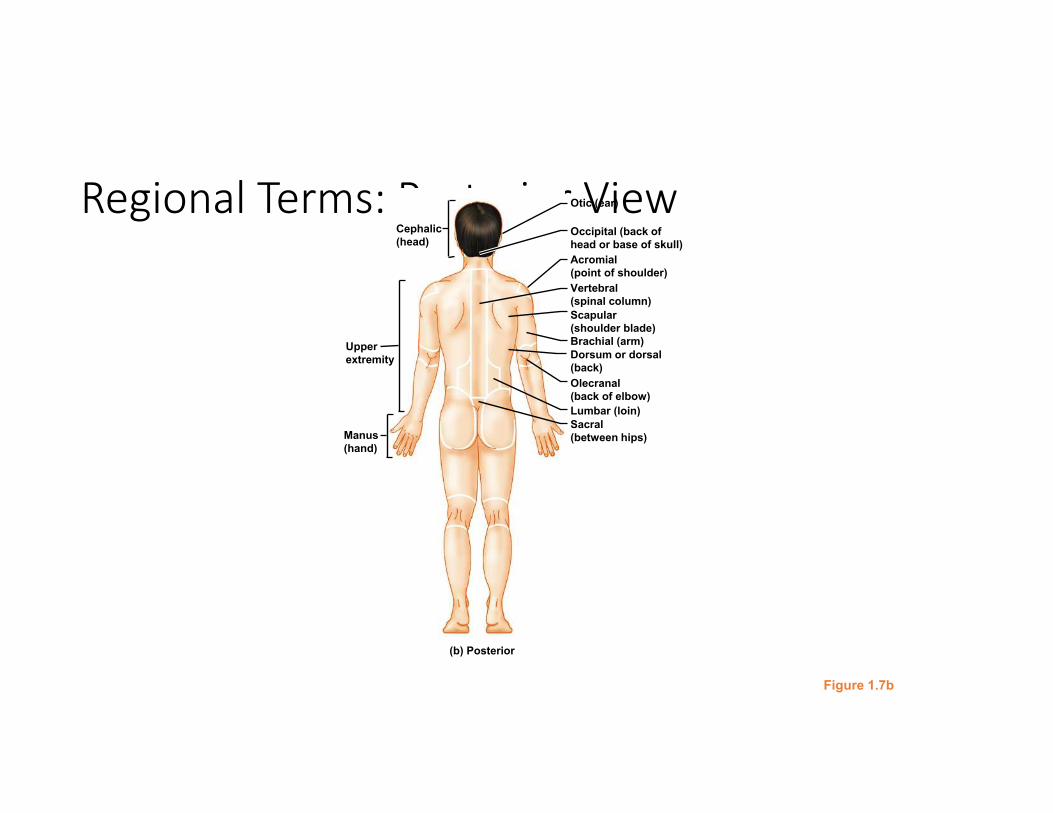
Regional Terms: Posterior View
Figure 1.7b
Otic (ear)
Occipital (back of
head or base of skull)
Cephalic
(head)
(b) Posterior
Regional Terms: Posterior View
Figure 1.7b
Brachial (arm)
Otic (ear)
Occipital (back of
head or base of skull)
Acromial
(point of shoulder)
Vertebral
(spinal column)
Scapular
(shoulder blade)
Dorsum or dorsal
(back)
Olecranal
(back of elbow)
Lumbar (loin)
Sacral
(between hips)Manus
(hand)
Upper
extremity
Cephalic
(head)
(b) Posterior
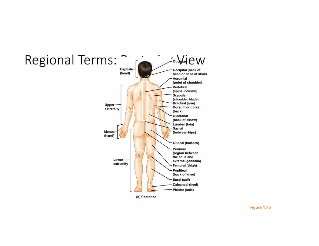
Regional Terms: Posterior View
Figure 1.7b
Brachial (arm)
Otic (ear)
Occipital (back of
head or base of skull)
Acromial
(point of shoulder)
Vertebral
(spinal column)
Scapular
(shoulder blade)
Dorsum or dorsal
(back)
Olecranal
(back of elbow)
Lumbar (loin)
Sacral
(between hips)
Gluteal (buttock)
Perineal
(region between
the anus and
external genitalia)
Femoral (thigh)
Popliteal
(back of knee)
Sural (calf)
Calcaneal (heel)
Plantar (sole)
Manus
(hand)
Upper
extremity
Cephalic
(head)
Lower
extremity
(b) Posterior
Copyright © 2010 Pearson Education, Inc.
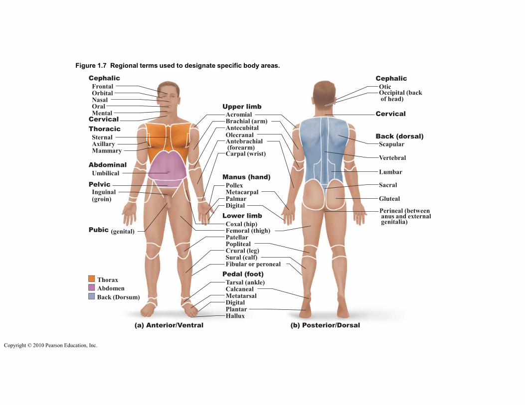
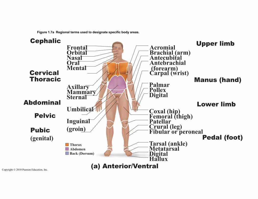
Figure 1.7 Regional terms used to designate specific body areas.
CervicalCervical
Back (dorsal)
(a) Anterior/Ventral (b) Posterior/Dorsal
Pubic (genital)
Cephalic
FrontalOrbitalNasalOralMental
Thoracic
SternalAxillaryMammary
Scapular
Vertebral
Lumbar
Sacral
Gluteal
Perineal (betweenanus and externalgenitalia)
Abdominal
Umbilical
PelvicInguinal(groin)
Upper limb
AcromialBrachial (arm)AntecubitalOlecranalAntebrachial(forearm)
Carpal (wrist)
Manus (hand)
PollexMetacarpalPalmarDigital
Lower limb
Coxal (hip)Femoral (thigh)PatellarPoplitealCrural (leg)Sural (calf)Fibular or peroneal
Pedal (foot)
Tarsal (ankle)CalcanealMetatarsalDigitalPlantarHallux
Cephalic
OticOccipital (backof head)
Thorax
Abdomen
Back (Dorsum)
Copyright © 2010 Pearson Education, Inc.
Figure 1.7a Regional terms used to designate specific body areas.
Cervical
(a) Anterior/Ventral
Pubic
(genital)
CephalicFrontalOrbitalNasalOralMental
Thoracic
AxillaryMammarySternal
AbdominalUmbilical
PelvicInguinal
(groin)
Upper limbAcromialBrachial (arm)AntecubitalAntebrachial(forearm)Carpal (wrist)
Manus (hand)PalmarPollexDigital
Lower limbCoxal (hip)Femoral (thigh)PatellarCrural (leg)Fibular or peroneal
Pedal (foot)Tarsal (ankle)MetatarsalDigitalHallux
Thorax
Abdomen
Back (Dorsum)
Copyright © 2010 Pearson Education, Inc.
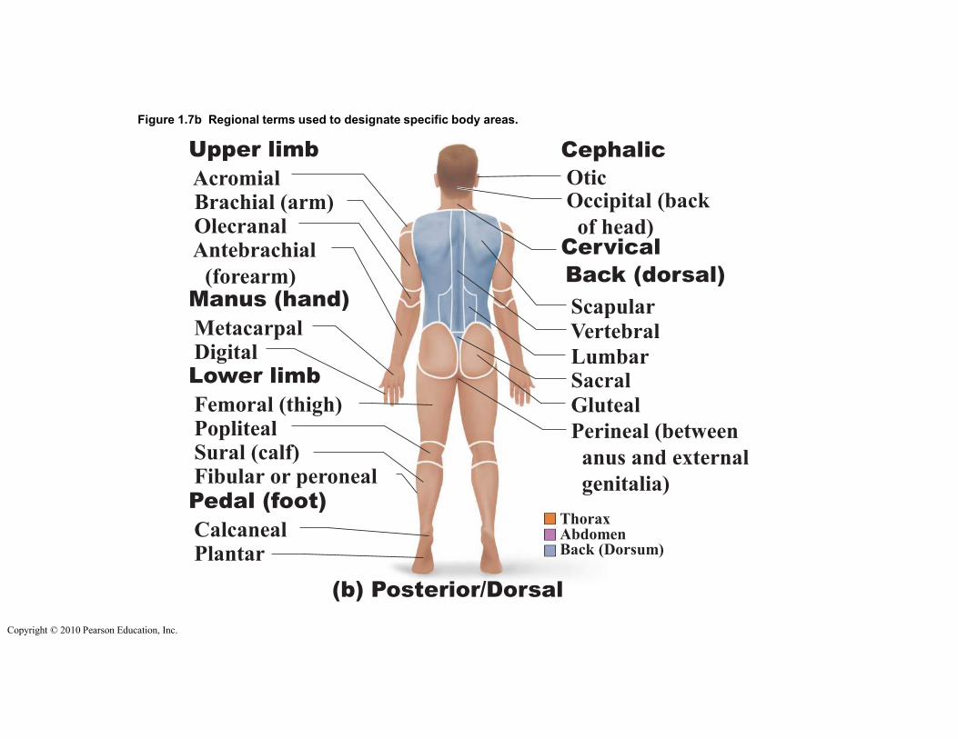
Figure 1.7b Regional terms used to designate specific body areas.
Cervical
Back (dorsal)
(b) Posterior/Dorsal
Scapular
Vertebral
LumbarSacral
Gluteal
Perineal (between
anus and external
genitalia)
Upper limb
AcromialBrachial (arm)OlecranalAntebrachial
(forearm)Manus (hand)
MetacarpalDigitalLower limb
Femoral (thigh)PoplitealSural (calf)Fibular or peroneal
Pedal (foot)
CalcanealPlantar
Cephalic
OticOccipital (back
of head)
ThoraxAbdomenBack (Dorsum)
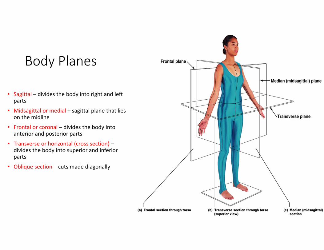
Body Planes
• Sagittal – divides the body into right and left parts
• Midsagittal or medial – sagittal plane that lies on the midline
• Frontal or coronal – divides the body into anterior and posterior parts
• Transverse or horizontal (cross section) –divides the body into superior and inferior parts
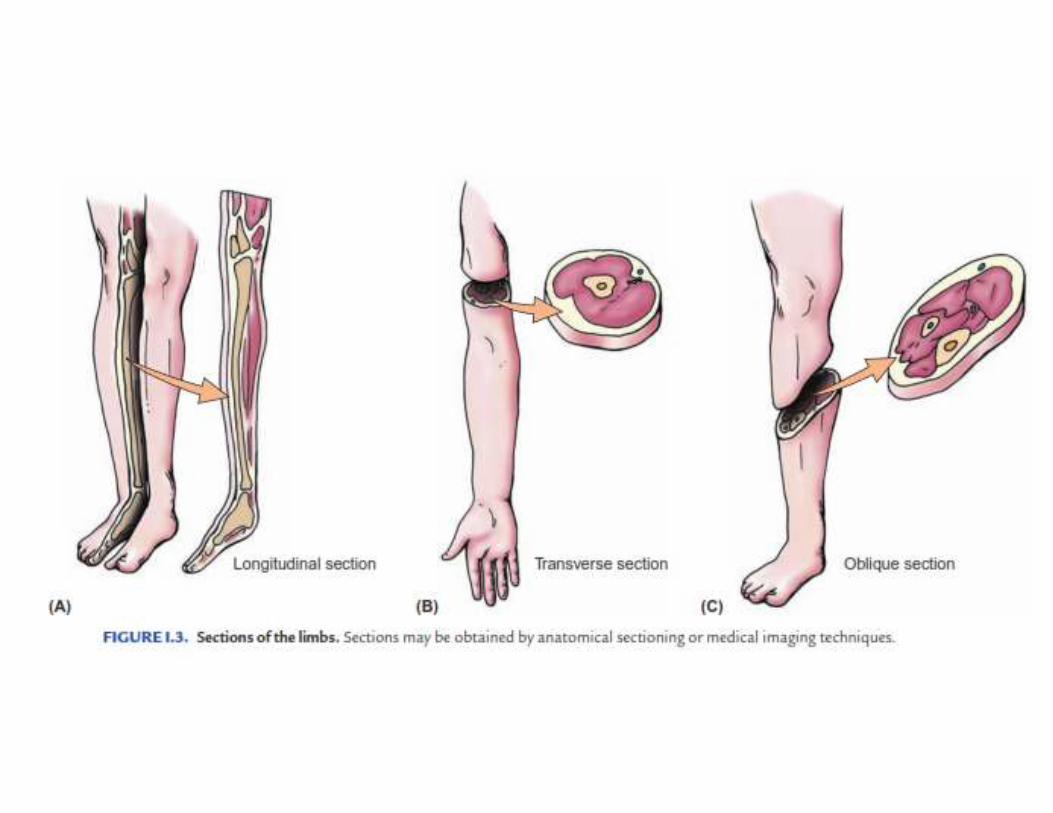
• Oblique section – cuts made diagonally
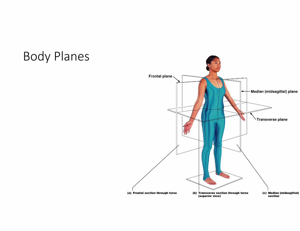
Body Planes
Figure 1.8
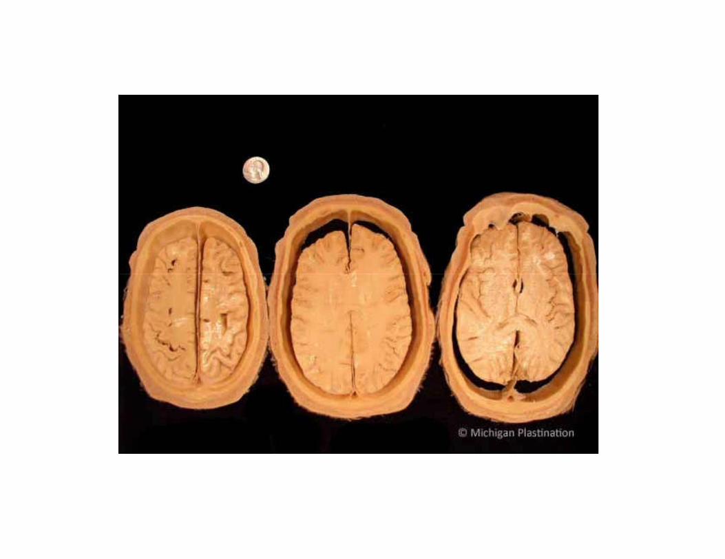
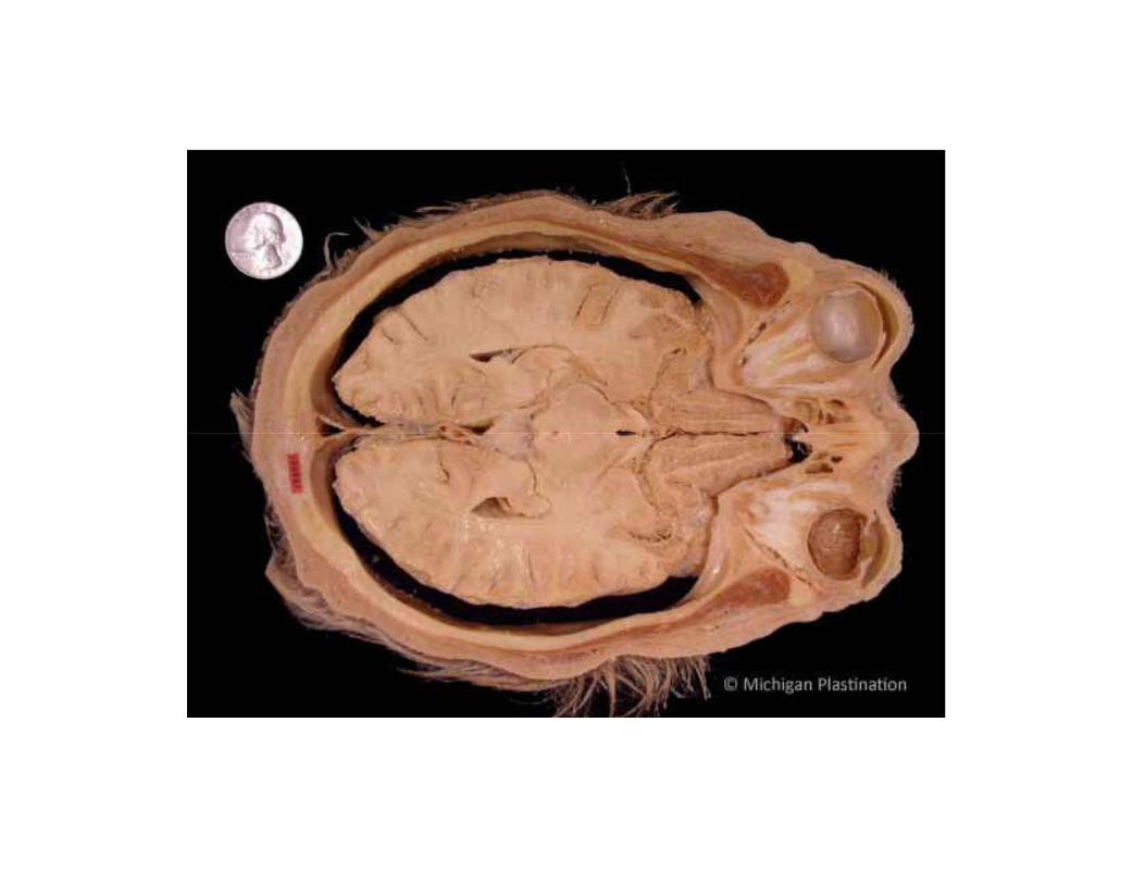
Body Cavities
• Dorsal cavity protects the nervous system, and is divided into two subdivisions
• Cranial cavity – within the skull; encases the brain
• Vertebral cavity – runs within the vertebral column; encases the spinal cord
• Ventral cavity houses the internal organs (viscera), and is divided into two subdivisions
• Thoracic
• Abdominopelvic
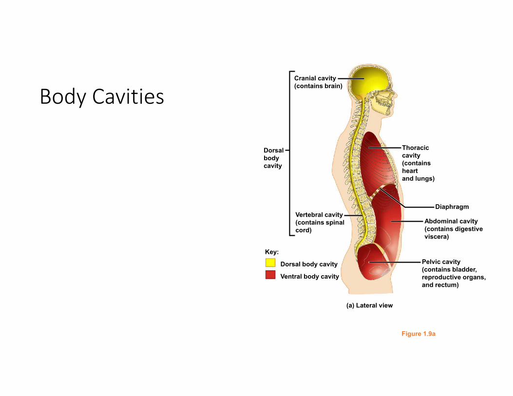
Body Cavities
Figure 1.9a
Cranial cavity
(contains brain)
Dorsal
body
cavity
Diaphragm
Abdominal cavity
(contains digestive
viscera)
Pelvic cavity
(contains bladder,
reproductive organs,
and rectum)
Vertebral cavity
(contains spinal
cord)
Key:
Dorsal body cavity
Ventral body cavity
Thoracic
cavity
(contains
heart
and lungs)
(a) Lateral view
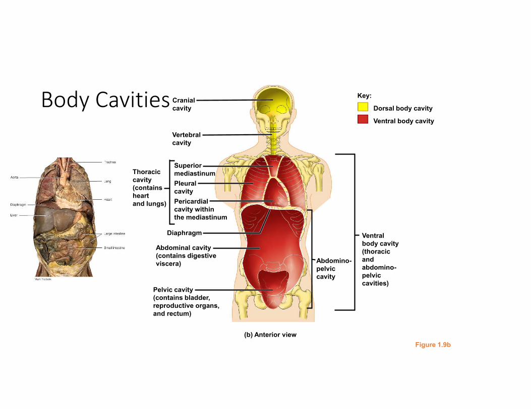
Body Cavities
Figure 1.9b
Ventral
body cavity
(thoracic
and
abdomino-
pelvic
cavities)
Abdomino-
pelvic
cavity
Superior
mediastinum
Pleural
cavity
Cranial
cavity
Vertebral
cavity
Pericardial
cavity within
the mediastinum
Diaphragm
Abdominal cavity
(contains digestive
viscera)
Pelvic cavity
(contains bladder,
reproductive organs,
and rectum)
Thoracic
cavity
(contains
heart
and lungs)
(b) Anterior view
Key:
Dorsal body cavity
Ventral body cavity
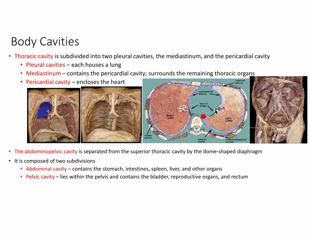
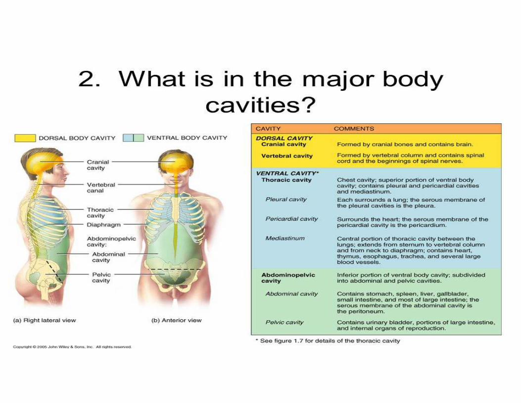
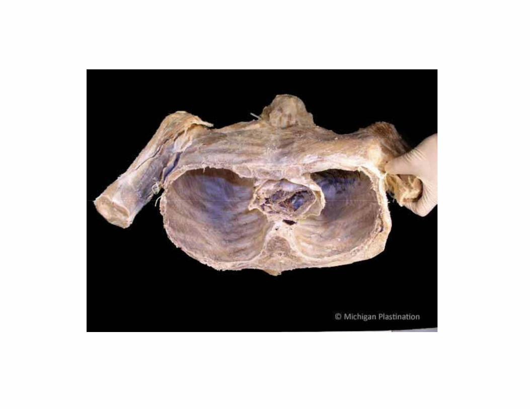
Body Cavities• Thoracic cavity is subdivided into two pleural cavities, the mediastinum, and the pericardial cavity
• Pleural cavities – each houses a lung
• Mediastinum – contains the pericardial cavity; surrounds the remaining thoracic organs
• Pericardial cavity – encloses the heart
• The abdominopelvic cavity is separated from the superior thoracic cavity by the dome-shaped diaphragm
• It is composed of two subdivisions
• Abdominal cavity – contains the stomach, intestines, spleen, liver, and other organs
• Pelvic cavity – lies within the pelvis and contains the bladder, reproductive organs, and rectum
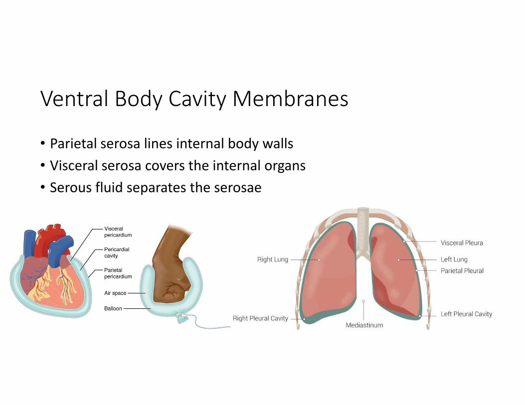
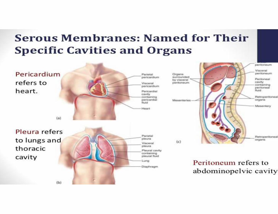
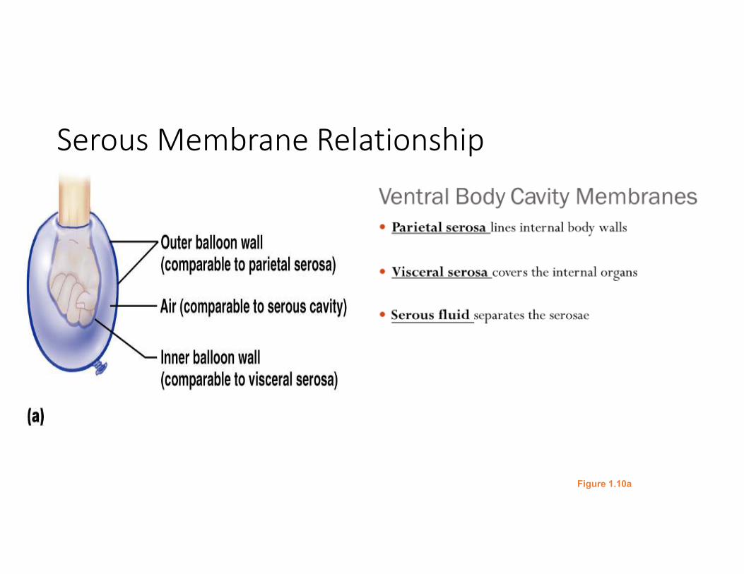
Ventral Body Cavity Membranes
• Parietal serosa lines internal body walls
• Visceral serosa covers the internal organs
• Serous fluid separates the serosae
Body Cavities� Thoracic cavity is subdivided into two pleural cavities, the
mediastinum, and the pericardial cavity
� Pleural cavities – each houses a lung
� Mediastinum – contains the pericardial cavity; surrounds the remaining thoracic organs
� Pericardial cavity – encloses the heart
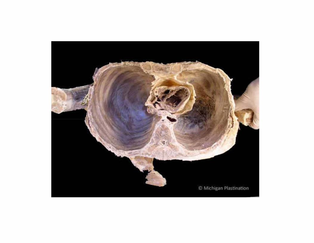
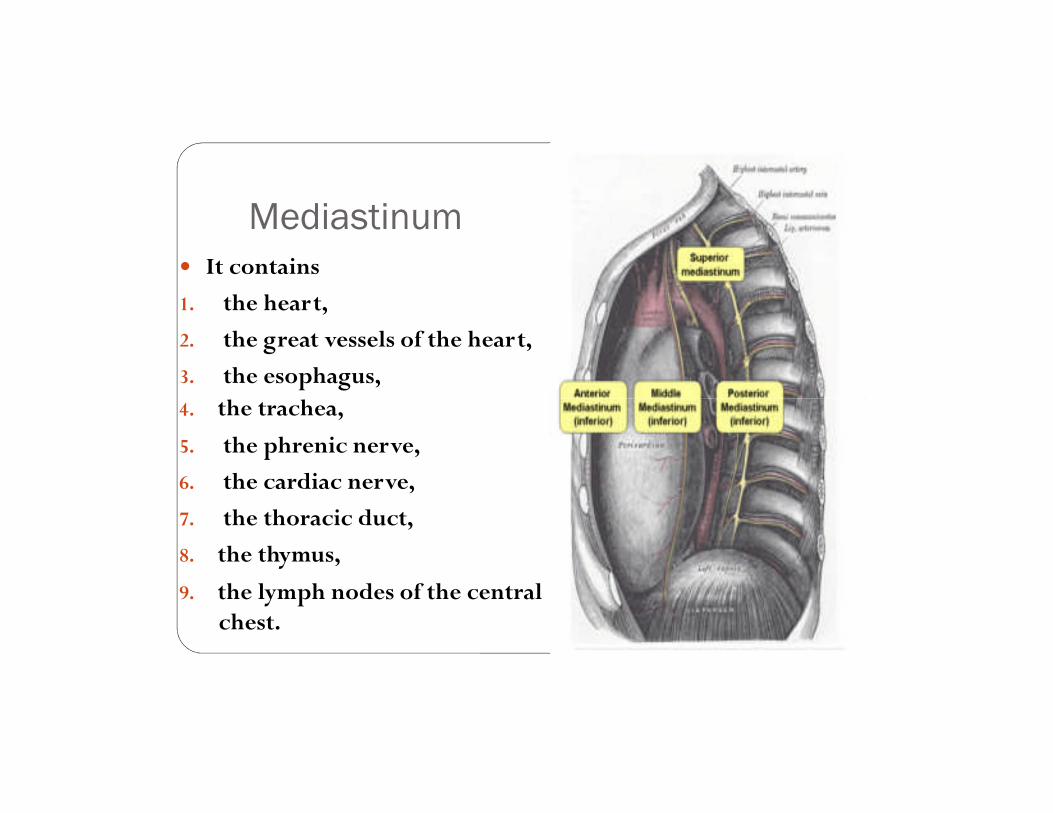
Mediastinum� It contains
1. the heart,
2. the great vessels of the heart,
3. the esophagus,4. the trachea,
5. the phrenic nerve,
6. the cardiac nerve,
7. the thoracic duct,
8. the thymus,
9. the lymph nodes of the central chest.
Serous Membrane Relationship
Figure 1.10a
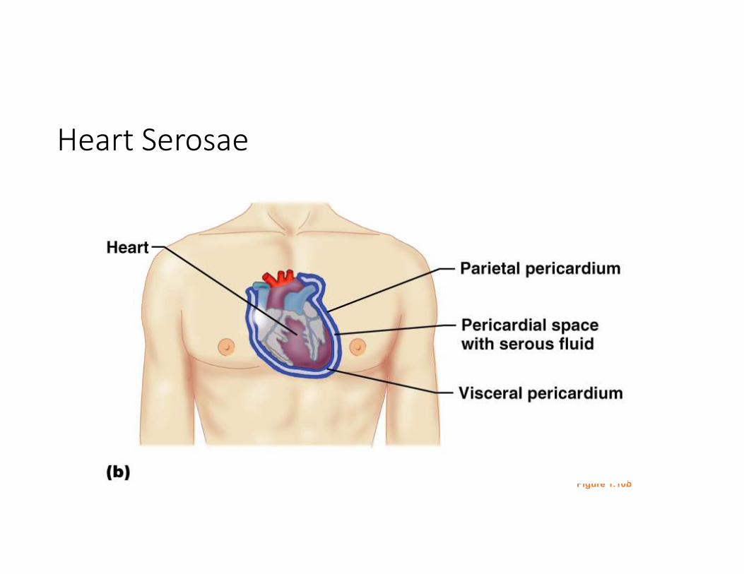
Heart Serosae
Figure 1.10b
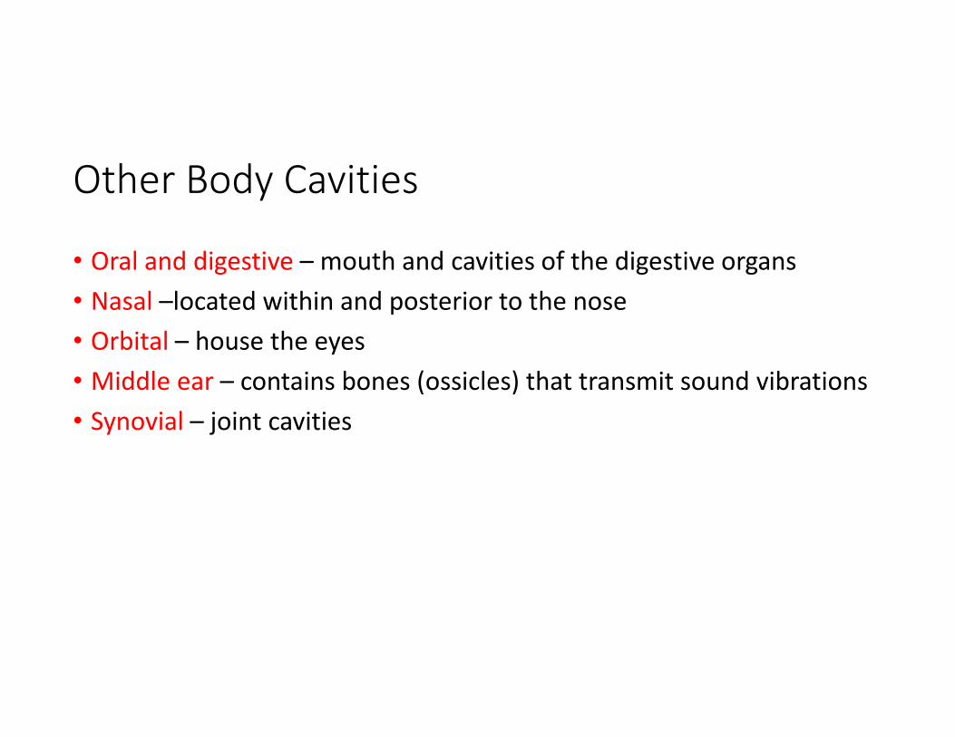
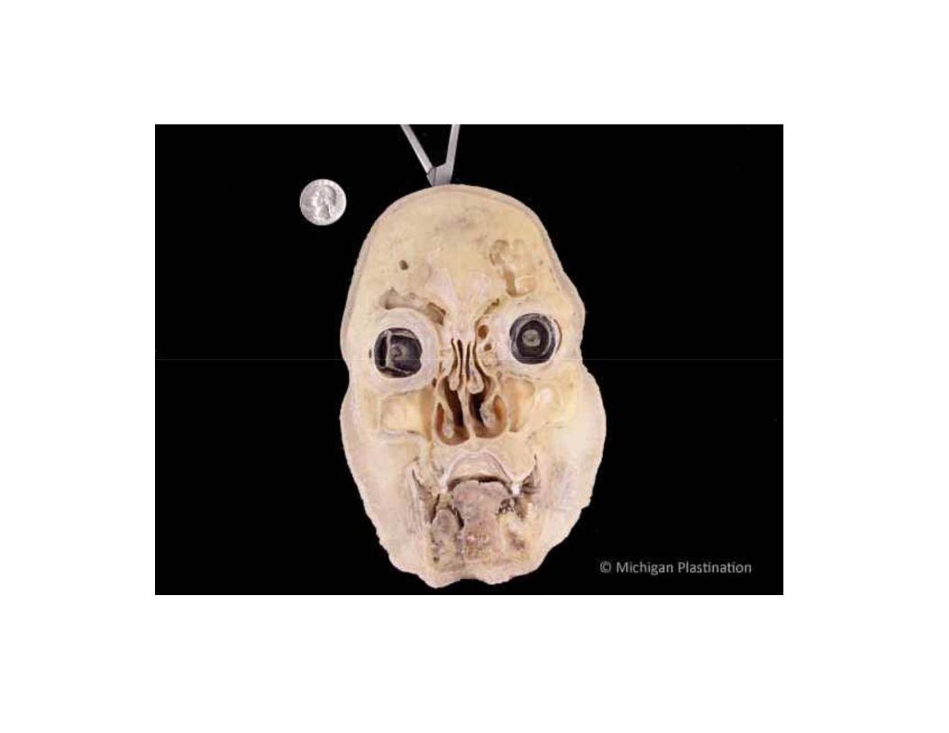
Other Body Cavities
• Oral and digestive – mouth and cavities of the digestive organs
• Nasal –located within and posterior to the nose
• Orbital – house the eyes
• Middle ear – contains bones (ossicles) that transmit sound vibrations
• Synovial – joint cavities
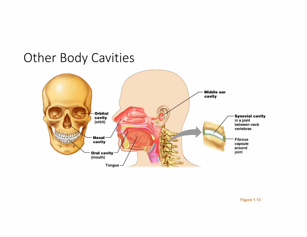
Other Body Cavities
Figure 1.13