Embed Size (px)
Citation preview
Int J Clin Exp Pathol 2017;10(1):446-452www.ijcep.com /ISSN:1936-2625/IJCEP0041249
Original ArticleAn inbred family with pulmonary alveolar microlithiasis in China: a genome-wide SNP study
Duchao Zhang1*, Kun Xiao1*, Wei Guan1*, Xiaohong Hu2, Peng Yan1, Lixin Xie1
1Department of Pulmonary & Critical Care Medicine Chinese PLA General Hospital, Beijing, China; 2Department of Pediatrics, Hospital Affiliated to Chinese PLA General Hospital (304 Hospital), Beijing, China. *Co-first authors.
Received October 6, 2016; Accepted October 20, 2016; Epub January 1, 2017; Published January 15, 2017
Abstract: Pulmonary alveolar microlithiasis (PAM) is a rare genetic disease that is characterized by the accumula-tion of calcium phosphate deposits in the alveolar spaces of the lung. The clinical characteristics of the patients with PAM in Mainland China were analyzed, and a high-density single nucleotide polymorphism (SNP) was used to analysis genome-wide of the patients’ genomic DNA. The two patients were sisters of an inbred family whose parents were cousins and presented typical manifestation of recurrent cough, progressive dyspnea. High resolu-tion computed tomography (HRCT) demonstrated the pulmonary was full of high density reflection of intraalveolar microliths especially in double lower lobe, and calcification was found in the pericardial, aorta and pleural. We found homozygous mutation of the SLC34A2 gene, c.910A>T (p.K304X) in exon 8 in two patients, and heterozygous muta-tion in consanguineous marriage of parents and the other family members. We concluded that a patient with an inbred family history and typical radiological features of high density intra-alveolar microlith, PAM should be highly suspected. The homozygous mutation in SLC34A2 gene, leading to a premature stop codon and a truncated protein, was responsible for PAM in the inbred family.
Keywords: Pedigree research, pulmonary alveolar microlithiasis, whole exon sequencing, single nucleotide poly-morphism
Introduction
Pulmonary alveolar microlithiasis (PAM) is a rare chronic lung disease with many microliths of calcium phosphate accumulate in intra-alve-olar [1]. Many patients with asymptomatic or only minor recurrent cough have normal pulmo-nary function or a mild restrictive pattern. Typical chest radiograph reveals sand-like micronodulation of calcified densities bilateral-ly, mainly in the middle and lower zones [2-4]. Recently, mutation of SLC34A2 gene, which encodes the sodium phosphate co-transporter (NaPi-IIb), is considered to be responsible for PAM [5-7]. However, its mutation symbols in dif-ferent cases are not investigated yet for its lim-ited data.
In this study we conducted human whole exon sequencing for an inbred family with pulmonary alveolar microlithiasis, screened the related gene mutation, in order to discover disease associated mutations of SLC34A2 gene and
provide meaningful references for the study of etiology and diagnosis of rare disease PAM.
Materials and methods
Subjects
Two PAM patients and other members of an inbred family (Figure 1) were recruited for this study. Patients were diagnosed based on char-acteristic computer tomography (CT) and pathology findings. Written informed consent was obtained from either the patient or from an authorized family member. This study was approved by the Ethics Committee of Chinese People’s Liberation Army General Hospital (approval number, S2015-067-01).
Case one
The proband (V4) was a 52-year-old female. In 2003, the patient was admitted to the hospital complaining of recurrent cough and dyspnea.
Pulmonary alveolar microlithiasis in China
447 Int J Clin Exp Pathol 2017;10(1):446-452
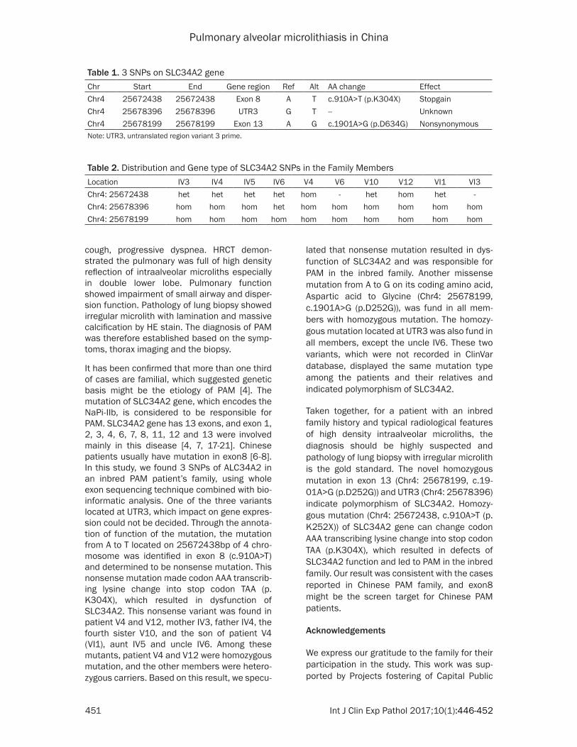
She was healthy in the past and denied history of smoking or medication. Her parents are con-sanguineous, and she has a son and four sis-ters. One sister (V12) of the patient had the same symptom, neither of the other family members complained of discomfort and their chest CT scans were normal. Physical examina-tion revealed crackle rales in both lung fields, without cyanosis of lips, venous varicose or bulb fingers. Laboratory tests revealed a decreased level of PaO2 (84.3 mmHg). The tumor index and concentration of serum calci-um were within the normal range. Spirometry examination showed slight restrictive ventilato-ry disturbances and moderate decreased dif-fusing function (vital capacity, 77.2% of predict-ed, forced expiratory volume in 1 s, 82.5% of predicted, carbon monoxide transfer factor-sin-gle breath, 47.0% of predicted). Chest comput-ed tomography (CT) scan showed high density reflection of intraalveolar sand-stones espe-cially in double lower lobe, and calcification of the mediastinal and interlobular pleura as well as the pericardium. Pathology of lung biopsy showed irregular microlith with lamination and massive calcification by hematoxylin-eosin (HE) stain. The final diagnosis was PAM based on characteristic CT and pathology findings.
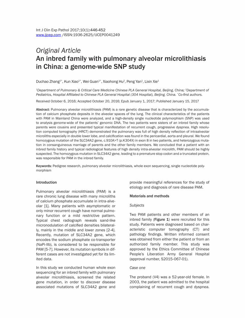
within the normal range. Pulmonary function test demonstrated moderate impairment of ventilation function (vital capacity, 95.2% of predicted, forced expiratory volume in 1 s, 94.8% of predicted, carbon monoxide transfer factor-single breath, 56.1% of predicted). CT scan showed high density reflection of intraal-veolar sand-stones especially in double lower lobe, pericardium and subpleural calcification shadow, and multiple calcified plaque of aortic and coronary artery (Figure 2A). Pathology of lung biopsy showed irregular microlith with lam-ination and massive calcification by HE stain (Figure 2B). The final diagnosis was PAM based on characteristic CT and pathology findings.
Genome whole exon sequencing
Blood samples were collected from the pa- tients, their parents, sisters, and the children. Genome DNA was extracted using the human blood DNA extraction kit (QIAGEN). The exon regions were enriched by SeqCap EZ human whole exon capture system of NimbleGen (Roche). After database setup, pair-end (double ends) sequencing was conducted following the instruction brochure using Illumina HiSeq2500 sequencing system. Preliminary data analysis and quality control were conducted for sequenc-ing results.
Figure 1. Pedigree Chart of Family History.
Case two
The proband (V12) was a 39-year-old female. In 2015, the patient was admitted to the hospital with the same complaining of recurrent cou- gh and exertional dyspnea. She was healthy in the past and denied history of smoking or medication. Physical exam-ination revealed normal brea- th sound. Laboratory tests revealed a decreased level of PaO2 (86.6 mmHg). Classi- fication of bronchoalveolar lavage fluid (BALF) reve- aled increased lymphocyte (19%, normal <13%) and de- creased macrophage (78%, normal >84%). The tumor in- dex, rheumatoid factor, immu-noglobulin, C-reactive protein, and concentration of serum calcium and phosphorus were
Pulmonary alveolar microlithiasis in China
448 Int J Clin Exp Pathol 2017;10(1):446-452
Figure 2. A. Pulmonary CT Results of Patient V12. Lung window image of CT scan of the chest showing intraalveolar sand-stones especially in double lower lobe, pericardium and subpleural calcification shadow, and multiple calcified plaque of aortic. Mediastinal window image showing calcification of the mediastinal pleura as well as the pericardium. B. Lung biopsy specimen of patient V12. Lung biopsy specimen of patient V12 stained with hema-toxylin and eosin (×200) showing calcium in alveolar spaces and in the lung parenchyma (red triangle).
Bioinformatic analysis of SNPs of SLA34A2
Based on the above analyzed results, low qual-ity SNPs were eliminated. The variants pub-lished in normal control individuals were also eliminated, including the common variant car-ried by normal individuals in the public genetic
mutation database 1000 ge- momes, Hapmap and dbSNP. Analysis of related functional pathway regulation and the history of PAM revealed that SLC34A2 gene mutation was probably related to encode the sodium phosphate co-transporter (NaPi-IIb), which is considered to be responsible for PAM.
Results
Data analysis of whole exon sequencing
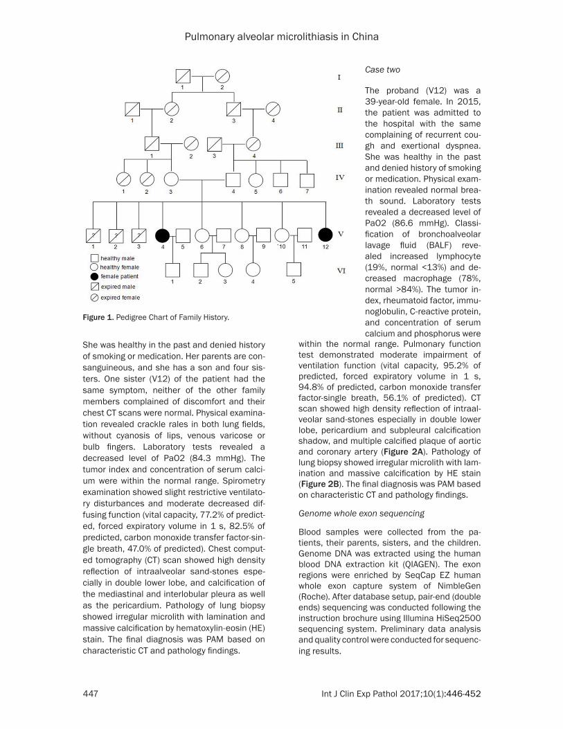
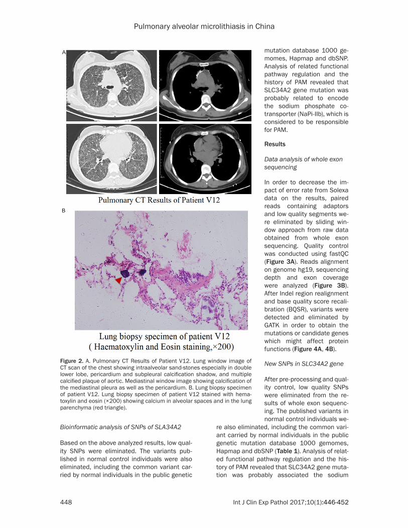
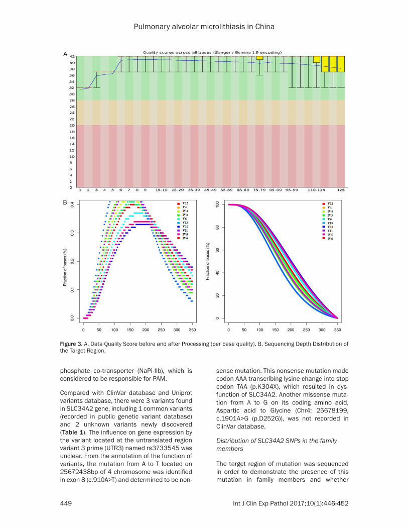
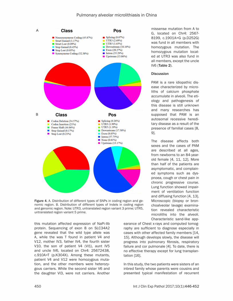
In order to decrease the im- pact of error rate from Solexa data on the results, paired reads containing adaptors and low quality segments we- re eliminated by sliding win-dow approach from raw data obtained from whole exon sequencing. Quality control was conducted using fastQC (Figure 3A). Reads alignment on genome hg19, sequencing depth and exon coverage were analyzed (Figure 3B). After Indel region realignment and base quality score recali-bration (BQSR), variants were detected and eliminated by GATK in order to obtain the mutations or candidate genes which might affect protein functions (Figure 4A, 4B).
New SNPs in SLC34A2 gene
After pre-processing and qual-ity control, low quality SNPs were eliminated from the re- sults of whole exon sequenc-ing. The published variants in normal control individuals we-
re also eliminated, including the common vari-ant carried by normal individuals in the public genetic mutation database 1000 gemomes, Hapmap and dbSNP (Table 1). Analysis of relat-ed functional pathway regulation and the his-tory of PAM revealed that SLC34A2 gene muta-tion was probably associated the sodium
Pulmonary alveolar microlithiasis in China
449 Int J Clin Exp Pathol 2017;10(1):446-452
phosphate co-transporter (NaPi-IIb), which is considered to be responsible for PAM.
Compared with ClinVar database and Uniprot variants database, there were 3 variants found in SLC34A2 gene, including 1 common variants (recorded in public genetic variant database) and 2 unknown variants newly discovered (Table 1). The influence on gene expression by the variant located at the untranslated region variant 3 prime (UTR3) named rs3733545 was unclear. From the annotation of the function of variants, the mutation from A to T located on 25672438bp of 4 chromosome was identified in exon 8 (c.910A>T) and determined to be non-
sense mutation. This nonsense mutation made codon AAA transcribing lysine change into stop codon TAA (p.K304X), which resulted in dys-function of SLC34A2. Another missense muta-tion from A to G on its coding amino acid, Aspartic acid to Glycine (Chr4: 25678199, c.1901A>G (p.D252G)), was not recorded in ClinVar database.
Distribution of SLC34A2 SNPs in the family members
The target region of mutation was sequenced in order to demonstrate the presence of this mutation in family members and whether
Figure 3. A. Data Quality Score before and after Processing (per base quality). B. Sequencing Depth Distribution of the Target Region.
Pulmonary alveolar microlithiasis in China
450 Int J Clin Exp Pathol 2017;10(1):446-452
this mutation affected expression of NaPi-IIb protein. Sequencing of exon 8 on SLC34A2 gene revealed that the wild type allele was A, while the was T found in patient V4 and V12, mother IV3, father IV4, the fourth sister V10, the son of patient V4 (VI1), aunt IV5 and uncle IV6, located on Chr4: 25672438, c.910A>T (p.K304X). Among these mutants, patient V4 and V12 were homozygous muta-tion, and the other members were heterozy-gous carriers. While the second sister V6 and the daughter VI3, were not carriers. Another
earance of Chest x-rays and computed tomog-raphy are sufficient to diagnose especially in cases with other affected family members [14, 15]. Although develops slowly, the disease will progress into pulmonary fibrosis, respiratory failure and cor pulmonale [4]. To date, there is no effective therapy except for lung transplan-tation [16].
In this study, the two patients were sisters of an inbred family whose parents were cousins and presented typical manifestation of recurrent
Figure 4. A. Distribution of different types of SNPs in coding region and ge-nomic region. B. Distribution of different types of Indels in coding region and genomic region. Note: UTR3, untranslated region variant 3 prime; UTR5, untranslated region variant 5 prime.
missense mutation from A to G, located on Chr4: 2567- 8199, c.1901A>G (p.D252G) was fund in all members with homozygous mutation. The homozygous mutation locat-ed at UTR3 was also fund in all members, except the uncle IV6 (Table 2).
Discussion
PAM is a rare idiopathic dis-ease characterized by micro-liths of calcium phosphate accumulate in alveoli. The eti-ology and pathogenesis of this disease is still unknown and many researches has supposed that PAM is an autosomal recessive heredi-tary disease as a result of the presence of familial cases [8, 9].
The disease affects both sexes and the cases of PAM are described at all ages, from newborns to an 84-year-old female [4, 11, 12]. More than half of the patients are asymptomatic, and complain- ed symptoms such as dys-pnoea, cough or chest pain in chronic progressive course. Lung function showed impair-ment of ventilation function and diffusing function [4, 13]. Microscopic (biopsy or bron-choalveolar lavage) examina-tion revealed characteristic microliths into the alveoli. Characteristic sand-like app-
Pulmonary alveolar microlithiasis in China
451 Int J Clin Exp Pathol 2017;10(1):446-452
cough, progressive dyspnea. HRCT demon-strated the pulmonary was full of high density reflection of intraalveolar microliths especially in double lower lobe. Pulmonary function showed impairment of small airway and disper-sion function. Pathology of lung biopsy showed irregular microlith with lamination and massive calcification by HE stain. The diagnosis of PAM was therefore established based on the symp-toms, thorax imaging and the biopsy.
It has been confirmed that more than one third of cases are familial, which suggested genetic basis might be the etiology of PAM [4]. The mutation of SLC34A2 gene, which encodes the NaPi-IIb, is considered to be responsible for PAM. SLC34A2 gene has 13 exons, and exon 1, 2, 3, 4, 6, 7, 8, 11, 12 and 13 were involved mainly in this disease [4, 7, 17-21]. Chinese patients usually have mutation in exon8 [6-8]. In this study, we found 3 SNPs of ALC34A2 in an inbred PAM patient’s family, using whole exon sequencing technique combined with bio-informatic analysis. One of the three variants located at UTR3, which impact on gene expres-sion could not be decided. Through the annota-tion of function of the mutation, the mutation from A to T located on 25672438bp of 4 chro-mosome was identified in exon 8 (c.910A>T) and determined to be nonsense mutation. This nonsense mutation made codon AAA transcrib-ing lysine change into stop codon TAA (p.K304X), which resulted in dysfunction of SLC34A2. This nonsense variant was found in patient V4 and V12, mother IV3, father IV4, the fourth sister V10, and the son of patient V4 (VI1), aunt IV5 and uncle IV6. Among these mutants, patient V4 and V12 were homozygous mutation, and the other members were hetero-zygous carriers. Based on this result, we specu-
lated that nonsense mutation resulted in dys-function of SLC34A2 and was responsible for PAM in the inbred family. Another missense mutation from A to G on its coding amino acid, Aspartic acid to Glycine (Chr4: 25678199, c.1901A>G (p.D252G)), was fund in all mem-bers with homozygous mutation. The homozy-gous mutation located at UTR3 was also fund in all members, except the uncle IV6. These two variants, which were not recorded in ClinVar database, displayed the same mutation type among the patients and their relatives and indicated polymorphism of SLC34A2.
Taken together, for a patient with an inbred family history and typical radiological features of high density intraalveolar microliths, the diagnosis should be highly suspected and pathology of lung biopsy with irregular microlith is the gold standard. The novel homozygous mutation in exon 13 (Chr4: 25678199, c.19- 01A>G (p.D252G)) and UTR3 (Chr4: 25678396) indicate polymorphism of SLC34A2. Homozy- gous mutation (Chr4: 25672438, c.910A>T (p.K252X)) of SLC34A2 gene can change codon AAA transcribing lysine change into stop codon TAA (p.K304X), which resulted in defects of SLC34A2 function and led to PAM in the inbred family. Our result was consistent with the cases reported in Chinese PAM family, and exon8 might be the screen target for Chinese PAM patients.
Acknowledgements
We express our gratitude to the family for their participation in the study. This work was sup-ported by Projects fostering of Capital Public
Table 1. 3 SNPs on SLC34A2 geneChr Start End Gene region Ref Alt AA change EffectChr4 25672438 25672438 Exon 8 A T c.910A>T (p.K304X) StopgainChr4 25678396 25678396 UTR3 G T -- UnknownChr4 25678199 25678199 Exon 13 A G c.1901A>G (p.D634G) NonsynonymousNote: UTR3, untranslated region variant 3 prime.
Table 2. Distribution and Gene type of SLC34A2 SNPs in the Family MembersLocation IV3 IV4 IV5 IV6 V4 V6 V10 V12 VI1 VI3Chr4: 25672438 het het het het hom - het hom het -Chr4: 25678396 hom hom hom het hom hom hom hom hom homChr4: 25678199 hom hom hom hom hom hom hom hom hom hom
Pulmonary alveolar microlithiasis in China
452 Int J Clin Exp Pathol 2017;10(1):446-452
Health by the Science and Technology Com- mission of Beijing (No. Z11110706730000).
Disclosure of conflict of interest
None.
Address correspondence to: Lixin Xie, Department of Pulmonary & Critical Care Medicine, Chinese PLA General Hospital, Beijing 100853, China. Tel: 86-10-55499128; Fax: 86-10-55499128; E-mail: xielx- [email protected]
References
[1] Santos MK. Diagnosis of pulmonary alveolar microlithiasis. Radiol Bras 2015; 48: IX-X.
[2] Ganesan N, Ambroise MM, Ramdas A, Kisku KH, Singh K and Varghese RG. Pulmonary al-veolar microlithiasis: an interesting case re-port with systematic review of Indian literature. Front Med 2015; 9: 229-238.
[3] Jönsson ÅL, Simonsen U, Hilberg O and Bend-strup E. Pulmonary alveolar microlithiasis: two case reports and review of the literature. Eur Respir Rev 2012; 21: 249-256.
[4] Castellana G, Castellana G, Gentile M, Castel-lana R and Resta O. Pulmonary alveolar micro-lithiasis: review of the 1022 cases reported worldwide. Eur Respir Rev 2015; 24: 607-20.
[5] Vismara MF, Colao E, Fabiani F, Bombardiere F, Tamburrini O, Alessio C, Manti F, Pelaia G, Ro-meo P, Iuliano R and Perrotti N. The sodium-phosphate co-transporter SLC34A2, and pul-monary alveolar microlithiasis: Presentation of an inbred family and a novel truncating muta-tion in exon 3. Respir Med Case Rep 2015; 16: 77-80.
[6] Wang H, Yin X, Wu D and Jiang X. SLC34A2 gene compound heterozygous mutation identi-fication in a patient with pulmonary alveolar microlithiasis and computational 3D protein structure prediction. Meta Gene 2014; 2: 557-64.
[7] Yin X, Wang H, Wu D, Zhao G, Shao J and Dai Y. SLC34A2 Gene mutation of pulmonary alveo-lar microlithiasis: report of four cases and re-view of literatures. Respir Med 2013; 107: 217-22.
[8] Zhong YQ, Hu CP, Cai XD and Nie HP. A novel mutation of the SLC34A2 gene in a Chinese pedigree with pulmonary alveolar microlithia-sis. Zhonghua Yi Xue Yi Chuan Xue Za Zhi 2009; 26: 365-368.
[9] Dogan OT, Ozsahin SL, Gul E, Arslan S, Koksal B, Berk S, Ozdemir O and Akkurt I. A frame-shift mutation in the SLC34A2 gene in three patients with pulmonary alveolar microlithiasis in an inbred family. Intern Med 2010; 49: 45-49.
[10] Stefani M. La microlitiasi alveolare polmonare. Anat Patol Oncol 1968; 34: 485-526.
[11] Dahabreh M, Najada A. Pulmonary alveolar mi-crolithiasis in an 8-month-old infant. Ann Trop Paediatr 2009; 29: 55-59.
[12] Krishnakurup J, Abdelsayed G. The calcareous lung. Mayo Clin Proc 2011; 86: 85.
[13] Ferreira Francisco FA, Pereira e Silva JL, Hoch-hegger B, Zanetti G and Marchiori E. Pulmo-nary alveolar microlithiasis. State-of-the-art re-view. Respir Med 2013; 107: 1-9.
[14] Ch’ng LS, Bux SI, Liam CK, Rahman NA and Ho CY. Sandstorm appearance of pulmonary alve-olar microlithiasis incidentally detected in a young, asymptomatic male. Korean J Radiol 2013; 14: 859-862.
[15] Francisco FA, Rodrigues RS, Barreto MM, Es-cuissato DL, Araujo Neto CA, Silva JL, Silva CS, Hochhegger B, Souza AS Jr, Zanetti G and Mar-chiori E. Can chest high-resolution computed tomography findings diagnose pulmonary al-veolar microlithiasis? Radiol Bras 2015; 48: 205-210.
[16] Güçyetmez B, Ogan A, Cimet Ayyıldız A, Yalçın Güder B and Klepetko W. Lung transplantation in an intensive care patient with pulmonary al-veolar microlithiasis-a case report. F1000Res 2014; 3: 118.
[17] Corut A, Senyigit A, Ugur SA, Altin S, Ozcelik U, Calisir H, Yildirim Z, Gocmen A and Tolun A. Mutations in SLC34A2 cause pulmonary alveo-lar microlithiasis and are possibly associated with testicular microlithiasis. Am J Hum Genet 2006; 79: 650-656.
[18] Ishihara Y, Hagiwara K, Zen K, Huqun, Hoso-kawa Y and Natsuhara A. A case of pulmonary alveolar microlithiasis with an intragenetic de-letion in SLC34A2 detected by a genome-wide SNP study. Thorax 2009; 64: 365-367.
[19] Dogan OT, Ozsahin SL, Gul E, Arslan S, Koksal B, Berk S, Ozdemir O and Akkurt I. A frame-shift mutation in the SLC34A2 gene in three patients with pulmonary alveolar microlithiasis in an inbred family. Intern Med 2010; 49: 45-9.
[20] Özbudak IH, Başsorgun CI, Ozbılım G, Lülecı G, Sarper A, Erdoğan A, Taylan F and Altiok E. Pul-monary alveolar microlithiasis with homozy-gous c.316G>C (p.G106R) mutation: a case report. Turk Patoloji Derg 2012; 28: 282-285.
[21] Jönsson ÅL, Hilberg O, Bendstrup EM, Mo-gensen S and Simonsen U. SLC34A2 gene mu-tation may explain comorbidity of pulmonary alveolar microlithiasis and aortic valve sclero-sis. Am J Respir Crit Care Med 2012; 185: 464.