Embed Size (px)
Citation preview

Death Protein 5 and p53-Upregulated Modulator ofApoptosis Mediate the Endoplasmic ReticulumStress–Mitochondrial Dialog Triggering LipotoxicRodent and Human b-Cell ApoptosisDaniel A. Cunha,
1Mariana Igoillo-Esteve,
1Esteban N. Gurzov,
1Carla M. Germano,
1
Najib Naamane,1Ihsane Marhfour,
1Makiko Fukaya,
1Jean-Marie Vanderwinden,
2Conny Gysemans,
3
Chantal Mathieu,3Lorella Marselli,
4Piero Marchetti,
4Heather P. Harding,
5David Ron,
5
Décio L. Eizirik,1and Miriam Cnop
1,6
Environmental factors such as diets rich in saturated fats contributeto dysfunction and death of pancreatic b-cells in diabetes. Endo-plasmic reticulum (ER) stress is elicited in b-cells by saturated fattyacids. Here we show that palmitate-induced b-cell apoptosis is me-diated by the intrinsic mitochondrial pathway. By microarray anal-ysis, we identified a palmitate-triggered ER stress gene expressionsignature and the induction of the BH3-only proteins death protein5 (DP5) and p53-upregulated modulator of apoptosis (PUMA).Knockdown of either protein reduced cytochrome c release, cas-pase-3 activation, and apoptosis in rat and human b-cells. DP5induction depends on inositol-requiring enzyme 1 (IRE1)–depen-dent c-Jun NH2-terminal kinase and PKR–like ER kinase (PERK)–induced activating transcription factor (ATF3) binding to its pro-moter. PUMA expression is also PERK/ATF3-dependent, throughtribbles 3 (TRB3)–regulated AKT inhibition and FoxO3a activation.DP5
2/2 mice are protected from high fat diet–induced loss ofglucose tolerance and have twofold greater pancreatic b-cell mass.This study elucidates the crosstalk between lipotoxic ER stressand the mitochondrial pathway of apoptosis that causes b-celldeath in diabetes. Diabetes 61:2763–2775, 2012
The global prevalence of type 2 diabetes (T2D)has reached 250 million and is projected to in-crease to nearly 400 million by 2030 (1). Theconsumption of a hypercaloric diet rich in satu-
rated fats, in the setting of an unfavorable genetic back-ground, is causal in the pathogenesis of T2D. Insulinresistance in peripheral tissues is a prime feature of T2D,but diabetes only develops in patients who are unable to
sustain a compensatory increase in insulin release by thepancreatic b-cells. The loss of b-cell mass by apoptosiscontributes to the progressive b-cell failure in T2D (2).Dietary or adipocyte-derived free fatty acids (FFAs) con-tribute both to peripheral insulin resistance and to b-celldysfunction and death in T2D. Chronic high-fat diet (HFD)leads to hyperglycemia as a consequence of impaired in-sulin secretion and insulin resistance (3,4), and in vitroexposure to saturated and, to a lesser extent, unsaturatedFFAs induces b-cell dysfunction and death (5,6).
How FFAs cause b-cell apoptosis is not well under-stood. Several studies have indicated endoplasmic reticulum(ER) stress as a potential mediator (7–10). Perturbations inER function initiate the unfolded protein response (UPR)governed by three ER transmembrane proteins: PKR–likeER kinase (PERK), inositol-requiring kinase-1 (IRE1), andactivating transcription factor (ATF) 6. The prime functionof the UPR is to restore ER homeostasis by reducingprotein load and increasing ER folding capacity and mis-folded protein degradation. Attenuation of protein trans-lation is executed by PERK through phosphorylation of theeukaryotic translation initiation factor 2a (eIF2a), whereasthe two other branches increase ER function by upregu-lating ER chaperones and the ER-associated protein deg-radation machinery. Nonresolved ER stress induces celldeath (11,12). ER stress may contribute to b-cell failure inT2D, because increased expression of markers of ERstress, including ATF3 and C/EBP homologous protein(CHOP; also known as GADD153 or DDIT3), has beenfound in islets from T2D patients (11). Prolonged eIF2aphosphorylation induces b-cell apoptosis through the mi-tochondrial pathway of cell death (13–15), but how lip-otoxic ER stress response culminates in the triggering andexecution of b-cell apoptosis remains unknown.
The intrinsic or mitochondrial pathway of cell death istightly modulated by proteins of the B-cell lymphoma (Bcl)2 family, composed of antiapoptotic (Bcl-2, Bcl-XL, Bcl-w,myeloid cell leukemia sequence [Mcl] 1, and A1) and proa-poptotic members that are further subdivided in multido-main (Bax, Bak, and Bok) and BH3-only proteins (deathprotein 5 [DP5, also known as harakiri], Bim, Bid, p53-upregulated modulator of apoptosis [PUMA, also known asBBC3], Bad, and Noxa) (16). Apoptosis starts with Baxtranslocation from the cytosol to the mitochondrial mem-brane, where it oligomerizes with Bak. The pore formed byBax/Bak causes mitochondrial outer membrane permea-bilization, allowing soluble proteins such as cytochrome c to
From the 1Laboratory of Experimental Medicine, Université Libre de Bruxelles,Brussels, Belgium; the 2Laboratory of Neurophysiology, Université Libre deBruxelles, Brussels, Belgium; the 3Laboratory of Experimental MedicineEndocrinology (LEGENDO), Faculty of Medicine, Katholieke UniversiteitLeuven, Leuven, Belgium; the 4Department of Endocrinology and Metabo-lism, University of Pisa, Pisa, Italy; the 5University of Cambridge MetabolicResearch Laboratories and NIHR Cambridge Biomedical Research Centre,Cambridge, U.K.; and the 6Division of Endocrinology, Erasmus Hospital,Université Libre de Bruxelles, Brussels, Belgium.
Corresponding author: Miriam Cnop, [email protected] 7 February 2012 and accepted 4 May 2012.DOI: 10.2337/db12-0123This article contains Supplementary Data online at http://diabetes
.diabetesjournals.org/lookup/suppl/doi:10.2337/db12-0123/-/DC1.E.N.G. is currently affiliated with the Department of Biochemistry and Molecular
Biology, School of Biomedical Sciences, Monash University, Melbourne,Australia.
� 2012 by the American Diabetes Association. Readers may use this article aslong as the work is properly cited, the use is educational and not for profit,and the work is not altered. See http://creativecommons.org/licenses/by-nc-nd/3.0/ for details.
diabetes.diabetesjournals.org DIABETES, VOL. 61, NOVEMBER 2012 2763
ORIGINAL ARTICLE

diffuse to the cytosol. The subsequent formation of theapoptosome leads to caspase 9 activation and caspase3–mediated cell death. The BH3-only proteins induce ap-optosis in a cell type- and stimulus-dependent fashion.These proteins are essential initiators of apoptosis by theirability to bind and inhibit prosurvival Bcl-2 members andby their direct activation of Bax and Bak (17). The BH3-only proteins are transcriptionally regulated, and some,including Bim and Bid, are posttranslationally modified.The c-Jun NH2-terminal kinase (JNK) plays an importantrole in the regulation of the mitochondrial pathway ofapoptosis (18). JNK activates Bax, Bim, and Bad by phos-phorylation (19–21) and upregulates Bim, PUMA, and DP5 inhepatocytes and neurons (22,23).
In this study, we have used global gene expression an-alyses followed by a comprehensive series of focusedexperiments to characterize the pathways of apoptosis inlipotoxic b-cell death and their regulation by the ER stressresponse. We demonstrate that the activation of JNK andPERK by palmitate contributes to induction of the BH3-only proteins DP5 and PUMA, and we clarify the ER stress–mitochondrial dialog triggering lipotoxic b-cell apoptosis,thus suggesting novel targets for the prevention of b-celldemise in early T2D.
RESEARCH DESIGN AND METHODS
Culture of INS-1E and primary fluorescence activated cell sorter–purified
rat b- and human islet cells and functional studies. The culture of INS-1Eand primary fluorescence activated cell sorter–purified rat b-cells is describedin the Supplementary Data.
Human islets (from 4 donors aged 63 6 7 years, BMI 25 6 1 kg/m2, cause ofdeath cerebral hemorrhage) were isolated by collagenase digestion and den-sity gradient purification. The islets were cultured, dispersed, and transfectedas described previously (24). The percentage of b-cells, examined by insulinimmunofluorescence (9), was 69 6 11%.
For FFA exposure, primary b-cells were cultured in medium with 1% BSAwithout FBS, and INS-1E cells were cultured in medium with 1% FBS and 1%BSA (5). Oleate and palmitate (Sigma, Schnelldorf, Germany) were dissolvedin 90% ethanol (6) and used at a final concentration of 0.5 mmol/L, which in thepresence of 1% BSA results in unbound FFA concentrations in the nanomolarrange (5). The chemical JNK inhibitor SP600125 (Sigma) and the peptide JNKinhibitor L-TAT-JNKi were used at 10 mmol/L for 2 h before and during FFAexposure (9). The IRE1 inhibitor 4m8C was used at 25 mmol/L during FFAexposure (25).
Details on the microarray analysis, real-time PCR primers, RNA interfer-ence, chromatin immunoprecipitation (ChIP), promoter reporter assay, andantibodies are provided in the Supplementary Data.Assessment of b-cell apoptosis. Apoptotic b-cells and INS-1E cells werecounted in fluorescence microscopy after staining with the DNA-binding dyespropidium iodide (5 mg/mL) and Hoechst 33342 (10 mg/mL) (15). Apoptosiswas confirmed by additional methods, including Bax translocation, caspase9 and 3 cleavage, and cytochrome c release, measured as described else-where (13).Mouse studies. Male DP5
2/2 and wild type (WT) C57BL/6 J mice were givenstandard chow (fat 10% of caloric intake, D12450B) or HFD (fat 60% of caloricintake, D12492; Research Diets). After 12 weeks, intraperitoneal glucose tol-erance tests were done in 16-h fasted mice by injecting glucose (2 mg/g bodyweight). Blood was collected from the tail at times 0, 15, 30, 60, 90, and 120min, and glucose was measured with a FreeStyle Lite glucometer (AbbottDiabetes Care). Plasma was separated by centrifugation (10 min, 5000g) andstored at 220°C. Plasma insulin was measured with the Ultrasensitive MouseELISA Kit (Crystal Chem, Downers Grove, IL). For the intraperitoneal insulintolerance test, 0.75 mU/g body weight Actrapid (Novo Nordisk) was injectedafter a 4-h fast, and glucose was measured at the time points listed previously.b-Cell mass. Pancreases were taken from mice killed after 25 weeks on thedifferent diets. Three nonconsecutive 5-mm thick pancreatic sections 150 mmapart were labeled with a standard immunoperoxidase method for paraffinsections with a mouse anti-insulin antibody (1/4000; Sigma). The b-cell masswas quantified blindly as b-cell volume density multiplied by pancreas weight(26). To assess b-cell size, nuclear crowding was determined as the number ofb-cell nuclei per 100 mm2 b-cell cytoplasm (27). The b-cell proliferation wasexamined as described in the Supplementary Data.
Glucose-stimulated insulin secretion. After isolation, DP52/2 and WT
mouse islets were cultured for 30 min in M199 medium containing 5.6 mmol/Lglucose and washed with modified Krebs-Ringer bicarbonate HEPES solution(150 mmol/L NaCl, 3.5 mmol/L KCl, 0.5 mmol/L MgCl2, 1.5 mmol/L CaCl2,5 mmol/L NaHCO3, 0.5 mmol/L NaH2PO4, 10 mmol/L HEPES, and 0.1% BSA atpH 7.4). Insulin secretion was induced by 1 h of incubation with Krebs-Ringerbicarbonate HEPES solution containing 1.7 or 16.7 mmol/L glucose. Insulinwas measured by ELISA in cell-free supernatants and acid-ethanol extractedcell lysates.Statistical analysis. Data are presented as means 6 SE of the indicatednumber (n) of independent experiments. Comparisons were performed byANOVA, followed by paired t test with the Bonferroni correction for multiplecomparisons. P , 0.05 was considered statistically significant.
RESULTS
Palmitate induces b-cell death through the mitochon-drial pathway of apoptosis. Palmitate induced cyto-chrome c release from the mitochondria (Fig. 1A–C), Baxtranslocation from the cytosol to the mitochondria (Fig.1B-D), and caspase 9 and 3 activation (Fig. 1E), demon-strating that palmitate engages the intrinsic pathway ofapoptosis in pancreatic b-cells. Oleate induces less celldeath (5,13) and does not stimulate cytochrome c release.In a time-course study, palmitate decreased Bcl-2 proteinexpression from 16 h on, independently of BCL2 mRNAexpression (Supplementary Fig. 1). Knockdown of Bcl-2induced apoptosis and sensitized cells to FFAs (Supple-mentary Fig. 1B). Palmitate also reduced Bcl-XL proteinlevels though at a late time-point of 24 h only (Supple-mentary Fig. 2A). Similar to Bcl-2, Bcl-XL knockdown in-duced b-cell apoptosis (Supplementary Fig. 2B). Theseresults confirm the antiapoptotic role of Bcl-2 and Bcl-XLin b-cells under basal and lipotoxic conditions. The mito-chondrial morphology of palmitate-treated cells was punc-tate, as opposed to the reticular adenosine 5�-triphosphate(ATP) synthase b staining in control cells (SupplementaryFig. 1C and D), and this coincided with the marked de-crease in Bcl-2 protein expression, suggesting that Bcl-2depletion by palmitate contributes to disruption of the mi-tochondrial network.Palmitate modulates cell death and other gene net-works in b-cells. We next profiled the global gene ex-pression of INS-1E cells exposed to palmitate for 6 and 14 h.In the array analysis, 20,405 probe sets corresponding to10,524 genes were detected as present in control or pal-mitate-treated b-cells. Palmitate modified the expressionof 6.7% (792 genes) and 8.2% (1,074 genes) of probe sets at6 and 14 h, respectively. The top functional clusters in-cluded gene expression, lipid metabolism, cell growth,cellular function and maintenance, and cellular compromiseat 6 h and cell death, cellular function, cellular compromise,protein synthesis, and lipid metabolism at 14 h. Two net-works were enriched in proapoptotic signaling molecules.At 6 h, palmitate upregulated markers of ER stress, in-cluding ATF4, ATF3, CHOP, growth arrest and DNA damageprotein 34, and TRB3 (Supplementary Fig. 3), which is inkeeping with our previous findings on the proapoptoticsignaling in the PERK branch of the UPR (9,15). At 14 h,we detected a network of mitochondrial cell death genescontaining DP5 and PUMA, PERK, and Jun upregulations(Supplementary Fig. 4).
Genes were also classified by a previously describedmanual curation (28) according to their potential role inb-cell dysfunction and death (Supplementary Table 1).Palmitate decreased the expression of glucose metabolismgenes involved in glycolysis and the citric acid cycle, andupregulated lipid metabolism genes such as Acsl, Cpt1a,
DP5 AND PUMA LINK LIPOTOXIC ER STRESS TO APOPTOSIS
2764 DIABETES, VOL. 61, NOVEMBER 2012 diabetes.diabetesjournals.org

FIG. 1. Palmitate induces apoptosis through the mitochondrial pathway. A: Representative immunofluorescence images of INS-1E cells exposed topalmitate for 24 h and stained for cytochrome c (green) and DNA (with Hoechst 33342, blue) (bar 20 mm). B: Representative immunofluorescencepictures of INS-1E cells treated with palmitate for 24 h and stained for Bax (red), ATP synthase b (green), and DNA (Hoechst, blue) (barrepresents 20 mm). Arrows point to cells with Bax translocation to the mitochondria. C: Percentage of INS-1E cells with mitochondrial Baxtranslocation or cytochrome c release after palmitate treatment for the indicated periods (n = 3–4).D: Confocal microscopy of INS-1E cells treatedwith palmitate for 24 h and stained for Bax (green), ATP synthase b (red), and DNA (Hoechst, blue) (top). Scatterplots of fluorescence intensitiesmeasured by line scanning in confocal microscopy images for Bax and ATP synthase b of 3 control and 4 palmitate-treated cells. Linear regressionshows a tight correlation in palmitate-treated cells (bottom right) compared with control condition (bottom left), suggestive of Bax translocationto the mitochondria. E: Time course of caspase 9 and 3 activation analyzed by Western blot in palmitate-treated INS-1E cells. A representative blotof 3 independent experiments is shown. *P < 0.05 against control. (A high-quality digital representation of this figure is available in the onlineissue.)
D.A. CUNHA AND ASSOCIATES
diabetes.diabetesjournals.org DIABETES, VOL. 61, NOVEMBER 2012 2765

and Fads1. The AP-1 family of transcription factors, in-cluding c-Jun and c-Fos, was induced at both time points.Genes mediating ER function and signal transduction inthe different branches of the UPR were induced, includingATF3, ATF4, CHOP, and TRB3 and also Sec61a1 andDnajc3 (Supplementary Table 1). The mRNA expressionsof prodeath genes such as Bax, Bak, and Bim were notsignificantly modified; however, the BH3-only proteinsDP5 and PUMA were markedly upregulated. On the basisof these gene expression analyses, these two Bcl-2 mem-bers were further studied for their role in mitochondrialb-cell death.The BH3-only proteins DP5 and PUMA contribute topalmitate-induced apoptosis. By real-time PCR, weconfirmed the array finding that palmitate induced DP5expression, with a maximum after 16 h (Fig. 2A). On theother hand, oleate did not inducedDP5 expression (Fig. 2A).Efficient DP5 knockdown by RNA interference (Fig. 2B)reduced cytochrome c release, caspase 3 activation, andapoptosis in palmitate-treated cells (Fig. 2C and D) but didnot prevent the mild oleate-induced apoptosis (Fig. 2D).Palmitate induced DP5 in primary rat b-cells (Fig. 2E), andDP5 knockdown partially protected b-cells against palmitate(Fig. 2E). In human islet cells, palmitate also inducedDP5 (Fig. 2F), and DP5 knockdown protected cells fromlipotoxicity (Fig. 2F).
PUMA was markedly induced by palmitate, peaking at16 h, but not by oleate (Fig. 3A). Knockdown of PUMA(Fig. 3B) decreased palmitate-induced cytochrome c re-lease, caspase 3 activation, and apoptosis (Fig. 3C and D),whereas it did not affect oleate toxicity (Fig. 3D). Palmi-tate also upregulated PUMA expression in fluorescenceactivated cell sorter–purified rat b-cells and human isletcells (Fig. 3E and F). PUMA RNA interference partially(Fig. 3E) and nearly completely (Fig. 3F) prevented pal-mitate toxicity in, respectively, primary rat b-cells andhuman islet cells. As a negative control, we studied a genethat was not detected as changed in the array analysis,namely Bim. Unlike DP5 and PUMA, Bim was not inducedby palmitate (Supplementary Fig. 2C), and knockdown ofits protein product did not modify apoptosis rates (Sup-plementary Fig. 2D), confirming that Bim does not playa major role in lipotoxic b-cell death.JNK mediates the lipotoxic induction of DP5 but notPUMA. To assess the mechanism by which palmitateinduces DP5 expression, a DP5 promoter luciferase re-porter was used. The promoter sequence between 2125 to285 is important for DP5 induction by palmitate, becauseits deletion reduced promoter activity by 42% comparedwith the full construct (Fig. 4A). This promoter region hasa conserved ATF site where c-Jun binds after phosphory-lation by JNK (23). Palmitate activated JNK and its targetc-Jun from 30 min onward, peaking at 4–8 h (Supplemen-tary Fig. 5A and B). JNK inhibition by SP600125 reducedc-Jun phosphorylation (Fig. 4B) and abrogated palmitate-induced DP5 promoter activation and mRNA expressionboth in INS-1E cells (Fig. 4C and D) and in primary b-cells(Fig. 4D). The small peptide JNK inhibitor L-TAT-JNKi,previously shown to inhibit JNK and protect b-cells againstpalmitate treatment (9), produced a similar reduction ofc-Jun phosphorylation and decreased palmitate-inducedDP5 expression (Supplementary Fig. 5C and D). The p-c-Jun binding to the DP5 promoter was increased by pal-mitate as observed in ChIP experiments (Fig. 4E), suggestingthat p-c-Jun transcriptionally induces DP5. IRE1a knock-down (by 766 3%) reduced XBP1 splicing (by 46 6 6%) and
decreased palmitate-induced JNK phosphorylation (Fig. 4F).Additional support for the role of IRE1 was provided byusing the novel IRE1 inhibitor 4m8C (25). This small mol-ecule very effectively blocked IRE1, inhibiting splicedXBP1 expression by 96 6 1%, and significantly decreasedJNK phosphorylation and DP5 expression (Fig. 4G and H),confirming that IRE1-JNK mediates the DP5 induction.4m8C did not inhibit JNK phosphorylation by interleukin-1b and interferon-g, suggesting it does not directly in-activate JNK (data not shown).
PUMA expression is controlled by different transcrip-tion factors, including p53 and nuclear factor-kB (NF-kB)(29,30). Time course experiments showed that p53 is notinduced by palmitate in INS-1E cells, suggesting a p53-independent PUMA regulation in b-cells (SupplementaryFig. 5E). The activation of the PUMA promoter by palmi-tate was not affected by mutation of the NF-kB binding site(Supplementary Fig. 5F), previously shown to be impor-tant for cytokine-mediated PUMA upregulation (31). Fur-thermore, PUMA promoter activity and PUMA mRNAinduction were not modified by JNK inhibitors (Supple-mentary Fig. 5G–I), suggesting a different role for JNK inthe regulation of the DP5 and PUMA promoters.PERK-ATF3 signaling induces DP5 and PUMA. ThePERK pathway and its downstream effectors CHOP andTRB3 are proapoptotic in palmitate-treated b-cells (9,15,32).Knockdown of PERK by small interfering RNA (siRNA)decreased basal and palmitate-induced eIF2a phosphory-lation (Supplementary Fig. 6A–C) but did not prevent JNKactivation (Supplementary Fig. 6D). PERK knockdownreduced DP5 and PUMA inductions by palmitate (Fig. 5Aand B). Knockdown of ATF4 or CHOP did not affect theinduction of either BH3-only protein by palmitate (Sup-plementary Fig. 6E–H). ATF3 siRNA, however, mimickedthe effect of PERK siRNA on DP5 and PUMA expressions(Fig. 5A and B), suggesting that PERK-ATF3 activation bypalmitate leads to DP5 and PUMA expression. In primaryb-cells, knockdown of PERK or ATF3 also decreased pal-mitate induction of DP5 and PUMA (Fig. 5C). This isconsistent with the presence of a putative ATF3 bindingsite in the DP5 promoter region 2125 to 285. Exposure ofINS-1E cells to palmitate stimulated the association ofATF3 to the DP5 promoter, showing that this BH3-onlyprotein is a direct target of the PERK-ATF3 pathway (Fig. 5D).The PUMA promoter, on the other hand, does not containan ATF3 binding site, suggesting that ATF3 indirectlymodulates PUMA.TRB3-AKT-FoxO3a signaling contributes to the in-duction of PUMA by palmitate. In the microarray anal-ysis, palmitate markedly upregulated TRB3 (SupplementaryFig. 3 and Supplementary Table 1), an ER stress–inducibleand proapoptotic gene in the PERK pathway (33). Becauseknockdown of CHOP did not change TRB3 expression(Supplementary Fig. 6G), we examined the role of ATF3.ATF3 knockdown attenuated TRB3 induction by palmitate(Fig. 5E). TRB3 is a pseudokinase that inhibits AKT phos-phorylation, leading to the activation of death pathwaysincluding GSK-3b. Because AKT inactivates the forkheadbox O (FoxO) family of transcription factors, among whichFoxO3a has been shown to induce PUMA-dependent apo-ptosis, we examined whether the TRB3-AKT-FoxO3a path-way plays a role in the induction of DP5 and PUMA bypalmitate. The induction of TRB3 mRNA by palmitate wasconfirmed at the protein level (Fig. 5F), and the FFA de-creased both AKT and FoxO3a phosphorylation (Fig. 5Fand Fig. 6A). The dephosphorylation of FoxO3a (Fig. 6A)
DP5 AND PUMA LINK LIPOTOXIC ER STRESS TO APOPTOSIS
2766 DIABETES, VOL. 61, NOVEMBER 2012 diabetes.diabetesjournals.org

was accompanied by its nuclear translocation (Fig. 6B and C),confirming activation of the transcription factor. Theknockdown of TRB3 partially prevented early changes inAKT and FoxO3a phosphorylation (Fig. 5F). In parallel, TRB3knockdown decreased palmitate-induced PUMA expression(but not DP5 expression), and reduced lipotoxic b-cell apo-ptosis (Fig. 5G and H). Additional TRB3/AKT-independentmechanisms may regulate FoxO3a activation at later time
points. In silico analysis of the DP5 and PUMA promotersidentified putative FoxO3a binding sites in both BH3-only genes, and ChIP demonstrated that palmitate inducesFoxO3a binding to both promoters (Fig. 6D). FoxO3aknockdown partially prevented induction of DP5 and PUMAby palmitate (Fig. 6E–F), showing that the AKT-FoxO3apathway downstream of ER stress controls the BH3-onlygene expression and contributes to b-cell death.
FIG. 2. Palmitate-induced DP5 expression contributes to b-cell death. A: Time-course analysis of DP5 mRNA expression in oleate- or palmitate-treated INS-1E cells (n = 4). B: DP5 mRNA expression of INS-1E cells transfected with negative or DP5 siRNA and treated with palmitate for 16 h(n = 5). C: Cytoplasmic cytochrome c and cleaved caspase 3 levels of INS-1E cells transfected with negative (N) or DP5 (D) siRNA and palmitatetreated for 16 h. Apoptosis-inducing factor (AIF) expression was used as mitochondrial control, b-actin was used as cytoplasmic control, anda-tubulin was used as a control for protein loading for the caspase 3 blot. Separate blots on the right show the noncytosolic fraction, includingmitochondria, used as a positive control for cytochrome c and AIF blotting. A representative blot of 5 independent experiments is shown. D:Apoptosis in INS-1E cells transfected with negative or DP5 siRNA and then treated with oleate or palmitate for 16 h (n = 3–4). E: DP5 mRNAexpression in primary rat b-cells transfected with negative or DP5 siRNA and treated with palmitate for 24 h, after which apoptosis was assessed(n = 4). F: Protection from palmitate-induced cell death by DP5 knockdown in dispersed human islet cells. Cells were transfected with DP5 siRNAand 2 days later exposed to palmitate for 24 h (n = 4). GAPDH, glyceraldehyde-3-phosphate dehydrogenase. *P < 0.05 against untreated cells.#P < 0.05.
D.A. CUNHA AND ASSOCIATES
diabetes.diabetesjournals.org DIABETES, VOL. 61, NOVEMBER 2012 2767

DP52/2mice are protected from HFD-induced loss of
glucose tolerance and have increased b-cell mass.Because DP5 is upstream of PUMA (17) and DP5 but notPUMA knockdown provides complete b-cell protectionfrom palmitate at early time points (data not shown), weselected DP5
2/2 mice for in vivo studies. DP52/2 and WTmice were given standard chow or HFD (fat 60% of caloric
intake). After 12 weeks, the two HFD-fed genotypes hadsimilar weight gain and insulin sensitivity (Fig. 7A and B).In line with this, similar weight gain, perirenal fat accu-mulation and hepatic insulin-induced AKT phosphoryla-tion were observed in WT or DP5 knockout mice after 25weeks of chow or HFD (data not shown). DP5 mRNA ex-pression increased threefold in islets from HFD-fed WT mice
FIG. 3. Palmitate-induced PUMA expression contributes to b-cell death. A: Time-course analysis of PUMA mRNA expression in oleate- andpalmitate-treated INS-1E cells (n = 4). B: PUMA mRNA expression of INS-1E cells transfected with negative or PUMA siRNA and treated withpalmitate for 16 h (n = 5). C: Cytoplasmic cytochrome c and cleaved caspase 3 levels in INS-1E cells transfected with negative (N) or PUMA (P)siRNA and palmitate treated for 16 h. Apoptosis-inducing factor (AIF) expression was used as mitochondrial control, b-actin was used as cyto-plasmic control, and a-tubulin was used as a control for protein loading for the cleaved caspase 3 blot. Separate blots on the right show thenoncytosolic fraction, including mitochondria, used as a positive control for cytochrome c and AIF blotting. A representative blot of 5 independentexperiments is shown. D: Apoptosis in INS-1E cells transfected with negative or PUMA siRNA and then treated with oleate or palmitate for 16 h(n = 3–4). E: PUMA mRNA expression in primary rat b-cells transfected with negative or PUMA siRNA and treated with palmitate for 24 h, afterwhich apoptosis was assessed (n = 4). F: Protection from palmitate-induced cell death by PUMA knockdown in dispersed human islet cells. Cellswere transfected with PUMA siRNA and 2 days later exposed to palmitate for 24 h (n = 4). GAPDH, glyceraldehyde-3-phosphate dehydrogenase.*P < 0.05 against untreated cells. #P < 0.05.
DP5 AND PUMA LINK LIPOTOXIC ER STRESS TO APOPTOSIS
2768 DIABETES, VOL. 61, NOVEMBER 2012 diabetes.diabetesjournals.org

FIG. 4. JNK activation contributes to DP5 induction by palmitate in b-cells. A: DP5 promoter study of INS-1E cells transfected with DP5 promoterfragments of different lengths (left) and the control plasmid CMV-RL and then treated with palmitate for 16 h. Firefly luciferase (LUC) activity wasnormalized to Renilla luciferase activity and expressed as fold induction of control (n = 4). B: JNK and c-Jun phosphorylation in INS-1E cells treatedwith palmitate or the JNK inhibitor SP600125 (10 mmol/L) for 16 h. A representative blot of 3 independent experiments is shown. C: DP5 promoterstudy of INS-1E cells transfected with the full DP5 promoter and treated with the JNK inhibitor SP600125 as in B (n = 4).D: DP5mRNA expression ofINS-1E cells (left) or primary b-cells (right) exposed to palmitate for 16 h or 24 h, respectively, in the presence or absence of SP600125 (n = 4). E:ChIP showing the binding of p-c-Jun to theDP5 promoter in INS-1E cells treated with palmitate (PAL) or left untreated (CTL) for 4 h (n = 3). Sampleswere incubated with p-c-Jun antibody (IP) or goat serum (serum, negative control). Input is total DNA from the samples. A representative image of 3independent experiments is shown. The glyceraldehyde-3-phosphate dehydrogenase (GAPDH) promoter was used as a negative control. F: INS-1E cellswere transfected with negative (N) or IRE1a (I) siRNA and after 3 days treated with palmitate for the indicated times. A representative blot for IRE1aprotein expression and JNK protein phosphorylation is shown (n = 4). G and H: JNK phosphorylation (G) and DP5 mRNA expression (H) (n = 3–4) inINS-1E cells treated with palmitate and/or the IRE1 inhibitor 4m8C (25 mmol/L) for 16 h. InG, a representative image of 4 independent experiments isshown. *P < 0.05 against untreated cells. #P < 0.05.
D.A. CUNHA AND ASSOCIATES
diabetes.diabetesjournals.org DIABETES, VOL. 61, NOVEMBER 2012 2769

FIG. 5. The PERK-ATF3 pathway modulates DP5 and PUMA expressions. A: PERK and ATF3 protein expressions of INS-1E cells transfected withnegative, ATF3, or PERK siRNA and treated 3 days after with palmitate (PAL) or left untreated (CTL) for 16 h. A representative blot of 3 in-dependent experiments is shown. B: DP5 and PUMA mRNA expressions of INS-1E cells transfected and treated as in A (n = 4). C: DP5 and PUMAmRNA expressions of primary rat b-cells transfected with negative, ATF3, or PERK siRNA and 3 days after treated with palmitate for 24 h. D: ChIPshowing the binding of ATF3 to the DP5 promoter in INS-1E cells treated with palmitate (PAL) or left untreated (CTL) for 8 h (n = 3). Sampleswere incubated with ATF3 antibody (IP) or goat serum (serum, negative control). Input is total DNA from the samples. The glyceraldehyde-3-phosphate dehydrogenase (GAPDH) promoter was used as a negative control. A representative image of 3 independent experiments is shown. E:ATF3 and TRB3 protein expression of INS-1E cells transfected with negative (N) or ATF3 (A3) siRNA and treated with palmitate for 16 h. Arepresentative blot of 3 independent experiments is shown. F: FoxO3a and AKT protein phosphorylation and TRB3 protein expression in INS-1Ecells transfected with negative (N) or TRB3 (T) siRNA and treated with palmitate (PAL) or left untreated (CTL) for the indicated times. Arepresentative blot of 4 independent experiments is shown. G: DP5 and PUMA mRNA expressions in INS-1E cells transfected with negative orTRB3 siRNA and treated with palmitate for 16 h (n = 4). H: Apoptosis in INS-1E cells transfected and treated as in G (n = 4). *P < 0.05 againstuntreated cells. #P < 0.05.
DP5 AND PUMA LINK LIPOTOXIC ER STRESS TO APOPTOSIS
2770 DIABETES, VOL. 61, NOVEMBER 2012 diabetes.diabetesjournals.org
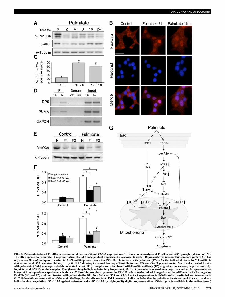
FIG. 6. Palmitate-induced FoxO3a activation modulates DP5 and PUMA expressions. A: Time-course analysis of FoxO3a and AKT phosphorylation of INS-1E cells exposed to palmitate. A representative blot of 5 independent experiments is shown. B and C: Representative immunofluorescence picture (B, barrepresents 20 mm) and quantification (C) of FoxO3a-positive nuclei in INS-1E cells treated with palmitate (PAL) for the indicated times. In B, FoxO3a isstained red and DNA is stained blue (n = 3). D: ChIP showing increased binding of FoxO3a to the DP5 and PUMA promoters in INS-1E cells treated for 4 hwith palmitate (PAL) as compared with untreated cells (CTL). Samples were incubated with FoxO3a antibody (IP) or goat serum (serum, negative control).Input is total DNA from the samples. The glyceraldehyde-3-phosphate dehydrogenase (GAPDH) promoter was used as a negative control. A representativeimage of 3 independent experiments is shown. E: FoxO3a protein expression in INS-1E cells transfected with negative or two different siRNAs targetingFoxO3a (F1 and F2) and then treated with palmitate for 16 h (n = 3–4). F:DP5 and PUMAmRNA expressions in INS-1E cells transfected and treated as inE. G: Schematic representation of the main findings; for details see text. Thick arrow up indicates induction by palmitate treatment and thick arrow downindicates downregulation. *P < 0.05 against untreated cells. #P < 0.05. (A high-quality digital representation of this figure is available in the online issue.)
D.A. CUNHA AND ASSOCIATES
diabetes.diabetesjournals.org DIABETES, VOL. 61, NOVEMBER 2012 2771

FIG. 7. DP5 knockout mice are partially protected from HFD-induced impaired glucose tolerance. A: Weight gains of WT or DP52/2mice receiving
standard chow (C, dotted line) and HFD (full line) were similar. B: Blood glucose levels during intraperitoneal insulin tolerance test (n = 8–15). Cand D: Blood glucose (C) and plasma insulin (D) levels during intraperitoneal glucose tolerance test (n = 8–15). E and F: Glucose-stimulatedinsulin secretion (E) and insulin content (F) in isolated islets, normalized by total protein content. Insulin secretion at 16.7 mmol/L glucose wassignificantly higher than that at 1.7 mmol/L for all conditions (P< 0.01; n = 4). G: Cell death in WT and DP52/2
mouse islets treated for 2 days with0.5 mmol/L palmitate (n = 3). H: Pancreatic b-cell mass was assessed in WT and DP52/2
mice maintained on chow or HFD for a total period of 25weeks (n = 5–6). I: The number of Ki67-positive (dividing) b-cells per islet increased in the HFD-fed mice (n = 3–4). J: b-Cell nuclear crowding wasmeasured to evaluate b-cell hypertrophy (n = 3–4). *P< 0.05 for the comparison between genotypes on the same diet. #P< 0.05 for the comparisonbetween chow and HFD. §P < 0.05 as indicated.
DP5 AND PUMA LINK LIPOTOXIC ER STRESS TO APOPTOSIS
2772 DIABETES, VOL. 61, NOVEMBER 2012 diabetes.diabetesjournals.org
compared with chow-fed WT mice (P = 0.17, n = 5–6).DP5
2/2 mice were protected from HFD-induced diabetes(Fig. 7C) as a result of strikingly increased insulin secre-tion (Fig. 7D). Islets isolated from HFD-fed DP5
2/2 micecontained and secreted twofold more insulin after glucosestimulation (Fig. 7E-F), suggesting a protective effect atthe b-cell level. The lack of the DP5 gene also protectedmouse islets in vitro from palmitate-induced cell death(Fig. 7G). This resistance to lipotoxicity was paralleled bya doubling of b-cell mass in HFD-fed DP5
2/2 mice but notin WT mice (Fig. 7H), confirming the physiological role ofDP5 in vivo. We could not detect apoptotic b-cells in vivoin HFD-fed WT mice (data not shown) and were thereforeunable to assess the putative protective role of DP5 de-ficiency. Increased b-cell proliferation, assessed by Ki67staining (Fig. 7I), and b-cell hypertrophy (Fig. 7J) likelycontribute to the greater b-cell mass of DP52/2 mice. ThusDP5 may not only play a role in b-cell apoptosis but alsoserve as a brake on b-cell growth and proliferation.
DISCUSSION
Failure of the pancreatic b-cells to compensate for highinsulin needs is central to the pathogenesis of T2D, andseveral studies have reported a significant decrease in b-cellmass in T2D (2,26). Chronic exposure to FFAs causes lossof functional b-cell mass (34,35) and may contribute to T2D.Saturated lipids are harmful to b-cells, and pronounced ERstress signaling (especially in the PERK pathway) has beenproposed to mediate lipotoxic apoptosis (7–10,15). Execu-tion of FFA-induced apoptosis occurs through the mito-chondrial pathway, as indicated by cytochrome c releasefrom the mitochondria after palmitate treatment (13,36,37),but it remained to be clarified how palmitate-induced ERstress crosstalks with the mitochondria to culminate inb-cell death. We have now answered this question, showingthat palmitate transcriptionally induces the BH3-only pro-teins DP5 and PUMA through PERK-dependent ATF3 ex-pression (Fig. 6G).
In the hierarchical model for Bax/Bak activation, theBH3-only “sensitizer” proteins DP5, Bad, Bik, Bnip3, Bmf,and Noxa neutralize the prosurvival Bcl-2 proteins, allowingthe BH3-only “activators” PUMA, Bim, and tBid to be re-leased to activate Bax/Bak (38,39). The use of the differentpro- and antiapoptotic proteins after a proapoptotic stimu-lus is cell and context dependent.
Palmitate decreases Bcl-2 expression (13); however itscontribution to lipotoxic b-cell death had not been exam-ined. We now show that the decreased Bcl-2 protein ex-pression in palmitate-exposed b-cells occurs in conjunctionwith mitochondrial fragmentation and apoptosis. A reduc-tion in Bcl-XL protein was seen only after 24 h of palmitateexposure, suggesting that decreased Bcl-XL levels are notpivotal in palmitate-induced apoptosis. A role for Mcl-1protein downregulation has also been reported in palmitate-and ER stress–induced b-cell apoptosis (40).
The BH3-only prodeath sensitizers and activators inlipotoxicity are essentially unknown. Gene expression pro-filing of palmitate-treated b-cells indicated that palmitateinduced a gene expression signature of ER stress at theearly time-point (6 h). At a later time-point (14 h), palmitateupregulated apoptosis-related genes, shifting from anadaptive response to one geared for cell death. As part ofthe proapoptotic signal network, there was upregulation ofDP5 and PUMA, genes for two BH3-only proteins previouslyshown to play a role in apoptosis of b-cells exposed to
cytokines (31,41). Knockdown of the BH3-only sensitizerDP5 and the activator PUMA protected b-cells from palmi-tate, establishing their contribution to saturated FFA-in-duced apoptosis. Because the protection is partial in ratb-cells, other BH3-only proteins or alternative death mech-anisms may play additional roles. Of note, oleate did notincrease DP5 or PUMA expression, in keeping with the ob-servation that this FFA is much less toxic (5,9).
We next set out to identify the mechanism of inductionof these prodeath proteins by palmitate. Although the up-stream UPR has been characterized in detail, the down-stream transduction mechanisms through which ER stressconnects to mitochondrial apoptosis have remained elu-sive. By promoter analysis, we identified a putative c-Junbinding site in the DP5 promoter, and confirmed p-c-Junbinding by ChIP. Inhibition of IRE1-dependent JNK acti-vation reduced DP5 promoter activation and mRNA in-duction by palmitate. These results are consistent with ourprevious finding that JNK inhibition protects againstlipotoxic b-cell death (9). JNK regulates the intrinsic deathpathway through transcriptional as well as posttranscriptionalmodifications of Bcl-2 family members (42,43).
The BH3-only activator PUMA was not induced by JNK,p53, or NF-kB, previously shown to modulate PUMA ex-pression (29–31,44). We therefore examined the role of ERstress in PUMA expression. Of the three branches of theUPR, sustained activation of the PERK pathway plays alarge part in b-cell apoptosis (14,15). Downstream ofPERK, CHOP contributes to palmitate-induced apoptosis(9). CHOP deletion also improves glycemic control andprevents b-cell loss in mouse models of T2D (45). ATF3 isalso induced by PERK; its role in b-cell death is morecontroversial. ATF3 knockout confers minor protectionagainst cytokine-induced islet cell apoptosis (46) which mayinvolve the regulation of the insulin receptor substrate2 (47). In HFD-fed mice, on the other hand, ATF3 is protectiveby regulating insulin synthesis (48). We have now demon-strated that PERK-induced ATF3, but not ATF4-CHOP,induces both PUMA and DP5. Interestingly, the mecha-nisms by which ATF3 regulates DP5 and PUMA expres-sions are not similar. ATF3 directly modulates DP5 mRNAexpression, as suggested by its binding to the DP5 pro-moter, whereas PUMA regulation depends on a complexpathway involving TRB3, AKT, and FoxO3a (Fig. 6G).
TRB3 was identified as a proapoptotic effector of CHOPand ATF4 (49). Increased TRB3 expression was observedin islets from T2D donors and HFD-fed mice (50). In thesame study, interaction between TRB3 and ATF4 de-creased insulin secretion by reducing the expression ofexocytosis-related genes. We show here that TRB3 is in-duced by ATF3 but not CHOP and causes FoxO3a-dependent PUMA induction and cell death. In other celltypes, FoxO3a has been shown to induce PUMA expres-sion independently of p53 (51). We also demonstrated thatDP5 upregulation is partially FoxO3a dependent, which isconsistent with the presence of a putative FoxO3a bindingsite in its promoter. This DP5 induction by FoxO3a is ap-parently TRB3-independent, suggesting alternative FoxO3aactivation mechanism.
The physiological relevance of these findings was ex-amined in DP5
2/2 mice exposed to HFD. Compared withWT mice, the DP5
2/2 mice were resistant to lipotoxic lossof glucose tolerance as a result of significantly greaterglucose-stimulated insulin secretion and an adaptive in-crease in b-cell mass. Interestingly, b-cell proliferation andhypertrophy were increased in HFD-fed DP5
2/2 mice,
D.A. CUNHA AND ASSOCIATES
diabetes.diabetesjournals.org DIABETES, VOL. 61, NOVEMBER 2012 2773

pointing to a hitherto unknown inhibitory role of DP5 incell growth and the cell cycle. Because the mice werewhole-body knockouts, it cannot be excluded that DP5 de-ficiency also exerts beneficial effects in peripheral tissues.
In conclusion, lipotoxic ER stress causes JNK- andPERK-dependent ATF3 and TRB3-FOXO3a activation, andthese transcription factors regulate expression of Bcl-2family members. Palmitate engages the mitochondrial path-way of cell death through induction of the BH3-only sensitizerDP5, loss of antiapoptotic Bcl-2 and Mcl-1, and upregula-tion of the BH3-only activator PUMA, culminating in theactivation of Bax/Bak and mitochondrial permeabilization(Fig. 6G). These results provide insight into the mechanismsof lipotoxic b-cell ER stress and identify hitherto unknowntranscriptional regulation and signal transduction betweenER stress and mitochondrial apoptosis. The findings alsodelineate potential new areas for b-cell therapy in the pre-vention and treatment of T2D.
ACKNOWLEDGMENTS
This work was supported by the European Union (Collab-orative Projects CEED3 and BetaBat in the FrameworkProgram 7), a European Foundation for the Study of Diabetes(EFSD)/Lilly grant, the Fonds National de la RechercheScientifique (FNRS), Fonds de la Recherche ScientifiqueMédicale (FRSM) and Actions de Recherche Concertées dela Communauté Française (ARC), Belgium. D.A.C. was sup-ported by an FNRS post-doctoral fellowship, E.N.G. was sup-ported by an EMBO long-term fellowship, and J.-M.V. isresearch director at the FNRS.
No potential conflicts of interest relevant to this articlewere reported.
D.A.C., E.N.G., D.L.E, and M.C. contributed to theexperimental design of the study. D.A.C., M.I.-E., E.N.G.,C.M.G., N.N., I.M., M.F., J.-M.V., C.G., and D.R. carriedout experiments, helped with data analysis, or both. C.M.,L.M., P.M., H.P.H., and D.R. contributed materials and datainterpretation. D.A.C., M.I.-E., E.N.G., D.L.E, and M.C.analyzed and interpreted the data. D.A.C., D.L.E, and M.C.wrote the manuscript. M.C. is the guarantor of this workand, as such, had full access to all the data in the study andtakes responsibility for the integrity of the data and theaccuracy of the data analysis.
The authors thank Mingtao Li, Sun Yat-sen University,for providing the DP5 promoter; Lin Zhang, University ofPittsburgh, for the PUMA promoter; Andrea Jurisicova,University of Toronto, and Gabriel Nuñez, University ofMichigan, for the DP5
2/2 mice; Miriam HernangomezHerrero, Université Libre de Bruxelles, for help with themouse experiments; and Gilbert Vandenbroeck, Marie-Anne Neef, Maryse Urbain, Anyishai Musuaya, StephanieMertens, and Rachid Makhnas, all from Université Librede Bruxelles, for expert technical assistance.
REFERENCES
1. International Diabetes Federation. IDF Diabetes Atlas. 4th ed. Brussels,International Diabetes Federation Executive Office, 2009
2. Butler AE, Janson J, Bonner-Weir S, Ritzel R, Rizza RA, Butler PC. b-celldeficit and increased b-cell apoptosis in humans with type 2 diabetes.Diabetes 2003;52:102–110
3. Swinburn BA, Boyce VL, Bergman RN, Howard BV, Bogardus C. De-terioration in carbohydrate metabolism and lipoprotein changes inducedby modern, high fat diet in Pima Indians and Caucasians. J Clin EndocrinolMetab 1991;73:156–165
4. Kaiyala KJ, Prigeon RL, Kahn SE, Woods SC, Porte D Jr, Schwartz MW.Reduced b-cell function contributes to impaired glucose tolerance in
dogs made obese by high-fat feeding. Am J Physiol 1999;277:E659–E667
5. Cnop M, Hannaert JC, Hoorens A, Eizirik DL, Pipeleers DG. Inverse re-lationship between cytotoxicity of free fatty acids in pancreatic islet cellsand cellular triglyceride accumulation. Diabetes 2001;50:1771–1777
6. Zhou YP, Grill VE. Long-term exposure of rat pancreatic islets to fattyacids inhibits glucose-induced insulin secretion and biosynthesis througha glucose fatty acid cycle. J Clin Invest 1994;93:870–876
7. Kharroubi I, Ladrière L, Cardozo AK, Dogusan Z, Cnop M, Eizirik DL. Freefatty acids and cytokines induce pancreatic b-cell apoptosis by differentmechanisms: role of nuclear factor-kB and endoplasmic reticulum stress.Endocrinology 2004;145:5087–5096
8. Karaskov E, Scott C, Zhang L, Teodoro T, Ravazzola M, Volchuk A.Chronic palmitate but not oleate exposure induces endoplasmic reticulumstress, which may contribute to INS-1 pancreatic b-cell apoptosis. Endo-crinology 2006;147:3398–3407
9. Cunha DA, Hekerman P, Ladrière L, et al. Initiation and execution of lip-otoxic ER stress in pancreatic b-cells. J Cell Sci 2008;121:2308–2318
10. Laybutt DR, Preston AM, Akerfeldt MC, et al. Endoplasmic reticulumstress contributes to beta cell apoptosis in type 2 diabetes. Diabetologia2007;50:752–763
11. Eizirik DL, Cardozo AK, Cnop M. The role for endoplasmic reticulumstress in diabetes mellitus. Endocr Rev 2008;29:42–61
12. Ron D, Walter P. Signal integration in the endoplasmic reticulum unfoldedprotein response. Nat Rev Mol Cell Biol 2007;8:519–529
13. Cunha DA, Ladrière L, Ortis F, et al. Glucagon-like peptide-1 agonistsprotect pancreatic b-cells from lipotoxic endoplasmic reticulum stressthrough upregulation of BiP and JunB. Diabetes 2009;58:2851–2862
14. Ladrière L, Igoillo-Esteve M, Cunha DA, et al. Enhanced signaling down-stream of ribonucleic acid-activated protein kinase-like endoplasmic re-ticulum kinase potentiates lipotoxic endoplasmic reticulum stress inhuman islets. J Clin Endocrinol Metab 2010;95:1442–1449
15. Cnop M, Ladriere L, Hekerman P, et al. Selective inhibition of eukaryotictranslation initiation factor 2 a dephosphorylation potentiates fatty acid-induced endoplasmic reticulum stress and causes pancreatic b-cell dys-function and apoptosis. J Biol Chem 2007;282:3989–3997
16. Youle RJ, Strasser A. The BCL-2 protein family: opposing activities thatmediate cell death. Nat Rev Mol Cell Biol 2008;9:47–59
17. Kim H, Rafiuddin-Shah M, Tu HC, et al. Hierarchical regulation of mito-chondrion-dependent apoptosis by BCL-2 subfamilies. Nat Cell Biol 2006;8:1348–1358
18. Tournier C, Hess P, Yang DD, et al. Requirement of JNK for stress-inducedactivation of the cytochrome c-mediated death pathway. Science 2000;288:870–874
19. Kim BJ, Ryu SW, Song BJ. JNK- and p38 kinase-mediated phosphorylationof Bax leads to its activation and mitochondrial translocation and toapoptosis of human hepatoma HepG2 cells. J Biol Chem 2006;281:21256–21265
20. Lei K, Davis RJ. JNK phosphorylation of Bim-related members of the Bcl2family induces Bax-dependent apoptosis. Proc Natl Acad Sci USA 2003;100:2432–2437
21. Donovan N, Becker EB, Konishi Y, Bonni A. JNK phosphorylation andactivation of BAD couples the stress-activated signaling pathway to thecell death machinery. J Biol Chem 2002;277:40944–40949
22. Akazawa Y, Cazanave S, Mott JL, et al. Palmitoleate attenuates palmitate-induced Bim and PUMA up-regulation and hepatocyte lipoapoptosis.J Hepatol 2010;52:586–593
23. Ma C, Ying C, Yuan Z, et al. dp5/HRK is a c-Jun target gene and required forapoptosis induced by potassium deprivation in cerebellar granule neurons.J Biol Chem 2007;282:30901–30909
24. Santin I, Moore F, Colli ML, et al. PTPN2, a candidate gene for type 1diabetes, modulates pancreatic b-cell apoptosis via regulation of the BH3-only protein Bim. Diabetes 2011;60:3279–3288
25. Cross BC, Bond PJ, Sadowski PG, et al. The molecular basis for selectiveinhibition of unconventional mRNA splicing by an IRE1-binding smallmolecule. Proc Natl Acad Sci USA 2012;109:E869–E878
26. Rahier J, Guiot Y, Goebbels RM, Sempoux C, Henquin JC. Pancreatic b-cellmass in European subjects with type 2 diabetes. Diabetes Obes Metab2008;10(Suppl. 4):32–42
27. Sempoux C, Guiot Y, Lefevre A, et al. Neonatal hyperinsulinemic hypo-glycemia: heterogeneity of the syndrome and keys for differential di-agnosis. J Clin Endocrinol Metab 1998;83:1455–1461
28. Ortis F, Naamane N, Flamez D, et al. Cytokines interleukin-1b and tumornecrosis factor-a regulate different transcriptional and alternative splicingnetworks in primary b-cells. Diabetes 2010;59:358–374
29. Nakano K, Vousden KH. PUMA, a novel proapoptotic gene, is induced byp53. Mol Cell 2001;7:683–694
DP5 AND PUMA LINK LIPOTOXIC ER STRESS TO APOPTOSIS
2774 DIABETES, VOL. 61, NOVEMBER 2012 diabetes.diabetesjournals.org

30. Wang P, Qiu W, Dudgeon C, et al. PUMA is directly activated by NF-kB andcontributes to TNF-a-induced apoptosis. Cell Death Differ 2009;16:1192–1202
31. Gurzov EN, Germano CM, Cunha DA, et al. p53 up-regulated modulator ofapoptosis (PUMA) activation contributes to pancreatic b-cell apoptosisinduced by proinflammatory cytokines and endoplasmic reticulum stress.J Biol Chem 2010;285:19910–19920
32. McCullough KD, Martindale JL, Klotz LO, Aw TY, Holbrook NJ. Gadd153sensitizes cells to endoplasmic reticulum stress by down-regulatingBcl2 and perturbing the cellular redox state. Mol Cell Biol 2001;21:1249–1259
33. Qian B, Wang H, Men X, et al. TRIB3 is implicated in glucotoxicity- andendoplasmic reticulum stress-induced b-cell apoptosis [published correc-tion appears in J Endocrinol 2009;200:243 and 2009;203:399]. J Endocrinol2008;199:407–416
34. Cnop M, Igoillo-Esteve M, Cunha DA, Ladrière L, Eizirik DL. An update onlipotoxic endoplasmic reticulum stress in pancreatic b-cells. Biochem SocTrans 2008;36:909–915
35. Poitout V. Glucolipotoxicity of the pancreatic beta-cell: myth or reality?Biochem Soc Trans 2008;36:901–904
36. Maedler K, Spinas GA, Dyntar D, Moritz W, Kaiser N, Donath MY. Distincteffects of saturated and monounsaturated fatty acids on b-cell turnoverand function. Diabetes 2001;50:69–76
37. Maestre I, Jordán J, Calvo S, et al. Mitochondrial dysfunction is involved inapoptosis induced by serum withdrawal and fatty acids in the b-cell lineINS-1. Endocrinology 2003;144:335–345
38. Kuwana T, Bouchier-Hayes L, Chipuk JE, et al. BH3 domains of BH3-onlyproteins differentially regulate Bax-mediated mitochondrial membranepermeabilization both directly and indirectly. Mol Cell 2005;17:525–535
39. Ren D, Tu HC, Kim H, et al. BID, BIM, and PUMA are essential for acti-vation of the BAX- and BAK-dependent cell death program. Science 2010;330:1390–1393
40. Allagnat F, Cunha D, Moore F, Vanderwinden JM, Eizirik DL, Cardozo AK.Mcl-1 downregulation by pro-inflammatory cytokines and palmitate is an
early event contributing to b-cell apoptosis. Cell Death Differ 2011;18:328–337
41. Gurzov EN, Ortis F, Cunha DA, et al. Signaling by IL-1b+IFN-g and ERstress converge on DP5/Hrk activation: a novel mechanism for pancreaticb-cell apoptosis. Cell Death Differ 2009;16:1539–1550
42. Pierucci D, Cicconi S, Bonini P, et al. NGF-withdrawal induces apoptosisin pancreatic beta cells in vitro. Diabetologia 2001;44:1281–1295
43. Harris CA, Johnson EM Jr. BH3-only Bcl-2 family members are co-ordinately regulated by the JNK pathway and require Bax to induce apo-ptosis in neurons. J Biol Chem 2001;276:37754–37760
44. Cazanave SC, Mott JL, Elmi NA, et al. JNK1-dependent PUMA expressioncontributes to hepatocyte lipoapoptosis. J Biol Chem 2009;284:26591–26602
45. Song B, Scheuner D, Ron D, Pennathur S, Kaufman RJ. Chop deletionreduces oxidative stress, improves b cell function, and promotes cellsurvival in multiple mouse models of diabetes. J Clin Invest 2008;118:3378–3389
46. Hartman MG, Lu D, Kim ML, et al. Role for activating transcription factor 3in stress-induced b-cell apoptosis. Mol Cell Biol 2004;24:5721–5732
47. Li D, Yin X, Zmuda EJ, et al. The repression of IRS2 gene by ATF3, a stress-inducible gene, contributes to pancreatic b-cell apoptosis. Diabetes 2008;57:635–644
48. Zmuda EJ, Qi L, Zhu MX, Mirmira RG, Montminy MR, Hai T. The roles ofATF3, an adaptive-response gene, in high-fat-diet-induced diabetes andpancreatic b-cell dysfunction. Mol Endocrinol 2010;24:1423–1433
49. Ohoka N, Yoshii S, Hattori T, Onozaki K, Hayashi H. TRB3, a novel ERstress-inducible gene, is induced via ATF4-CHOP pathway and is involvedin cell death. EMBO J 2005;24:1243–1255
50. Liew CW, Bochenski J, Kawamori D, et al. The pseudokinase tribbleshomolog 3 interacts with ATF4 to negatively regulate insulin exocytosis inhuman and mouse b cells. J Clin Invest 2010;120:2876–2888
51. You H, Pellegrini M, Tsuchihara K, et al. FOXO3a-dependent regulation ofPuma in response to cytokine/growth factor withdrawal. J Exp Med 2006;203:1657–1663
D.A. CUNHA AND ASSOCIATES
diabetes.diabetesjournals.org DIABETES, VOL. 61, NOVEMBER 2012 2775