Embed Size (px)
Citation preview

Int J Clin Exp Med 2016;9(10):19052-19063www.ijcem.com /ISSN:1940-5901/IJCEM0027818
Original Article Effects of calcitriol on the growth of PANC-1 cell line with vitamin D receptor (VDR) gene silencing
Yan-Rong Zhao*, Wei-Jian Hu*, Chun-Guang Zhou, Wen-Biao Duan, Ke-Long Sun, Cang Li, Hua-Jun Yu, Yi-Hu Zheng, Qi-Yu Zhang
Department of General Surgery, The First Affiliated Hospital of Wenzhou Medical University, Wenzhou, China. *Equal contributors and co-first authors.
Received November 28, 2015; Accepted August 13, 2016; Epub October 15, 2016; Published October 30, 2016
Abstract: Treatment with calcitriol or its analogues may be a potential effective chemotherapy for pancreatic cancer. Vitamin D Receptor (VDR) plays an important role in antitumor. However, the precise function and mechanism of VDR in Pancreatic cancer remain undefined. To address this question, we investigated the effects of calcitriol on PANC-1 cell line with VDR gene silencing or not. We found evidence that calcitriol could induce growth inhibition, block the G1/S transition and result in apoptosis in PANC-1 cell line. But those effects were weakened in the VDR gene silencing group. Caspase-3, p21 and p27 levels were noticeably up-regulated after treatment with calcitriol, while Bax/Bcl-2 ratio changed not obviously. In VDR gene silencing groups, caspase-3 expression was equally up-regulated after treatment with calcitriol, but not obviously; Bax/Bcl-2 ratio declined after treatment with calcitriol; p21 and p27 levels were down-regulated. These results were further confirmed at the mRNA levels. Therefore, our results revealed that calcitriol exerted anticancer effects may be mainly via VDR pathway. The VDR gene expression may have a close relationship with the expression of cycle and apoptosis-related molecules, like caspase-3, p21 and p27. However, further precise antitumor mechanisms of calcitriol still need to be elucidated.
Keywords: Calcitriol, apoptosis, VDR, cell cycle, pancreatic cancer
Introduction
Pancreatic carcinoma is the fourth leading cause of cancer death and one of the most deadly malignancies worldwide [1]. The high case-fatality rate of pancreatic cancer is due to the extremely poor prognosis, the absence of specific symptoms and the less effective response to conventional therapy. The 5-year relative survival rate of pancreatic cancer patients is only about 5%, and the median sur-vival time after diagnosis is about 6 months [2, 3]. In the present, surgical resection of pancre-atic cancer is still the most effective treatment, but only 15%-20% of the cases are resectable [4, 5]. Alternatively, radiotherapy and chemo-therapy are utilized to treat pancreatic cancer in high proportion of patients. However, neither radiotherapy nor chemotherapy substantially improve 5-year survival rates [6-8]. Thus, novel effective therapeutic modalities are critical to treat those nonsurgical patients. There has been rising evidence showed that activated
vitamin D and its analogues are effective agents controlling cellular differentiation, prolif-eration and apoptosis in many different kinds of cancers [9-11].
Vitamin D3 and its analogues coupled with their low toxicity, have been considered to be a ther-apeutic agent in the treatment of cancers, including pancreatic cancer [12]. The vitamin D receptor (VDR) mediating the biological activity has been identified in malignant tissue [13-15]. Furthermore, It also has been demonstrated that pancreatic cancer cells which express the VDR responded to the treatment with these agents as a general phenomenon [16, 17]. But high and systematically administered doses treatment with vitamin D3 always leads to a risk of hypercalcemia which delay their clinical application [18]. Previous studies indicated that some new analogues are efficient in reduc-ing pancreatic cancer cell growth without induc-ing hypercalcemia [19, 20]. However, to be used as therapy, the precise drug action mechanism

Effects of calcitriol on PANC-1 cells with VDR gene silencing
19053 Int J Clin Exp Med 2016;9(10):19052-19063
is still unclear. Previous studies show that Vitamin D3 and its analogues controlling the cellular differentiation and proliferation of some pancreatic cancer cell lines is associated with G1 phase cell cycle arrest [19]. Some re- ports also suggest that the differentiation-inducing activity of vitamin D has a close rela-tionship with the signaling system of cell cycle-regulating agents, such as retinoblastoma pro-tein and p21/27 protein [17, 21-23]. Accum- ulating evidence demonstrates that Vitamin D3 and its analogues induced apoptosis might be mainly involved in the mitochondria-mediated pathway [11, 24, 25]. This pathway is triggered by Bak or Bax and inhibited by Bcl-2 and its family members. Calcitriol, an active form of vitamin D3, has been declared to induce differ-entiation and inhibit the proliferation of various types of cancer cells [26-29]. Rely on these premises, we supposed to determine the grow- th inhibitory effects of calcitriol on pancreatic cancer cells and to clarify the possible relation-ships between VDR gene expression. To clarify the precise effects, we studied PANC-1 cell line which is one type of pancreatic cancer cell lines.
The aim of the present study was to investigate the antitumor activity and mechanisms of cal-citriol on PANC-1 cell line and to clarify their possible relationship between VDR gene expre- ssion.
Materials and methods
Cell line and reagents
Human pancreatic cancer cell line PANC-1 was purchased from Shanghai Institutes for Biolo- gical Sciences. PANC-1 was maintained in RPMI 1640 (Gibco) containing 10% FBS (Hyclone) and incubated at 37°C in 5% CO2 humidified in- cubator. Revert Aid First Strand cDNA Synthesis Kit was obtained from Fermentas (Thermo Fisher Scientific Inc, Waltham, USA). Antibodies against Bcl-2 (sc492), Bax (sc6236) and cas-pase-3 (sc7148) were purchased from Santa Cruz (Santa Cruz, CA, USA). Antibodies against p21 (2947) and p27 (3688) were purchased from Cell Signaling Technology (Danvers, MA, USA). Antibodies against VDR (ab8756) were purchased from Abcam (Cambridge, MA, USA). Calcitriol Injection was bought from Abbott Laboratories Limited (Canada). Lipofectami- ne 2000, and TRIzol were purchased from Invitrogen (Carlsbad, CA, USA).
Cell proliferation and apoptosis analysis
The cell viability was measured by 3-(4,5-dim- ethylthiazol-2-yl)-2,5-diphenyltetrazoli um-bro-mide (MTT; Cell Proliferation Kit I) assay accord-ing to the manufacturer’s protocol (Roche, Mannheim, Germany). Briefly, 5×10³ cells were plated in 96-well plates in growth media for 24 h before the serum starvation procedure. After starving with the serum-free medium (SFM) for 24 h, cells were treated with different concen-trations of calcitriol for 24 h, 48 h, and 72 h as indicated. Then, cells were co-incubated with MTT reagent (5 mg/ml) for approx. 4 h. The reaction was stopped by the addition of acidi-fied sodium dodecyl sulfate (10% SDS) buffer and the plates were incubated overnight in the humidified atmosphere (37°C, 5% CO2). Cell viability was determined on a plate reader (BioTek Instruments. USA) by measuring the absorbance (test wavelength 595 nm, refer-ence wavelength 650 nm). The percentages of cell viability compared with control group cells were considered as cell growth proliferation rates.
VDR gene silencing
All the siRNA molecules were chemically syn-thesized and purchased from Shanghai Gene- Pharma (Shanghai, China). Three siRNA target-ing to VDR was designed according to the char-acterization of siRNA by Shanghai GenePharma. siRNA VDR-homo-1103 (sense: 5’-CCU GCU CAG AUC ACU GUA UTT-3’ and antisense: 5’-AUA CAG UGA UCU GAG CAG GTT-3’), siRNA VDR-homo-1346 (sense: 5’-GUG CCA UUG AGG UCA UCA UTT-3’ and antisense: 5’-AUG AUG ACC UCA AUG GCA CTT-3’), and siRNA VDR-ho- mo-643 (sense: 5’-CCA CUG GCU UUC ACU UCA ATT-3’ and antisense: 5’-UUG AAG UGA AAG CCA GUG GTT-3’) targeted to human VDR mRNA sequences, respectively. Negative control du- plexes of siRNA (sense: 5’-UUC UCC GAA CGU GUC ACG UTT-3’ and antisense: 5’-ACG UGA CAC GUU CGG AGA ATT-3’) did not target any known mammalian genes. Transfection was performed with Lipofectamine 2000 according to the manufacturer procedures.
Morphological studies of apoptosis
Following transfection for 24 h, PANC-1 cells in logarithmic growth were seeded in 24-well plates by density of 5×10⁴/ml. Calcitriol treat-ment group (50 nmol/L) and control group were

Effects of calcitriol on PANC-1 cells with VDR gene silencing
19054 Int J Clin Exp Med 2016;9(10):19052-19063
cultured for 24, 48 h. Cells were fixed and sta- ined with 2.5 mg/ml Hoechst 33258. Variations in cell morphology were observed by fluores-cence microscopy (Olympus, Tokyo, Japan).
Flow cytometric analysis of apoptosis
The PANC-1 cells were seeded at 1×10⁶ in per 3.5 cm cell culture dishes and transfected as described before. 24 hours later, cells were treated with calcitriol (50 nM) for the indicated periods of time. After the treatment, the cells were harvested, stained with Annexin V-FITC/PI (1 μg/ml) (Sigma) to analyze apoptosis on the FACS Cali-bur flow cytometer (BD, CA, USA) using Cell Quest software. Cells with FITC (-) and PI (-) were deemed viable cells. Cells with FITC (+) and PI (-) were deemed early apoptosis. Cells with both FITC (+) and PI (+) were deemed late apoptosis. All of the samples were carried out in triplicate. The cell apoptosis rate was cal-culated following the formula: (Napoptosis cells /Ntotal
cells)×100%.
Flow cytometric analysis of the cell cycle
To analyze cell-cycle distribution, cells were treated with 50 nM calcitriol for 24 h and 48 h. After trypsinization, cells were washed with PBS and fixed in 75% ethanol at 4°C for 2 hours. After fixation, the cells were washed with PBS and incubated with propidium iodide solu-tion containing 10 μg/ml of DNase-free RNase A for 15 min. A total of 1×10⁶ cells were then subjected to cell-cycle analysis by flow cytome-try according to the manufacturer’s instruc-tions. The cell-cycle distribution was illustrated
Real-time-PCR Master Mix (Toyobo) on 7500 Real-time-PCR System (Applied Biosystems, Carlsbad, California, USA) with an initial dena-turation step at 95°C for 3 min, followed by 40 cycles of denaturation at 95°C for 5 s and 60°C for 30 s. At the end of the amplification, a melt-ing curve was executed to ensure that only a single specific product was amplified. The prim-ers were designed and synthesized by Shanghai Sangon Biotech (Shanghai, China) and were listed in Table 1.
Western blotting
Total cellular protein was extracted from cells using RIPA buffer containing protease and ph- osphatase inhibitor (Beyotime, China) accord-ing to the manufacturer’s instructions. Proteins were quantified by the BCA Protein Assay Kit (Beyotime, China). Then proteins were separat-ed by SDS gel electrophoresis and transferred to the PVDF membrane (Bio-Rad). After block-ing the membranes with 5% nonfat dry milk for about 2 h, proteins were incubated with rele-vant antibodies overnight at 4°C and then incu-bated with a secondary antibody conjugated with horseradish peroxidase. Target band sig-nals were detected using ECL (Advansta, Menlo Park, California, USA) and exposed on films. The density of protein bands was measured by Quantity One.
Statistical analysis
The obtained data were presented as means ± SD from all the experiments and statistically evaluated by one-way ANOVA among groups of
Table 1. Prime pairs used in Real-time-PCRGenes Primers (5’-3’) Product Size (BP)VDR Forward ATCTGTGGGGTGTGTGGAGAC 108
Reverse GAATAGTGCCTTCCGCTTCATBcl-2 Forward ATGTGTGTGGAGAGCGTCAAC 180
Reverse AGACAGCCAGGAGAAATCAAACBax Forward GGTTGTCGCCCTTTTCTACTTT 172
Reverse GTGAGGAGGCTTGAGGAGTCTCaspase-3 Forward TCCTTTTCCTTTGACGCTACTT 109
Reverse CCACCAACCAACCATTTCTTTAP21 Forward GGAAGGGACACACAAGAAGAAG 137
Reverse AACACAGAGATAACCCCACTCAAP27 Forward CTGCTGTGCCTCCAAATACA 195
Reverse TTCTCAGGGGCTTCTCTTAGTGGAPDH Forward TGACTTCAACAGCGACACCCA 121
Reverse CACCCTGTTGCTGTAGCCAAA
as the percentage of cells in G1, S, and G2.
Real-time PCR
Total RNA was extracted fr- om cultured cells with TRIz- ol Reagent following the ma- nufacturer’s protocol. The RNA quality was verified us- ing spectrophotometric and agarose gel electrophore-sis. cDNA was synthesized with the Revert Aid First Strand cDNA Synthesis Kit using 1 μg RNA. Quantitative analyses of VDR, Bax, Bcl-2, caspase-3, p21 and p27 mRNA expression were per-formed using SYBR Green

Effects of calcitriol on PANC-1 cells with VDR gene silencing
19055 Int J Clin Exp Med 2016;9(10):19052-19063
cells. All analyses were conducted using SPSS 16.0 statistical software (IBM). The level of sta-tistical significance was set at P<0.05.
Results
Calcitriol affect cell viability and morphology
In MTT based cell proliferation experiment, cal-citriol had a significant inhibitory effect on the growth of PANC-1 cell line in a time- and dose-dependent manner. The results revealed that the treatment of calcitriol (1, 5, 10 nM) for 24 h and 48 h did not change the viability of PANC-1 significantly. Cell viability was declined signifi-cantly after treatment of calcitriol at 50 nM (**P<0.001), the proliferation rate of PANC-1 cells decreased to 80% at 24 h, decreased to
creased in siRNA VDR-1103 group (**P<0.001) (Figure 2B). Additionally, the morphological appearances of cells were still normal (Figure 2D).
Hoechst 33258 and Annexin V/PI double staining test cell apoptosis
To demonstrate the role of calcitriol in inducing cell apoptosis, we examined the morphologic changes by Hoechst 33258 staining (Figure 3A). When negative control group cells were cultured with 50 nM calcitriol, apoptotic mor-phologic changes were observed with a time-dependent manner compared with the medium control group. In medium control group, cells were round and homogeneously stained, while they were cultured with calcitriol for 24 h and
Figure 1. Calcitriol affect cell viability and morphology. Calcitriol time and dose dependently reduce proliferation (A). Data are expressed as the mean of six replicate wells from one experiment repeated at least three times. Each bar represents the means ± SD (n = 6). Photos of cell morphology changes of PANC-1 (100×) (B). (**P<0.001).
30% at 48 h, and dropped to 20% or less at 72 h (Figure 1A). Calcitriol also changed cell morphology. Cells without calcitriol had smooth cell membrane and rapidly incre- asing number, while cells co-incubated with calcitriol exhib-ited a large number of cellular debris and shrinking morphol-ogy in a time-dependent man-ner (Figure 1B).
VDR gene silencing
Inhibition of VDR expression was achieved by Lipofectamine 2000 transfers of VDR siRNA. Successfully transfected Ce- lls shown green fluorescence which indicated high transfec-tion efficiency (Figure 2C). In order to select one most effec-tive siRNA from 3 candidates, VDR mRNA expression was tested by Real-time-PCR. In our experiment, VDR expres-sion was reduced by approxi-mately 30%, 88% and 91% in siRNA VDR-1346, siRNA VDR-643 and siRNA VDR-1103 transfected groups compar- ed with the negative Control group (**P<0.001) (Figure 2A). The results of our analy-sis were also confirmed by Western blotting. VDR protein expression significantly de-
Effects of calcitriol on PANC-1 cells with VDR gene silencing
19056 Int J Clin Exp Med 2016;9(10):19052-19063
Figure 2. VDR Gene Silencing in PANC-1 cells. Effects of different siRNA transfection on expression of VDR mRNA (A). VDR protein expression levels in siRNA VDR-1103 transfected cells (B). Successfully transfected cells show green fluorescence (C). Photos of cell morphology changes of transfected PANC-1 cells (100×) (D). (**P<0.001).
48 h cells showed marked granular apoptotic bodies (in- dicated by yellow arrows in Figure 3A). However, In VDR gene silencing groups (siRNA VDR-1103); no obvious apop-tosis were seen no matter treated with or without cal- citriol. We also used Annexin V/PI based flow cytometry to test the apoptosis in PANC-1 cells (Figure 3B). Treatment with 50 nM calcitriol for 24 hours the percentage of ap- optosis obviously increased (23.47%), especially for 48 hours (45.26%) in negative siRNA group (Figure 3C). How- ever, the percentage of apop-totic cells in VDR gene silenc-ing group (siRNA VDR-1103) which treated with calcitriol were also significantly increas- ed compared with the control group (Figure 3D), but the per-centage of apoptotic cells were significantly decreased compared with the negative siRNA groups (Figure 3E).
VDR gene silencing affected the cell cycle arrest
We also investigated the eff- ects of calcitriol on the cell-cy- cle phase distributions by flow cytometric (Figure 4A). We discovered that calcitriol could delay cells in the G0/G1 ph- ase; from entering S phase in negative siRNA groups (Figure 4B). As shown, the percentage of G0/G1 phase cells treated with 50 nM calcitriol for 24 h and 48 h increased to 67.87% and 69.96% respectively rela-tive to 60.09% of the medium control group. Also, a marked increase of G0/G1 phase cells was observed in the VDR gene silencing group (siRNA VDR-1103) compared with the me- dium control group (Figure 4C). The increased percent-age of G0/G1 indicates that siRNA group cells were also

Effects of calcitriol on PANC-1 cells with VDR gene silencing
19057 Int J Clin Exp Med 2016;9(10):19052-19063
Figure 3. Hoechst 33258 staining of PANC-1 cells (A). Apoptotic cells were marked by yellow arrows. Hoechst 33258 stain. ×200. Annexin V/PI based flow cytom-etry to test the apoptosis in cells (B). The percentage of apoptosis obviously increased (23.47%), especially for 48 hours (45.26%) in negative siRNA group (C). The percentage of apoptotic cells also significantly increased in the VDR gene silencing groups (D). The percentage of apoptotic cells significantly decreased compared with the negative siRNA groups (E). (**P<0.001).

Effects of calcitriol on PANC-1 cells with VDR gene silencing
19058 Int J Clin Exp Med 2016;9(10):19052-19063
susceptible arrested (*P<0.05). However, the percentage of G0/G1 phase cells in siRNA groups was significantly decreased compared with the negative siRNA groups (Figure 4D). All increased proportion of cells in the G0/G1 ph- ase of the cell-cycle together with a decrease in S phase was statistically significant (*P<0.05).
VDR gene silencing effected the Bax/Bcl-2 ra-tio and the expressions of caspase-3
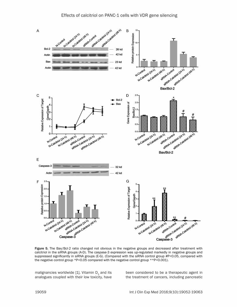
In negative groups, we found the Bax/Bcl-2 ratio changed not obvious which treated with calcitriol compared with the medium control group (Figure 5B-D). In VDR gene silencing groups, the amounts of Bax mRNA expression were slightly decreased, but not significantly; the expression levels of Bcl-2 mRNA were
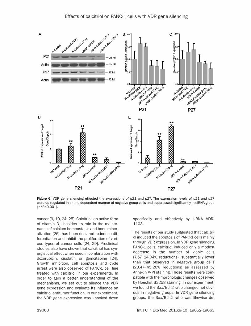
els of p21 mRNA were up-regulated 1.2 times in 24 h and 2.1 times in 48 h; the expression levels of p27 mRNA were up-regulated 4.2 times and 6.7 times (Figure 6D, 6E). VDR gene silencing resulted in significantly decreased lev-els of p21 and p27 compared to the control group (Figure 6B, 6C). The p21 and p27 mRNA expression levels also were suppressed signifi-cantly in siRNA group (Figure 6D, 6E). Our results showed that the amounts of p21 and p27 mRNA were reduced to 26% and 12% respectively compared to the negative control group (**P<0.001).
Discussion
Pancreatic cancer is the fourth leading cause of cancer death and it is one of the most deadly
Figure 4. Cell cycle arrest and inhibition in PANC-1 cells treated with 50 nM calcitriol. The percent cells in each phase are plotted and the histograms showing the percentage of different groups cells distribution in the G0/G1, S, and G2/M phases. (*P<0.05).
sharply increased (Figure 5C). So, the Bax/Bcl-2 ratio dimin-ished after treatment with cal-citriol (Figure 5D). These results were likewise confirmed at the protein levels (Figure 5B). In negative groups, we found the cells treated with calcitriol ex- pressed higher levels caspa- se-3 compared with the medi-um control group (Figure 5E). As shown, the caspase-3 mRNA expression was up-regulated 2.0 times within 24 h and 3.8 times within 48 h (Figure 5G). In siRNA groups, caspase-3 expre- ssion was dramatically sup-pressed (Figure 5F). The mRNA expression levels of caspase-3 were also suppressed remark-ably by calcitriol (Figure 5G). (Compared with the siRNA con-trol group #P<0.05, compared with the negative control group **P<0.001).
VDR gene silencing effected the expressions of p21 and p27
We also demonstrated that the expression levels of p21 and p27 were up-regulated in a time-dependent manner of neg-ative group cells treated with 50 nM calcitriol (Figure 6B, 6C). Those results were equally con-firmed at mRNA levels. In nega-tive group, the expression lev-
Effects of calcitriol on PANC-1 cells with VDR gene silencing
19059 Int J Clin Exp Med 2016;9(10):19052-19063
malignancies worldwide [1]. Vitamin D3 and its analogues coupled with their low toxicity, have
been considered to be a therapeutic agent in the treatment of cancers, including pancreatic
Figure 5. The Bax/Bcl-2 ratio changed not obvious in the negative groups and decreased after treatment with calcitriol in the siRNA groups (A-D). The caspase-3 expression was up-regulated markedly in negative groups and suppressed significantly in siRNA groups (E-G). (Compared with the siRNA control group #P<0.05, compared with the negative control group *P<0.05 compared with the negative control group **P<0.001).
Effects of calcitriol on PANC-1 cells with VDR gene silencing
19060 Int J Clin Exp Med 2016;9(10):19052-19063
cancer [9, 10, 24, 25]. Calcitriol, an active form of vitamin D3, besides its role in the mainte-nance of calcium homeostasis and bone miner-alization [26], has been declared to induce dif-ferentiation and inhibit the proliferation of vari-ous types of cancer cells [24, 29]. Preclinical studies also have shown that calcitriol has syn-ergistical effect when used in combination with doxorubicin, cisplatin or gemcitabine [24]. Growth inhibition, cell apoptosis and cycle arrest were also observed of PANC-1 cell line treated with calcitriol in our experiments. In order to gain a better understanding of the mechanisms, we set out to silence the VDR gene expression and evaluate its influence on calcitriol antitumor function. In our experiment, the VDR gene expression was knocked down
specifically and effectively by siRNA VDR- 1103.
The results of our study suggested that calcitri-ol induced the apoptosis of PANC-1 cells mainly through VDR expression. In VDR gene silencing PANC-1 cells, calcitriol induced only a modest decrease in the number of viable cells (7.57~14.04% reductions), substantially lower than that observed in negative group cells (23.47~45.26% reductions) as assessed by Annexin V/PI staining. Those results were com-patible with the morphologic changes observed by Hoechst 33258 staining. In our experiment, we found the Bax/Bcl-2 ratio changed not obvi-ous in negative groups. In VDR gene silencing groups, the Bax/Bcl-2 ratio was likewise de-
Figure 6. VDR gene silencing effected the expressions of p21 and p27. The expression levels of p21 and p27 were up-regulated in a time-dependent manner of negative group cells and suppressed significantly in siRNA group (**P<0.001).

Effects of calcitriol on PANC-1 cells with VDR gene silencing
19061 Int J Clin Exp Med 2016;9(10):19052-19063
creased after treatment with calcitriol. As far as we know, the mitochondria pathway is inhibited by Bcl-2 or its other family members and acti-vated by Bak or Bax [30, 31]. The cells survive or not after receiving apoptosis signals mainly depends on the ratio of Bax/Bcl-2 radio [32]. Those results support that calcitriol inducing the apoptosis of PANC-1 cells may not be main-ly through the mitochondria pathway in PANC-1 cell line. In our experiment, we also found cal-citriol could induce higher expression levels of caspase-3 in negative groups. However, in si- RNA groups, caspase-3 expression was dra-matically suppressed. These data suggest that the expression of the VDR gene, in majority part, renders cancer cells susceptible to cal-citriol. This is in accordance with a previously published study on BxPC-3, AsPC-1, Hs700T, and Hs766T cells [19, 33-36]. However, the accurately mechanism of the regulation of cas-pase-3 is presently unknown. These results indicated that calcitriol induced cells apoptosis and inhibited proliferation mainly involved in the VDR-mediated pathway. Accordingly, other factors besides VDR states may also partici-pate in the modulation of cell apoptosis induced by calcitriol.
VDR expression is under a close relationship with the signaling system of cell cycle-regulat-ing agents, such as p21 and p27 proteins. In our study, the percentage of G0/G1 phase cells in siRNA groups was significantly decreased compared with the negative groups. Those results demonstrated that the growth inhibitory effects of calcitriol were mainly linked to VDR level and the induction of p21 and p27. Both p21 and p27 are members of cyclin-dependent kinase2 (cdk2) which involved in G1 cell cycle arrest [22, 37-40]. In our experiment showed marked increases in p21 and p27 content after treatment with calcitriol in Negative groups. These results are also consistent with the pre-viously reported growth inhibitory effect of cal-citriol on pancreatic cancer cells, characterized by the up-regulation of p21 and p27 in the BxPC-3, Hs700T and Hs766T cell lines [22, 41]. Furthermore, the increased percentage of G0/G1 phase cells in the VDR gene silencing groups suggested that calcitriol arrest the cell-cycle of PANC-1 cells may have any further possible mechanisms.
In conclusion, calcitriol exerts a conspicuous antitumor effect on PANC-1 cells, which mainly
mediated via the expression of the VDR gene and a series of genes which involved in cell apoptosis and cell cycle. The expression levels of the VDR gene and the antineoplastic effects of calcitriol, suggested that calcitriol induce apoptosis and inhibit cell proliferation is mainly via VDR pathway, but may not be only via this way. Other possible mechanisms including the calcitriol antitumor effects as possible media-tors of these effects still were not elucidated. So, more antitumor mechanisms of calcitriol need to be elucidated further and should be confirmed in vivo studies.
Acknowledgements
This study was financed by Wenzhou Municipal Sci-Tech Bureau. Funding for this work was sup-ported by Wenzhou Municipal Sci-Tech Bureau. Project (No. Y20130097).
Disclosure of conflict of interest
None.
Address correspondence to: Drs. Yi-Hu Zheng and Qi-Yu Zhang, Department of General Surgery, The First Affiliated Hospital of Wenzhou Medical Univer- sity, Nan Bai Xiang Street, Ouhai District, Wenzhou 325000, Zhejiang, China. E-mail: [email protected] (YHZ); [email protected] (QYZ)
References
[1] Siegel R, Naishadham D and Jemal A. Cancer statistics, 2012. CA Cancer J Clin 2012; 62: 10-29.
[2] Jemal A, Siegel R, Ward E, Hao Y, Xu J, Murray T and Thun MJ. Cancer statistics, 2008. CA Cancer J Clin 2008; 58: 71-96.
[3] Rosemurgy AS and Serafini FM. New directions in systemic therapy of pancreatic cancer. Can- cer Control 2000; 7: 437-451.
[4] Beger HG, Rau B, Gansauge F, Poch B and Link KH. Treatment of pancreatic cancer: challenge of the facts. World J Surg 2003; 27: 1075-1084.
[5] Helm JF, Centeno BA, Coppola D, Druta M, Park JY, Chen DT, Hodul PJ, Kvols LK, Yeatman TJ, Carey LC, Karl RC and Malafa MP. Outcomes following resection of pancreatic adenocarci-noma: 20-year experience at a single institu-tion. Cancer Control 2008; 15: 288-294.
[6] Lowy AM. Neoadjuvant therapy for pancreatic cancer. J Gastrointest Surg 2008; 12: 1600-1608.
[7] Rajagopalan MS, Heron DE, Wegner RE, Zeh HJ, Bahary N, Krasinskas AM, Lembersky B,

Effects of calcitriol on PANC-1 cells with VDR gene silencing
19062 Int J Clin Exp Med 2016;9(10):19052-19063
Brand R, Moser AJ, Quinn AE and Burton SA. Pathologic response with neoadjuvant chemo-therapy and stereotactic body radiotherapy for borderline resectable and locally-advanced pancreatic cancer. Radiat Oncol 2013; 8: 254.
[8] Abrams RA, Grochow LB, Chakravarthy A, Sohn TA, Zahurak ML, Haulk TL, Ord S, Hruban RH, Lillemoe KD, Pitt HA, Cameron JL and Yeo CJ. Intensified adjuvant therapy for pancreatic and periampullary adenocarcinoma: survival re-sults and observations regarding patterns of failure, radiotherapy dose and CA19-9 levels. Int J Radiat Oncol Biol Phys 1999; 44: 1039-1046.
[9] Yamaji T, Iwasaki M, Sasazuki S, Sakamoto H, Yoshida T and Tsugane S. Association between plasma 25-hydroxyvitamin D and colorectal adenoma according to dietary calcium intake and vitamin D receptor polymorphism. Am J Epidemiol 2012; 175: 236-244.
[10] Imtiaz S, Siddiqui N, Raza SA, Loya A and Muhammad A. Vitamin D deficiency in newly diagnosed breast cancer patients. Indian J Endocrinol Metab 2012; 16: 409-413.
[11] Zhang J, Zhang H, Zhang X and Yu Z. Synergistic effect of retinoic acid and vitamin D analog EB1089-induced apoptosis of hepatocellular cancer cells. Cytotechnology 2013; 65: 457-465.
[12] Banerjee P and Chatterjee M. Antiproliferative role of vitamin D and its analogs--a brief over-view. Mol Cell Biochem 2003; 253: 247-254.
[13] Colston KW, James SY, Ofori-Kuragu EA, Bin- derup L and Grant AG. Vitamin D receptors and anti-proliferative effects of vitamin D deriva-tives in human pancreatic carcinoma cells in vivo and in vitro. Br J Cancer 1997; 76: 1017-1020.
[14] Albrechtsson E, Jonsson T, Moller S, Hoglund M, Ohlsson B and Axelson J. Vitamin D recep-tor is expressed in pancreatic cancer cells and a vitamin D3 analogue decreases cell number. Pancreatology 2003; 3: 41-46.
[15] Schwartz B, Smirnoff P, Shany S and Liel Y. Estrogen controls expression and bioresponse of 1,25-dihydroxyvitamin D receptors in the rat colon. Mol Cell Biochem 2000; 203: 87-93.
[16] Kawa S, Yoshizawa K, Tokoo M, Imai H, Oguchi H, Kiyosawa K, Homma T, Nikaido T and Fur- ihata K. Inhibitory effect of 220-oxa-1,25-dihy-droxyvitamin D3 on the proliferation of pancre-atic cancer cell lines. Gastroenterology 1996; 110: 1605-1613.
[17] Kawa S, Nikaido T, Aoki Y, Zhai Y, Kumagai T, Furihata K, Fujii S and Kiyosawa K. Vitamin D analogues up-regulate p21 and p27 during growth inhibition of pancreatic cancer cell lines. Br J Cancer 1997; 76: 884-889.
[18] Pols HA, Birkenhager JC and van Leeuwen JP. Vitamin D analogues: from molecule to clinical
application. Clin Endocrinol (Oxf) 1994; 40: 285-292.
[19] Kawa S, Yoshizawa K, Nikaido T and Kiyosawa K. Inhibitory effect of 22-oxa-1,25-dihydroxyvi-tamin D3, maxacalcitol, on the proliferation of pancreatic cancer cell lines. J Steroid Biochem Mol Biol 2005; 97: 173-177.
[20] Mouratidis PX, Dalgleish AG and Colston KW. Investigation of the mechanisms by which EB1089 abrogates apoptosis induced by 9-cis retinoic acid in pancreatic cancer cells. Pan- creas 2006; 32: 93-100.
[21] Schwartz GG, Eads D, Rao A, Cramer SD, Willingham MC, Chen TC, Jamieson DP, Wang L, Burnstein KL, Holick MF and Koumenis C. Pancreatic cancer cells express 25-hydroxyvi-tamin D-1 alpha-hydroxylase and their prolifer-ation is inhibited by the prohormone 25-hy-droxyvitamin D3. Carcinogenesis 2004; 25: 1015-1026.
[22] Schwartz GG, Eads D, Naczki C, Northrup S, Chen T and Koumenis C. 19-nor-1 alpha,25-dihydroxyvitamin D2 (paricalcitol) inhibits the proliferation of human pancreatic cancer cells in vitro and in vivo. Cancer Biol Ther 2008; 7: 430-436.
[23] Zhang M, Li J, Wang L, Tian Z, Zhang P, Xu Q, Zhang C, Wei F and Chen W. Prognostic signifi-cance of p21, p27 and survivin protein expres-sion in patients with oral squamous cell carci-noma. Oncol Lett 2013; 6: 381-386.
[24] Yu WD, Ma Y, Flynn G, Muindi JR, Kong RX, Trump DL and Johnson CS. Calcitriol enhances gemcitabine anti-tumor activity in vitro and in vivo by promoting apoptosis in a human pan-creatic carcinoma model system. Cell Cycle 2010; 9: 3022-3029.
[25] Baust JM, Klossner DP, Robilotto A, Vanbuskirk RG, Gage AA, Mouraviev V, Polascik TJ and Baust JG. Vitamin D(3) cryosensitization in-creases prostate cancer susceptibility to cryo-ablation via mitochondrial-mediated apoptosis and necrosis. BJU Int 2012; 109: 949-958.
[26] Reichel H, Koeffler HP and Norman AW. The role of the vitamin D endocrine system in health and disease. N Engl J Med 1989; 320: 980-991.
[27] Peehl DM, Skowronski RJ, Leung GK, Wong ST, Stamey TA and Feldman D. Antiproliferative ef-fects of 1,25-dihydroxyvitamin D3 on primary cultures of human prostatic cells. Cancer Res 1994; 54: 805-810.
[28] Hershberger PA, Yu WD, Modzelewski RA, Rueger RM, Johnson CS and Trump DL. Ca- lcitriol (1,25-dihydroxycholecalciferol) enhanc-es paclitaxel antitumor activity in vitro and in vivo and accelerates paclitaxel-induced apop-tosis. Clin Cancer Res 2001; 7: 1043-1051.
[29] Beer TM and Myrthue A. Calcitriol in cancer treatment: from the lab to the clinic. Mol Cancer Ther 2004; 3: 373-381.

Effects of calcitriol on PANC-1 cells with VDR gene silencing
19063 Int J Clin Exp Med 2016;9(10):19052-19063
[30] Kroemer G. The proto-oncogene Bcl-2 and its role in regulating apoptosis. Nat Med 1997; 3: 614-620.
[31] Chi X, Kale J, Leber B and Andrews DW. Reg- ulating cell death at, on, and in membranes. Biochim Biophys Acta 2014; 1843: 2100-2113.
[32] Cory S and Adams JM. Killing cancer cells by flipping the Bcl-2/Bax switch. Cancer Cell 2005; 8: 5-6.
[33] Pettersson F, Colston KW and Dalgleish AG. Differential and antagonistic effects of 9-cis-retinoic acid and vitamin D analogues on pan-creatic cancer cells in vitro. Br J Cancer 2000; 83: 239-245.
[34] Zugmaier G, Jager R, Grage B, Gottardis MM, Havemann K and Knabbe C. Growth-inhibitory effects of vitamin D analogues and retinoids on human pancreatic cancer cells. Br J Cancer 1996; 73: 1341-1346.
[35] Persons KS, Eddy VJ, Chadid S, Deoliveira R, Saha AK and Ray R. Anti-growth effect of 1,25-dihydroxyvitamin D3-3-bromoacetate alo- ne or in combination with 5-amino-imidazole-4-carboxamide-1-beta-4-ribofuranoside in pa- ncreatic cancer cells. Anticancer Res 2010; 30: 1875-1880.
[36] Kisker O, Onizuka S, Becker CM, Fannon M, Flynn E, D’Amato R, Zetter B, Folkman J, Ray R, Swamy N and Pirie-Shepherd S. Vitamin D binding protein-macrophage activating factor (DBP-maf) inhibits angiogenesis and tumor growth in mice. Neoplasia 2003; 5: 32-40.
[37] Jeffrey PD, Russo AA, Polyak K, Gibbs E, Hurwitz J, Massague J and Pavletich NP. Mechanism of CDK activation revealed by the structure of a cyclinA-CDK2 complex. Nature 1995; 376: 313-320.
[38] Russo AA, Jeffrey PD, Patten AK, Massague J and Pavletich NP. Crystal structure of the p27Kip1 cyclin-dependent-kinase inhibitor bound to the cyclin A-Cdk2 complex. Nature 1996; 382: 325-331.
[39] Hager G, Formanek M, Gedlicka C, Thurnher D, Knerer B and Kornfehl J. 1,25(OH)2 vitamin D3 induces elevated expression of the cell cy-cle-regulating genes P21 and P27 in squa-mous carcinoma cell lines of the head and neck. Acta Otolaryngol 2001; 121: 103-109.
[40] Chiang KC and Chen TC. Vitamin D for the pre-vention and treatment of pancreatic cancer. World J Gastroenterol 2009; 15: 3349-3354.
[41] Laurila E, Vuorinen E, Savinainen K, Rauhala H and Kallioniemi A. KPNA7, a nuclear transport receptor, promotes malignant properties of pancreatic cancer cells in vitro. Exp Cell Res 2014; 322: 159-167.


![Cambridge International Examinations Cambridge ... International... · Give the number of geometrical isomers of calcitriol, including calcitriol..... [1] (d) A sample of calcitriol](https://img.pdfslide.net/doc/110x75/5ebc295fe2a01012316c9a27/cambridge-international-examinations-cambridge-international-give-the-number.jpg)