Embed Size (px)
Citation preview

Int J Clin Exp Med 2016;9(3):5728-5736www.ijcem.com /ISSN:1940-5901/IJCEM0015746
Original ArticleHereditary spinal schwannomas disease: a report for pedigree cases
Chao Xie2, Xiangming Xu2, Xiandian Zou2, Mingnan Lu2, Chuanlin Mei2, Yishan Zhang1, Weitao Guo1, Yingjie Zhou1
1Luoyang Orthopedic-Traumatological Hospital, Luoyang, Henan, China; 2Guangdong Medical College, Zhanjiang, Guangdong, China
Received September 7, 2015; Accepted November 23, 2015; Epub March 15, 2016; Published March 30, 2016
Abstract: A 33-year-old male patient was found in clinic, which was hospitalized for surgical treatments due to the multiple tumors in vertebral canal. The earliest onset of the disease for him occurred when he was 19 years old. Up to now, he has been performed 7 times of surgical treatment. Tracing his family history, the pedigree contains 48 members. According to the diagnostic criteria proposed by Macollin and the International Schwannomatosis Work-shop in 2011, nine patients were diagnosed (five died), and two were suspected patients. Meanwhile, neither neu-rofibromatosis type I with café au lait spots on the skin, nor multiple neurofibromatosis type II history with vestibular nerve function deficiency or hearing impairment was found in the pedigree. The vestibular nerve space occupying lesion was also not found in cranial MRI examination. The excisional tumor was confirmed to be schwannomas by pathological examination. The preliminary gene sequencing for the NF2 and SMARCB1 gene demonstrated that there was a missense mutation (C.593G→A) in exon 6 codon 198 in blood of the proband (III:1), a nonsense muta-tion (AAG→TAA) in exon 4 codon 149 in blood of II:5 and II:2, a missense mutation (C.593G→A) in exon 6 codon 198 in tumor tissue of III:1, and a missense mutation (C.119T→G) in exon 2 codon 40 in tumor of III:2. The synonymous mutation (C.93G→A) in exon 1 codon 31 of the SMARCB1 gene was detected in the tumor and blood samples from many members of this pedigree. Hence, this family could be confirmed to be a certain hereditary spinal schwan-noma pedigree and the synonymous mutation (C.93G→A) in exon 1 codon 31 of the SMARCB1 gene may be the hereditary disease causing mutation.
Keywords: Schwannomas, schwannomas disease, diagnostic criteria, Non vestibular schwannoma, SMARCB1, NF2, genic mutation
Introduction
Schwannoma is a rare kind of Schwann cell derived benign tumor, with specific clinical ma- nifestations of multiple nerve sheath tumors on spinal cord or peripheral nerve fibers, neither intradermal and nor vestibular schwannomas (VS), as well as café au lait spots on the skin, neurofibromatosis type 1/2 (NF1/2) fundus Li- sch nodules, which was also called as NF3 [1]. In this paper, a certain hereditary spinal sch- wannoma pedigree was reported.
Materials and methods
Family members and clinical investigations
This pedigree containing 48 members came fr- om Guangdong Province of China (Figure 1). In accordance with previous diagnostic criteria
proposed by Macollin [2] and the International Schwannomatosis Workshop in 2011 [3], nine patients were diagnosed (five died), and two were suspected patients. After the family mem-bers signed informed consent forms, the de- tailed histories of their disease were collected. All family members received physical examina-tion and imaging tests (spinal canal myelogra-phy or lumbar and cranial MRI). Blood samples from all members were collected. Individuals in whom a tumor was detected by MRI underwent surgical treatment, and tumor samples were collected.
Proband III:1, 33-year-old male, had received four surgical treatments since the onset of dis-ease at the age of 19 years. In September 2006, the patient was first hospitalized because of neck, chest and back pain for 5 years and

Hereditary spinal schwannomas disease
5729 Int J Clin Exp Med 2016;9(3):5728-5736
walking dysfunction for 1 month. MRI revealed multiple tumors on the neck and thoracic verte-bra. Excision of neck and thoracic duct tumors was conducted. Yellowish oval circumscribed tumors (size: 2×0.8, 1×0.6, 2.5×0.8, 0.4×0.3 cm) were visible in the dorsal region of T3, the dorsal region of T1, the ventral region of C5 and the dorsal region of C6, respectively, adhesive with nerve branches. The Symptom was reli-
of the tumors. In March 2013, this patient was hospitalized for the third time because of later back pain with numbness in the lower limbs for 3 month. MRI revealed multiple tumors in the lumbosacral spinal canal, an extramedullary subdural stripped tumor at T11 and T12, a tumor to the right of the vertebral body at T6 and T7, and extramedullary subdural nodular lesions at T1 and T3. Thoracolumbar spinal tumor excision
Figure 1. Pedigree chart of the familial spinal schwannomas.
Figure 2. The MRI images for the hospitalized proband in 2013: (A) and (C) lum-bar vertebra; (B) thoracic vertebra; Multiple occupying lesions (D) on the side of the thoracic vertebra and (E) in pelvic cavity; (F) Pelvic CT image, displaying huge occupying lesion (10*9.5*11 cm); (G) B-type ultrasonography image on right lower limb, displaying cystic tumor with size of 5.8*4.7 cm on right sciatic nerve, not connected with canalis spinalis, pressing the bladder to left.
eved after surgical remov-al of the tumors. In January 2011, this patient was hospitalized again beca- use of later back pain with numbness in the lower limbs for 1 month. MRI re- vealed multiple space-occupying lesions in the cervical, thoracic and lum-bar spinal cords. Po- sterior neck and thoracic tumor excision was con-ducted. A 2.0×1.5 cm tumor was observed in the dorsal region of the spinal segment at T1. Four tumors (1.8×1.6 cm, 2.3×1.4 cm, 2.0×1.0 cm, 1.8×1.6 cm) were observed in the dor-sal region of spinal seg-ments at T10-L2, which were well circumscribed and adhesive nerve roots. The Symptom was relieved after completely removal
Hereditary spinal schwannomas disease
5730 Int J Clin Exp Med 2016;9(3):5728-5736
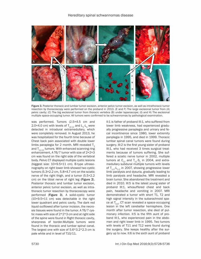
was performed. Tumors (2.5×4.5 cm and 2.0×4.0 cm) with levels of T10-12 and L2-L4 were detected in intradural extramedullary, which were completely removed. In August 2013, he was hospitalized for the fourth time because of Chest back pain associated with double lower limbs paraplegia for 2 month. MRI revealed T3 and T11/12 tumors. With enhanced scanning ring enhancement, A T6/7 tumor with size of 2×2×3 cm was found on the right side of the vertebral body. Pelvic CT displayed multiple cystic lesions (biggest size: 10×9.5×11 cm). B-type ultraso-nography on right lower limb showed two cystic tumors (5.3×2.2 cm, 5.8×4.7 cm) on the sciatic nerve of the right thigh, and a tumor (5.5×2.2 cm) on the tibial nerve of right leg (Figure 2). Posterior thoracic and lumbar tumor excision, anterior pelvic tumor excision, as well as intra-thoracic tumor resection by thoracoscopy were performed (Figure 3). A solid-cystic tumor (10×9.5×11 cm) was detectable in the right lower quadrant and pelvic cavity. The dark red liquid outflowed after tumor incision, the necro-sis tissues were found in the tumor. A T6/7 cys-tic mass with size of 2*2*3 cm and at right side of the spine were found in Right thoracic cavity, sharpness of border.Multiple tumors were found in the thoracic and lumbar spinal canal. The largest one with size of 5.6*3.2*2.3 cm is pale white and in level of T10/11.
II:1 is father of proband III:1, who suffered from lower limb weakness, had experienced gradu-ally progressive paraplegia and urinary and fe- cal incontinence since 1980, lower extremity panplegia in 1995, and died in 1999. Thoracic lumbar spinal canal tumors were found during surgery. III:2 is the first young sister of proband III:1, who had received 3 times surgical treat-ments because of tumors suffering. She suf-fered a sciatic nerve tumor in 2002, multiple tumors at C12 and T12-S1 in 2004, and extra-medullary subdural multiple tumors with levels of T1/2-L1/2 in 2007, showing progressive lower limb paralysis and dysuria, gradually leading to limb paralysis and headache. MRI revealed a brain tumor. She abandoned the treatment and died in 2010. III:5 is the latest young sister of proband III:1, whosuffered chest and back pain, headache and vomiting in 2007. MRI demonstrated a tumor with short T1, T2 and high signal intensity in the subarachnoid spa- ce at T6-8. CT scan revealed a space-occupying lesion in the left cerebellar hemisphere. One month after tumor resection, she died of pul-monary infection. II:5 is the fifth aunt of pro-band III:1, who experienced pain in the abdo-men and right lower limb in 1995. Two tumors with levels of T11 and T12 were found during the surgery. She keeps healthy after the sur-gery up to now. II:6 is the sixth aunt of proband
Figure 3. Posterior thoracic and lumbar tumor excision, anterior pelvic tumor excision, as well as intrathoracic tumor resection by thoracoscopy were performed on the proband in 2013. (E and F) The large excisional tumor from (A) pelvic cavity; (C) The big excisional tumor from thoracic vertebra (B) under laparoscope; (G and H) The excisional multiple space-occupying tumor. All tumors were confirmed to be schwannomas by pathological examination.

Hereditary spinal schwannomas disease
5731 Int J Clin Exp Med 2016;9(3):5728-5736
Table 1. Data on family members with rare hereditary spinal schwannomatosisNo. III:1 II:1 III:2 III:5 II:5 II:6 II:7 III:18 III:21Gender Male Male Female Female Female Female Female Female Female
Age of onset 19 24 13 16 37 17 26 24 20
Age of death - 43 27 17 - 24 32 - -
Pain +++ ++ ++ ++++ ++ + ++++ ++++ +++
Weakness/paraplegia +++ ++++ ++++ ++++ + ++ ++++ - -
Bowel and bladder disorders × √ √ × × √ √ × ×
Vestibular schwannomatosis × × × × × × × × ×
Milk coffee spot/Lischnodules × × × × × × × × ×
Times of surgery 6 > 5 3 1 1 3 2 2 1
Number of tumor 17 > 5 5 2 2 > 4 5 2 1
Position of tumor Spinal cord, thoracic cord, caudaequina, pelvic cavity, thigh
Thoracic vertebra, caudaequina, right
shoulder
Thigh, neck, thoracic cord, caudaequina
Thoracic vertebra, left cerebellum
Thoracic cord Thoracic cord, caudaequina
Thoracic cord, caudaequina
Cervical cord Lumbar vertebra, caudaequina
Complication - - - Pneumonia Lumber disc herniation
Urinary tract infection
- - -
Hereditary spinal schwannomas disease
5732 Int J Clin Exp Med 2016;9(3):5728-5736
III:1, who was hospitalized three times from 1981 to 1987, presenting with progressive lo- wer limb paraplegia and bowel and bladder dis-orders. The T11, L2 and L3 Tumors were observed. Symptoms were not lessened after the third surgery. She had a urinary tract infection and died in 1988. II:7 is the latest aunt of proband III:1, whopresented with lower back pain and progressive lower limb paraplegia, and received
twice surgeries. Multiple tumors were observed in levels of T12-L4. Paraplegia was not relieved after the secondary surgery. Bowel and bladder disorders appeared. She died in 2001. III:18, III:19 and III:19 are daughters of II:6. III:18 received twice cervical cord tumor resection. III:21 received lumbar multiple tumor resection. III:19 suffered cervical cord space-occupying lesion (MRI), but refused surgical treatment.
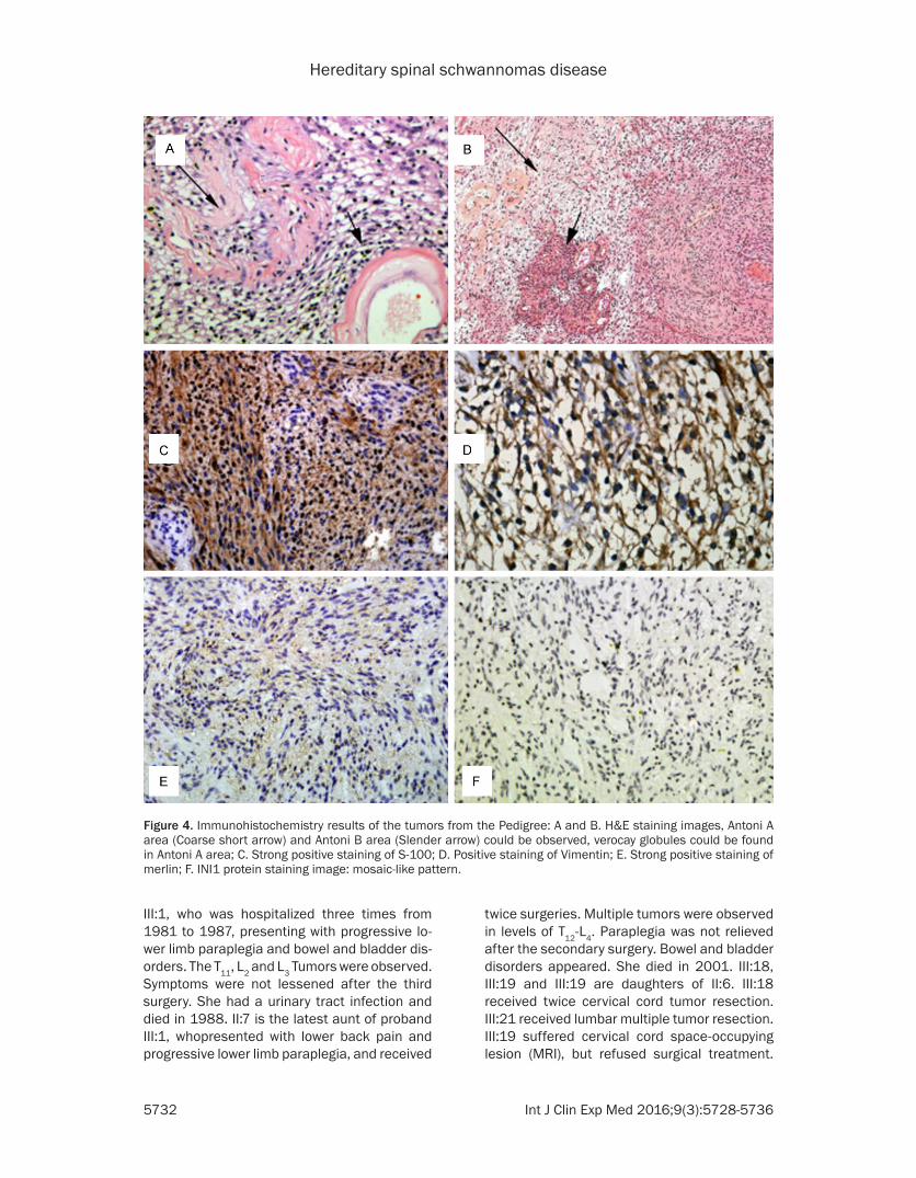
Figure 4. Immunohistochemistry results of the tumors from the Pedigree: A and B. H&E staining images, Antoni A area (Coarse short arrow) and Antoni B area (Slender arrow) could be observed, verocay globules could be found in Antoni A area; C. Strong positive staining of S-100; D. Positive staining of Vimentin; E. Strong positive staining of merlin; F. INI1 protein staining image: mosaic-like pattern.

Hereditary spinal schwannomas disease
5733 Int J Clin Exp Med 2016;9(3):5728-5736
III:20 is son of II:7, who received multiple tumor resection lateral to the inferior extremity of the right leg, but refused pathological examination. Pathological examination demonstrated that the obtained tumors from diagnosed patients during surgery were schwannomas.
Blood and tumor samples
Blood samples of 43 family members were col-lected, except the five dead patients. The re- moved fresh tumor samples during the surgery included thoracic cord, thoracic cavity and pel-vic cavity from III:1, and the cervical cord tumor from III:18. Fresh tumor samples were stored at -80°C. The following tissues were embedded in paraffin: cervical cord of III:1, thoracic cord of II:1, thoracic cord of III:2, and thoracic cord of III:5. Unfortunately, blood samples could only be obtained from II:5 and III:21. No blood or tumor samples were obtained from II:6 and II:7, who were dead. All tumor samples were stain- ed with H&E, DAB visualized for immunohisto-chemical staining. The applied Antibodies were as follows: vimentin and S-100 (Dako Cytoma- tion Denmark A/S), SMARCB1 gene expression
product INI1 (BD Biosciences San Jose, CA, USA), and NF2 gene expression product Merlin (Abcam, Cambridge, Great Britain).
DNA extraction, amplification and mutation analysis
DNA was extracted from 43 blood samples and eight tumor samples. Tumor samples included thoracic cord, thoracic cavity and pelvic cavity of III:1, and fresh cervical cord tumor of III:18, as well as paraffin-embedded tumor samples of cervical cord of III:1, thoracic cord of II:1, tho-racic cord of III:2, and thoracic cord of III:5. After DNA extraction, primers were designed using 17 exons of the NF2 gene and nineexons of the SMARCB1 gene in accordance with pre- viously described methods [1, 4]. After PCR amplification, nine exons from each of the NF2 and SMARCB1 genes were directly sequenced by the Sanger method. The copy number of NF2 and SMARCB1 genes was analyzed using mul-tiple ligation-dependent probe amplification (MLPA). The study was approved by the Ethics Committee of Affiliated Hospital of Guangdong Medical College in China.
Figure 5. The synonymous mutation (C.93G→A) in exon 1 codon 31 of the SMARCB1 gene detected in the tumor and blood samples from many members of this pedigree. Form A to I: SMARCB1 gene sequence diagram of exon 1 from the Blood, cervical spinal tumor, thoracic spinal tumor, pelvic tumor of the proband, blood of the oldest daugh-ter of the proband (IV:1), thoracic spinal tumor of the father of the proband (II:2), as well as blood of III:18, III:20, and III:21. All Mutations occurred in exon 1 codon 31, nucleic acid sequence from GAG to GAA.

Hereditary spinal schwannomas disease
5734 Int J Clin Exp Med 2016;9(3):5728-5736
Results
This pedigree is a rare family with hereditary spinal schwannomatosis. Multiple schwanno-mas were found during surgery and observed by MRI. There were no café au lait spots on the skin, Lisch nodules or vestibular schwanno-mas. Clinical manifestations present limb pain, weakness and numbness. Symptoms could not be lessened by oral non-steroidal analgesics. Progressive lower limb paralysis occurred. So- me patients suffered bowel and bladder disor-ders. These diseases were easy to relapse after surgery and in poor prognosis. III:5 had severe pneumonia, and died within 1 month after dis-charge from hospital, while II:6 had aurinary tract infection (see Table 1). The hematoxylin and eosin staining results demonstrated that all tumor samples were schwannomas. Immu- nohistochemistry showed positive expression of S-100 and vimentin. INI1 protein staining results revealed a mosaic-like pattern, indicat-ing schwannomatosis. Positive staining for mer-lin protein indicated normal expression of the NF2 gene (Figure 4).
The diverse mutations were shown in SMARCB1 gene analysis. No abnormal copy number for theNF2 or SMARCB1 genes were observed in MLPA analysis. Direct gene sequencing for the NF2 gene demonstrated that there was a mis-sense mutation (C.593G→A) in exon 6 codon 198 in blood of the proband (III:1), a nonsense mutation (AAG→TAA) in exon 4 codon 149 in blood of II:5 and II:2, a missense mutation (C.593G→A) in exon 6 codon 198 in tumor of III:1, and a missense mutation (C.119T→G) in exon 2 codon 40 in tumor of III:2. SMARCB1 gene exon sequencing suggested various muta-tions, as shown in Figure 5.
In blood samples from the proband, his olde- st daughter (IV:1), father (II:2), two younger fe- male cousins (II:18, 11:21) and a younger ma- le cousin (II:20), and three tumor samples fr- om the proband, a synonymous mutation (C. 93G→A) was detected in exon 1 codon 31 of the SMARCB1 gene. In a blood sample from the son (IV:3) of the proband, a synonymous mu- tation (C.897G→A) was detectable in exon 7 codon 299 of the SMARCB1 gene. These syn-onymous mutations were firstly detected in this family with schwannomatosis, but the patho-genic mechanism remains unclear.
A missense mutation was detected in exon 9 codon 383 of the SMARCB1 gene in blood sam-ples of IV:1 and IV:3, which would induce an amino acid substitutions (P.Pro383Leu). A mis-sense mutation was detected in exon 1 codon 18 of the SMARCR1 gene in a tumor sample from the thoracic cord of III:2, which would lead to an amino acid substitution (P.Gln18Leu). A missense mutation was found in exon 1 codon 7 in the tumor sample of lumbar vertebra of III:5, which would also cause an amino acid substitution (P.Ser7Ile).
A nonsense mutation (C.91G→T) was found in exon 1 codon 31 in the tumor sample from tho-racic cord of III:2. This mutation would induce premature termination of peptide chain synthe-sis (P. Glu31X).
Discussion
Schwannomatosis is the third independent ty- pe for multiple nerve sheath tumors, with the characterizations of neither intradermal nor ve- stibular schwannomas. It is significantly differ-ent with NF1 and NF2 both in genetics and clini-cal manifestations. However, the neurinoma- tosis in clinical is easily confused with NF2, especially the chimeric NF2. In the literature about NF2 published from 1990 to 2003, there were no distinguished schwannomatosis, as they are very similar in clinical. In 2003, the results of the genetics research showed that no direct connection of NF2 gene and familial schwannomas disease. In 2007, Hulsebos et al. reported a pedigree of schwannomas and genetic studies suggest that the pathogenesis may be related to SMARCB1 germline muta-tions. Then, the SMARCB1 gene became the focus of the schwannomas disease pathog- enesis. However, many studies confirmed the SMARCB1 gene mutation was only found in 40%-50% of familial schwannomatosis. More- over, the mutation were only found in 10% cases of sporadic schwannomatosis [1, 5, 6]. According to the previous diagnostic criteria proposed by Macollin [2] and the Internation- al Schwannomatosis Workshop in 2011 [3], nine patients were diagnosed (five died), and two were suspected patients in this report. SMARCB1 gene mutations were found in 6 patients of them, occupying 54% of family sch- wannomatosis cases. Considering that not all family members were taken spinal MRI exami-

Hereditary spinal schwannomas disease
5735 Int J Clin Exp Med 2016;9(3):5728-5736
nation, there may be some missed diagnosis, resulting in inaccurate data. The synonymous mutation (C.176G→A) in exon 1 codon 31 of the SMARCB1 gene was detected in the blood samples from the proband, his oldest daughter (IV:1), father (II:2), two younger female cousins (II:18, 11:21) and a younger male cousin (II:20), as well as three tumor samples from the pro-band. These synonymous mutations were firstly detected in pedigree with schwannomatosis. According to the degeneration rule for amino acid codon, the synonymous mutation does not cause the amino acid encoding error. However, the above members (except the children of pro-band) are all in this disease, indicating that syn-onymous mutation may also lead to a mutation effect. Despite few literature for the synony-mous mutation, it could nowadays be con-firmed that the mutation effect is usually harm-ful, which is more prone to promoting cancer gene rather than restraining cancer gene [7], leading to skipping of exon [8], variation of splicing donor site [9], etc. Up to now, the mech-anism of mutation effect caused by the synony-mous replacement of the codon is not clear. Studies have indicated that the synonymous substitution of codon may affect the accuracy and speed of translation, especially through the enhancement the C-ending content of the four-fold codons [10]. More research works are required for the exploration of the mechanism that schwannomas disease caused by synony-mous mutations. For the schwannomas dise- ases caused by germline mutations in the SMARCB1 gene, it could be speculated that mutation may be inherited to offspring through the “two-hit” mode. In the “two-hit” procedure, the first hit occurred in the gametids, leading to germ line mutation without clinical symptoms. When the mutation the gametids were inherit-ed to the next generation, germline mutation and somatic mutation simultaneously occur- red (“two-hit”), presenting clinical symptoms. According to the “two-hit” theory, the first hit in this pedigree occurred in the grandfather or grandmother of the proband, neither clinical symptoms, nor SMARCB1 gene mutations de- tected from the blood (somatic cells) samples. After the combination of their gametes, muta-tion was inherited to the father and the aunts of the proband by the “two-hit”, leading to spinal schwannomas diseases. The mutations were found in both blood and tumor samples by genetic testing. Then, the mutations continue to be inherited to third and fourth generations,
leading to the diseases on the proband, his sis-ter, and cousins. Although the oldest daughter of the proband has not yet clinical symptoms, the SMARCB1 gene mutation in the same site has been found in her blood testing. It is report-ed [11, 12] that the young children may be risk to the malignant rhabdomyosarcoma in the cases of SMARCB1 gene mutation and the ri- sk also increases with age. The Heterozygous mutations in the survivors may promote the susceptibility of schwannomas disease. Despi- te some research works found that the somatic mutations for the NF2 gene appears in the cases of schwannomas disease [5, 6, 13], the NF2 gene mutations in different sites were detected in blood and tumor tissues from only few patients of the pedigree, indicating that NF2 gene mutation is not the necessary condi-tions for schwannomas disease [1].
As the SMARCB1 gene mutations were detect-ed only in half of the members in the pedigree suffered the schwannomas disease, it is obvi-ous that the SMARCB1 gene germline muta-tions have been unable to fully explain the genetic basis of pedigree of schwannomas dis-ease. With the deepening of the research, so- me scholars have explore the genetic mecha-nism of schwannomas disease from the fields outside of the smarcb1 gene and even 22q. Piotrowski et al. [4] found a new LZTR1 gene located at the long arm of chromosome 22, which have confirmed to be a cancer promoting gene and could induce schwannomas disease in the patients without SMARCB1 gene muta-tion. These two pathogenic genes LZTR1 and SMARCB1 show that there is a potential func-tional association with chromatin remodeling mechanism, which plays a vital role in the adaptation of cellular differentiation and adap-tation to environmental stimuli. Zhang et al. [14] found the relationship between missense mutation of COQ6 gene and the susceptibility of familial schwannomatosis through full geno- me sequencing method, suggesting that the familial schwannomatosis may be not a single-gene disease. It is worth to mention that, al- though more and more research works have been focused on the genetics mechanism of schwannomas disease caused by SMARCB1 gene mutation, there is still rare contributions devoted to the influence of the SMARCB1 gene mutation on the function of the expressed INI1 protein, as well as the downstream molecular mechanisms.

Hereditary spinal schwannomas disease
5736 Int J Clin Exp Med 2016;9(3):5728-5736
We report a pedigree of hereditary spinal sc- hwannomas. The family affected members suf-fered schwannomas disease meet diagnostic criteria proposed by Macollin, which will help to increase the understanding of schwannomas. As the genetic schwannomas pedigree in China are rarely reported, the preliminary gene se- quencing indicates that the synonymous muta-tion (C.93G→A) in exon 1 codon 31 of the SMARCB1 gene may be the hereditary disease causing mutation, and this is direction for our future works.
Acknowledgements
This project was supported by the Natural Science Foundation of Guangdong Province in China (Grant No. S2013010012473), and sup-ported by Zhengzhou technology research and development funds support project No. 153- PKJGG063.
Disclosure of conflict of interest
None.
Address correspondence to: Drs. Weitao Guo and Yingjie Zhou, Luoyang Orthopedic-Traumatological Hospital, 100 Yongping RD., Luoyang, Henan, China. Tel: +86-371-88820000; Fax: +86-371-88820000; E-mail: [email protected] (WTG); [email protected] (YJZ)
References
[1] Boyd C, Smith MJ, Kluwe L, Balogh A, Maccollin M and Plotkin SR. Alterations in the SMARCB1 (INI1) tumor suppressor gene in familial schwannomatosis. Clin Genet 2008; 74: 358-366.
[2] MacCollin M, Chiocca EA, Evans DG, Friedman JM, Horvitz R, Jaramillo D, Lev M, Mautner VF, Niimura M, Plotkin SR, Sang CN, Stemmer-Ra-chamimov A and Roach ES. Diagnostic criteria for schwannoma-tosis. Neurology 2005; 64: 1838-1845.
[3] Plotkin SR, Blakeley JO, Evans DG, Hanemann CO, Hulsebos TJ, Hunter-Schaedle K, Kalpana GV, Korf B, Messiaen L, Papi L, Ratner N, Sher-man LS, Smith MJ, Stemmer-Rachamimov AO, Vitte J and Giovannini M. Update from the 2011 International Schwannomatosis Work-shop: From genetics to diagnostic criteria. Am J Med Genet A 2013; 161A: 405-16.
[4] Piotrowski A, Xie J, Liu YF, Poplawski AB, Gomes AR, Madanecki P, Fu C, Crowley MR, Crossman DK, Armstrong L, Babovic-Vuksanovic D, Ber- gner A, Blakeley JO, Blumenthal AL, Daniels MS, Feit H, Gardner K, Hurst S, Kobelka C, Lee C, Nagy R, Rauen KA, Slopis JM, Suwannarat P,
Westman JA, Zanko A, Korf BR and Messia- en LM. Germline loss-of-function mutations in LZTR1 predispose to an inherited disorder of multiple schwannomas. Nat Genet 2014; 46: 182-187.
[5] Sestini R, Bacci C, Provenzano A, Genuardi M and Papi L. Evidence of a four-hit mechanism involving SMARCB1 and NF2 in schwanno- matosis-associated schwannomas. Hum Mu-tat 2008; 29: 227-31.
[6] Hadfield KD, Newman WG, Bowers NL, Walla- ce A, Bolger C, Colley A, McCann E, Trump D, Prescott T and Evans DG. Molecular charac-terisation of SMARCB1 and NF2 in familial and sporadic schwannomatosis. J Med Genet 2008; 45: 332-339.
[7] Karamatic CV, Poole J, Burton N and Daniels G. Three uncommon KEL alleles in one family with unusual Kell phenotypes explain a 35-year old conundrum. Vox Sang 2014; 106: 242-247.
[8] Rice GI, Reijns MA, Coffin SR,Forte GM, Ander-son BH, Szynkiewicz M, Gornall H, Gent D, Leitch A, Botella MP, Fazzi E, Gener B, Lagae L, Olivieri I, Orcesi S, Swoboda KJ, Perrino FW, Jackson AP and Crow YJ. Synonymous muta-tions in RNASEH2A create cryptic splice sites impairing RNase H2 enzyme function in Aicar-di-Goutières syndrome. Hum Mutat 2013; 34: 1066-1070.
[9] Supek F, Miñana B, Valcárcel J, Gabaldón T and Lehner B. Synonymous mutations fre-quently act as driver mutations in human can-cers. Cell 2014; 156: 1324-1335.
[10] Du J, Dungan SZ, Sabouhanian A and Chang BS. Selection on synonymous codons in ma- mmalian rhodopsins: a possible role in opti-mizing translational processes. BMC Evol Biol 2014; 14: 96.
[11] Hulsebos TJ, Plomp AS, Wolterman RA. Germ-line mutation of INI1/SMA- RCB1 in familial schwannomatosis. Am J Hum Genet 2007; 80: 805-810.
[12] Janson K, Nedzi LA, David O, Schorin M, Walsh JW, Bhattacharjee M, Pridjian G, Tan L, Judkins AR and Biegel JA. Predisposition to atypical teratoid/rhabdoid tumor due to an inherited INI1 mutation. Pediatr Blood Cancer 2006; 47: 79-84.
[13] Landi A, Dugoni DE, Marotta N, Mancarella C and Delfini R. Spinal schwannomatosis in the absence of neurofibromatosis: A very rare con-dition. Int J Surg Case Rep 2011; 2: 36-39.
[14] Zhang K, Lin JW, Wang J, Wu X, Gao H, Hsieh YC, Hwu P, Liu YR, Su L, Chiou HY, Wang D, Yuan YC, Whang-Peng J, Chiu WT and Yen Y. A germline missense mutation in COQ6 is asso-ci- ated with susceptibility to familial schwan-nomatosis. Genet Med 2014; 16: 787-792.


![Imaging of Hereditary Hemorrhagic Telangiectasia · Spinal and cerebral vascular malformations are mani-festations of underlying vascular dysplasia [12]. These lesions represent abnormal](https://img.pdfslide.net/doc/110x75/5ed59c731b7fdd786a1b540e/imaging-of-hereditary-hemorrhagic-telangiectasia-spinal-and-cerebral-vascular-malformations.jpg)