Embed Size (px)
Citation preview
Int J Clin Exp Pathol 2014;7(8):4981-4990www.ijcep.com /ISSN:1936-2625/IJCEP0001040
Original Article Subtotal splenectomy for splenomegaly in cirrhotic patients
Haibo Chu1, Xiaofang Liu2, Jianhua Zhao1, Yongbo Xu1, Lei Wang2, Tao Wang1, Wenjun Guo2, Shengming Zhang3, Xiaoji Zhu1
1Center of General Surgery, The 89th Hospital of People’s Liberation Army, Weifang, China; 2Department of Pathol-ogy, Weifang Medical University, Weifang, China; 3Department of Electron Microscope, Weifang Medical University, Weifang, China
Received June 8, 2014; Accepted July 22, 2014; Epub July 15, 2014; Published August 1, 2014
Abstract: Background: In recent years, the spleen has become to be recognized as the “control center” of the immune-metabolic-endocrine network. However, It is controversial that splenomegaly due to portal hypertension is treated by subtotal splenectomy. The aim of this study was to evaluate the distribution of fibrous tissue, morphology of cells as well as splenic size, hemodynamics, hematological and immunological indexes in the residual spleen after subtotal splenectomy. This information may help finding the basis for the operation of subtotal splenectomy. Methods: Ten cases of splenomegaly due to portal hypertension were investigated. Two groups were created: Sple-nomegaly and Residual spleen. Control group was 10 cases of trauma-induced splenic rupture. Samples were sliced, and morphological changes were observed under light microscopy and electron microscopy. Indexes of splen-ic size, hemodynamics, hematology and immunology of the spleen were measured. Results: Under light microscopy, the number of collagen fibers and elastic fibers was increased, and the number of reticular fibers was decreased in the residual spleen and splenomegaly groups. Under electron microscopy, the ultrastructure of endothelial cells, lymphocytes, macrophages, and reticular cells in the residual spleen group were noticeably improved more than in the splenomegaly group. Flow volume in the residual spleen and portal vein decreased obviously, with number of platelet rising to normal, and there was no significant difference in the indexes of immunology. Conclusion: Af-ter subtotal splenectomy, the residual spleen will not experience a high-pressure environment, and the fibrosis of splenic tissue and remodelling of corpuscular morphology will cease.
Keywords: Histopathology, cytomorphology, fiber tissues, residual spleen, cirrhosis
Introduction
The spleen is the largest secondary lymphoid organ in the human body. With its location in the circulatory system and with the unusual structure and function of its compartments, the spleen is a unique organ. The spleen also contains about one-fourth of the body’s lym-phocytes and initiates immune responses against blood-borne antigens [1-3]. Various immune cells ensure the complex function of the spleen as a filter of the blood as well as a lymphoid organ [4, 5].
In recent years, the spleen has become to be recognized as the “control center” of the immune-metabolic-endocrine network [6]. The prevalence of infection and mortality after sple-
nectomy is 3.2% and 1.4%, respectively [5]. The asplenic state or hyposplenism may be impor-tant features of low immune function [7, 8]. The duration of overwhelming post-splenectomy infection can range from < 1 week to a much longer period [4]. Splenomegaly due to portal hypertension is treated by subtotal splenecto-my, which is a challenge to conventional sple-nectomy. How the fibrous tissue and cellular morphology of the residual spleen are changed upon carrying out this treatment is controver-sial [9-11]. Advocates for splenectomy believe that, with fibrosis and little immune function in splenomegaly, recurrence of splenomegaly and hypersplenism may happen in residual spleen [12]. However, advocates inclined to preserve spleen suggest that subtotal splenectomy may reduce portal venous pressure, correct hyper-
Morphological and functional assessment of residual spleen
4982 Int J Clin Exp Pathol 2014;7(8):4981-4990
tissue samples were randomly collected from patients in the 89th Hospital of the People’s Liberation Army (Weifang, China). Ten cases (6 males and 4 females; mean age, 32 (27-38) years)) with splenomegaly due to portal hyper-tension who had received subtotal splenecto-my (with preservation the upper pole) plus fixa-tion of the posterior sternal omentum majors were included in this group. Selection criteria: (1) Hepatitis-B infection patients (HBV-DNA< 5 × 10² copy/ml) who were confirmed to have cir-rhosis, and were classified as Grade A or B according to the Child-Pugh classification. (2) Cirrhosis was accompanied by hypersplenism with moderate or severe varicose veins of the lower esophagus, plus a history of digestive tract hemorrhage. (3) The fibrosis level in the spleen was III [19]. (4) All patients were followed up continually. Spleen organization obtained by operating formed splenomegaly group. Patients who had spleen puncture to check for spleen organization 6 years after surgery formed the residual spleen group. Ten cases (7 males 3 females; mean age, 31 (26-35) years) who underwent splenectomy without splenomegaly formed the control group. Guided by color Doppler ultrasound, hollow-needle biopsy was used to obtain samples of residual splenic tis-sue [20].
Thirty tissue samples were collected. Speci- mens were fixed in 10% formalin, dehydrated, embedded and sliced. Each specimen was made into 15 slices (5 slices per group). After staining (elastic-van Gieson (EVG), Masson, and improved ammonia silver staining), slices were observed under light microscope (BX51; Olympus, Tokyo, Japan) for histomorphological analyses. A total of 1 mm3 of fresh specimens from the splenomegaly group and residual spleen group were taken. They were fixed in 3% glutaraldehyde for 24-48 h, dehydrated, embe- dded and dried. Specimens were then made into ultra-thin (70 nm) slices. Slices were cleansed with water, and soaked in a saturated aqueous solution of uranyl acetate. They were then cleaned with double-distilled water and soaked in lead citrate solution. The histomor-phology of splenic cells was observed using a Hitachi H-7500 Transmission Electron Micro- scope (Hitachi, Tokyo, Japan).
Quantitative analyses of fibers [21]
The percentage composition of the three fibers in each sample was calculated under high mag-
splenism and retain the immune function of spleen-which means killing two birds with one stone [13]. Besides, incidence of portal venous thrombosis in patients after subtotal splenec-tomy for portal hypertension was obviously high, and this may affect the flow of blood into liver [14, 15]. Subtotal splenectomy includes two ways, i.e., with preserving the upper pole and the lower pole of the spleen supplied by the gastrosplenic vessels and splen-omentum and splen-colon vessels [13, 16]. Currently, subto-tal splenectomy was mainly performed in giant splenomegaly patients with hereditary sphero-cytosis and portal hypertension due to cirrhosis [17, 18]. In order to decrease the portal pres-sure and reserve the splenic function, we have operated subtotal splenectomy of preserving the lower splenic pole (residual spleen about 11 × 7 × 4 cm), for splenomegaly owing to por-tal hypertension since 1984. Our surgical method is that, via transthoracic approach, residual spleen (supplied by splenic omentum and splenocolic vessels) and partial omentum were fixed with left lung, which formed porto-pulmary shunt; or by transperitoneal approach, residual spleen (supplied by splenic omentum and splenocolic vessels) and partial omentum were fixed with left retroperitoneal, forming por-tosystemic shunt.
In the present study, the distribution of fibrous tissue, morphology of cells, splenic size, hemo-dynamics, hematological and immunological in the residual spleen was contracted. This infor-mation may help finding the basis for the opera-tion of subtotal splenectomy.
Materials and methods
Ethical approval of the study protocol was obtained from the Human Research Ethics Committee of Weifang Medical University (Wei- fang, China). All individuals provided written informed consent before induction into the study.
Patients and methods
Our studies relating to subtotal splenectomy started in 1984. Up to 2012, we have conduct-ed 752 operations of subtotal splenectomy (preserving the lower pole, normal spleen size of splenic tissue, splenic omentum and spleno-colic vessels for the purpose of blood-supply), with 117 cases for splenic trauma and 635 for splenomegaly due to portal hypertension. Ten
Morphological and functional assessment of residual spleen
4983 Int J Clin Exp Pathol 2014;7(8):4981-4990
nification (× 200). Image-Pro Plus was used to calculate the cumulative area of the relative content of each fiber. Three high-magnification images with the most visible fibers (relative content of collagen fibers, elastic fibers and reticular fibers) were calculated under light microscope (× 200) and the three fields aver-aged. EVG staining (Beisuo, Beijing, China) and improved ammonia silver staining (Shiji He Li, Beijing, China) was then carried out.
In addition, patients underwent complete phys-ical examination before and after the opera-tion, which included abdominal ultrasonogra-
phy (splenic size: splenic length, splenic thick- ness, splenic square area; hemodynamics: splenic artery flow volume, portal venous diam-eter, portal venous flow volume), abdominal CT scan, splenic scintigraphy with 99mTc sulfur col-loid, laboratory hematological (platelets) and immunological (IgA, IgM, IgG and Tuftsin) evaluation.
Statistical analysis
Data analyses were carried out using SPSS ver17.0 (SPSS, Chicago, IL, USA). All values are mean ± standard deviation (SD). Study groups
Figure 1. (A) Hyperplasia of collagen fibers were seen in the cord of the red pulp and surrounded the central ar-tery of the white pulp. (B) Collagen fibers of the splenic cord showed “cord-like” or “small pieces” hyperplasia. The central artery was surrounded by hyperplasic collagen fibers. The white pulp, like the red pulp, was seen. (C) The central arterial wall in white pulp was defined clearly, but collagenous fibers were not observed around (A-C, Masson staining, × 100). (D) The number of elastic fibers around the central artery increased. These fibers were lamellar or loose; capillary change was observed. (E) Hyperplastic elastic fibers were around the central artery, which demon-strates “white pulp vascularization”. (F) The elastic fibers of central arterial wall were clear, but elastic fibers were not observed around. (D-F, EVG staining, × 100). (G) The number of reticular fibers in the splenic cord decreased and they arranged like threads. (H)The number of reticular fibers in the splenic cord decreased, and they showed as irregular filaments. (I) Reticular fibers around endothelial cells in sinus lienis spread to splenic cord, and sinus lienis were separated like mesh and presented like wire netting (red pulp: WP, central artery: arrow, reticular fiber: arrowhead) (G-I, silver staining, × 100).
Morphological and functional assessment of residual spleen
4984 Int J Clin Exp Pathol 2014;7(8):4981-4990
were compared with the control group by the rank-sum test and Nemenyi test. P < 0.05 (two-sided) was considered significant.
Results
Images of splenic tissue under light micro-scope in residual spleen, splenomegaly and normal spleen
Collagen fibers: In residual spleen group, hyper-plastic collagen fibers with slender collagen fibers around blood vessels were clearly observed. Collagen fibers surrounding the cen-
tral artery in the white pulp extended all around (Figure 1A). Hyaline degeneration of the splenic capsule was also found due to the hyperplasia of collagenous fibers. In splenomegaly group, collagen fibers hyperplasia in splenic cord were “cord-like” or “small pieces”. The central artery surrounded by hyperplasia of collagen fibers made the white pulp look like the red pulp (Figure 1B). Elastic fibers were replaced by col-lagen fibers in the spleen capsule, the hyaline change occurred, in other words, “rock candy spleen” happened. In control group, the central arterial wall in the white pulp was clearly defi- ned, and collagenous fibers were not observed (Figure 1C).
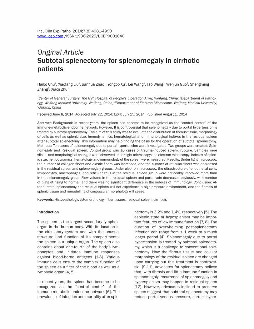
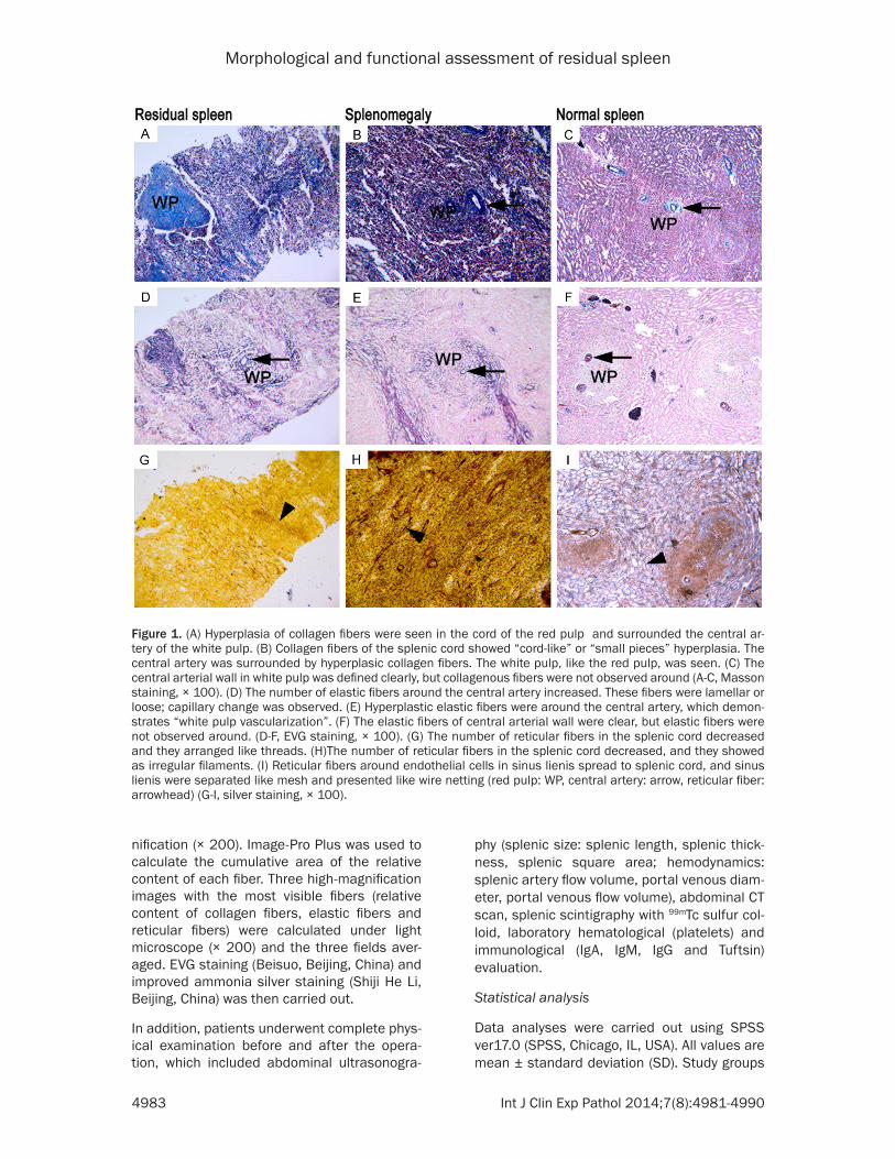
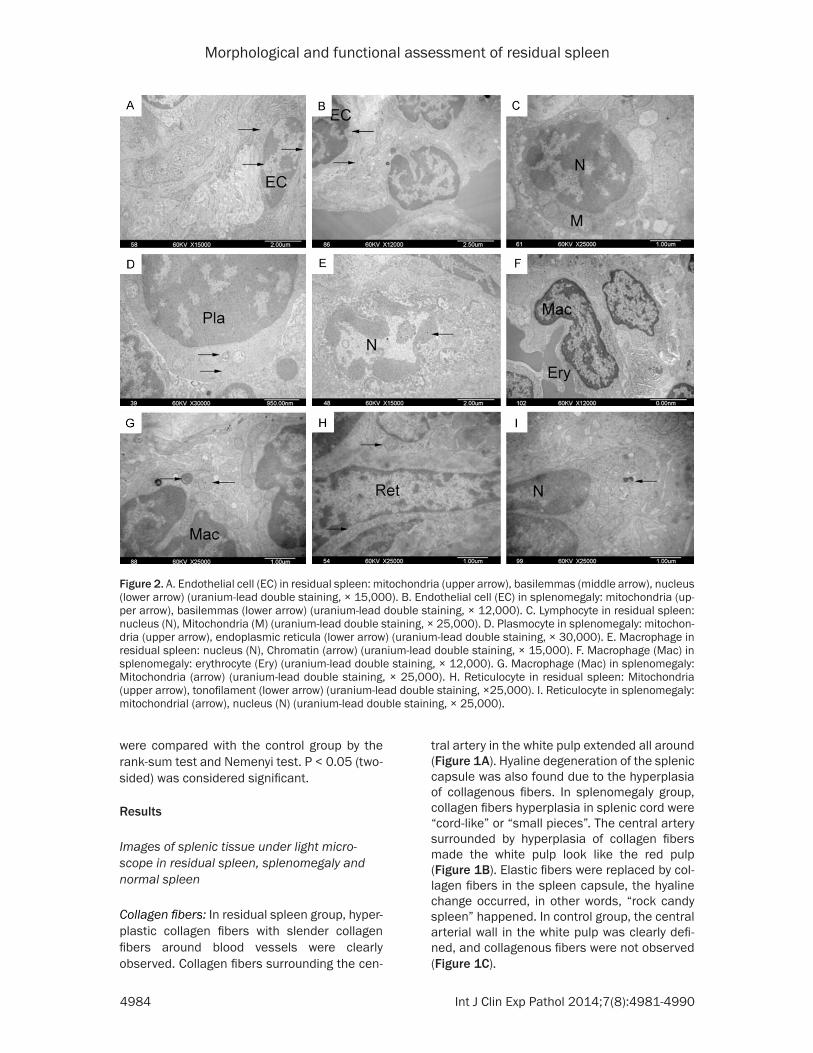
Figure 2. A. Endothelial cell (EC) in residual spleen: mitochondria (upper arrow), basilemmas (middle arrow), nucleus (lower arrow) (uranium-lead double staining, × 15,000). B. Endothelial cell (EC) in splenomegaly: mitochondria (up-per arrow), basilemmas (lower arrow) (uranium-lead double staining, × 12,000). C. Lymphocyte in residual spleen: nucleus (N), Mitochondria (M) (uranium-lead double staining, × 25,000). D. Plasmocyte in splenomegaly: mitochon-dria (upper arrow), endoplasmic reticula (lower arrow) (uranium-lead double staining, × 30,000). E. Macrophage in residual spleen: nucleus (N), Chromatin (arrow) (uranium-lead double staining, × 15,000). F. Macrophage (Mac) in splenomegaly: erythrocyte (Ery) (uranium-lead double staining, × 12,000). G. Macrophage (Mac) in splenomegaly: Mitochondria (arrow) (uranium-lead double staining, × 25,000). H. Reticulocyte in residual spleen: Mitochondria (upper arrow), tonofilament (lower arrow) (uranium-lead double staining, ×25,000). I. Reticulocyte in splenomegaly: mitochondrial (arrow), nucleus (N) (uranium-lead double staining, × 25,000).

Morphological and functional assessment of residual spleen
4985 Int J Clin Exp Pathol 2014;7(8):4981-4990
um-sized and small lymphocytes were observed along with plasmocytes with abundant rough endoplasmic reticula and high secretion. How- ever, medullary degeneration of some mito-chondria was observed in the plasmocytes (Figure 2D). In control group, lymphocytes were oval with big, round and deeply dyed nuclei.
Macrophages: In residual spleen group, apop-tosis was observed in some macrophages. Chromatin gathered at the edge, karyopyknosis karyotheca had disappeared and “chromatin spillover” could also be observed (Figure 2E). In splenomegaly group, the electron concentra-tion in the macrophage cytoplasm was lower; also, macrophages were seen to phagocytize erythrocytes (Figure 2F). In cytoplasm, the number of mitochondria increased along with an increase in the size of the matrix space. Medullary degeneration and vacuolar degener-ation as well as cristae fragmentation were observed in some mitochondria (Figure 2G). In control group, the nuclei of macrophages with abundant cytoplasm were round or oval and deeply dyed.
Reticulocytes: In residual spleen group, the number of reticulocytes with large somas decreased; a considerable amount of nucleo-plasm and clear intracellular tonofilament was observed. Mitochondria and other cytoplasmic organelles were not observed (Figure 2H). In splenomegaly group, the number of reticulo-cytes with enlarged somas, appreciable nucleo-plasm, with various morphologies as well as protuberances increased. Karyoplasm and mitochondria increased, mitochondria crista ambiguity, and medullary degeneration was observed in some of them (Figure 2I). In control group, reticulocytes were oblong with protuber-ances, and the cytoplasm was abundant with a few pigmented granules.
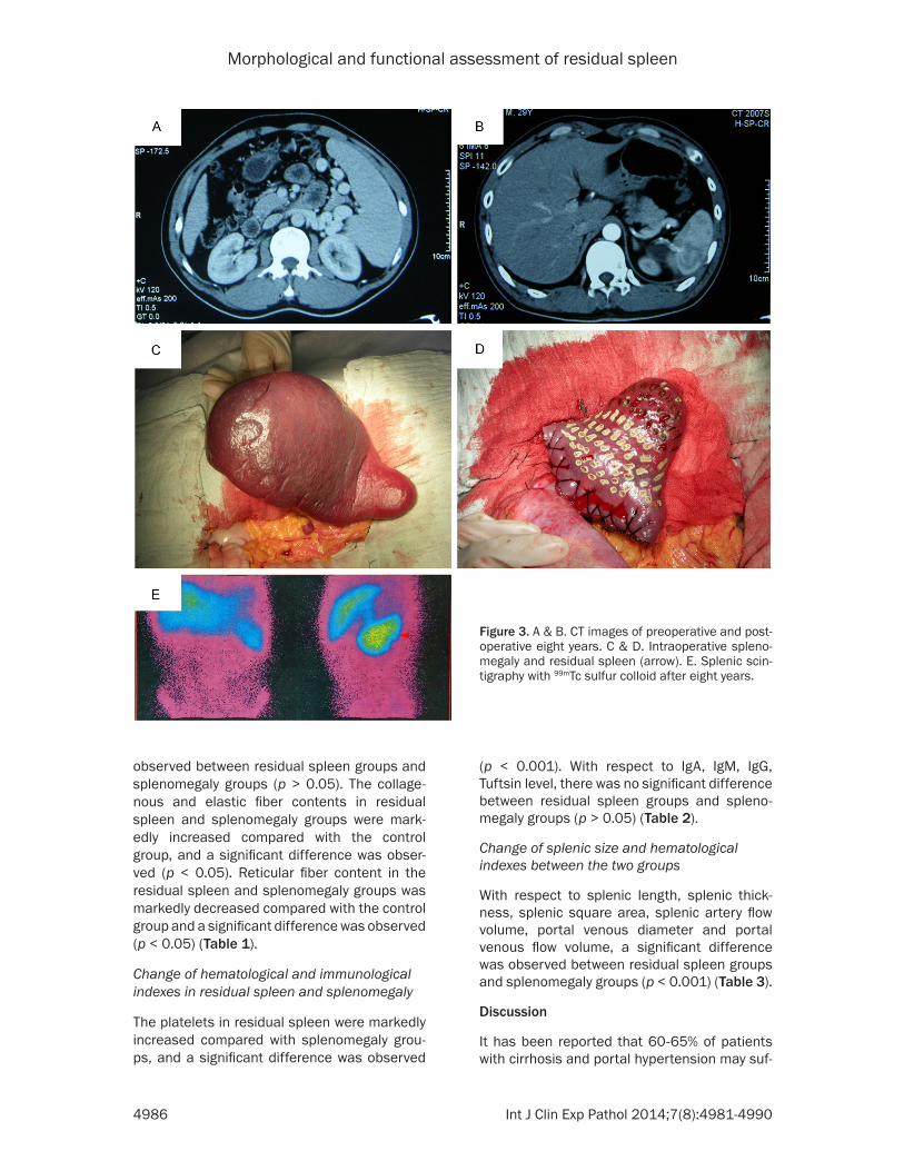
Images of clinical data
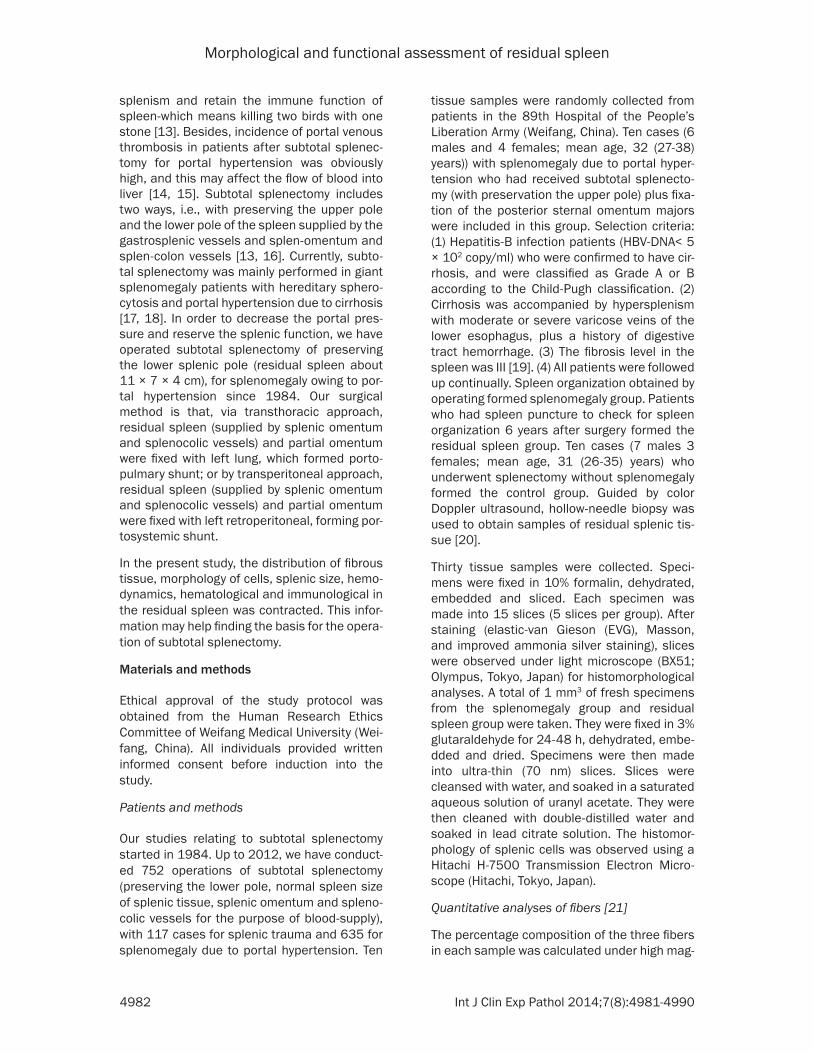
Residual spleen was preserved about 11 × 7 × 4 cm on operation. It was alive that was con-firmed by CT scan, and still had phagocytosis via splenic scintigraphy with 99mTc sulfur colloid after eight years (Figure 3A-E).
Measurement of collagenous, elastic and retic-ular fiber contents in spleen tissues
With respect to the content of collagenous and elastic fibers, a significant difference was not
Elastic fibers: In residual spleen group, slender and successive intimas were observed in the white pulp. The number of elastic fibers around the central artery increased, and they were lamellar or loose (i.e., capillary change was seen; Figure 1D). In splenomegaly group, the elastic fibers of the central artery in the white pulp demonstrated a sparse layer change, and the elastic membrane was irregular and not successive. The elastic fibers of the arterial wall were piled up and arranged irregularly. Hyperplastic elastic fibers were clearly obser- ved, which showed white pulp vascularization (Figure 1E). In control group, the elastic fibers of the central arterial wall were clear, but elas-tic fibers around the wall were not observed (Figure 1F).
Reticular fibers: In residual spleen group, the number of reticular fibers in the splenic cord was decreased and arranged like threads (Figure 1G). In splenomegaly group, the number of reticular fibers in the splenic cord was reduced and appeared like irregular filaments; distinguishing between the splenic cord and sinus lienis was difficult (Figure 1H). In control group, the reticular fibers around the endothe-lial cells in the sinus lienis spread to the splenic cord; the sinus lienis was separated like mesh and looked like wire netting (Figure 1I).
Images of cellular morphology under electron microscope in residual spleen and spleno-megaly
Endothelial cells: In residual spleen group, the endothelial cells had a spindle-shaped and their basilemmas were clear with fenestrations. The structures of mitochondria in the cyto-plasm were approximately normal (Figure 2A). In splenomegaly group, endothelial cells lost their spindle-shaped, were decreased in num-ber, and had a shrinking volume. In addition, the cytoplasm and nucleoplasm were scarce; the basement membrane was not clear and most of the fenestrations had disappeared. Vacuolar degeneration was observed in the mitochondria (Figure 2B). In control group, the endothelial cells had a spindle-shaped with well-developed golgiosome and rough endo-plasmic reticula in the cytoplasm.
Lymphocytes: In residual spleen group, medi-um-sized and small lymphocytes were observed with approximately normal morphologies (Figure 2C). In splenomegaly group, large, medi-
Morphological and functional assessment of residual spleen
4986 Int J Clin Exp Pathol 2014;7(8):4981-4990
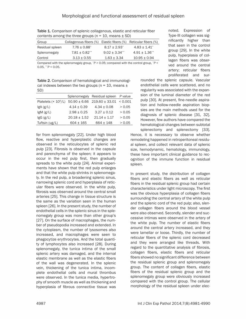
observed between residual spleen groups and splenomegaly groups (p > 0.05). The collage-nous and elastic fiber contents in residual spleen and splenomegaly groups were mark-edly increased compared with the control group, and a significant difference was obser- ved (p < 0.05). Reticular fiber content in the residual spleen and splenomegaly groups was markedly decreased compared with the control group and a significant difference was observed (p < 0.05) (Table 1).
Change of hematological and immunological indexes in residual spleen and splenomegaly
The platelets in residual spleen were markedly increased compared with splenomegaly grou- ps, and a significant difference was observed
(p < 0.001). With respect to IgA, IgM, IgG, Tuftsin level, there was no significant difference between residual spleen groups and spleno-megaly groups (p > 0.05) (Table 2).
Change of splenic size and hematological indexes between the two groups
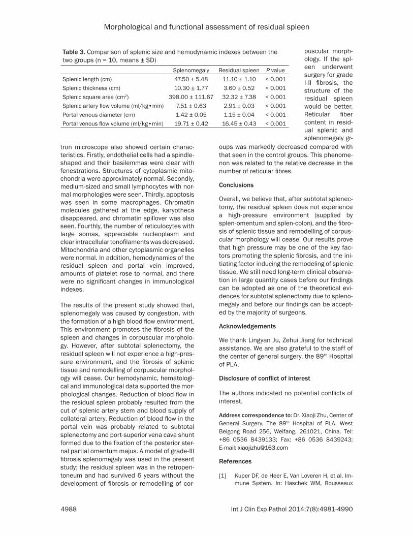
With respect to splenic length, splenic thick-ness, splenic square area, splenic artery flow volume, portal venous diameter and portal venous flow volume, a significant difference was observed between residual spleen groups and splenomegaly groups (p < 0.001) (Table 3).
Discussion
It has been reported that 60-65% of patients with cirrhosis and portal hypertension may suf-
Figure 3. A & B. CT images of preoperative and post-operative eight years. C & D. Intraoperative spleno-megaly and residual spleen (arrow). E. Splenic scin-tigraphy with 99mTc sulfur colloid after eight years.
Morphological and functional assessment of residual spleen
4987 Int J Clin Exp Pathol 2014;7(8):4981-4990
fer from splenomegaly [22]. Under high blood flow, reactive and hyperplastic changes are observed in the reticulocytes of splenic red pulp [23]. Fibrosis is observed in the capsule and parenchyma of the spleen; it appears to occur in the red pulp first, then gradually spreads to the white pulp [24]. Animal experi-ments have shown that the red pulp enlarges and that the white pulp shrinks in splenomega-ly. In the red pulp, a broadening splenic sinus, narrowing splenic cord and hyperplasia of retic-ular fibers were observed. In the white pulp, fibrosis was observed around the central small arteries [25]. This change in tissue structure is the same as the variation seen in the human spleen [26]. In the present study, the number of endothelial cells in the splenic sinus in the sple-nomegaly group was more than other group’s [27]. On the surface of macrophages, the num-ber of pseudopodia increased and extended. In the cytoplasm, the number of lysosomes also increased, and macrophages were seen to phagocytize erythrocytes. And the total quanti-ty of lymphocytes also increased [28]. During splenomegaly, the tunica intima of the small splenic artery was damaged, and the internal elastic membrane as well as the elastic fibers of the wall was degenerated. In the splenic vein, thickening of the tunica intima, incom-plete endothelial cells and mural thrombus were observed. In the tunica media, hypertro-phy of smooth muscle as well as thickening and hyperplasia of fibrous connective tissue was
Hence, it is necessary to observe whether remodeling happened in retroperitoneal residu-al spleen, and collect relevant data of splenic size, hemodynamic, hematology, immunology, these have important clinical guidance to rec-ognition of the immune function in residual spleen.
In present study, the distribution of collagen fibers and elastic fibers as well as reticular fibers in the residual splenic group had certain characteristics under light microscopy. The first was the obvious hyperplasia of collagen fibers surrounding the central artery of the white pulp and the splenic cord of the red pulp; also, slen-der collagen fibers around the blood vessel were also observed. Secondly, slender and suc-cessive intimas were observed in the artery of the white pulp. The number of elastic fibers around the central artery increased, and they were lamellar or loose. Thirdly, the number of reticular fibers of the splenic cord decreased and they were arranged like threads. With regard to the quantitative analysis of fibrosis, collagen fibers, elastic fibers and reticular fibers showed no significant difference between the residual splenic group and splenomegaly group. The content of collagen fibers, elastic fibers of the residual splenic group and the splenomegaly group were obviously increased compared with the control group. The cellular morphology of the residual spleen under elec-
Table 1. Comparison of splenic collagenous, elastic and reticular fiber contents among the three groups (n = 10, means ± SD)Group Collagenous fibers (%) Elastic fibers (%) Reticular fibers (%)Residual spleen 7.76 ± 0.88* 8.17 ± 2.93* 4.83 ± 1.41*
Splenomegaly 7.81 ± 0.82** 9.02 ± 3.34** 4.91 ± 1.36**
Control 3.13 ± 0.55 1.63 ± 3.34 10.95 ± 0.94Compared with the splenomegaly group, *P > 0.05; compared with the control group, *P < 0.05, **P < 0.05.
Table 2. Comparison of hematological and immunologi-cal indexes between the two groups (n = 10, means ± SD)
Splenomegaly Residual spleen P valuePlatelets (× 109/L) 50.90 ± 6.66 219.60 ± 33.01 < 0.001IgA (g/L) 4.14 ± 0.39 4.34 ± 0.08 > 0.05IgM (g/L) 2.98 ± 0.25 3.27 ± 0.12 > 0.05IgG (g/L) 20.18 ± 1.52 21.14 ± 1.17 > 0.05Tuftsin (ug/L) 604 ± 165 664 ± 148 > 0.05
noted. Expression of Type-III collagen was sig-nificantly higher than that seen in the control group [29]. In the white pulp, hyperplasia of col-lagen fibers was obser- ved around the central artery; reticular fibers proliferated and sur-
rounded the splenic capsule. Vascular endothelial cells were scattered, and no regularity was associated with the expan-sion of the luminal diameter of the red pulp [30]. At present, fine-needle aspira-tion and hollow-needle aspiration biop-sies are the main methods used for the diagnosis of splenic disease [31, 32]. However, few authors have compared the hematological changes between subtotal splenectomy and splenectomy [33].
Morphological and functional assessment of residual spleen
4988 Int J Clin Exp Pathol 2014;7(8):4981-4990
tron microscope also showed certain charac-teristics. Firstly, endothelial cells had a spindle-shaped and their basilemmas were clear with fenestrations. Structures of cytoplasmic mito-chondria were approximately normal. Secondly, medium-sized and small lymphocytes with nor-mal morphologies were seen. Thirdly, apoptosis was seen in some macrophages. Chromatin molecules gathered at the edge, karyotheca disappeared, and chromatin spillover was also seen. Fourthly, the number of reticulocytes with large somas, appreciable nucleoplasm and clear intracellular tonofilaments was decreased. Mitochondria and other cytoplasmic organelles were normal. In addition, hemodynamics of the residual spleen and portal vein improved, amounts of platelet rose to normal, and there were no significant changes in immunological indexes.
The results of the present study showed that, splenomegaly was caused by congestion, with the formation of a high blood flow environment. This environment promotes the fibrosis of the spleen and changes in corpuscular morpholo-gy. However, after subtotal splenectomy, the residual spleen will not experience a high-pres-sure environment, and the fibrosis of splenic tissue and remodelling of corpuscular morphol-ogy will cease. Our hemodynamic, hematologi-cal and immunological data supported the mor-phological changes. Reduction of blood flow in the residual spleen probably resulted from the cut of splenic artery stem and blood supply of collateral artery. Reduction of blood flow in the portal vein was probably related to subtotal splenectomy and port-superior vena cava shunt formed due to the fixation of the posterior ster-nal partial omentum majus. A model of grade-III fibrosis splenomegaly was used in the present study; the residual spleen was in the retroperi-toneum and had survived 6 years without the development of fibrosis or remodelling of cor-
oups was markedly decreased compared with that seen in the control groups. This phenome-non was related to the relative decrease in the number of reticular fibres.
Conclusions
Overall, we believe that, after subtotal splenec-tomy, the residual spleen does not experience a high-pressure environment (supplied by splen-omentum and splen-colon), and the fibro-sis of splenic tissue and remodelling of corpus-cular morphology will cease. Our results prove that high pressure may be one of the key fac-tors promoting the splenic fibrosis, and the ini-tiating factor inducing the remodeling of splenic tissue. We still need long-term clinical observa-tion in large quantity cases before our findings can be adopted as one of the theoretical evi-dences for subtotal splenectomy due to spleno-megaly and before our findings can be accept-ed by the majority of surgeons.
Acknowledgements
We thank Lingyan Ju, Zehui Jiang for technical assistance. We are also grateful to the staff of the center of general surgery, the 89th Hospital of PLA.
Disclosure of conflict of interest
The authors indicated no potential conflicts of interest.
Address correspondence to: Dr. Xiaoji Zhu, Center of General Surgery, The 89th Hospital of PLA, West Beigong Road 256, Weifang, 261021, China. Tel: +86 0536 8439133; Fax: +86 0536 8439243; E-mail: [email protected]
References
[1] Kuper DF, de Heer E, Van Loveren H, et al. Im-mune System. In: Haschek WM, Rousseaux
Table 3. Comparison of splenic size and hemodynamic indexes between the two groups (n = 10, means ± SD)
Splenomegaly Residual spleen P valueSplenic length (cm) 47.50 ± 5.48 11.10 ± 1.10 < 0.001Splenic thickness (cm) 10.30 ± 1.77 3.60 ± 0.52 < 0.001Splenic square area (cm2) 398.00 ± 111.67 32.32 ± 7.38 < 0.001Splenic artery flow volume (ml/kg•min) 7.51 ± 0.63 2.91 ± 0.03 < 0.001Portal venous diameter (cm) 1.42 ± 0.05 1.15 ± 0.04 < 0.001Portal venous flow volume (ml/kg•min) 19.71 ± 0.42 16.45 ± 0.43 < 0.001
puscular morph- ology. If the spl- een underwent surgery for grade I-II fibrosis, the structure of the residual spleen would be better. Reticular fiber content in resid-ual splenic and splenomegaly gr-
Morphological and functional assessment of residual spleen
4989 Int J Clin Exp Pathol 2014;7(8):4981-4990
CG, Wallig MA, editors. Handbook of Toxicolog-ic Pathology. San Diego: Academic Press; 2002. pp. 585-646.
[2] Nolte MA, Hamann A, Kraal G, Mebius RE. The strict regulation of lymphocyte migration to splenic white pulp does not involve common homing receptors. Immunology 2002; 106: 299-307.
[3] Balogh P, Horváth G, Szakal AK. Immunoarchi-tecture of distinct reticular fibroblastic do-mains in the white pulp of mouse spleen. J Histochem Cytochem 2004; 52: 1287-98.
[4] Bisharat N, Omari H, Lavi I, Raz R. Risk of infec-tion and death among post-splenectomy pa-tients. J Infect 2001; 43: 182-86.
[5] Okabayashi T, Hanazaki K. Overwhelming post-splenectomy infection syndrome in adults-a clinically preventable disease. World J Gastro-enterol 2008; 14: 176-79.
[6] Tarantino G, Savastano S, Capone D, Colao A. Spleen: A new role for an old player? World J Gastroenterol 2011; 17: 3776-84.
[7] Mourtzoukou EG, Pappas G, Peppas G, Fala-gas ME. Vaccination of asplenic or hyposplenic adults. Br J Surg 2008; 95: 273-80.
[8] Di Sabatino A, Carsetti R, Corazza GR. Post-splenectomy and hyposplenic states. Lancet 2011; 378: 86-97.
[9] Rezende-Neto JB, Petroianu A, Santana SK. Subtotal splenectomy and central splenorenal shunt for treatment of bleeding from Roux en Y jejunal loop varices secondary to portal hyper-tension. Dig Dis Sci 2008; 53: 539-43.
[10] Yao HS, Wang WJ, Wang Q, Gao WC, Xiang HG, Hu ZQ, Gao JD, Chen XY, Wang WM. Random-ized clinical trial of vessel sealing system (Liga-Sure) in esophagogastric devascularization and splenectomy in patients with portal hyper-tension. Am J Surg 2011; 202: 82-90.
[11] Sretenovic ALJ, Perišić V, Krstić Z, Vujović D, Pavićević P, Stanisavljević D, Radević B. Erra-tum to: Warren shunt combined with partial splenectomy for children with extrahepatic portal hypertension, massive splenomegaly, and severe hypersplenism. Surg Today 2013; 43: 526.
[12] Andraus W, Pinheiro RS, Haddad LB, Herman P, D’Albuquerque LAC. The best approach for splenectomy in portal hypertension. Surgery 2011; 149: 853.
[13] Li E, Zhao L, Zhu L, Lin A, Ge L, Wang F, Shi B. Treating portal hypertension by subtotal sple-nectomy with retroperitoneal splenic transpo-sition and devascularization: clinical study. Zhonghua Wai Ke Za Zhi 1998; 36: 333-35.
[14] Senzolo M, Rodriguez K, Nadal E, Burra P. Risk factors for portal venous thrombosis after sple-nectomy in patients with cirrhosis and portal hypertension. Br J Surg 2010; 97: 910-16.
[15] Hirashita T, Ohta M, Kai S, Masuda T, Eguchi H, Iwashita Y, Ogawa T, Kitano S. Implications of portal vein thrombosis after splenectomy for patients with idiopathic portal hypertension. Surg Today 2011; 41: 1475-80.
[16] Petroianu A, Resende V, Da Silva RG. Late fol-low-up of patients submitted to subtotal sple-nectomy. Int Surg 2006; 4: 172-78.
[17] Petroianu A. Treatment of portal hypertension by subtotal splenectomy and central splenore-nal shunt. Postgrad Med J 1988; 64: 38-41.
[18] Bader-Meunier B, Gauthier F, Archambaud F, Cynober T, Miélot F, Dommergues JP, Warsza-wski J, Mohandas N, Tchernia G. Long-term evaluation of the beneficial effect of subtotal splenectomy for management of hereditary spherocytosis. Blood 2001; 97: 399-403.
[19] Tejima K, Masuzaki R, Ikeda H, Yoshida H, Tateishi R, Sugioka Y, Kume Y, Okano T, Iwai T, Gotoh H, Katoh S, Suzuki A, Koike Y, Yatomi Y, Omata M, Koike K. Thrombocytopenia is more severe in patients with advanced chronic hep-atitis C than B with the same grade of liver stiffness and splenomegaly. J Gastroenterol 2010; 45: 876-84.
[20] Gómez-Rubio M, López-Cano A, Rendón P, Mu-ñoz-Benvenuty A, Macías M, Garre C, Segura-Cabral JM. Safety and diagnostic accuracy of percutaneous ultrasound-guided biopsy of the spleen: a multicenter study. J Clin Ultrasound 2009; 37: 445-50.
[21] Abe T, Hashiguchi A, Yamazaki K, Ebinuma H, Saito H, Kumada H, Izumi N, Masaki N, Saka-moto M. Quantification of collagen and elastic fibers using whole-slide images of liver biopsy specimens. Pathol Int 2013; 63: 305-10.
[22] Gibson PR, Gibson RN, Ditchfield MR, Donlan JD. Splenomegaly-an insensitive sign of portal hypertension. Aust N Z J Med 1990; 20: 771-74.
[23] Ruehl-Fehlert C, Hartmann E, Rinke M. Reac-tive and proliferative changes of splenic reticu-lum cells of rats investigated with special staining methods and immunohistochemistry. Exp Toxicol Pathol 2008; 59: 281-90.
[24] Misawa T. Induction of experimental spleno-megaly with portal hypertension in rats with liver cirrhosis. Nihon Shokakibyo Gakkai Zasshi 1991; 88: 2653-62.
[25] Suttie AW. Histopathology of the spleen. Toxi-col Pathol 2006; 34: 466-503.
[26] Yamamoto K. Morphological studies of the spleen in idiopathic portal hypertension (so-called Banti’s syndrome without liver cirrhosis) using light microscopy, scanning electron mi-croscopy and histometry. Acta Pathol Jpn 1979; 29: 1-19.
[27] Saitoh K, Kamiyama R, Hatakeyama S, Sugiura M. In vitro labeling and tissue autoradiography
Morphological and functional assessment of residual spleen
4990 Int J Clin Exp Pathol 2014;7(8):4981-4990
of splenomegaly associated with portal hyper-tension. Virchows Arch B Cell Pathol Incl Mol Pathol 1986; 52: 33-40.
[28] Li ZF, Zhang S, Lv GB, Huang Y, Zhang W, Ren S, Yang J, Dang SS. Changes in count and func-tion of splenic lymphocytes from patients with portal hypertension. World J Gastroenterol 2008; 14: 2377-82.
[29] Li T, Ni JY, Qi YW, Li HY, Zhang T, Yang Z. Splen-ic vasculopathy in portal hypertension pa-tients. World J Gastroenterol 2006; 12: 2737-41.
[30] Li ZF, Zhang S, Huang Y, Xia XM, Li AM, Pan D, Zhang W, Wang J. Morphological changes of blood spleen barrier in portal hypertensive spleen. Chin Med J (Engl) 2008; 121: 561-65.
[31] Friedlander MA, Wei XJ, Iyengar P, Moreira AL. Diagnostic pitfalls in fine needle aspiration bi-opsy of the spleen. Diagn Cytopathol 2008; 36: 69-75.
[32] Lal A, Ariga R, Gattuso P, Nemcek AA, Nayar R. Splenic fine needle aspiration and core biopsy. A review of 49 cases. Acta Cytol 2003; 47: 951-59.
[33] Petroianu A, De Oliveira AE, Alberti LR. Hyper-splenism in schistosomatic portal hyperten-sion. Arch Med Res 2005; 36: 496-501.