Embed Size (px)
Citation preview
43http://www.ecevr.org/
CLINICAL EXPERIMENTALVACCINERESEARCH
Original article
Introduction
Respiratory syncytial virus (RSV) is a Pneumoviridae family, negative-sense, single-
stranded RNA virus that can cause respiratory diseases such as pneumonia, bronchi-
olitis, and asthma in infants and elderly or immunocompromised patients [1]. It is
known that in the United States alone, more than 500,000 people visit the emergency
room every year and more than 50,000 are hospitalized due to RSV. Worldwide, ap-
proximately 66,000-199,000 people die annually due to RSV infection, with most fatali-
ties occurring in developing countries [2,3]. Since the discovery of RSV in 1956, only
© Korean Vaccine Society.This is an Open Access article distributed under the terms of the Creative Commons Attribution Non-Com-mercial License (http://creativecommons.org/licenses/by-nc/4.0) which permits unrestricted non-commercial use, distribution, and reproduction in any medium, pro-vided the original work is properly cited.
K O R E A N V A C C I N E S O C I E T Y
K O R E A N V A C C I N E S O C I E T Y
K O R E A N A C C I N E O C I E T Y
VS
Clin Exp Vaccine Res 2019;8:43-53https://doi.org/10.7774/cevr.2019.8.1.43pISSN 2287-3651 • eISSN 2287-366X
Yeong-Min Jo, Jungwoo Kim, Jun ChangGraduate School of Pharmaceutical Sciences, Ewha Womans University, Seoul, Korea
Received: December 29, 2018Revised: January 12, 2019Accepted: January 25, 2019
Corresponding author: Jun Chang, PhDGraduate School of Pharmaceutical Sciences, Ewha Womans University, 52 Ewhayeodae-gil, Seoul 03760, KoreaTel: +82-2-3277-2549Fax: +82-2-3277-3051E-mail: [email protected]
No potential conflict of interest relevant to this article was reported.
We would like to express our gratitude to Jung-Yoon Lee and other members of the Immunology Laboratory for providing technical support to complete the experiments.
Purpose: Respiratory syncytial virus (RSV) can cause serious respiratory illnesses such as pneumonia, asthma, and bronchiolitis in infants and elderly or immunocompromised individu-als. An RSV vaccine has yet to be developed; only prophylactic anti-RSV antibody is com-mercially available. So, we investigated whether our vaccine candidate is able to induce type 1 CD4+ T helper (Th1), CD8+ T-cell responses, and protective immunity without vaccine-enhanced disease (VED) against RSV.Materials and Methods: We used RSV G protein fragment (Gcf A) with recombinant baculo-virus capable of expressing the RSV M2 protein (Bac M2) as a vaccine candidate, and injected this vaccine (Gcf A/Bac M2) intramuscularly, and challenged with RSV intranasally into mice. Enzyme-linked immunosorbent assay, flow cytometry, plaque assay, and weight measurement were performed to confirm humoral immunity, cellular immunity, and protective immunity.Results: The Gcf A/Bac M2 formulation induced a stronger IgG response to Gcf A than Gcf A inoculation alone, and the ratio of IgG1/IgG2a indicated that the responses shifted predomi-nantly to Th1. In addition, both RSV G-specific Th1 responses and RSV M2-specific CD8+ T-cell responses were induced, and G protein-associated eosinophilic infiltration was suppressed compared to the control group. Moreover, the Gcf A/Bac M2 group showed effective protec-tion after an RSV challenge. Conclusion: Bac M2 could serve as a vaccine with intrinsic adjuvant activity, and the Gcf A/Bac M2 shows promise as a vaccine candidate for inducing protective immunity without incit-ing VED.
Keywords: Respiratory syncytial virus, Vaccine, Recombinant baculovirus, M2, G protein
Vaccine containing G protein fragment and recombinant baculovirus expressing M2 protein induces protective immunity to respiratory syncytial virus
1 / 1CROSSMARK_logo_3_Test
2017-03-16https://crossmark-cdn.crossref.org/widget/v2.0/logos/CROSSMARK_Color_square.svg
Yeong-Min Jo et al • Vaccine against respiratory syncytial virus
44 http://www.ecevr.org/ https://doi.org/10.7774/cevr.2019.8.1.43
the prophylactic antibodies palivizumab (Synagis) and RSV
immunoglobulin (RSV-IVIG, RespiGam) have been commer-
cially available, while no vaccine or medicine has been devel-
oped as yet [4,5]. In the 1960s, there were reports of deaths of
children vaccinated with formalin-inactivated RSV (FI-RSV)
vaccine due to vaccine-enhanced disease (VED), which is
characterized by excessive eosinophil infiltration and type 2
CD4+ T helper (Th2)-like responses [6]. From this perspective,
it is generally recognized that monitoring for elicited Th2-like
and eosinophilic responses is important in the development
of RSV vaccines.
The RSV G protein is a surface glycoprotein composed of
298 amino acids and is one of the main target proteins in RSV
vaccine research. This protein is known to induce neutraliz-
ing antibodies, and to have a CX3C chemokine motif (a.a. 182-
186) in the central conserved region capable of binding to
CX3CR1, thereby influencing T-cell responses in RSV-infect-
ed lung [3,7]. Previously, a Gcf A of 131-230 amino acids from
an RSV A2 strain was produced and evaluated as a vaccine
with a cholera toxin (CT) adjuvant. As a result, specific IgG
was induced, and protective efficacy against RSV A2 challenge
was demonstrated [8,9]. However, because the CT cannot be
used as an adjuvant in humans, our experiment was conduct-
ed using Bac M2, which has intrinsic adjuvant activity.
Baculovirus is a double stranded DNA insect virus, known
to be a safe vaccine platform for human use because it can-
not be replicated in mammalian cells. Several studies of the
baculovirus platform have been conducted to investigate the
expression of proteins in mammalian cells, using strong pro-
moters including CMV, CAG, SV40, and HBV. Based on this
research, Bac M2 was generated. The RSV M2 protein is ex-
pressed from two overlapping frames (ORFs) to M2-1 and
M2-2, respectively. The M2-1 protein has a strong epitope for
CD8+ T cells (a.a. 82-90), effectively inducing a cytotoxic T
lymphocyte (CTL) response and contributing to virus clear-
ance [10,11]. It has been shown that the M2-specific CTL re-
sponses are induced in BALB/c mice infected with vaccinia
virus RSV-M2 (vac-M2) [12]. Previous studies have shown
that M2-specific CTL responses are induced by Bac M2 im-
munization via intranasal or intramuscular routes [10,13].
In this study, we investigated whether Bac M2 can play an
adjuvant role when used in combination with Gcf A by mea-
suring humoral and cellular immune responses, bronchoal-
veolar lavage (BAL) cell responses, and viral titers in the lung
as an indicator of protective immunity, as well as immunopa-
thology-related weight loss.
Materials and Methods
Construction of a plasmid capable of expressing Gcf A and the production of Gcf AThe method for constructing a plasmid in which Gcf A was
encrypted was as described in a previously published report
[9]. Features of Gcf A include a central conserved region and
cysteine residues (Cys-172, Cys-176, Cys-182, and Cys-186)
[9]. The plasmid was transformed into ClearColi BL21 (DE3)
(Lucigen, Middleton, WI, USA), then spread on a Luria-Ber-
tani (LB) agar plate containing ampicillin and cultured over-
night at 37°C. The single colony of Gcf A-transformed Clear-
Coli was taken, added to fresh LB (+ampicillin) media (13 mL),
and placed in a 37°C shaking incubator overnight. Fresh LB
media (+ampicillin) (500 mL) was filled with 5 mL of ClearColi
(+Gcf plasmid) that had been cultured the day before and
grown up to 0.5 of OD 600 during shaking incubation. After
the addition of 1 mM IPTG and shaking incubation at 37°C
for 4 more hours, cells were obtained by centrifugation at
8,750 rpm and 4°C for 20 minutes. After cell resuspension us-
ing a binding buffer (20 mM KPO4, 500 mM NaCl, 10 mM im-
idazole), the cells were disrupted with a sonicator. After add-
ing a binding buffer to HisTrap HP (5 mL, GE Healthcare,
Pittsburgh, PA, USA) containing Ni2+ Sepharose, the proteins
that had not reacted to Ni2+ were removed. Elution buffer (20
mM KPO4, 500 mM NaCl, 500 mM imidazole) was applied to
obtain Gcf A reacting with Ni2+. Gcf A treated with 4× sodium
dodecyl sulfate (SDS) (+DTT) was run on 15% polyacrylamide
gel electrophoresis (PAGE) gel after staining with coma blue
reagent, confirming the presence of a band of Gcf A. Using
Sephacryl S-200 (GE Healthcare), pure Gcf A was separated,
and the concentration was measured using a BCA assay kit
(Thermo Scientific, Waltham, MA, USA). Gcf A treated with
4× SDS (+DTT, -DTT) was run on 15% PAGE gel and was con-
firmed as pure Gcf A via banding after staining with coma blue
reagent. The Gcf A was stored at -70°C.
Production of Bac M2The method for production of a Bac M2 was as described in a
previously published report [10]. Bac M2 was grown in Spodo
ptera frugiperda 9 (Sf-9) insect cells (Invitrogen, Carlsbad,
CA, USA) at 28°C using SF-900 II serum-free medium (Gibco
BRL, Rockville, MD, USA). In order to obtain Bac M2, the final
culture supernatants of infected Sf-9 cells were collected, treat-
ed with sucrose cushion (25% w/w sucrose in 5 mM NaCl, 10
mM EDTA in phosphate buffered saline [PBS]), and centri-
Yeong-Min Jo et al • Vaccine against respiratory syncytial virus
45http://www.ecevr.org/https://doi.org/10.7774/cevr.2019.8.1.43
fuged at 24,000 rpm for 75 minutes at 4°C. The pellets were
resuspended in 0.25 M sucrose and stored in LN2. The viral ti-
ter was determined by plaque assay using Sf-9 cells according
to the manufacturer’s protocol (Invitrogen).
Production of the RSV A2 strainTo obtain RSV A2, HEp-2 cells (ATCC, Manassas, VA, USA)
were cultured in minimal essential medium (MEM; Welgene,
Gyeongsan, Korea) containing 10% fetal bovine serum (FBS).
RSV A2 was proliferated in MEM containing 3% heat-inacti-
vated FBS, 2 mM glutamine, and 20 mM HEPES. After 4 days,
syncytia formation was verified and the viruses were harvest-
ed by ultra-centrifugation (25,000 rpm for 1 hour at 4°C). The
pellets were resuspended with a 25-gauge needle and serum-
free MEM and then sonicated. Viral titers were performed by
standard RSV plaque assay and the viruses were stored at
-70°C.
Mouse immunization and RSV challengeSix-week-old female BALB/cAnNCrljOri mice (n=5/group)
were purchased from Orient Bio Inc. (Seongnam, Korea) and
kept in a specific pathogen free room for a week-long adapta-
tion period. Three vaccines were used: Gcf A (20 μg/mouse)
alone, Gcf A with added Bac control (live baculovirus; Gcf A/
Bac control, 1×10⁷ plaque-forming unit [PFU]/mouse), and
Bac M2 (baculovirus expressing M2 protein; Gcf A/Bac M2,
1×10⁷ PFU/mouse) in Gcf A (20 μg/mouse). Immunization
was performed on days 0 and 14 through intramuscular injec-
tion of the right leg (volume, 100 μL). Viral challenge was per-
formed 23 days after the second immunization with RSV at a
dose of 1×10⁶ PFU/mouse. The mice were anesthetized with iso-
flurane (Ifran, Hana Pharm, Seongnam, Korea) inhalation and
the RSV (volume, 50 μL) was administered via the left intrana-
sal route. The condition of the mice was monitored through
weight measurement for 5 days. The vaccines and RSV were
diluted in PBS. All mice were monitored for the duration of the
experiment and efforts were made to minimize pain. The re-
search was carried out after approval by and in accordance
with the guidelines of the Ewha Womans University Animal
Experimental Ethics Committee (IACUC: 18-002).
IgG and IgG subtype titration specific to RSV G protein Blood collection was performed two days before the first im-
munization and 21 days after the second immunization. Blood
was drawn via the retro-orbital plexus using a capillary tube
containing heparin and collected in an Eppendorf tube. The
blood was centrifuged for 15 minutes at 5,600 rpm and 4°C,
and the sera were separated and stored at -70°C. The Gcf A-
specific antibody titer was measured by direct enzyme-linked
immunosorbent assay (ELISA). The Gcf A was diluted in PBS,
held overnight in aliquots of 100 μL/well (50 ng/well) in a
96-well plate, washed 3 times with PBS (+0.05% Tween-20),
and blocked with PBS (+0.05% Tween-20) containing 1% skim
milk for 2 hours at room temperature (RT). After 2 additional
washes with PBS (+0.05% Tween-20), 2-fold serial diluted se-
rum was added to the coating antigen and incubated for 2
hours at RT. After 4 washes with PBS (+0.05% Tween-20), horse-
radish peroxidase-conjugated affinity-purified rabbit anti-
mouse total IgG secondary antibody (Zymed Laboratory, San
Francisco, CA, USA) was applied. For the second immuniza-
tion serum, rabbit anti-mouse IgG1, IgG2a secondary anti-
body, horseradish peroxidase (Invitrogen) was applied. Incu-
bation at RT for 1 hour was followed by washing 6 times with
PBS. After the addition of tetramethylbenzidine peroxidase
substrate (1:1; TMB, KPL, Gaithersburg, MD, USA), the reac-
tion was stopped with 1 M H3PO4, and the analysis was car-
ried out at OD 450 nm using a Thermo ELISA plate reader.
Cut-off values were calculated with serum-free wells.
Sampling for investigation of cellular responsesOn day 5 after the RSV challenge, the mice were sacrificed by
CO2 anesthesia. BAL fluids were collected with a catheter (BD
Angiocath Plus, Becton-Dickinson, Franklin Lakes, NJ, USA)
and 1 mL of PBS. Cells and supernatants were separated by
centrifugation and leukocyte recruitment was measured in
the BAL cell samples. Lung tissues of the immunized mice
were collected, homogenized using a 70 μm cell strainer (SPL),
and centrifuged to separate lung mononuclear cells and su-
pernatants. Then, the proportions of immune cells were mea-
sured and the supernatants were used to measure viral repli-
cation in the lungs.
Lung viral titration specific for the RSV A2 strainOn day 5 after the RSV challenge, all mice were sacrificed by
CO2 euthanasia and single-cell suspensions of lung tissue
were obtained using MEM (Welgene) and a 70 μm cell strain-
er (SPL). The cells and supernatants were separated by cen-
trifugation at 1,600 rpm and 4°C and the supernatants were
incubated at 37°C for 5 days with HEp-2 cells (ATCC). Viral ti-
ters were recorded as PFU/g of lung tissue, with a limit of de-
tection of 100 PFU/g.
Yeong-Min Jo et al • Vaccine against respiratory syncytial virus
46 http://www.ecevr.org/ https://doi.org/10.7774/cevr.2019.8.1.43
Flow cytometric analysisTo measure the granulocyte population in the BAL specimens,
the BAL cells were treated with rat anti-mouse CD16/CD32
blocking antibody (BD Biosciences, San Jose, CA, USA) and
reacted at RT for 10 minutes. BAL cells were stained in the
dark for 30 minutes at 4°C with anti-Siglec-F antibody (E50-
2440, BD Biosciences), anti-Gr-1 antibody (RB6-8C5, BioLeg-
end, San Diego, CA, USA), anti-CD11c antibody (N418, Bio-
Legend), and anti-CD45 antibody (30-F11, BioLegend). To
analyze the RSV M2-tetramer binding activity of CD8+ T cells,
lymphocytes isolated from lung tissue were treated with CD16/
32 blocking antibody (BD Biosciences) and reacted at RT for
10 minutes. Staining was performed at 4°C for 30 minutes in
the dark using with anti-CD8 antibody (53-6.7, BioLegend),
anti-CD44 antibody (IM7, BioLegend), and MHC class I H-
2Kd tetramer complexes (H-2Kd/SYIGSINNI) presented with
RSV M282-90 peptide. All stained cells were fixed with fluores-
cence-activated cell sorting (FACS) lysing solution (BD Bio-
sciences) and analysis was performed with a BD FACSCali-
bur (BD Biosciences).
Th1 interferon γ (IFN-γ)+ cells and CD8+ IFN-γ+ T cells were
analyzed by intracellular staining (ICS). First, lung lympho-
cytes were resuspended in IMDM (Welgene) containing 10%
heat-inactivated FBS. The cells were stimulated by adding 50
μg/mL phorbol myristate acetate (1:1,000, Sigma-Aldrich, St.
Louis, MO, USA), 500 μg/mL ionomycin (1:1,000, Sigma-Al-
drich), and either 10 μM RSV M2 peptide (82-90: SYIGSINNI)
or 10 μM RSV G peptide (183-195: WAICKRIPNKKPG) to IM-
DM (+10% FBS) (Welgene) containing β-mercaptoethanol
(1:1,000, Sigma-Aldrich), recombinant human IL-2 (BioLeg-
end), and Brefeldin A (1:1,000, Invitrogen), followed by incu-
bation in the dark for 5 hours at 37°C. Stimulated cells were
stained with anti-CD3 antibody (17A2, BioLegend), anti-CD44
antibody (IM7, BioLegend), and either anti-CD8 antibody
(53-6.7, BioLegend) or anti-CD4 antibody (RM 4-5, BioLeg-
end) at 4°C for 30 minutes in the dark and fixed with FACS
lysing solution (BD Biosciences). For the intracellular cyto-
kine staining, first the fixed cells were treated with FACS buf-
fer (0.5% FBS, 0.09% NaN3 in PBS) containing 0.5% saponin
(Sigma-Aldrich) and then they were permeabilized at RT for
15 minutes in the dark. The permeabilized cells were stained
with anti-IFN-γ antibody (XMG 1.2, BioLegend) at RT for 30
minutes in the dark. The stained cells were analyzed using a
BD FACSCalibur (BD Biosciences) and all flow cytometry da-
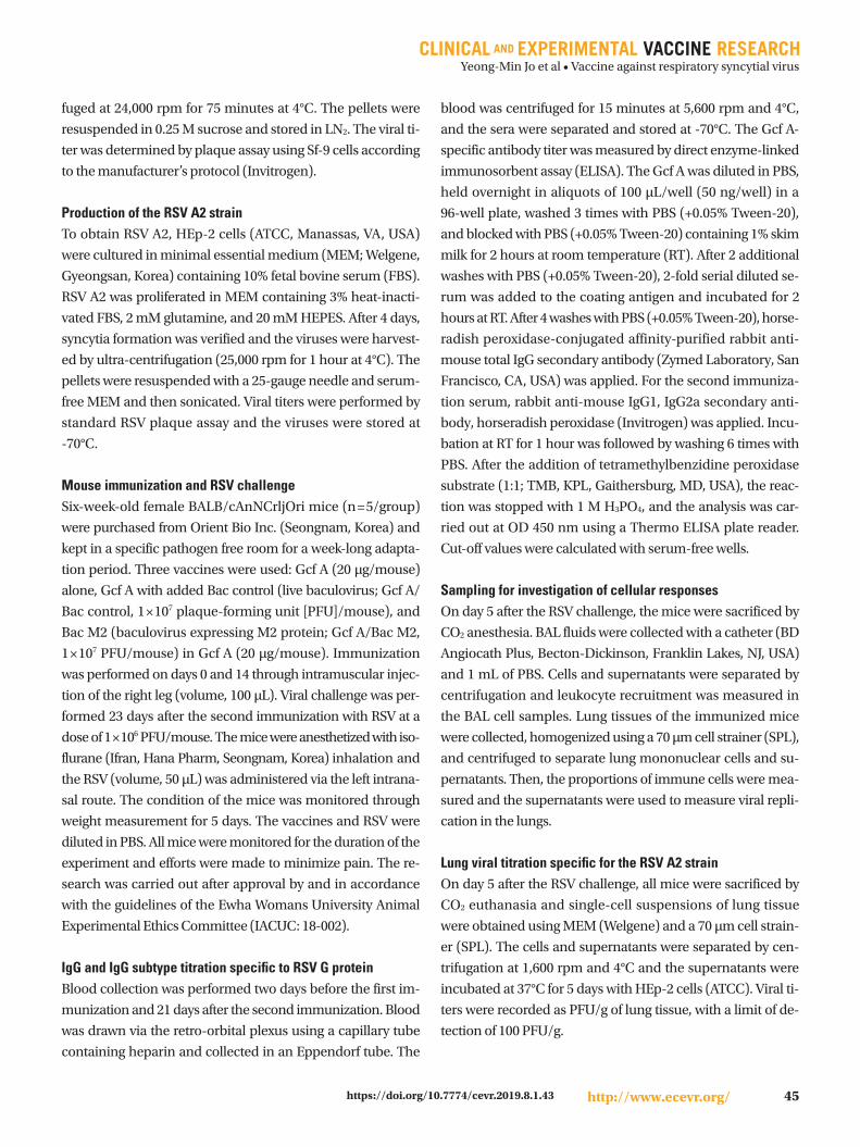
Fig. 1. Experiment schedule and assessment of the vaccine-induced humoral immune responses. (A) Experiment schedule for investigating the efficacy of the manufactured vaccine. (B) Serum IgG antibody titers specific for Gcf A were measured by enzyme-linked immunosorbent assay (ELISA) using sera obtained two days before and at days 0 and 14 after immunization with Gcf A, Gcf A/Bac control, and Gcf A/Bac M2 (20 ug Gcf A and 107 plaque-forming unit Bac control and Bac M2 were used). (C) IgG subtypes (IgG1, IgG2a) specific for Gcf A were measured by ELISA using serum IgG antibodies obtained after the second immunization. RSV, respiratory syncytial virus. Results indicate Log2 endpoint val-ues from individual mice (n=5/group). Statistically significant values are marked with an asterisk. *p<0.05, **p<0.01, ***p<0.001.
Fig 1.
20
15
10
5
0Pre immunization 2nd immunization
RSV
Gcf
A s
peci
fic Ig
G ti
ter
(-log
2 dilu
tion)
***
***
Fig 1.
20
15
10
5
0IgG1 IgG2a
RSV
Gcf
A s
peci
fic Ig
G
subt
ypes
tite
r (-lo
g 2 d
ilutio
n)
*
** Gcf AGcf A/Bac controlGcf A/Bac M2
Not detected
CB
Fig 1.
A D-2 D0 D14 D35 D37 D42
Bleed (Pre immunization)
Immunization (1st)
Immunization (2nd) Bleed
RSVchallenge Sacrifice
Body weight monitoring
Yeong-Min Jo et al • Vaccine against respiratory syncytial virus
47http://www.ecevr.org/https://doi.org/10.7774/cevr.2019.8.1.43
ta were analyzed using Flowjo software (TreeStar Inc., San
Carlos, CA, USA).
Statistical analysisANOVA and Bonferroni multiple comparisons test were used
to determine the statistical difference between each group.
The p-value was considered to be less than 0.05 when there
was more than 95% confidence interval. Statistically signifi-
cant values are marked with an asterisk.
Results
Experiment scheme and vaccine-induced antibody responses specific to Gcf A We conducted the study according to the experimental plan
shown in Fig. 1A and investigated the immunogenicity of each
vaccine by comparing the Gcf A-specific IgG titer in each group
through ELISA using sera obtained before immunization and
after two immunizations (Fig. 1A, B). We found that the mean
titers of serum antibodies increased considerably after the
second immunization, and confirmed that the Gcf A/Bac
control and Gcf A/Bac M2 groups had higher titers than the
Gcf A group (Fig. 1B). After the failure of the FI-RSV vaccine,
it has generally been thought that the Th2 response must be
avoided and inducing the Th1 response is important for pre-
venting VED [14]. Therefore, before verifying the responses of
Th1 IFN-γ+ cells, we compared the IgG subtype titers that can
indicate the responses of Th1 or Th2 cells using the sera from
each group. Doing so, we confirmed that the Gcf A/Bac con-
trol and Gcf A/Bac M2 groups (reciprocal log2 titers, 14 and
15, respectively) showed higher mean titers of serum IgG1
antibodies than the Gcf A group (reciprocal log2 titer, 11). Im-
portantly, no IgG2a response was observed in the Gcf A group,
while it was evident in the other two groups (reciprocal log2
titers, 14 and 13, respectively). These results indicated that
total IgG response was upregulated by Bac control or Bac M2
when co-immunized with Gcf A, and importantly, significant
IgG2a responses were induced only in Bac co-immunization
groups (Fig. 1C).
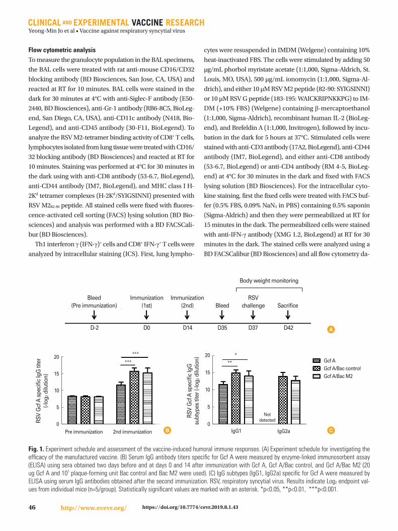
Th1 IFN-γ+ cell responses induced by vaccination IL-2 secretion from Th1 cells activates T cells and induces an-
tibody production by promoting B cell maturation. In addi-
tion, Th1 cells secrete IFN-γ, which can be helpful for increas-
ing CTL activity and suppressing virus replication [14-16]. In
order to identify the Th1 INF-γ+ cells specific for RSV G when
the Bac M2 was used as an adjuvant, we prepared lung cells
of immunized mice on day 5 after the RSV challenge, stimu-
lated them with RSV G peptide (183-195: WAICKRIPNKKPG),
and enumerated the percentages of Th1 cells secreting IFN-γ.
Th1 INF-γ+ cells were significantly more strongly induced in
the Gcf A/Bac M2 group (mean Th1 cells, 4.5%) compared
with the Gcf A group (Fig. 2). Interestingly, the Gcf A/Bac
control group also showed a significant Th1 INF-γ+ cell re-
sponse compared with the response in the Gcf A group (Fig.
2B). Even, the percentage of cell population was slightly high-
er in comparison with the Gcf A/Bac M2 group, though the
difference was not statistically significant (Fig. 2).
Fig. 2. Identification of CD4+ Th1 interferon γ (IFN-γ)+ cells specific for RSV G (183-195) peptide induced by vaccine inoculation. Lung mono-nuclear cells were isolated from mice vaccinated twice and sacrificed 5 days after RSV challenge. Isolated cells were stimulated with G pep-tide (183-195: WAICKRIPNKKPG) for 5 hours in vitro. Cells were stained with anti-CD3 antibody, anti-CD4 antibody, and anti-CD44 antibody and then fixed. Permeabilized cells were stained with anti-IFN-γ antibody and analyzed by flow cytometry. (A) The dot plots in the gated area represent CD4+ CD44+ T cells (Th1 cells) that can secrete IFN-γ. (B) The percentage of CD4+ CD44+ T cells (Th1 cells) that can secrete IFN-γ for each vaccine-immunized group. Data are expressed as mean± standard deviation (n=5/group). Statistically significant values are marked with an asterisk. ***p<0.001.
Fig 2.
Gcf A
Gcf A/Bac control
Gcf A/Bac M2
8
6
4
2
0No stimulation G peptide stimulation
RSV
G p
eptid
e sp
ecifi
c
CD4+ C
D44
+ IFN
-gam
ma+ c
ells
(%)
***
***
B
Fig 2.
No stimulation
G peptide stimulation
CD44
Gcf A Gcf A/Bac control Gcf A/Bac M2
IFN
- γ
A

Yeong-Min Jo et al • Vaccine against respiratory syncytial virus
48 http://www.ecevr.org/ https://doi.org/10.7774/cevr.2019.8.1.43
CD8+ IFN-γ+ T-cell responses specific for RSV M2 induced by vaccination IFN-γ secreted by CD8+ T cells during RSV infection is impor-
tant because it can block the pulmonary eosinophils; this has
been observed in experiments with mice immunized with a
recombinant vaccinia virus (vacv M2) capable of expressing
M2 protein [17] . In order to investigate the CTL response
specific to RSV M2, we confirmed the percentages of CD8+
IFN-γ+ T cells by stimulation with M2 peptide (82-90: SYIG-
SINNI) in lung cells obtained on day 5 after the RSV challenge.
When Gcf A-Gcf A/Bac M2 and Gcf A/Bac control-Gcf A/Bac
M2 groups were compared, the response of CD8+ IFN-γ+ T cells
specific to M2 peptide was shown to be significantly much
higher in the Gcf A/Bac M2 group (~6.2% vs. ~22.0%) (Fig. 3).
There was no significant difference in the responses of CD8+
IFN-γ+ T cells between the Gcf A group (mean CD8+ T cells,
6.5%) and Gcf A/Bac control group (mean CD8+ T cells, 5.7%).
Identification of CD8+ M2 Tet+ T cells after RSV challengeFollowing identification of CD8+ IFN-γ+ T-cell responses, lung
cells obtained 5 days after RSV challenge were also checked
to confirm whether they are also reactive to M2 peptide-load-
ed MHC class I H-2Kd tetramer (H-2Kd/SYIGSINNI). The per-
centage of CD8+ Tet+ cells in the Gcf A/Bac M2 group (~58.8%)
appeared to be about 2- to 3-fold higher than in the other two
groups, while the percentages of these cells in the Gcf A group
Fig. 3. Comparison of CD8+ interferon γ (IFN-γ)+ T cells specific for RSV M2 (82-90) peptide after vaccination. Mice were challenged with RSV after 2 vaccinations and lung mononuclear cells were harvested at day 5 to identify CD8+ CD44+ T cells capable of secreting IFN-γ. The harvest-ed lung mononuclear cells were stimulated with M2 peptide (82-90: SYIGSINNI) for 5 hours in vitro. Stimulated lung cells were stained with anti-CD3, anti-CD8, and anti-CD44 antibodies and then fixed. Permeabilized cells were stained with anti-IFN-γ antibody and analyzed by flow cytometry. (A) The dot plots in the gated area represent the CD8+ CD44+ T cells that can secrete IFN-γ. (B) The percentage of CD8+ CD44+ T cells that can secrete IFN-γ for each vaccine-immunized group. Data are expressed as mean± standard deviation (n=5/group). Statistically significant values are marked with an asterisk. ***p<0.001.
Fig 3.
No stimulation
M2 peptide stimulation
CD44
Gcf A Gcf A/Bac control Gcf A/Bac M2
IFN
- γ
A
Fig 3.
Gcf A
Gcf A/Bac control
Gcf A/Bac M2
30
20
10
0No
stimulationM2 peptide stimulation
RSV
M2
pept
ide
spec
ific
CD
8+ CD
44+ IF
N-g
amm
a+ cel
ls (%
)
***
B
***
Fig. 4. CD8+ Tet+ cells specific for RSV M2 after vaccination. To determine the binding capacity between CD8+ T cells and the RSV M2 tetramer, lung mononuclear cells were isolated from mice sacrificed at day 5 after RSV challenge. Isolated cells were stained with anti-CD8 antibody, anti-CD44 antibody, and H-2Kd RSV M2 tetramer and then fixed. Fixed cells were analyzed by flow cytometry. (A) Gating represents the popula-tion of CD8+ CD44+ T cells that can bind to the RSV M2 tetramer. (B) The percentage of CD8+ CD44+ T cells specific for the RSV M2 tetramer is shown for each group. Data are expressed as mean±standard deviation (n=5/group). Statistically significant values are marked with asterisks. ***p<0.001.
Fig 4.
B.
CD44
Gcf A Gcf A/Bac control Gcf A/Bac M2
RSV
M2-
tetra
mer
A
B
Fig 4.
B. 80
60
40
20
0Gcf A Gcf
A/Bac control
Gcf A/Bac
M2
RSV
M2
tetra
mer
spe
cific
CD
8+ CD
44+ T
cells
(%)
******
Yeong-Min Jo et al • Vaccine against respiratory syncytial virus
49http://www.ecevr.org/https://doi.org/10.7774/cevr.2019.8.1.43
(~23.8%) and Gcf A/Bac control group (~17.7%) in the same
gated cell population were similar (Fig. 4A). The similar per-
centages of CD8+ Tet+ cells in the Gcf A and Gcf A/Bac control
groups and the significantly higher mean percentage of CD8+
Tet+ cells in the Gcf A/Bac M2 group were confirmed (Fig. 4B).
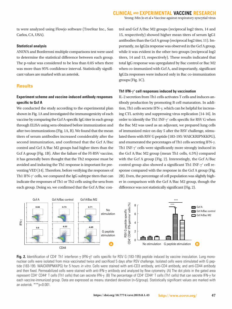
Identification of VED by measurement of BAL cellsThe detection of VED in has been integral to RSV vaccine de-
velopment since the failure of the FI-RSV vaccine, which was
found to cause lung inflammation due to hypersecretion of
type II cytokines. Therefore, our study included investigation
of the extent of eosinophil, alveolar macrophage, and neutro-
phil response for each group in the BAL cells collected from
the airway. First, we found that the numbers of eosinophils
(CD11c-, Siglec-F+) in Gcf A and Gcf A/Bac M2 groups appeared
to be lower, whereas the Gcf A/Bac control group had a slight-
ly greater number (Fig. 5A, C). Next, we observed that the al-
veolar macrophage (CD11c+, Siglec-F+) responses followed
Fig. 5. Determination of vaccine-enhanced disease through analysis of bronchoalveolar lavage (BAL) cells. After 5 days of RSV challenge, BAL cells were harvested from the airways of all immunized mice. BAL cells were stained with an anti-CD45 antibody, anti-CD11c antibody, anti-Siglec-F antibody, and anti-Gr-1 antibody. The stained cells were fixed and analyzed by flow cytometry. (A) CD11c-, Siglec-F+ cells are eosino-phils and CD11c+, Siglec-F+ cells are alveolar macrophages. (B) CD11c+, Gr-1+ cells are neutrophils. (C-E) Percentage of BAL cells for each group: eosinophils (C), alveolar macrophages (D), and neutrophils (E). Data are expressed as mean±standard deviation (n=5/group). Statistically signifi-cant values are marked with an asterisk. *p<0.05, ***p<0.001.
Fig 5.
CD11c
Gcf A Gcf A/Bac control Gcf A/Bac M2
Gr-1
B
Fig 5.
CD11c
Gcf A Gcf A/Bac control Gcf A/Bac M2
Sigl
ec-F
A
C
Fig 5.
15
10
5
0Gcf A Gcf
A/Bac control
Gcf A/Bac
M2
Eosi
noph
il (%
)
**
D
Fig 5.
80
60
40
20
0Gcf A Gcf
A/Bac control
Gcf A/Bac
M2
Alv
eola
r mac
roph
age
(%) ***
*
E
Fig 5.
15
10
5
0Gcf A Gcf
A/Bac control
Gcf A/Bac
M2
Neu
troph
il (%
)
******
Yeong-Min Jo et al • Vaccine against respiratory syncytial virus
50 http://www.ecevr.org/ https://doi.org/10.7774/cevr.2019.8.1.43
the order Gcf A group>Gcf A/Bac control group>Gcf A/Bac
M2 group. Statistical significance was confirmed for the Gcf
A/Bac control and Gcf A/Bac M2 groups compared with the
Gcf A group, and for the Gcf A/Bac M2 group compared with
the Gcf A/Bac controls (Fig. 5A, D). Finally, we found that the
neutrophil (CD11c+, Gr-1+) response was highest in the Gcf
A/Bac M2 group compared to a level of approximately 4%-5%
in the Gcf A and Gcf A/Bac control groups (Fig. 5B, E).
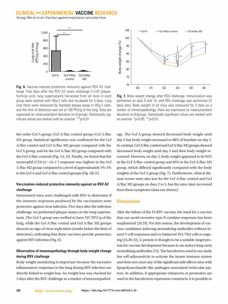
Vaccination-induced protective immunity against an RSV A2 challenge Immunized mice were challenged with RSV to determine if
the immune responses produced by the vaccination were
protective against virus infection. Five days after the infection
challenge, we performed plaque assays on the lung superna-
tants. The Gcf A group was verified to have 537 PFU/g of the
lung, while the Gcf A/Bac control and Gcf A/Bac M2 groups
showed no sign of virus replication (results below the limit of
detection), indicating that these vaccines provide protection
against RSV infection (Fig. 6).
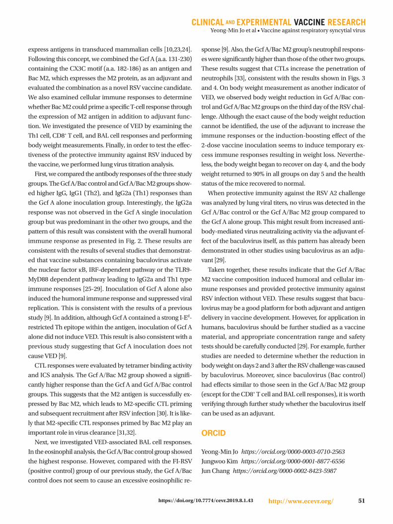
Observation of immunopathology through body weight change during RSV challengeBody weight monitoring is important because the excessive
inflammatory responses in the lung during RSV infection are
directly linked to weight loss. So, weight loss was checked for
5 days after the RSV challenge as a marker of immunopathol-
ogy. The Gcf A group showed decreased body weight until
day 2 but body weight increased to 98% of baseline on day 5.
In contrast, Gcf A/Bac control and Gcf A/Bac M2 groups showed
decreased body weight until day 3 and then body weight re-
covered. However, on day 3, body weight appeared to be 81%
in the Gcf A/Bac control group and 83% in the Gcf A/Bac M2
group, which differed significantly compared with the body
weights of the Gcf A group (Fig. 7). Furthermore, clinical dis-
ease scores were also low for the Gcf A/Bac control and Gcf
A/Bac M2 groups on days 2 to 3, but the mice later recovered
from these symptoms (data not shown).
Discussion
After the failure of the FI-RSV vaccine, the need for a vaccine
that can avoid excessive type II cytokine responses has been
emphasized [18,19]. For this reason, the development of vac-
cine candidates inducing neutralizing antibodies without bi-
ased T-cell responses and/or balanced Th1/Th2 cells is ongo-
ing [16,20-22]. G protein is thought to be a suitable target pro-
tein for vaccine development because it can induce long-term
neutralizing antibodies [15]. The baculovirus used in our study
has self-adjuvanticity to activate the innate immune system
and does not cause any of the significant side effects seen with
lipopolysaccharide-like pathogen-associated molecular pat-
tern. In addition, if appropriate enhancers or promoters are
used in the baculovirus expression constructs, it is possible to
Fig. 6. Vaccine-induced protective immunity against RSV A2 chal-lenge. Five days after the RSV A2 strain challenge (1×10⁶ plaque-forming unit), lung supernatants harvested from all mice in each group were reacted with HEp-2 cells and incubated for 5 days. Lung viral titers were measured by standard plaque assay in HEp-2 cells, and the limit of detection was set at 100 PFU/g of the lung. Data are expressed as mean±standard deviation (n=5/group). Statistically sig-nificant values are marked with an asterisk. **p<0.01.
Fig 6.
Gcf A
Gcf A/B
ac co
ntrol
Gcf A/B
ac M
20
200
400
600
800
1000 ****
Lung
vira
l tite
r(P
FU/g
of L
ung)
1,000
800
600
400
200
0Gcf A Gcf A/Bac
controlGcf A/Bac
M2
Lung
vira
l tite
r (P
FU/g
of l
ung)
**
**
Fig. 7. Body weight change after RSV challenge. Immunization was performed on days 0 and 14, and RSV challenge was performed 23 days later. Body weight of all mice was measured for 5 days as a marker of immunopathology. Data are expressed as mean±standard deviation (n=5/group). Statistically significant values are marked with an asterisk. *p<0.05, **p<0.01.
Fig 7.
105
100
95
90
85
80
75
70 D0 D1 D2 D3 D4 D5
Fig 7.
Gcf A
Fig 7.
Gcf A/Bac control
Fig 7.
Gcf A/Bac M2
Body
wei
ght c
hang
e (%
)
Yeong-Min Jo et al • Vaccine against respiratory syncytial virus
51http://www.ecevr.org/https://doi.org/10.7774/cevr.2019.8.1.43
express antigens in transduced mammalian cells [10,23,24].
Following this concept, we combined the Gcf A (a.a. 131-230)
containing the CX3C motif (a.a. 182-186) as an antigen and
Bac M2, which expresses the M2 protein, as an adjuvant and
evaluated the combination as a novel RSV vaccine candidate.
We also examined cellular immune responses to determine
whether Bac M2 could prime a specific T-cell response through
the expression of M2 antigen in addition to adjuvant func-
tion. We investigated the presence of VED by examining the
Th1 cell, CD8+ T cell, and BAL cell responses and performing
body weight measurements. Finally, in order to test the effec-
tiveness of the protective immunity against RSV induced by
the vaccine, we performed lung virus titration analysis.
First, we compared the antibody responses of the three study
groups. The Gcf A/Bac control and Gcf A/Bac M2 groups show-
ed higher IgG, IgG1 (Th2), and IgG2a (Th1) responses than
the Gcf A alone inoculation group. Interestingly, the IgG2a
response was not observed in the Gcf A single inoculation
group but was predominant in the other two groups, and the
pattern of this result was consistent with the overall humoral
immune response as presented in Fig. 2. These results are
consistent with the results of several studies that demonstrat-
ed that vaccine substances containing baculovirus activate
the nuclear factor кB, IRF-dependent pathway or the TLR9-
MyD88 dependent pathway leading to IgG2a and Th1 type
immune responses [25-29]. Inoculation of Gcf A alone also
induced the humoral immune response and suppressed viral
replication. This is consistent with the results of a previous
study [9]. In addition, although Gcf A contained a strong I-Ed-
restricted Th epitope within the antigen, inoculation of Gcf A
alone did not induce VED. This result is also consistent with a
previous study suggesting that Gcf A inoculation does not
cause VED [9].
CTL responses were evaluated by tetramer binding activity
and ICS analysis. The Gcf A/Bac M2 group showed a signifi-
cantly higher response than the Gcf A and Gcf A/Bac control
groups. This suggests that the M2 antigen is successfully ex-
pressed by Bac M2, which leads to M2-specific CTL priming
and subsequent recruitment after RSV infection [30]. It is like-
ly that M2-specific CTL responses primed by Bac M2 play an
important role in virus clearance [31,32].
Next, we investigated VED-associated BAL cell responses.
In the eosinophil analysis, the Gcf A/Bac control group showed
the highest response. However, compared with the FI-RSV
(positive control) group of our previous study, the Gcf A/Bac
control does not seem to cause an excessive eosinophilic re-
sponse [9]. Also, the Gcf A/Bac M2 group’s neutrophil respons-
es were significantly higher than those of the other two groups.
These results suggest that CTLs increase the penetration of
neutrophils [33], consistent with the results shown in Figs. 3
and 4. On body weight measurement as another indicator of
VED, we observed body weight reduction in Gcf A/Bac con-
trol and Gcf A/Bac M2 groups on the third day of the RSV chal-
lenge. Although the exact cause of the body weight reduction
cannot be identified, the use of the adjuvant to increase the
immune responses or the induction-boosting effect of the
2-dose vaccine inoculation seems to induce temporary ex-
cess immune responses resulting in weight loss. Neverthe-
less, the body weight began to recover on day 4, and the body
weight returned to 90% in all groups on day 5 and the health
status of the mice recovered to normal.
When protective immunity against the RSV A2 challenge
was analyzed by lung viral titers, no virus was detected in the
Gcf A/Bac control or the Gcf A/Bac M2 group compared to
the Gcf A alone group. This might result from increased anti-
body-mediated virus neutralizing activity via the adjuvant ef-
fect of the baculovirus itself, as this pattern has already been
demonstrated in other studies using baculovirus as an adju-
vant [29].
Taken together, these results indicate that the Gcf A/Bac
M2 vaccine composition induced humoral and cellular im-
mune responses and provided protective immunity against
RSV infection without VED. These results suggest that bacu-
lovirus may be a good platform for both adjuvant and antigen
delivery in vaccine development. However, for application in
humans, baculovirus should be further studied as a vaccine
material, and appropriate concentration range and safety
tests should be carefully conducted [29]. For example, further
studies are needed to determine whether the reduction in
body weight on days 2 and 3 after the RSV challenge was caused
by baculovirus. Moreover, since baculovirus (Bac control)
had effects similar to those seen in the Gcf A/Bac M2 group
(except for the CD8+ T cell and BAL cell responses), it is worth
verifying through further study whether the baculovirus itself
can be used as an adjuvant.
ORCID
Yeong-Min Jo https://orcid.org/0000000307102563
Jungwoo Kim https://orcid.org/0000000188776556
Jun Chang https://orcid.org/0000000284235987

Yeong-Min Jo et al • Vaccine against respiratory syncytial virus
52 http://www.ecevr.org/ https://doi.org/10.7774/cevr.2019.8.1.43
References
1. Lee JY, Chang J. Universal vaccine against respiratory syn-
cytial virus A and B subtypes. PLoS One 2017;12:e0175384.
2. Jeong KI, Piepenhagen PA, Kishko M, et al. CX3CR1 is ex-
pressed in differentiated human ciliated airway cells and
co-localizes with respiratory syncytial virus on cilia in a G
protein-dependent manner. PLoS One 2015;10:e0130517.
3. Schmidt ME, Varga SM. Modulation of the host immune
response by respiratory syncytial virus proteins. J Micro-
biol 2017;55:161-71.
4. Openshaw PJ, Chiu C, Culley FJ, Johansson C. Protective
and harmful immunity to RSV infection. Annu Rev Im-
munol 2017;35:501-32.
5. Citron MP, Patel M, Purcell M, et al. A novel method for
strict intranasal delivery of non-replicating RSV vaccines
in cotton rats and non-human primates. Vaccine 2018;36:
2876-85.
6. Killikelly AM, Kanekiyo M, Graham BS. Pre-fusion F is ab-
sent on the surface of formalin-inactivated respiratory
syncytial virus. Sci Rep 2016;6:34108.
7. Boyoglu-Barnum S, Todd SO, Meng J, et al. Mutating the
CX3C motif in the G protein should make a live respirato-
ry syncytial virus vaccine safer and more effective. J Virol
2017;91:e02059-16.
8. Cheon IS, Shim BS, Park SM, et al. Development of safe
and effective RSV vaccine by modified CD4 epitope in G
protein core fragment (Gcf). PLoS One 2014;9:e94269.
9. Kim S, Joo DH, Lee JB, et al. Dual role of respiratory syn-
cytial virus glycoprotein fragment as a mucosal immuno-
gen and chemotactic adjuvant. PLoS One 2012;7:e32226.
10. Lee JY, Chang J. Recombinant baculovirus-based vaccine
expressing M2 protein induces protective CD8(+) T-cell
immunity against respiratory syncytial virus infection. J
Microbiol 2017;55:900-8.
11. Bar-Haim E, Erez N, Malloy AM, Graham BS, Ruckwardt
TJ. CD8+ TCR transgenic strains expressing public versus
private TCR targeting the respiratory syncytial virus K(d)
M2(82-90) epitope demonstrate similar functional pro-
files. PLoS One 2014;9:e99249.
12. Kulkarni AB, Morse HC 3rd, Bennink JR, Yewdell JW, Mur-
phy BR. Immunization of mice with vaccinia virus-M2 re-
combinant induces epitope-specific and cross-reactive
Kd-restricted CD8+ cytotoxic T cells. J Virol 1993;67:4086-
92.
13. Kim S, Chang J. Baculovirus-based vccine dsplaying rspi-
ratory sncytial vrus glycoprotein iduces potective imunity
against RSV ifection without vccine-ehanced dsease. Im-
mune Netw 2012;12:8-17.
14. Varga SM, Welsh RM. High frequency of virus-specific in-
terleukin-2-producing CD4(+) T cells and Th1 dominance
during lymphocytic choriomeningitis virus infection. J Vi-
rol 2000;74:4429-32.
15. Li C, Zhou X, Zhong Y, et al. A recombinant G protein plus
cyclosporine A-based respiratory syncytial virus vaccine
elicits humoral and regulatory T cell responses against in-
fection without vaccine-enhanced disease. J Immunol
2016;196:1721-31.
16. Hua Y, Jiao YY, Ma Y, et al. DNA vaccine encoding central
conserved region of G protein induces Th1 predominant
immune response and protection from RSV infection in
mice. Immunol Lett 2016;179:95-101.
17. Castilow EM, Varga SM. Overcoming T cell-mediated im-
munopathology to achieve safe RSV vaccination. Future
Virol 2008;3:445-54.
18. Blanco JC, Boukhvalova MS, Shirey KA, Prince GA, Vogel
SN. New insights for development of a safe and protective
RSV vaccine. Hum Vaccin 2010;6:482-92.
19. Connors M, Giese NA, Kulkarni AB, Firestone CY, Morse
HC 3rd, Murphy BR. Enhanced pulmonary histopatholo-
gy induced by respiratory syncytial virus (RSV) challenge
of formalin-inactivated RSV-immunized BALB/c mice is
abrogated by depletion of interleukin-4 (IL-4) and IL-10. J
Virol 1994;68:5321-5.
20. Durant LR, Makris S, Voorburg CM, Loebbermann J, Jo-
hansson C, Openshaw PJ. Regulatory T cells prevent Th2
immune responses and pulmonary eosinophilia during
respiratory syncytial virus infection in mice. J Virol 2013;
87:10946-54.
21. Graham BS. Vaccines against respiratory syncytial virus:
the time has finally come. Vaccine 2016;34:3535-41.
22. Mazur NI, Higgins D, Nunes MC, et al. The respiratory syn-
cytial virus vaccine landscape: lessons from the graveyard
and promising candidates. Lancet Infect Dis 2018;18:e295-
311.
23. Boyce FM, Bucher NL. Baculovirus-mediated gene trans-
fer into mammalian cells. Proc Natl Acad Sci U S A 1996;
93:2348-52.
24. Hervas-Stubbs S, Rueda P, Lopez L, Leclerc C. Insect bac-
uloviruses strongly potentiate adaptive immune respons-
es by inducing type I IFN. J Immunol 2007;178:2361-9.
25. Bielefeldt-Ohmann H, Beasley DW, Fitzpatrick DR, Aas-

Yeong-Min Jo et al • Vaccine against respiratory syncytial virus
53http://www.ecevr.org/https://doi.org/10.7774/cevr.2019.8.1.43
kov JG. Analysis of a recombinant dengue-2 virus-den-
gue-3 virus hybrid envelope protein expressed in a secre-
tory baculovirus system. J Gen Virol 1997;78(Pt 11):2723-
33.
26. Ruitenberg KM, Walker C, Love DN, Wellington JE, Whal-
ley JM. A prime-boost immunization strategy with DNA
and recombinant baculovirus-expressed protein enhanc-
es protective immunogenicity of glycoprotein D of equine
herpesvirus 1 in naive and infection-primed mice. Vaccine
2000;18:1367-73.
27. Chen CY, Lin SY, Cheng MC, et al. Baculovirus vector as
an avian influenza vaccine: hemagglutinin expression and
presentation augment the vaccine immunogenicity. J Bio-
technol 2013;164:143-50.
28. Grabowska AK, Lipinska AD, Rohde J, Szewczyk B, Bien-
kowska-Szewczyk K, Rziha HJ. New baculovirus recombi-
nants expressing Pseudorabies virus (PRV) glycoproteins
protect mice against lethal challenge infection. Vaccine
2009;27:3584-91.
29. Heinimaki S, Tamminen K, Malm M, Vesikari T, Blazevic V.
Live baculovirus acts as a strong B and T cell adjuvant for
monomeric and oligomeric protein antigens. Virology
2017;511:114-22.
30. Hsu SC, Chargelegue D, Steward MW. Reduction of respi-
ratory syncytial virus titer in the lungs of mice after intra-
nasal immunization with a chimeric peptide consisting of
a single CTL epitope linked to a fusion peptide. Virology
1998;240:376-81.
31. Chang J. Current progress on development of respiratory
syncytial virus vaccine. BMB Rep 2011;44:232-7.
32. Nicholas JA, Rubino KL, Levely ME, Adams EG, Collins
PL. Cytolytic T-lymphocyte responses to respiratory syn-
cytial virus: effector cell phenotype and target proteins. J
Virol 1990;64:4232-41.
33. Openshaw PJ, Culley FJ, Olszewska W. Immunopathogen-
esis of vaccine-enhanced RSV disease. Vaccine 2001;20
Suppl 1:S27-31.