Embed Size (px)
Citation preview
Proc. Natl. Acad. Sci. USAVol. 90, pp. 2197-2201, March 1993Biochemistry
A widely expressed human protein-tyrosine phosphatase containingsrc homology 2 domains
(dephosphorylation/cloning/expression/purification)
SULTAN AHMAD*, DENIS BANVILLE*, ZHIZHUANG ZHAOt, EDMOND H. FISCHERt, AND SHI-HSIANG SHEN***Section of Molecular Biology, Biotechnology Research Institute, National Research Council of Canada, 6100 Royalmount Avenue, Montreal, PQ, CanadaH3A 2R2; and tDepartment of Biochemistry, University of Washington, Seattle, WA 98195
Contributed by Edmond H. Fischer, December 9, 1992
ABSTRACT A cDNA encoding a nontransmembrane pro-tein-tyrosine phosphatase (PTP; EC 3.1.3.48), termed PTP2C,was isolated from a human umbilical cord cDNA library. Theenzyme contains a single phosphatase domain and two adjacentcopies of the src homology 2 (SH2) domain at its aminoterminus. A variant of PTP2C (PTP2Ci) which has four extraamino acid residues within the catalytic domain has beenidentified also. PTP2C is widely expressed in human tissues andis particularly abundant in heart, brain, and skeletal muscle.The catalytic domain ofPTP2C was expressed as a recombinantenzyme in Escherichia coli and purified to near homogeneity bytwo chromatographic steps. The recombinant enzyme wastotally specific toward phosphotyrosine residues. The struc-tural similarity between PTP2C and the previously describedPTP1C suggests the existence of a subfamily ofSH2-containingPTPs; these may play an important role in signal transductionthrough interaction of their SH2 domains with phosphoty-rosine-containing proteins.
Phosphorylation of several intracellular proteins on tyrosineresidues has been implicated in a number of critical physio-logical and pathological processes, including cell growth,differentiation, and neoplastic transformation (1-5). Thestate of such phosphorylation depends upon the relativeactivities of two families of enzymes with opposing actions,namely, protein-tyrosine kinases (PTKs; EC 2.7.1.112) andprotein-tyrosine phosphatases (PTPs; EC 3.1.3.48). As withPTKs, there are two classes of PTPs: transmembrane andnontransmembrane enzymes (for review, see refs. 6 and 7).All the nontransmembrane PTPs reported to date have asingle highly conserved catalytic domain of ca. 230 aminoacid residues, while the transmembrane PTPs have two, withthe exception of HPTP-,B (8) and DPTP1OD (9), which haveonly one. Segments outside of the catalytic domain displayconsiderable structural diversity.
Recently, we and others (10-13) have identified a non-transmembrane PTP (PTP1C) which contains two copies ofthe src homology 2 (SH2) domain at itsN terminus. Likewise,a Drosophila gene, designated corkscrew, also encodes aPTP with two SH2 domains (14); it differs from PTPlC inhaving an insert interrupting the catalytic segment. SH2domains were originally found in various nonreceptor PTKsand other cytoplasmic signaling proteins (for review, see refs.15 and 16). They bind strongly to peptide segments containingtyrosine phosphate, but the selectivity of the interaction isdetermined by the nature of the residues surrounding thephosphotyrosine residue, particularly those at positions +1and +3. These domains play a major role in signal transduc-tion by allowing the interaction of intracellular tyrosinekinases or other SH2-containing proteins with growth factorreceptors, once these have undergone autophosphorylation.
The specificity of the interaction will determine which par-ticular signal pathway may become activated. In the presentreport, we describe another soluble phosphatase, designatedas PTP2C; it also contains two SH2 domains and is widelyexpressed in human tissues.§ We further identified a variantof this enzyme having an in-frame insertion of 12 base pairswithin the catalytic domain.
MATERIALS AND METHODSIsolation of PTP2C Clones. For the polymerase chain reac-
tion (PCR), a set ofdegenerate primers was designed from twohighly conserved regions within the catalytic domain ofPTPs.One primer corresponded to the amino acid sequence KC(A/D)QYWP and the other, to VHCSAGV. PCR fragments ofabout 250 bp were amplified from a human umbilical cordcDNA library. PCRs were performed for 30 cycles with a 94°Cdenaturation and a 50°C annealing temperature under standardconditions. The products were purified by electrophoresis ona 1.6% agarose gel and cloned in the pBluescript II KS (+)vector (Stratagene). One of the cloned PCR fragments wasused as a probe to screen the cDNA library at 50°C in 4xSSC/2x Denhardt's solution/0.5% SDS/0.1 M sodium phos-phate buffer, pH 7.0, containing sodium dextran sulfate at 100mg/ml, and sonicated and denatured salmon sperm DNA at100 ,g/ml (lx SSC = 0.15 M NaCl/0.015 M sodium citrate;lx Denhardt's solution = 0.02% bovine serum albumin/0.02% Ficoll/0.02% polyvinylpyrrolidone). Positive cloneswere subcloned in the pBluescript KS vector and sequencedwith oligonucleotides or internal restriction fragment primers(17), using a T7 DNA polymerase sequencing kit (Pharmacia).Northern Blot Analysis. A human multiple-tissue Northern
blot system (Clontech) was used to determine the level ofexpression of PTP2C in various tissues. The blot was pre-hybridized for 3 hr and then hybridized overnight at 50°C in50% (vol/vol) formamide/2.5x Denhardt's solution/25 mMpotassium phosphate buffer, pH 7.4/0.1% SDS/10% sodiumdextran sulfate/5x SSPE containing sonicated and dena-tured salmon spermDNA at 250 ,ug/ml and yeast tRNA at 500,ug/ml (1 x SSPE = 0.15 M NaCl/0.01 M sodium phosphate,pH 7.4/1 mM EDTA). The probes (PTP2C and ,B-actin) werelabeled with the T7 Quick Prime kit (Pharmacia). The blotwas washed twice with 2x SSPE/0.1% SDS, then with lxSSPE/0.1% SDS, and finally with 0.2x SSPE/0.5% SDS atroom temperature for 15 min each before exposure to KodakX-Omat AR film at -80°C with an intensifying screen.
Expression, Purification, and Assay of Recombinant En-zyme. The PTP2C cDNA clone was cleaved with Bgl II (at
Abbreviations: PTK, protein-tyrosine kinase; PTP, protein-tyrosinephosphatase; SH2, src homology 2; MAP kinase, mitogen-acti-vated protein kinase.*To whom reprint requests should be addressed.§The sequence reported in this paper has been deposited in theGenBank data base (accession no. L08807).
2197
The publication costs of this article were defrayed in part by page chargepayment. This article must therefore be hereby marked "advertisement"in accordance with 18 U.S.C. §1734 solely to indicate this fact.
Proc. Natl. Acad. Sci. USA 90 (1993)
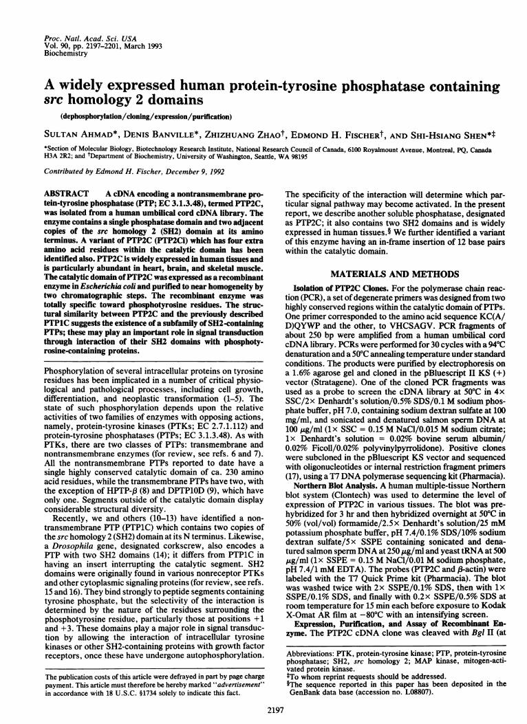
GGAGGAGCGAGCCGGGCCGCTGGCTCTGCCCCGCGTCCGGTCCCGAGC GGGCCTCCCTCGGGCCAGCCCGATGTGACC
GGGGGGCAGCTGCACAGTCTCCGGGATCCC CAGGCCTGGAGGGGGGTCTGTGCGCGGCCGGAGCCCAGCGGAGCCTGAGCAAGGAGCGGG
ATGACATCGCGGAGATG?'CACCCAAAT ATCACTSGGTGTGAGGCAGAAAACCTACTG ?rGACAAGAGGAGTTGATGGCAG?TTTT GCAAGGCCTAGTAAAAGTAACCCTGGAGAC 3181 M T S R R W FN P N I T G V Z A Z N L L L T R O V D SB F L A R P 8 8 N P G D
TTCACACTTTCCGTTAGAAGAAATGGAGCT GTCACCCACATCAAGATTCAGAACACTGGT GATTACTATGACCTGTATGGAGGGGAGAAA TTTGCCACTT'TGGCTGAGTTGGTCCAGTAT 43841 F T L S V R R N G A V T N I X I Q N T G D Y Y D L Y G G 3 K F A T L A Z L V Q Y
TACATGGAACATCACGGGCAATTAAAAGAG AAGAATGGAGATGTCATTGAGCTTAAATAT CCTCTGAACTGTGCAGATCCTACCTCTGAA AGGTGGTTTCATGGACATCTCTCTGGGAAA 55881 Y N Z H N G Q L X Z K N G D V I Z L K Y P L N C A D P T S E R W F H G N L S G K
GAAGCAGAGAAATTATTAACT<3AAAAAGGA AAACATGGTAGTTTTCTTGTACGAGAGAGC CAGAGCCACCCTGGAGATTrTTCTTTCT GTGCGCACTGGTGATGACAAAGGGGAGAGC 678121 Z A Z K L L T Z K 0 X H 0 8 F L V R N S Q 8 N P G D P V L S V R T 0 D D K G N S
AATGACGGCAAGTCTAAAGTGACCCATGTT ATGATTCGCTGTCAGGAACTGAAATACGAC GTTGGTGGAGGAGAACGGTTTGATTCTTTG ACAGATCTTGTGGAACATTATAAGAAGAAT 798161 N D 0 X S X V T N V X I R C Q N L K Y D V 0 0 G Z R F D S L T D L V Z H Y K X N
CCTATGGTGGAAACATTGGGTACAGTACTA CAACTCAAGCAGCCCCTTAACACGACTCGT ATAAATGCIGCTGAATAGAAAGCAGAGTT CGAGAACTAAGCAAATTAGCTGAGACCACA 918201 P X V Z T L 0 T V L Q L X Q P L N T T R I N A A E I E S R V R E L S K L A E T T
GATAAAGTCAAACAAGGCTTTTGGGAAGAA TTTGAGACACTACAACAACAGGAGTGCAAA CTTCTCTACAGCCGAAAAGAGGGTCAAAGG CAAGAAAACAAAAACAAAAATAGATATAAA241 D K V K Q G F W E E F E T L Q Q Q E C K L L Y S R K E G Q R Q E N K N K N R Y K
1038
AACATCCTGCCCTTTGATCATACCAGGGTT GTCCTACACGATGGTGATCCCAATGAGCCT G7TTCAGATTACATCAATGCAAATATCATC ATGCCTGAATTTGAAACCAAGTGCAACAAT 1158281 N I L P F D H T R V V L H D G D P N E P V S D Y I N A N I I M P E F E T K C N N
TCAAAGCCCAAAAAGAGTTACATTGCCACA CAAGGCTGCCTGCAAAACACGGTGAATGAC 7TrGGCGGATGGTGTCCAAGAAAACTCC CGAGTGATTGTCATGACAACGAAAGAAGTG 1278321 S K P K K S Y I A T Q G C L Q N T V N D F W R M V F Q E N S R V I V M T T K E V
GAGAGAGGAAAGAGTAAATGTGTCAAATAC TGGCCTGATGAGTATGCTCTAAAAGAATAT GGCGTCATGCGTGTTAGGAACGTCAAAGAA AGCGCCGCTCATGACTATACGCTAAGAGAA 1398361 E R G K S K C V K Y W P D E Y A L K E Y C V M R V R N V K E S A A H D Y T L R E
VCTTAAACTTTCAAAGGTTGGACAAGGGAAT ACGGAGAGAACGGTCTGGCAATACCACTTT CGGACCTGGCCGGACCACGGCGTGCCCAGC GACCCIGGGGGCGTGCTGGACTTCCTGGAG 1518
401 L K L S K V G Q G N T E R T V W Q Y H F R T W P D H G V P S D P G G V L D F L E
GAGGTGCACCATAAGCAGGAGAGCATCATG GATGCAGGGCCGGTCGTGGTGCACTGCAGT GCTGGAATTGGCCGGACAGGGACGTTCATT GTGATTGATATTCTTATTGACATCATCAGA 1638441 E V H H K Q E S I M D A G P V V V H C 8 A 0 I G R T G T F I V I D I L I D I I R
GAGAAAGGTGTTGACTGCGATATTGACGTT CCCAAAACCATCCAGATGGTGCGGTCTCAG AGGTCAGGGATGGTCCAGACAGAAGCACAG TACCGATTTATCTATATGGCGGTCCAGCAT481 E K G V D C D I D V P K T I Q M V R S Q R S G M V 0 T E A Q Y R F I Y M A V Q H
17 58
TATATTGAAACACTACAGCGCAGGATTGAA GAAGAGCAGAAAAGCAAGAGGAAAGGGCAC GAATATACAAATATTAAGTATTCTCTAGCG GACCAGACGAGTGGAGATCAGAGCCCTCTC 1878521 Y I E T L Q R R I E E E Q K S K R K G H E Y T N I K Y S L A D Q T S G D Q S P L
CCGCCTTGTACTCCAACGCCACCCTGTGCA GAAATGAGAGAAGACAGTGCTAGAGTCTAT GAAAACGTGGGCCTGATGCAACAGCAGAAA AGTTTCAGATGAGAAAACCTGCCAAAACTT 1998561 P P C T P T P P C A E M R E D S A R V Y E N V G L M Q Q 0 K S F R -
CAGCACAGAAATAGATGTGGACTT'CACCC TCTCCCTAAAAAGATCAAGAACAGACGCAA GAAAGTTTATGTGAAGACAGAATTTGGATT TGGGAAGGCTTGCAATGTGGTTGACTACCT 2118TT'GATAAGCAAAAN'PGAAACCATTTAAA GACCACTGTATTTTAACTCAACAATACCTG CTTCCCAATTACTCATrTCCTCAGATAAGA AGAAATCATCTCTACAATGTAGACAACATT 2238ATATTTTATAGAATTPGVTGSAAATTGACG AAGCAGTTAAATTGTGCGCTGTATnTTTCA GAGGATTA7rGGGATTCAAATTCTAGTAAT AGGCCTTTrT// 2338
FIG. 1. Nucleotide sequence of the entire coding region plus part of the noncoding regions of the PTP2C cDNA clone and the amino acidsequence deduced. Amino acid residues are numbered on the left, nucleotide positions on the right. The two adjacent copies of the SH2 domainare underlined and boldfaced. The active site of the PTP is boldfaced (HCSAG, residues 458-462). The position of the 12-bp insert in PTP2Ciis indicated by an inverted triangle. A putative phosphorylation site for mitogen-activated protein (MAP) kinase is indicated by a brokenunderline.
nucleotide 771) and the ends were filled with the Klenowfragment ofDNA polymerase. The DNA was redigested withHindIII. The expression vector pET-3c (18) was cleaved withEcoRI, filled in, and redigested with HindIII. The truncatedPTP2C fragment was inserted in frame into the treated vectorpET-3c. The resultant plasmid, designated pASH2-PTP2C,was used to transform Escherichia coli BL21 (DE3) cells.Expression of the recombinant enzyme (ASH2-PTP2C) wascarried out as described (10). The recombinant protein con-sists of 406 amino acids in which the first four residues(MARI) were derived from the vector. For purification, E.coli cells containing the recombinant enzyme were sonicatedin a buffer consisting of 25 mM Tris HCl at pH 7.5, 10 mM2-mercaptoethanol, 2.0mM EDTA, 1.0mM benzamidine, 0.1mM phenylmethylsulfonyl fluoride, leupeptin at 20 ,ug/ml,pepstatin A at 1.0 ,uM, and aprotinin at 0.027 trypsin inhibitor
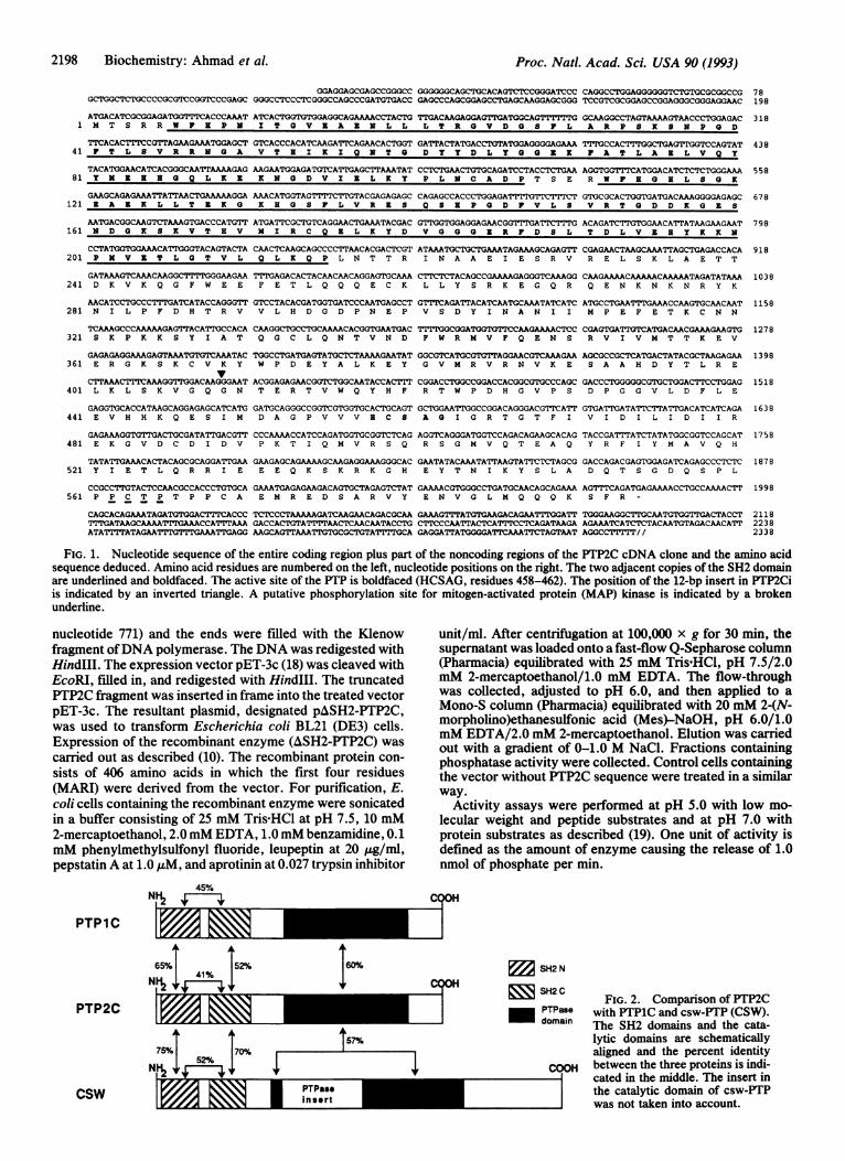
45%Nl g
PTP1C
unit/ml. After centrifugation at 100,000 x g for 30 min, thesupernatant was loaded onto a fast-flow Q-Sepharose column(Pharmacia) equilibrated with 25 mM Tris HCl, pH 7.5/2.0mM 2-mercaptoethanol/1.0 mM EDTA. The flow-throughwas collected, adjusted to pH 6.0, and then applied to aMono-S column (Pharmacia) equilibrated with 20 mM 2-(N-morpholino)ethanesulfonic acid (Mes)-NaOH, pH 6.0/1.0mM EDTA/2.0 mM 2-mercaptoethanol. Elution was carriedout with a gradient of 0-1.0 M NaCl. Fractions containingphosphatase activity were collected. Control cells containingthe vector without PTP2C sequence were treated in a similarway.
Activity assays were performed at pH 5.0 with low mo-lecular weight and peptide substrates and at pH 7.0 withprotein substrates as described (19). One unit of activity isdefined as the amount of enzyme causing the release of 1.0nmol of phosphate per min.
C?OH
1z -_65% 52%
Nit r-
I60%rnnw
v*I +* ,v.rm~C)]75% 70%
152
NS 1~ PTPas*insert
SH2 N
SH2C
PTPasedomain
COOH
FIG. 2. Comparison of PTP2Cwith PTP1C and csw-PTP (CSW).The SH2 domains and the cata-lytic domains are schematicallyaligned and the percent identitybetween the three proteins is indi-cated in the middle. The insert inthe catalytic domain of csw-PTPwas not taken into account.
78198
PTP2C
csw
2198 Biochemistry: Ahmad et al.
r"I'll
Proc. Natl. Acad. Sci. USA 90 (1993) 2199
_
_.i -Iq
0
heart
brain
:W
.It
.k S.
lung
liver
skeletal muscle
kidney
pancreas
M% b
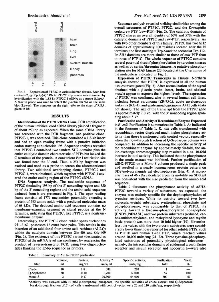
FIG. 3. Expression ofPTP2C in various human tissues. Each lanecontains 2 ,ug of poly(A)+ RNA. PTP2C expression was examined byhybridization with the 1.83-kb PTP2C-1 cDNA as a probe (Upper).A 1-actin probe was used to detect the l3-actin mRNA on the sameblot (Lower). The numbers on the right refer to the sizes of RNA,given in kb.
RESULTS
Identification of the PTP2C cDNA Clone. PCR amplificationof the human umbilical cord cDNA library yielded a fragmentof about 250 bp as expected. When the same cDNA librarywas screened with the PCR fragment, one positive clone,PTP2C-1, was obtained. This clone contained a 1.8-kb insertand had an open reading frame with a putative initiationcodon starting at nucleotide 198. Sequence analysis revealedthat PTP2C-1 contained two tandem SH2 domains plus theentire catalytic domain characteristic of PTPs but lacked theC terminus of the protein. A convenient Pst I restriction sitewas found near the 3' end. Thus, a 254-bp fragment was
excised and used as a probe to screen the aforementionedlibrary. Two additional overlapping clones, PTP2C-2 andPTP2C-3, were obtained, which together with PTP2C-1 cov-ered the entire coding region of the PTP2C cDNA.DNA Sequence Analysis. The nucleotide sequence of
PTP2C (including 198 bp of the 5' noncoding region and 350bp of the 3' noncoding region) and the amino acid sequencededuced from it are presented in Fig. 1. The open readingframe consists of 1779 nucleotides potentially encoding aprotein of 593 amino acids with a predicted molecular massof 68 kDa. The deduced amino acid sequence contains nomembrane-spanning segment or signal peptide at the Nterminus, indicating that PTP2C, like PTP1C, is a nontrans-membrane enzyme.
Interestingly, the PTP2C-2 clone, which spans nucleotides951-2162, possesses 12 extra base pairs. This results in theinsertion of an additional four amino acid residues (ALLQ)within the catalytic domain between Gln-408 and Gly-409(Fig. 1). The existence of this PTP2C variant (designated asPTP2Ci) at the mRNA level was confirmed by sequencing theproduct of reverse-transcript PCR, using two oligonucleo-tides flanking the 12-bp sequence as primers.
Sequence analysis revealed striking similarities among theoverall structures of PTP1C, PTP2C, and the Drosophilacorkscrew PTP (csw-PTP) (Fig. 2). The catalytic domain ofPTP2C shares an overall identity of 60% and 57% with thecatalytic domains of PTP1C and csw-PTP, respectively. Aswith two other members of this family, PTP2C has two SH2domains of approximately 100 residues located near the Nterminus, the first starting at Trp-6 and the second at Trp-112.Its SH2 domains are more similar to those of csw-PTP thanto those of PTP1C. The whole sequence of PTP2C containsseveral potential sites of phosphorylation by tyrosine kinasesas well as by serine/threonine kinases. A putative phosphor-ylation site for MAP kinase (20) located at the C terminus ofthe molecule is indicated in Fig. 1.
Expression of PTP2C Transcripts in Tissues. Northernanalysis showed that PTP2C is expressed in all the humantissues investigated (Fig. 3). After normalization of the signalobtained with a p-actin probe, heart, brain, and skeletalmuscle appear to express the highest levels. The expressionof PTP2C was confirmed also in several human cell lines,including breast carcinoma (ZR-75-1), acute myelogenousleukemia (KG-1), and epidermoid carcinoma A431 cells (datanot shown). The size of the transcript from the PTP2C geneis approximately 7.0 kb, with the 3' noncoding region span-ning about 5 kb.
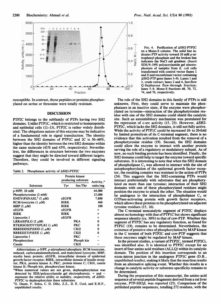
Purification and Activity ofRecombinant Enzyme Expressedin E. coli. Purification is summarized in Table 1. As indicatedin the footnote of Table 1, E. coli cells transformed withrecombinant vector displayed much higher phosphatase ac-tivity than those transformed with control vector, especiallywhen the flow-through fraction of Q-Sepharose column wascompared. In addition to increasing the specific activity ofthe recombinant enzyme by approximately 50-fold, the an-ion-exchange chromatography step resulted in a ca. 10-foldincrease in total activity. This indicated that the phosphatasein the crude extract was inhibited. Further purification ofASH2-PTP2C on a Mono-S column produced a single peakand resulted in a nearly homogeneous protein pattern onSDS/polyacrylamide gel electrophoresis (Fig. 4). A molec-ular mass of 46 kDa calculated from its mobility on SDS gelwas consistent with the size predicted from the amino acidsequence.Table 2 illustrates the phosphatase activity of ASH2-
PTP2C toward a variety of substrates. As expected, theenzyme was entirely specific for the dephosphorylation oftyrosine residues. While its activity toward two low-molecular-weight substrates, p-nitrophenyl phosphate andphosphotyrosine, was comparable to that of PTP1C, itsactivity toward a tyrosine-phosphorylated nonapeptide[ENDY(P)INASL] and two protein substrates (reduced, car-boxamidomethylated, and maleylated lysozyme and myelinbasic protein) was more than 10-fold higher (19). Neverthe-less, the values with the two protein substrates were consid-erably lower than those reported for other soluble PTPs, suchas PTP1B and human T-cell PTP, which reached valuesaround 10,000 units/mg (21, 22). Three tyrosine-phosphory-lated substrates of potentially physiological relevance-namely, the intracellular domains of epidermal growth factorreceptor and insulin receptor and lipocortin I-were also
Table 1. Summary of ASH2-PTP2C purificationVolume, Protein, Activity,* Specific activity, Purification, Yield,
Step ml mg/ml units/ml units/mg fold %Crude 10 1.8 380 210 1Q-Sepharose 30 0.10 1,200 12,000 57 100Mono-S 2 0.17 10,900 64,000 300 61
*Activity was assayed with 10 mM p-nitrophenyl phosphate; the specific activities of crude extract and Q-Sepharosebreak-through fraction of E. coli cells transformed with control vector were 20 and 110 units/mg, respectively.
Biochemistry: Ahmad et al.
Proc. Natl. Acad. Sci. USA 90 (1993)
0.6
0.4 0z
FIG. 4. Purification of ASH2-PTP2Con a Mono-S column. The solid line in-dicates PTP activity toward 10 mM p-ni-trophenyl phosphate and the broken lineindicates the NaCl salt gradient. (Inset)SDS/9-19% polyacrylamide gel electro-phoresis of samples from E. coli cellstransformed with control vector (lanes 1and 2) and recombinant vector containingA&SH2-PTP gene (lanes 3-9). Lanes 1 and3, crude extract; lanes 2 and 4, fast-flowQ-Sepharose flow-through fraction;lanes 5-9, Mono-S fractions 68, 70, 72,74, and 76, respectively.
susceptible. In contrast, those peptides or proteins phosphor-ylated on serine or threonine were totally resistant.
DISCUSSIONPTP2C belongs to the subfamily of PTPs having two SH2domains. Unlike PTP1C, which is restricted to hematopoieticand epithelial cells (11-13), PTP2C is rather widely distrib-uted. The ubiquitous nature of this enzyme may be indicativeof a fundamental role in signal transduction. The identitybetween the SH2 domains of PTP1C and 2C is 50-60%,higher than the identity between the two SH2 domains withinthe same molecule (41% and 45%, respectively). Neverthe-less, the differences in structure between the two enzymes
suggest that they might be directed toward different targets.Therefore, they could be involved in different signalingpathways.
Table 2. Phosphatase activity of ASH2-PTP2CProtein kinase
producing substrate Activity,*Substrate Tyr Ser/Thr units/mg
p-NPP, 10 mM 64,000Phosphotyrosine (2 mM) 15,000ENDY(P)INASLt (5 ,AM) sEGFR 7,800RCM-lysozyme (1 ,uM) BIRK 460MBP (1 AM) BIRK 100sEGFR sEGFR +BIRK BIRK +Lipocortin I sEGFR +LRRAS(P)LG (2 ,uM) PKA 0RTKRSGS(P)VYEPLKI (1 ,uM) PKC 0RRRDDDS(P)DDD (2 ,M) CKII 0RRREEET(P)EEE (1 AM) CKII 0Lipocortin I PKC -
Phosphorylase Phosph kinCasein CKII
Abbreviations: p-NPP, p-nitrophenyl phosphate; RCM-lysozyme,reduced, carboxamidomethylated, and maleylated lysozyme; MBP,myelin basic protein; sEGFR, intracellular domain of epidermalgrowth factor receptor; BIRK, intracellular domain of insulin recep-tor; PKA, protein kinase A; PKC, protein kinase C; CKII, caseinkinase II; Phosph kin, phosphorylase kinase.*When numerical values are not given, dephosphorylation wasdetected by SDS/polyacrylamide gel electrophoresis. + and -represent the relative ability or inability, respectively, of ASH2-PTP2C to dephosphorylate these substrates.
tG. Daum, F. Solca, C. D. Diltz, Z.Z., D. E. Cool, and E.H.F.,unpublished results.
The role of the SH2 domains in this family of PTPs is stillunknown. First, they could serve to maintain the phos-phatases in an inactive state, if the enzyme were phosphor-ylated on tyrosine-interaction of the phosphotyrosine res-idue with one of the SH2 domains could shield the catalyticsite. Such an autoinhibitory mechanism was postulated forthe repression of c-src activity (15, 23). However, ASH2-PTP2C, which lacks the SH2 domains, is still not fully active.While the activity ofPTP1C could be increased 10- to 20-foldby limited proteolysis of its C-terminal segment, there is noevidence that this activation resulted from the removal of aphosphotyrosine residue (19). Second, the SH2 domainscould allow the enzyme to interact with another proteinserving the role of a regulatory or modulatory subunit. As ofnow, no such binding protein has been identified. Finally, theSH2 domains could help to target the enzyme toward specificsubstrates. It is interesting to note that when the SH2 domainof phospholipase CY was allowed to interact with the site ofautophosphorylation of the epidermal growth factor recep-tor, the resulting complex was resistant to the action of PTPs(24). This suggests that the SH2-containing PTPs wouldinteract preferentially with substrates that are phosphory-lated on more than one tyrosine; interaction of their SH2domains with one of these phosphorylated residues mightposition the enzyme to attack the other. The situation wouldbe analogous to the interaction of phospholipase C., orGTPase-activating protein with growth factor receptors,which allows these proteins to be phosphorylated on adjacenttyrosine residues (15, 16).The C-terminal noncatalytic segment of PTP2C displays
almost no homology with that ofPTP1C but shows significantsequence identity (ca. 30%) to that of csw-PTP. Whether thissegment of PTP2C has any regulatory function, as found forPTP1C (19), remains to be determined. Nevertheless, theexistence ofputative sites ofphosphorylation by MAP kinasein the C termini of both PTP2C and csw-PTP suggests thatthese enzymes might be regulated by MAP kinase.
In the present studies, a variant ofPTP2C, termed PTP2Ci,was identified also. It is identical to PTP2C except for aninsert offour amino acid residues (ALLQ) within its catalyticdomain. The position of this insertion corresponds to anexon-intron junction in the analogous PTP1C gene (D.B.,unpublished results), making it likely that the insertion resultsfrom an alternative splicing event. Whether this results in achange in catalytic activity or substrate specificity remains tobe determined.
During the preparation of this manuscript, the amino acidsequence of several peptide fragments derived from a rat liverenzyme, PTP-SH2f3, was reported (25). Comparison of thepublished peptide sequences, totalling 272 residues, with the
cl)
*C:%
:3
Fraction
2200 Biochemistry: Ahmad et al.
Proc. Natl. Acad. Sci. USA 90 (1993) 2201
sequence of human PTP2C reveals that they are identicalexcept for the substitution of Lys-389 in PTP2C for anarginine in PTP-SH2,8. It can be assumed, therefore, thatPTP-SH2p is the rat homologue of human PTP2C.
We are grateful to Dr. Morag Park for providing the humanumbilical cord cDNA library. We thank Su-Fan Pen, Denis L'Abbe,Robert Larocque, and Rino Stocco for their skillful technical assis-tance.
1. Aaronson, S. A. (1991) Science 254, 1146-1153.2. Cantley, L. C., Auger, K. R., Carpenter, C., Duckworth, B.,
Graziani, A., Kapeller, R. & Soltoff, S. (1991) Cell 64, 281-302.3. Nishibe, S., Wahl, M. I., Hernandez-Sotomayor, S. M. T.,
Tonks, N. K., Rhee, S. G. & Carpenter, G. (1990) Science 250,1253-1256.
4. Huganir, R. L., Delcour, A. H., Greengard, P. & Hess, G. P.(1986) Nature (London) 321, 775-776.
5. Hopfield, J. F., Tank, D. W., Greengard, P. & Huganir, R. L.(1988) Nature (London) 336, 677-680.
6. Fischer, E. H., Charbonneau, H. & Tonks, N. (1991) Science253, 401-406.
7. Pot, D. A. & Dixon, J. E. (1992) Biochim. Biophys. Acta 1136,35-43.
8. Krueger, N. X., Streuli, M. & Saito, H. (1990) EMBO J. 9,3241-3252.
9. Yang, X., Seow, K. T., Bahri, S. M., Oon, S. H. & Chia, W.(1992) Cell 67, 661-673.
10. Shen, S.-H., Bastien, L., Posner, B. & Chretien, P. (1991)Nature (London) 352, 736-739.
11. Mathews, R. J., Bowne, D. B., Flores, E. & Thomas, M. L.(1992) Mol. Cell. Biol. 12, 2396-2405.
12. Yi, T., Cleveland, J. L. & Ihle, J. N. (1992) Mol. Cell. Biol. 12,836-846.
13. Plutzki, J., Neel, B. G. & Rosenberg, R. D. (1992) Proc. Natl.Acad. Sci. USA 89, 1123-1127.
14. Perkins, L. A., Larsen, I. & Perrimon, N. (1992) Cell 70,225-236.
15. Koch, C. A., Anderson, D., Moran, M. F., Ellis, C. & Pawson,T. (1991) Science 252, 668-674.
16. Pawson, T. & Gish, G. D. (1992) Cell 71, 359-362.17. Chretien, P. & Shen, S.-H. (1992) DNA Cell Biol. 11, 337-343.18. Stidies, F. W., Rosenber, A. H., Dunn, J. J. & Dubendorff,
J. W. (1990) Methods Enzymol. 185, 60-89.19. Zhao, Z., Bouchard, P., Diltz, C. D., Shen, S.-H. & Fischer,
E. H. (1993) J. Biol. Chem., in press.20. Clark, L. I., Sanghera, J. S. & Pelech, S. L. (1991) J. Biol.
Chem. 266, 1580-1584.21. Tonks, N. K., Diltz, C. D. & Fischer, E. H. (1988) J. Biol.
Chem. 263, 6731-6737.22. Zander, N. K., Lorenzen, J. A., Cool, D. E., Tonks, N. K.,
Daum, G., Krebs, E. G. & Fischer, E. H. (1991) Biochemistry30, 6964-6970.
23. Roussel, R. R., Brodeur, S. R., Shalloway, D. & Laudano,A. P. (1991) Proc. Natl. Acad. Sci. USA 88, 10696-10700.
24. Rotin, D., Margolis, B., Mohammadi, M., Daly, R. J., Daum,G., Li, N., Fischer, E. H., Burgess, W. H., Ullrich, A. &Schlessinger, J. (1992) EMBO J. 11, 559-567.
25. Hiraga, A., Munakata, H., Hata, K., Suzuki, Y. & Tsuiki, S.(1992) Eur. J. Biochem. 209, 195-206.
Biochemistry: Ahmad et al.