Embed Size (px)
Citation preview
Int J Anat Res 2017, 5(1):3535-40. ISSN 2321-4287 3535
Original Research Article
STUDY OF THE PLACENTAL ATTACHMENT OF FUNICULUSUMBILICALIS IN NORMAL AND PRE-ECLAMPTIC PREGNANCIESAND ITS EFFECTS ON BIRTH WEIGHTAnkit Jain *1, Sonia Baweja 2, Rashmi Jain 3.
ABSTRACT
Address for Correspondence: Dr. Ankit Jain, S/O Subodh Kumar Jain, Near Jain mandir, NehaNagar (Shakti Nagar), Makronia, distt. SAGAR, (M.P.), Pin- 470004,Telephone: +91 9827570908, +91 9977799776. E-Mail: [email protected]
Introduction: Abnormalities in the insertion of umbilical cord is associated with a number of complications inpregnancy and these complications may adversely affect the fetus. The aim of this study was to evaluate thevariations in the attachment of umbilical cord in normal and pre-eclamptic pregnancies and to assess theeffects of variable cord insertions on fetal birth weight.Materials and Methods: Seventy placentae each of normotensive and pre-eclamptic pregnancies were studied(n=140). After delivery, weight of the baby was recorded by using weighing machine and the attachment ofumbilical cord on placenta was observed.Results: In the present study, commonest site of insertion of umbilical cord was central (60%) in normalpregnancies, whereas in pre-eclamptic pregnancies, a common site of insertions of umbilical cord were central(37.14%) and/or eccentric (34.28%). Marginal cord insertions were found 2.11 times more in pre-eclampticpregnancies as compared to normal pregnancies. A single case of velamentous insertion was found in the pre-eclamptic pregnancies. We found that 65.52% of placentae with abnormal cord insertions were associated withlow fetal birth weight and the association between cord insertion and fetal birth weight was found statisticallyhighly significant.Discussion: Abnormal cord insertions are significantly associated with pre-eclampsia. Mean fetal birth weightdecreases as the site of cord insertion moves towards the periphery. Conclusively, abnormalities in the site ofinsertion of umbilical cord have an adverse effect on fetal health. Therefore, early detection of abnormal cordinsertion may provide sufficient information to take additional care in such conditions.KEY WORDS: Birth weight, Cord insertion, Pre-eclampsia, Placenta, Umbilical cord.
INTRODUCTION
International Journal of Anatomy and Research,Int J Anat Res 2017, Vol 5(1):3535-40. ISSN 2321-4287
DOI: https://dx.doi.org/10.16965/ijar.2017.107
Access this Article online
Quick Response code Web site: International Journal of Anatomy and ResearchISSN 2321-4287
www.ijmhr.org/ijar.htm
DOI: 10.16965/ijar.2017.107
*1 M.S., Ex. Resident, Department of Anatomy, Gandhi Medical College, Bhopal (M.P.), India.2 M.S., Associate Professor, Department of Anatomy, Gandhi Medical College, Bhopal (M.P.), India.3 M.D., Lab Head, Consultant Pathologist, SRL Diagnostics Ltd, Malviya Nagar, Bhopal (M.P.), India.
Received: 06 Jan 2017Peer Review: 06 Jan 2017Revised: None
Accepted: 13 Feb 2017Published (O): 28 Feb 2017Published (P): 28 Feb 2017
fetal surface of the placenta [1]. The umbilicalcord delivers oxygen and nutrients to the devel-oping fetus throughout pregnancy. Thus, thegrowth of the fetus is highly dependent on the
The umbilical cord is also referred to as Funicu-lus umbilicalis or Birth cord. It is a flexible struc-ture that connects the developing embryo to the
Int J Anat Res 2017, 5(1):3535-40. ISSN 2321-4287 3536
Ankit Jain, Sonia Baweja, Rashmi Jain. STUDY OF THE PLACENTAL ATTACHMENT OF FUNICULUS UMBILICALIS IN NORMAL AND PRE-ECLAMPTIC PREGNANCIES AND ITS EFFECTS ON BIRTH WEIGHT.
development of the umbilical cord [2]. Normally,the umbilical cord is inserted at the center ornear the center (eccentric) of the placenta. Othertypes of attachments of umbilical cords aremarginal, velamentous and furcated [3]. Inmarginal cord insertion, the umbilical cord isinserted within 2 cm from the placental edge[4]. In velamentous cord insertion, the umbili-cal cord is inserted into the chorio-amnioticmembranes rather than on to the placental mass[5]. In furcated insertion, umbilical cord branchbefore its insertion on the fetal surface of theplacenta [6]. Variations in the site of insertionof umbilical cords are explained by two differ-ent theories. First is “placental migration theoryor trophotropism”, in which the placentamigrates towards the richly vascularised areawith advancing gestation to achieve betterperfusion [7]. Another is the “blastocyst polar-ity theory”, which hypothesizes that abnormalcord insertion results from malpositioning ofblastocyst during implantation [8].Abnormal cord insertion is associated with poorobstetric outcomes. Increased rates of fetalmalformation, low birth weight, preterm labor,fetal growth restriction, vasa previa, low APGAR(appearance, pulse, grimace, activity and respi-ratory rate) scores and intrapartum complica-tions have been noted with velamentous cordinsertions [7,9,10]. In velamentous cord inser-tion, umbilical vessels are inserted into themembranes, therefore these vessels lack theprotection of Wharton’s jelly and are prone torupture and/or compression, which results inacute cessation of umbilical blood flow. Thusthe risk of perinatal death is increased in preg-nancies with velamentous cord insertions [11].Various studies suggest that compression ofumbilical vessels reduces cardiac output andincreases the risk of pulmonary complicationsafter birth [12, 13]. Marginal cord insertion hasalso been associated with fetal growth restric-tion and preterm delivery [9, 14]. Because ofpoor obstetric outcomes, evaluation of theattachment of umbilical cord deserves attentionright from the first trimester. Sonographic visu-alization of the site of cord insertion becomesmore difficult with advancing gestation; there-fore, it should be evaluated at 15-20 weeks ofgestation [15, 16].
The purpose of this study was to observe thevariations in the attachment of umbilical cordin normal and pre-eclamptic pregnancies and todetermine whether the umbilical cord insertionsite could be linked to fetal birth weight.
MATERIALS AND METHODS
The present study was an observationalcomparative study, which was carried out in theDepartment of Anatomy, Gandhi MedicalCollege, Bhopal (M.P). A total of 140 placentaewith umbilical cord were collected frompregnant women delivered in Sultania ZananaHospital associated to G.M.C. Bhopal, afterpermission from institutional ethics committee.All mothers were properly explained about thestudy and their written consent was taken.Women were diagnosed with pre-eclampsia ifthey had systolic BP > 140mmHg and diastolicBP > 90mmHg measured on two or moreoccasions, at least 4 hrs apart after the 20thweek of gestation with proteinuria. Proteinuriawas considered when there was a urine dipstickvalue of at least 1+ (>30mg/dl) on two separateoccasions at least 6 hours apart [17]. On thisbasis, subjects were divided into two groups.Group I consist of placentae obtained fromnormal pregnant women (n=70) with gestationalage 37-40 weeks. Group II consist of placentaeobtained from pre-eclamptic women (n=70) ofsimilar gestational age. Patients with essentialhypertension, diabetes mellitus, anemia, renaldisorders and other illness associated withpregnancy were excluded from this study.The mother’s and their neonates identified forthis study were given code numbers andstudied at the hospital. After delivery, fetal birthweight was recorded. The placentae werecollected soon after their expulsion and washedin the running tap water to clear all blood.Distance from the placental margin to the siteof attachment of the umbilical cord wasmeasured. The attachment of the umbilical cordon the fetal surface of the placenta wascategorized into central, eccentric, marginal andvelamentous insertion. Central cord insertionincludes the cord, which was inserted into thecenter of placenta, whereas cords which wereinserted near the center were included ineccentric cord insertion. Both central and eccen-
Int J Anat Res 2017, 5(1):3535-40. ISSN 2321-4287 3537
Ankit Jain, Sonia Baweja, Rashmi Jain. STUDY OF THE PLACENTAL ATTACHMENT OF FUNICULUS UMBILICALIS IN NORMAL AND PRE-ECLAMPTIC PREGNANCIES AND ITS EFFECTS ON BIRTH WEIGHT.
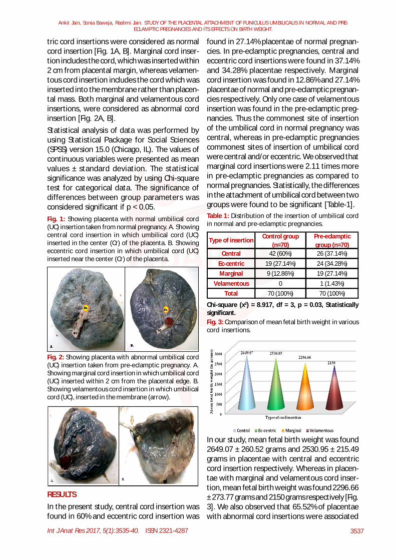
tric cord insertions were considered as normalcord insertion [Fig. 1A, B]. Marginal cord inser-tion includes the cord, which was inserted within2 cm from placental margin, whereas velamen-tous cord insertion includes the cord which wasinserted into the membrane rather than placen-tal mass. Both marginal and velamentous cordinsertions, were considered as abnormal cordinsertion [Fig. 2A, B].Statistical analysis of data was performed byusing Statistical Package for Social Sciences(SPSS) version 15.0 (Chicago, IL). The values ofcontinuous variables were presented as meanvalues ± standard deviation. The statisticalsignificance was analyzed by using Chi-squaretest for categorical data. The significance ofdifferences between group parameters wasconsidered significant if p < 0.05.Fig. 1: Showing placenta with normal umbilical cord(UC) insertion taken from normal pregnancy. A. Showingcentral cord insertion in which umbilical cord (UC)inserted in the center (Cr) of the placenta. B. Showingeccentric cord insertion in which umbilical cord (UC)inserted near the center (Cr) of the placenta.
Fig. 2: Showing placenta with abnormal umbilical cord(UC) insertion taken from pre-eclamptic pregnancy. A.Showing marginal cord insertion in which umbilical cord(UC) inserted within 2 cm from the placental edge. B.Showing velamentous cord insertion in which umbilicalcord (UC), inserted in the membrane (arrow).
found in 27.14% placentae of normal pregnan-cies. In pre-eclamptic pregnancies, central andeccentric cord insertions were found in 37.14%and 34.28% placentae respectively. Marginalcord insertion was found in 12.86% and 27.14%placentae of normal and pre-eclamptic pregnan-cies respectively. Only one case of velamentousinsertion was found in the pre-eclamptic preg-nancies. Thus the commonest site of insertionof the umbilical cord in normal pregnancy wascentral, whereas in pre-eclamptic pregnanciescommonest sites of insertion of umbilical cordwere central and/or eccentric. We observed thatmarginal cord insertions were 2.11 times morein pre-eclamptic pregnancies as compared tonormal pregnancies. Statistically, the differencesin the attachment of umbilical cord between twogroups were found to be significant [Table-1].
RESULTSIn the present study, central cord insertion wasfound in 60% and eccentric cord insertion was
Table 1: Distribution of the insertion of umbilical cordin normal and pre-eclamptic pregnancies.
Type of insertion Control group (n=70)
Pre-eclamptic group (n=70)
Central 42 (60%) 26 (37.14%)Ec-centric 19 (27.14%) 24 (34.28%)Marginal 9 (12.86%) 19 (27.14%)
Velamentous 0 1 (1.43%)Total 70 (100%) 70 (100%)
Chi-square (x2) = 8.917, df = 3, p = 0.03, Statisticallysignificant.Fig. 3: Comparison of mean fetal birth weight in variouscord insertions.
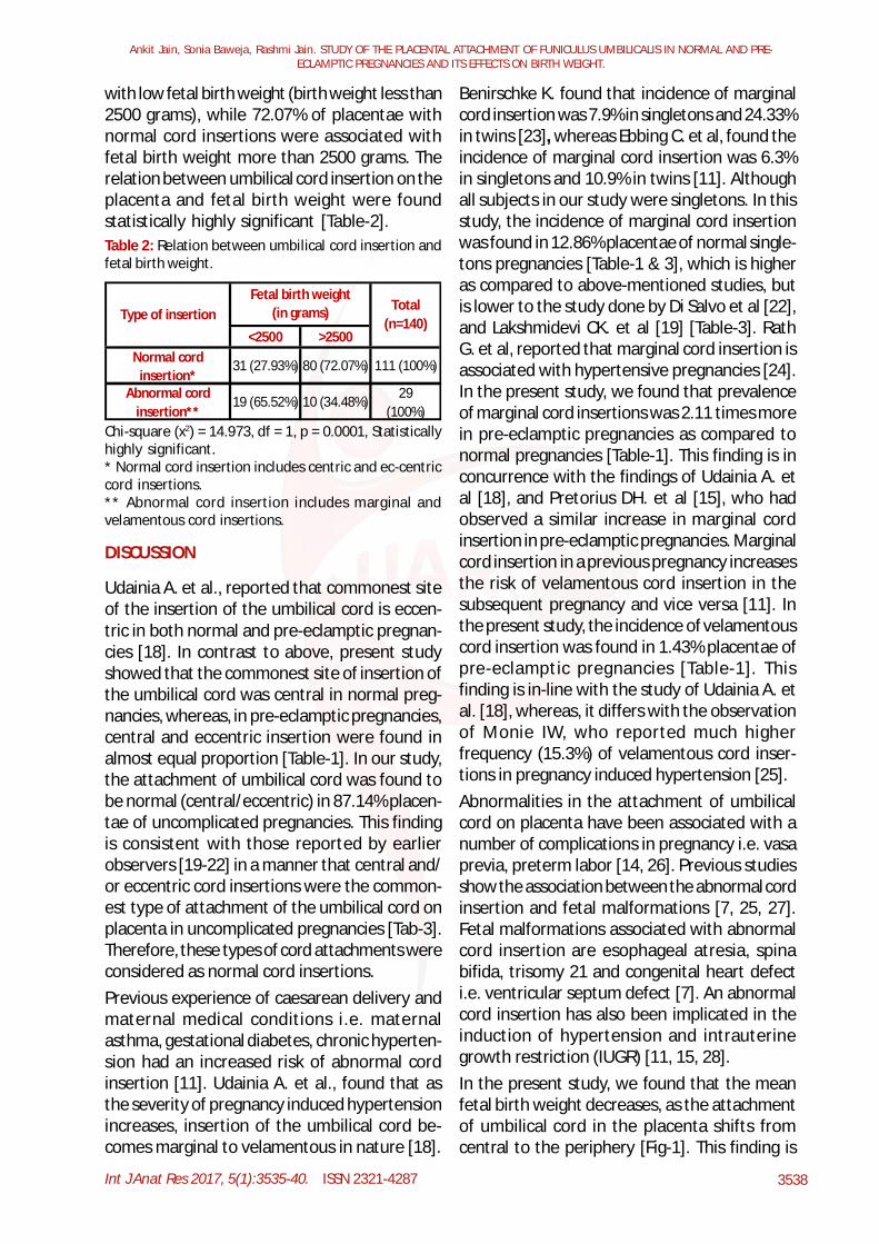
In our study, mean fetal birth weight was found2649.07 ± 260.52 grams and 2530.95 ± 215.49grams in placentae with central and eccentriccord insertion respectively. Whereas in placen-tae with marginal and velamentous cord inser-tion, mean fetal birth weight was found 2296.66± 273.77 grams and 2150 grams respectively [Fig.3]. We also observed that 65.52% of placentaewith abnormal cord insertions were associated
Int J Anat Res 2017, 5(1):3535-40. ISSN 2321-4287 3538
Ankit Jain, Sonia Baweja, Rashmi Jain. STUDY OF THE PLACENTAL ATTACHMENT OF FUNICULUS UMBILICALIS IN NORMAL AND PRE-ECLAMPTIC PREGNANCIES AND ITS EFFECTS ON BIRTH WEIGHT.
with low fetal birth weight (birth weight less than2500 grams), while 72.07% of placentae withnormal cord insertions were associated withfetal birth weight more than 2500 grams. Therelation between umbilical cord insertion on theplacenta and fetal birth weight were foundstatistically highly significant [Table-2].Table 2: Relation between umbilical cord insertion andfetal birth weight.
<2500 >2500Normal cord
insertion*31 (27.93%) 80 (72.07%) 111 (100%)
Abnormal cord insertion**
19 (65.52%) 10 (34.48%) 29 (100%)
Type of insertionFetal birth weight
(in grams) Total (n=140)
Chi-square (x2) = 14.973, df = 1, p = 0.0001, Statisticallyhighly significant.* Normal cord insertion includes centric and ec-centriccord insertions.** Abnormal cord insertion includes marginal andvelamentous cord insertions.
DISCUSSION
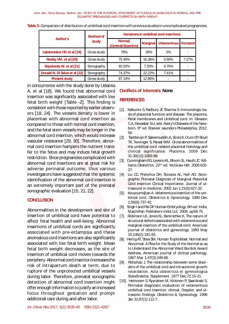
Udainia A. et al., reported that commonest siteof the insertion of the umbilical cord is eccen-tric in both normal and pre-eclamptic pregnan-cies [18]. In contrast to above, present studyshowed that the commonest site of insertion ofthe umbilical cord was central in normal preg-nancies, whereas, in pre-eclamptic pregnancies,central and eccentric insertion were found inalmost equal proportion [Table-1]. In our study,the attachment of umbilical cord was found tobe normal (central/eccentric) in 87.14% placen-tae of uncomplicated pregnancies. This findingis consistent with those reported by earlierobservers [19-22] in a manner that central and/or eccentric cord insertions were the common-est type of attachment of the umbilical cord onplacenta in uncomplicated pregnancies [Tab-3].Therefore, these types of cord attachments wereconsidered as normal cord insertions.Previous experience of caesarean delivery andmaternal medical conditions i.e. maternalasthma, gestational diabetes, chronic hyperten-sion had an increased risk of abnormal cordinsertion [11]. Udainia A. et al., found that asthe severity of pregnancy induced hypertensionincreases, insertion of the umbilical cord be-comes marginal to velamentous in nature [18].
Benirschke K. found that incidence of marginalcord insertion was 7.9% in singletons and 24.33%in twins [23], whereas Ebbing C. et al, found theincidence of marginal cord insertion was 6.3%in singletons and 10.9% in twins [11]. Althoughall subjects in our study were singletons. In thisstudy, the incidence of marginal cord insertionwas found in 12.86% placentae of normal single-tons pregnancies [Table-1 & 3], which is higheras compared to above-mentioned studies, butis lower to the study done by Di Salvo et al [22],and Lakshmidevi CK. et al [19] [Table-3]. RathG. et al, reported that marginal cord insertion isassociated with hypertensive pregnancies [24].In the present study, we found that prevalenceof marginal cord insertions was 2.11 times morein pre-eclamptic pregnancies as compared tonormal pregnancies [Table-1]. This finding is inconcurrence with the findings of Udainia A. etal [18], and Pretorius DH. et al [15], who hadobserved a similar increase in marginal cordinsertion in pre-eclamptic pregnancies. Marginalcord insertion in a previous pregnancy increasesthe risk of velamentous cord insertion in thesubsequent pregnancy and vice versa [11]. Inthe present study, the incidence of velamentouscord insertion was found in 1.43% placentae ofpre-eclamptic pregnancies [Table-1]. Thisfinding is in-line with the study of Udainia A. etal. [18], whereas, it differs with the observationof Monie IW, who reported much higherfrequency (15.3%) of velamentous cord inser-tions in pregnancy induced hypertension [25].Abnormalities in the attachment of umbilicalcord on placenta have been associated with anumber of complications in pregnancy i.e. vasaprevia, preterm labor [14, 26]. Previous studiesshow the association between the abnormal cordinsertion and fetal malformations [7, 25, 27].Fetal malformations associated with abnormalcord insertion are esophageal atresia, spinabifida, trisomy 21 and congenital heart defecti.e. ventricular septum defect [7]. An abnormalcord insertion has also been implicated in theinduction of hypertension and intrauterinegrowth restriction (IUGR) [11, 15, 28].In the present study, we found that the meanfetal birth weight decreases, as the attachmentof umbilical cord in the placenta shifts fromcentral to the periphery [Fig-1]. This finding is
Int J Anat Res 2017, 5(1):3535-40. ISSN 2321-4287 3539
in concurrence with the study done by UdainiaA. et al [18]. We found that abnormal cordinsertion was significantly associated with lowfetal birth weight [Table -2]. This finding isconsistent with those reported by earlier observ-ers [18, 24]. The vessels density is lower inplacentae with abnormal cord insertion ascompared to those with normal cord insertion,and the fetal stem vessels may be longer in theabnormal cord insertion, which would increasevascular resistance [29, 30]. Therefore, abnor-mal cord insertion hampers the nutrient trans-fer to the fetus and may induce fetal growthrestriction. Since pregnancies complicated withabnormal cord insertions are at great risk foradverse perinatal outcome, thus variousinvestigators have suggested that the systemicidentification of the abnormal cord insertion isan extremely important part of the prenatalsonographic evaluation [15, 21, 22].
CONCLUSION
Abnormalities in the development and site ofinsertion of umbilical cord have potential toaffect fetal health and well-being. Abnormalinsertions of umbilical cords are significantlyassociated with pre-eclampsia and theseanomalous cord insertions are also significantlyassociated with low fetal birth weight. Meanfetal birth weight decreases, as the site ofinsertion of umbilical cord moves towards theperiphery. Abnormal cord insertion increases therisk of intrapartum death at term, due torupture of the unprotected umbilical vesselsduring labor. Therefore, prenatal sonographicdetection of abnormal cord insertion mightoffer enough information to justify an increasedfocus throughout gestation and promptadditional care during and after labor.
Table 3: Comparison of distribution of umbilical cord insertion with previous studies in uncomplicated pregnancies.
Normal (Central/Eccentric)
Marginal Velamentous Furcated
Donald N. Di Salvo et al [22] Sonography 74.37% 22.22% 7.41% -Present study Gross study 87.14% 12.86% - -
-
Author’sMethod of
study
Variations in umbilical cord insertions
Lakshmidevi CK. et al [19] Gross study 78% 20% 2%
-
Reddy VM. et al [20] Gross study 75.45% 16.36% 0.90% 7.27%
Sepulveda W. et al [21] Sonography 92.02% 7.20% 0.75%
Conflicts of Interests: None
REFERENCES
[1]. Kalkunte S, Padbury JF, Sharma S. Immunologic ba-sis of placental function and disease: The placenta,Fetal membranes and Umbilical cord. In: GleasonC.A, Devaskar S.U, eds. Avery’s Diseases of the New-born. 9th ed. Elsevier saunders Philadelphia; 2012:50.
[2]. Tantbirojn P, Saleemuddin A, Sirois K, Crum CP, BoydTK, Tworoger S, Parast MM. Gross abnormalities ofthe umbilical cord: related placental histology andclinical significance. Placenta. 2009 Dec31;30(12):1083-88.
[3]. Cunningham FG, Leveno KL, Bloom SL, Hauth JC. Wil-liams Obstetrics. 22nd ed. McGraw-Hill; 2005:620-27.
[4]. Liu CC, Pretorius DH, Scioscia AL, Hull AD. Sono-graphic Prenatal Diagnosis of Marginal PlacentalCord Insertion Clinical Importance. Journal of ul-trasound in medicine. 2002 Jun 1;21(6):627-32.
[5]. Kouyoumdjian A. Velamentous insertion of the um-bilical cord. Obstetrics & Gynecology. 1980 Dec1;56(6):737-42.
[6]. Singh I and Pal GP. Human Embryology. 8th ed. India,Macmillan Publishers India Ltd. 2009, pp59-75.
[7]. Robinson LK, Jones KL, Benirschke K. The nature ofstructural defects associated with velamentous andmarginal insertion of the umbilical cord. Americanjournal of obstetrics and gynecology. 1983 May15;146(2):191-93.
[8]. Hertig AT, Shea SM. Human Trophoblast: Normal andAbnormal: A Plea for the Study of the Normal so asto Understand the Abnormal Ward Burdick AwardAddress. American journal of clinical pathology.1967 Mar 1;47(3):249-68.
[9]. Rolschau J. The relationship between some disor-ders of the umbilical cord and intrauterine growthretardation. Acta obstetricia et gynecologicaScandinavica. Supplement. 1977 Dec;72:15-21.
[10]. Heinonen S, Ryynänen M, Kirkinen P, Saarikoski S.Perinatal diagnostic evaluation of velamentousumbilical cord insertion: clinical, Doppler, and ul-trasonic findings. Obstetrics & Gynecology. 1996Jan 31;87(1):112-7.
Ankit Jain, Sonia Baweja, Rashmi Jain. STUDY OF THE PLACENTAL ATTACHMENT OF FUNICULUS UMBILICALIS IN NORMAL AND PRE-ECLAMPTIC PREGNANCIES AND ITS EFFECTS ON BIRTH WEIGHT.
Int J Anat Res 2017, 5(1):3535-40. ISSN 2321-4287 3540
[21]. Sepulveda W, Wong AE, Gomez L, Alcalde JL. Improv-ing sonographic evaluation of the umbilical cordat the second-trimester anatomy scan. Journal ofUltrasound in Medicine. 2009 Jun 1;28(6):831-5.
[22]. Di Salvo DN, Benson CB, Laing FC, Brown DL, FratesMC, Doubilet PM. Sonographic evaluation of theplacental cord insertion site. AJR. American jour-nal of roentgenology. 1998 May;170(5):1295-8.
[23]. Benirschke K. Abnormalities of the human placenta.NeoReviews. 2005 Sep 1; 6(9):e414-23.
[24]. Rath G, Garg K, Sood M. Insertion of umbilical cordon the placenta in hypertensive mother. J Anat SocIndia., 2000;49(2):149-52.
[25]. Monie IW. Velamentous insertion of the cord inearly pregnancy. American journal of obstetrics andgynecology. 1965 Sep 15;93(2):276-81.
[26]. Paavonen J, Jouttunpaa K, Kangasluoma P, Aro P,Heinonen PK. Velamentous insertion of the umbili-cal cord and vasa previa. International Journal ofGynecology & Obstetrics. 1984 Jun 30;22(3):207-11.
[27]. Raisanen S, Georgiadis L, Harju M, Keski-Nisula L,Heinonen S. Risk factors and adverse pregnancyoutcomes among births affected by velamentousumbilical cord insertion: a retrospective popula-tion-based register study. European Journal of Ob-stetrics & Gynecology and Reproductive Biology.2012;165(2):231-4.
[28]. Cai LY, Izumi SI, Koido S, Uchida N, Suzuki T,Matsubayashi H, Sugi T, Shida N, Kikuchi K,Yoshikata K. Abnormal placental cord insertion mayinduce intrauterine growth restriction in IVF-twinpregnancies. Human Reproduction. 2006 May1;21(5):1285-90.
[29]. Misra DP, Salafia CM, Miller RK, Charles AK. Non-linear and gender-specific relationships among pla-cental growth measures and the fetoplacental weightratio. Placenta. 2009 Dec 31;30(12):1052-7.
[30]. Salafia CM, Zhang J, Miller RK, Charles AK, Shrout P,Sun W. Placental growth patterns affect birth weightfor given placental weight. Birth Defects ResearchPart A: clinical and molecular teratology. 2007 Apr1;79(4):281-8.
[11]. Ebbing C, Kiserud T, Johnsen SL, Albrechtsen S,Rasmussen S. Prevalence, risk factors and outcomesof velamentous and marginal cord insertions: apopulation-based study of 634,741 pregnancies.PLoS One. 2013 Jul 30;8(7):e70380. Available athttp://dx.doi.org/10.1371/journal.pone. 0070380.
[12]. Itskovitz JO, LaGamma EF, Rudolph AM. Effects ofcord compression on fetal blood flow distributionand O2 delivery. American Journal of Physiology-Heart and Circulatory Physiology. 1987 Jan1;252(1):H100-9.
[13]. Soifer SJ, Kaslow D, Roman C, Heymann MA. Umbili-cal cord compression produces pulmonary hyper-tension in newborn lambs: a model to study thepathophysiology of persistent pulmonary hyperten-sion in the newborn. Journal of developmental physi-ology. 1987 Jun;9(3):239-52.
[14]. Brody S, Frenkel DA. Marginal insertion of the cordand premature labor. American journal of obstet-rics and gynecology. 1953 Jun 30; 65(6):1305-12.
[15]. Pretorius DH, Chau C, Poeltler DM, Mendoza A,Catanzarite VA, Hollenbach KA. Placental cord in-sertion visualization with prenatal ultrasonogra-phy. Journal of ultrasound in medicine. 1996 Aug1;15(8):585-93.
[16]. Hasegawa J, Matsuoka R, Ichizuka K, Sekizawa A,Farina A, Okai T. Velamentous cord insertion intothe lower third of the uterus is associated with in-trapartum fetal heart rate abnormalities. Ultra-sound in obstetrics & gynecology. 2006 Apr1;27(4):425-9.
[17]. Park K. Park’s textbook of Preventive and SocialMedicine. 22nd ed. Jabalpur: M/S BanarsidasBhanot India; 2013.
[18]. Udainia A, Mehta CD, Chauhan K, Suthar K, ChauhanK. Relation between umbilical cord insertion andfoetal outcome in pregnancy induced hypertension.International Journal of Basic and Applied MedicalSciences. 2014 Jan;4(1):332-7. Available at http://www.cibtech.org/jms.htm.
[19]. Lakshmi devi CK, Neelam S, Raghupathy NS. Mor-phological studies of normal human placenta atdifferent gestational periods. IOSR- Journal of den-tal and medical sciences. 2013 May;6(3):9-15.
[20]. Reddy VM, Geetha SP, Nim VK. Variations in Placen-tal attachment of umbilical cord. Journal of Anat.Soc. of India. 2012 Jun 1;61(1):1-4.
How to cite this article:
Ankit Jain, Sonia Baweja, Rashmi Jain. STUDY OF THE PLACENTALATTACHMENT OF FUNICULUS UMBILICALIS IN NORMAL AND PRE-ECLAMPTIC PREGNANCIES AND ITS EFFECTS ON BIRTH WEIGHT.Int J Anat Res 2017;5(1):3535-3540. DOI: 10.16965/ijar.2017.107
Ankit Jain, Sonia Baweja, Rashmi Jain. STUDY OF THE PLACENTAL ATTACHMENT OF FUNICULUS UMBILICALIS IN NORMAL AND PRE-ECLAMPTIC PREGNANCIES AND ITS EFFECTS ON BIRTH WEIGHT.