Embed Size (px)
Citation preview
RAD
No.87 (2020.2)
1. About our Hospital
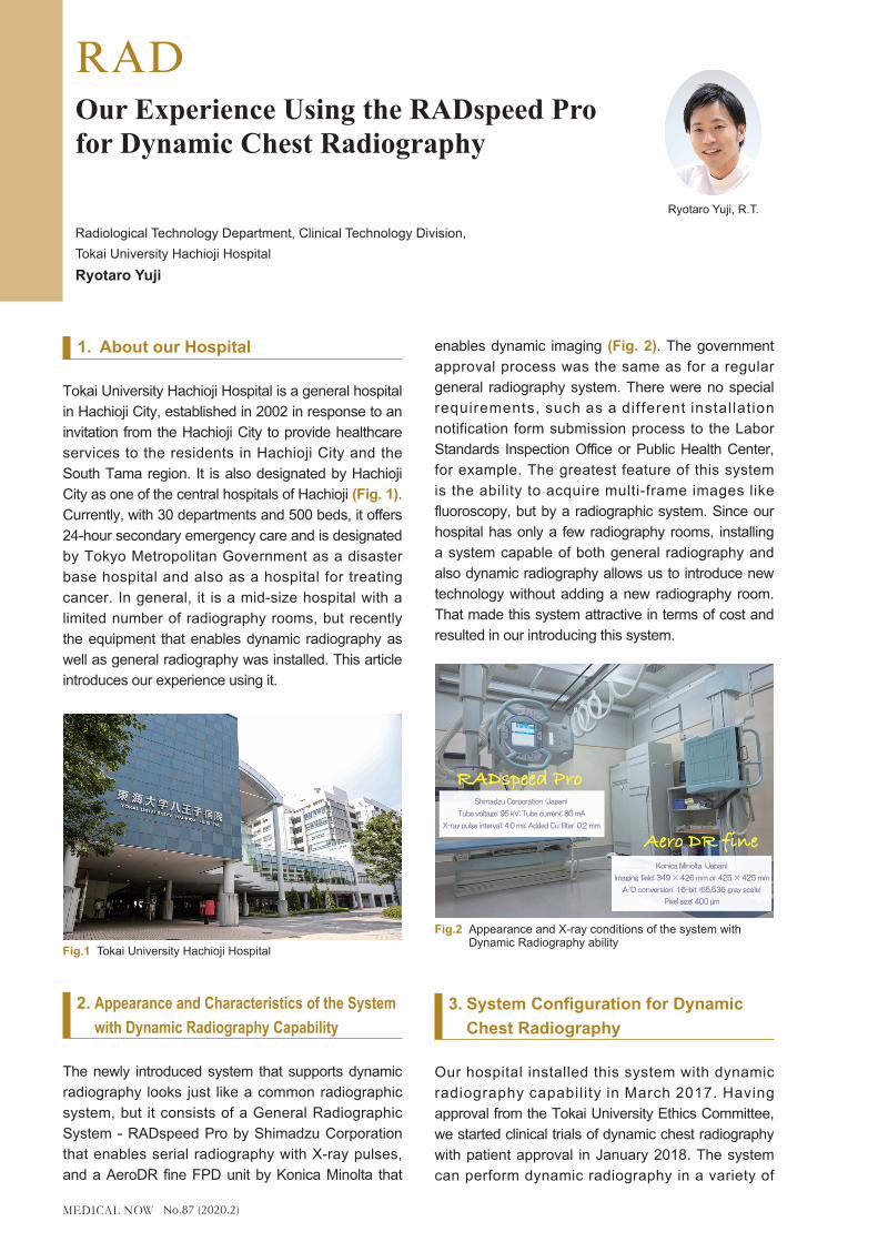
Tokai University Hachioji Hospital is a general hospital in Hachioji City, established in 2002 in response to an invitation from the Hachioji City to provide healthcare services to the residents in Hachioji City and the South Tama region. It is also designated by Hachioji City as one of the central hospitals of Hachioji (Fig. 1). Currently, with 30 departments and 500 beds, it offers 24-hour secondary emergency care and is designated by Tokyo Metropolitan Government as a disaster base hospital and also as a hospital for treating cancer. In general, it is a mid-size hospital with a limited number of radiography rooms, but recently the equipment that enables dynamic radiography as well as general radiography was installed. This article introduces our experience using it.
2. Appearance and Characteristics of the System with Dynamic Radiography Capability
The newly introduced system that supports dynamic radiography looks just like a common radiographic system, but it consists of a General Radiographic System - RADspeed Pro by Shimadzu Corporation that enables serial radiography with X-ray pulses, and a AeroDR fine FPD unit by Konica Minolta that
enables dynamic imaging (Fig. 2). The government approval process was the same as for a regular general radiography system. There were no special requirements, such as a different installation notification form submission process to the Labor Standards Inspection Office or Public Health Center, for example. The greatest feature of this system is the ability to acquire multi-frame images like fluoroscopy, but by a radiographic system. Since our hospital has only a few radiography rooms, installing a system capable of both general radiography and also dynamic radiography allows us to introduce new technology without adding a new radiography room. That made this system attractive in terms of cost and resulted in our introducing this system.
3. System Configuration for Dynamic Chest Radiography
Our hospital installed this system with dynamic radiography capability in March 2017. Having approval from the Tokai University Ethics Committee, we started clinical trials of dynamic chest radiography with patient approval in January 2018. The system can perform dynamic radiography in a variety of
Our Experience Using the RADspeed Pro for Dynamic Chest Radiography
Radiological Technology Department, Clinical Technology Division,Tokai University Hachioji HospitalRyotaro Yuji
Ryotaro Yuji, R.T.
Fig.1 Tokai University Hachioji Hospital
X-ray pulse interval: 4.0 ms; Added Cu filter: 0.2 mmX-ray pulse interval: 4.0 ms; Added Cu filter: 0.2 mm
Tube voltage: 95 kV; Tube current: 80 mATube voltage: 95 kV; Tube current: 80 mA
Shimadzu Corporation (Japan)Shimadzu Corporation (Japan)
Pixel size: 400 µmPixel size: 400 µmA/D conversion: 16-bit (65,536 gray scale)A/D conversion: 16-bit (65,536 gray scale)
Imaging field: 349 × 426 mm or 425 × 425 mmImaging field: 349 × 426 mm or 425 × 425 mm
Konica Minolta (Japan)Konica Minolta (Japan)
Fig.2 Appearance and X-ray conditions of the system with Dynamic Radiography ability
No.87 (2020.2)
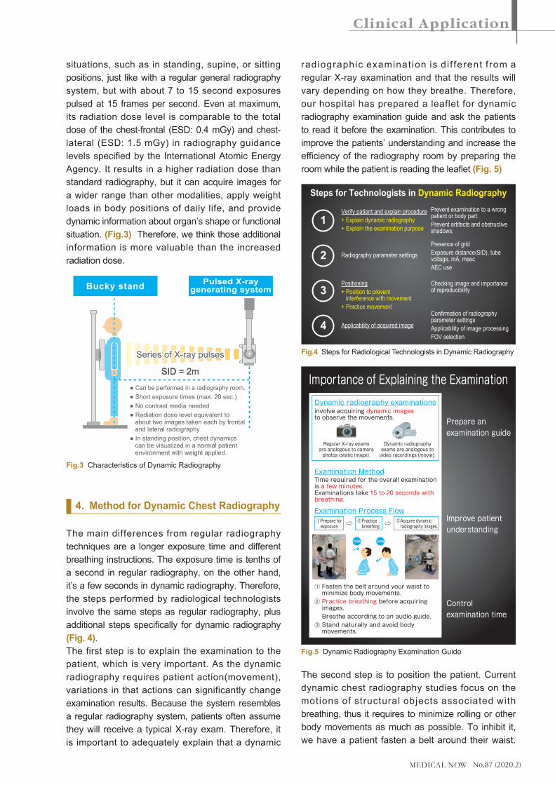
situations, such as in standing, supine, or sitting positions, just like with a regular general radiography system, but with about 7 to 15 second exposures pulsed at 15 frames per second. Even at maximum, its radiation dose level is comparable to the total dose of the chest-frontal (ESD: 0.4 mGy) and chest-lateral (ESD: 1.5 mGy) in radiography guidance levels specified by the International Atomic Energy Agency. It results in a higher radiation dose than standard radiography, but it can acquire images for a wider range than other modalities, apply weight loads in body positions of daily life, and provide dynamic information about organ’s shape or functional situation. (Fig.3) Therefore, we think those additional information is more valuable than the increased radiation dose.
4. Method for Dynamic Chest Radiography
The main differences from regular radiography techniques are a longer exposure time and different breathing instructions. The exposure time is tenths of a second in regular radiography, on the other hand, it’s a few seconds in dynamic radiography. Therefore, the steps performed by radiological technologists involve the same steps as regular radiography, plus additional steps specifically for dynamic radiography (Fig. 4).The first step is to explain the examination to the patient, which is very important. As the dynamic radiography requires patient action(movement), variations in that actions can significantly change examination results. Because the system resembles a regular radiography system, patients often assume they will receive a typical X-ray exam. Therefore, it is important to adequately explain that a dynamic
radiographic examinat ion is d i f ferent f rom a regular X-ray examination and that the results will vary depending on how they breathe. Therefore, our hospital has prepared a leaflet for dynamic radiography examination guide and ask the patients to read it before the examination. This contributes to improve the patients’ understanding and increase the efficiency of the radiography room by preparing the room while the patient is reading the leaflet (Fig. 5)
The second step is to position the patient. Current dynamic chest radiography studies focus on the motions of structural objects associated with breathing, thus it requires to minimize rolling or other body movements as much as possible. To inhibit it, we have a patient fasten a belt around their waist.
Series of X-ray pulsesSeries of X-ray pulses
SID = 2mSID = 2m
● Can be performed in a radiography room.● Short exposure times (max. 20 sec.)● No contrast media needed● Radiation dose level equivalent to
about two images taken each by frontal and lateral radiography
● In standing position, chest dynamics can be visualized in a normal patient environment with weight applied.
Bucky stand Pulsed X-raygenerating system
Fig.3 Characteristics of Dynamic Radiography
Importance of Explaining the Examination
Prepare anexamination guide
Improve patientunderstanding
Controlexamination time
involve acquiring dynamic images to observe the movements.
Dynamic radiography examinations
Examination Method
Examination Process Flow
Time required for the overall examinationis a few minutes.Examinations take 15 to 20 seconds withbreathing.
① Fasten the belt around your waist to minimize body movements.② Practice breathing before acquiring images. Breathe according to an audio guide.③ Stand naturally and avoid body movements.
Regular X-ray examsare analogous to cameraphotos (static image).
Dynamic radiographyexams are analogous tovideo recordings (movie).
①Prepare for exposure.
②Practice breathing.
③Acquire dynamic radiography images.
ExhaleInhale
Fig.5 Dynamic Radiography Examination Guide
Verify patient and explain procedure+ Explain dynamic radiography+ Explain the examination purpose
Radiography parameter settings
Positioning+ Position to prevent interference with movement+ Practice movement
Applicability of acquired image
Steps for Technologists in Dynamic RadiographyPrevent examination to a wrong patient or body part.Prevent artifacts and obstructive shadows.
Presence of gridExposure distance(SID), tube voltage, mA, msecAEC use
Checking image and importance of reproducibility
Confirmation of radiography parameter settingsApplicability of image processingFOV selection
1
2
3
4
Fig.4 Steps for Radiological Technologists in Dynamic Radiography

No.87 (2020.2)
However, because restricting movement does not provide a natural environment, the belt must be positioned on with attention to the respiratory and accessory muscles. At our hospital, we try to keep patients in a relaxed position, rather than positioning them to avoid the shoulder blades, which is the positioning typically used for regular radiography (Fig. 6).The third step is to give breathing instructions. A variety of breathing instructions are available, such as deep breathing, quiet breathing, or holding the breath, but and the exposure time and breathing instructions must be varied depending on the dynamic information needed. Currently, at our hospital, we instruct the patients to have deep breathing or hold breathing, but unlike regular radiography, the instructions are complicated (Fig. 7). Dynamic chest radiography examinations could also be used as functional examinations, so there is a possibility of performing examinations repeatedly. Considering
the radiological technologist may not be always the same, it is essential to use an automated voice generating device (“auto-voice” device) to avoid variations in reproducibility by the verbal instruction by the technologists. Therefore, the patients of our hospital practice breathing with the auto-voice device after hearing our explanation prior to the examination. Such auto-voice devices are normally associated with the exposure switch, so that the voice cannot be generated unless the technologist goes out of the examination room, However, in the RADspeed Pro system that we newly introduced, the auto-voice is triggered by its activation button on the X-ray tube control panel, which means the technologist can remain close to the patient to confirm if the patient is following breathing instructions. That is an important and effective functionality to confirm not only to follow breathing instructions, but also to implement the safety measures and body movement restraints properly. (Fig. 8).
Hand switch ON Hand switch OFF
Continue pressing hand switch (second level).
Auto-voice
Press hand switch(first level).
Voice:
Ready...inhale deeply...inhale, inhale, inhale.
Holdyour breath.
5 sec(without X-rays)
X-ray pulses
Breath held
Fully inhaledposition
Fully exhaledposition
Duration of Pulsed Exposure
Fully inhaledposition
Exhaling phase Breath held Inhaling phase
5 sec2 sec 5 sec2 sec
Holdyourbreath.
Slowly exhale. . .
OK,now slowly inhale. . .
Great.We’refinished.
Diaphragm level
Fig.7 Deep Breathing Instructions and Exposure Timing
PositioningPositioning to prevent tension in muscles used for breathing
Belt across pelvis
Hands level with elbows
Muscles Used for Breathing
Externalintercostalmuscle
Sternocleidomastoidmuscle
Scalene muscle
Trapezius muscle
Internal intercostal muscle
External abdominaloblique muscle
Transverseabdominal muscle
Internal abdominaloblique muscle
Diaphragm
Abdominalrectus muscle
Erectorspinae muscle
■Breathing muscles
Trapeziusmuscle
Front Rear
Fig.6 Positioning for Dynamic Chest Radiography

No.87 (2020.2)
5. Dynamic Radiography Data Analysis Technologies
After the image acquisition process, the data must be analyzed in the Konica Minolta KINOSIS dynamic X-ray image processing workstation, not in the main radiography system. A variety of analysis technologies is equipped in the workstation and the technology is advancing day by day. The current data analysis technologies can be generally classified either as dynamic chest imaging (DCI) for diagnosis for organs’ shape or movement or as pulmonary functional imaging (PFI) for diagnosis based on function (Fig. 9). DCI can analyze the distance and the positional relationship of the structure that is fluctuating dynamically and can show the relation with the time axis which was impossible until recently. PFI can provide information about periodic movement by analyzing trends in pixel value variations. That allows observing periodic relationships with breathing and heartbeat cycles.
6. Clinical Example of Dynamic Chest Radiography
Though dynamic radiography images are difficult to present on paper because they involve a time axis, the following describes a clinical example of a procedure performed at our hospital. Fig. 10 shows the positions of the right and left apices of the lung and the diaphragm detected from dynamic chest radiography images. Fig. 11 is the chart to show how their detected positions vary during breathing. In healthy lungs, the left and right diaphragms move together, but in this case, the right diaphragm (blue line) moves poorly, result ing in opposite movement from the left diaphragm (green line). This condition can only be shown instantaneously and cannot be determined from a regular X-ray image.Fig. 12 shows the extracted signals related to the fluctuation of pixel values in the lung field and the respiratory cycle. It shows the amount of signal change from the fully peak exhaled position, and the areas are colored in blue if the amount changes a lo t . Fig. 12a is f rom a pat ient wi th normal respiratory function, whereas Fig. 12b is from a patient with chronic obstructive pulmonary disease. The images show that less change quantity can be found in the upper middle lung field of the patient with the lung disease. This suggests that the degree of signal variation from the lung tissue during respiration might differ depending on the disorder, which can be expected to improve our understanding of the pathology and help improve detection of lung disease.
Image processing technologies expected to improve recognition capabilities
DynamicX-ray images
Dynamic Chest Imaging (DCI)Diagnosis of Shape/Movement
Pulmonary Functional Imaging (PFI)Diagnosis of Function
RibsuppressionBS-MODE
FE-MODE
BS : Bone Suppression
DM-MODE
Basic image measured
DM : Diaphragm Movement
PL-MODE
PH-MODE
PL : Pixel Value Measurement - low frequency
PH : Pooel Value Measurement - high frequency
FE : Frequency Enhancement
Diaphragmtracking
Lung fielddensity variations(breathing cycle)
Lung fielddensity variations(heartbeat cycle)
Measure distances/areasof lung field,airway diameter,heart, etc.
Frequencyenhancement
Image measurement technologies expected to improve quantitation of dynamic changes
Image analysis technologies expected for visualizing lung functional information
Fig.9 Dynamic Data Analysis Technologies
Breathing InstructionsAssociate withauto-voice device.
Breathing practice can be started inside the radiography room.
Breathing during practice can be confirmed near the patient.
Voice audibility and presence of any body movement can be confirmed.
Fig.8 Auto-Voice Activation Button Used during Breathing Practice

No.87 (2020.2)
7. Summary
Currently, the clinical researches of the dynamic radiography are proceeded in a wide variety of facilities, and its correlation with the nuclear medicine examinations has been recognized not only in animal studies but also in actual clinical studies, and r its value has been found in clinical field. The biggest advantage of dynamic radiography is its convenience. The dynamic radiography system we introduced can be used just as an extension of a general radiography system. Consequently, there has been little resistance from radiological technologists, who use radiology the most often. The minimal invasiveness and functional examination capability also offer revolutionary improvements in physical stress and cost for patients. Due to its low cost and simplicity of enabling functional examinations, the potential demands for dynamic radiography systems are presumably high, not only
among large hospitals, but also among smaller hospitals without nuclear medicine or CT . Use of dynamic radiography is expected to expand in the future for a wide variety of applications and other body areas. Consequently, it might even go beyond being simply another examination framework. That would require various knowledge and experience. From a multifaceted perspective, it’s possible that the new unique imaging methods of dynamic radiography will be developed. As medical imaging continues to transition from still images to dynamic imaging, radiological technologists will also need to increase their knowledge of physiology and functions so that they can better understand and comment on clinical information obtained from dynamic images.
All rights are reserved, including those to reproduce this article without permission from Shimadzu Corporation.
Fig.12 b) Patient with Chronic Obstructive Pulmonary Disease
Fig.12 a) Patient with Normal Lung Function
Fig.10 Position of Left/Right Lung Apex vs Diaphragm Fig.11 Height Variations Due to Breathing


![[PPT]Chest tube, thoracentesis and fibrinolyticschestgmcpatiala.weebly.com/uploads/8/3/5/5/8355281/chest... · Web viewDEFINITION A chest drain is a tube inserted through the chest](https://img.pdfslide.net/doc/110x75/5b403a5f7f8b9a4b3f8d15f4/pptchest-tube-thoracentesis-and-fibrinol-web-viewdefinition-a-chest-drain.jpg)


![Chest physiotherapy compared to no chest physiotherapy for ... · [Intervention Review] Chest physiotherapy compared to no chest physiotherapy for cystic fibrosis Cees P van der](https://img.pdfslide.net/doc/110x75/5cc2dd0188c99389538bb642/chest-physiotherapy-compared-to-no-chest-physiotherapy-for-intervention.jpg)