Embed Size (px)
Citation preview
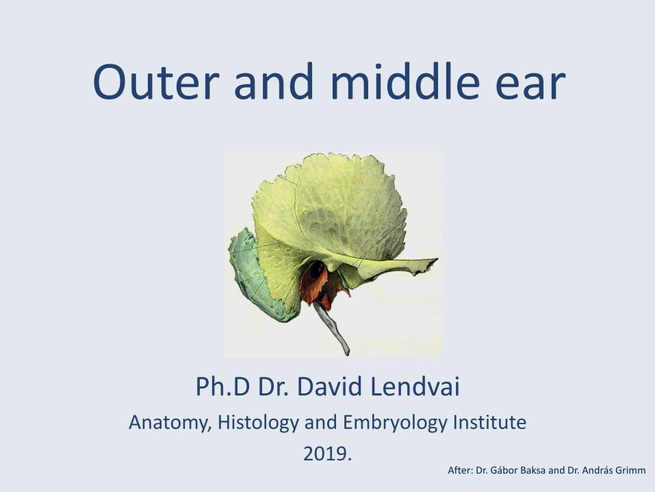
Outer and middle ear
Ph.D Dr. David LendvaiAnatomy, Histology and Embryology Institute
2019.After: Dr. Gábor Baksa and Dr. András Grimm
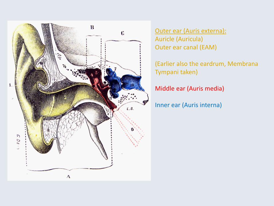
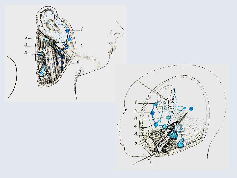
Outer ear (Auris externa):Auricle (Auricula)Outer ear canal (EAM)
(Earlier also the eardrum, MembranaTympani taken)
Middle ear (Auris media)
Inner ear (Auris interna)
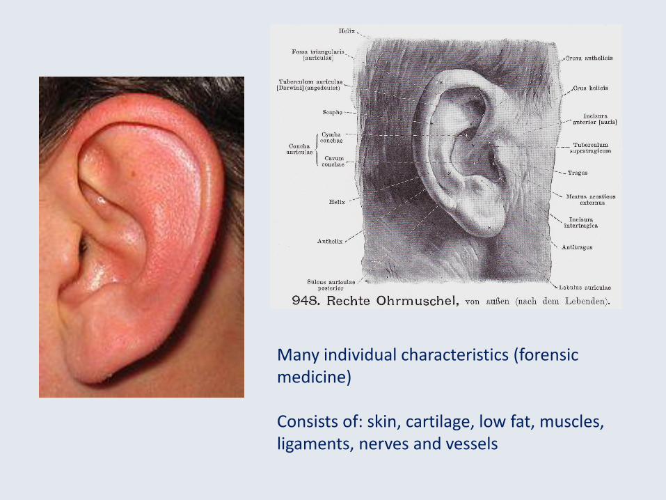
Many individual characteristics (forensicmedicine)
Consists of: skin, cartilage, low fat, muscles,ligaments, nerves and vessels
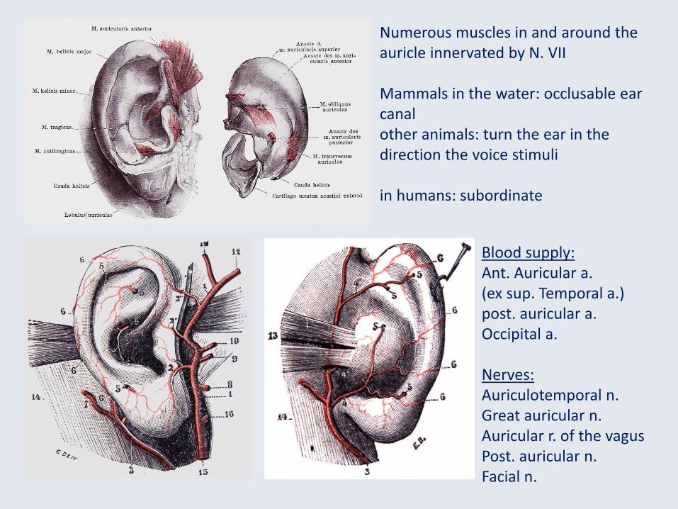
Numerous muscles in and around theauricle innervated by N. VII
Mammals in the water: occlusable earcanalother animals: turn the ear in the direction the voice stimuli
in humans: subordinate
Blood supply:Ant. Auricular a.(ex sup. Temporal a.)post. auricular a.Occipital a.
Nerves:Auriculotemporal n.Great auricular n.Auricular r. of the vagusPost. auricular n.Facial n.
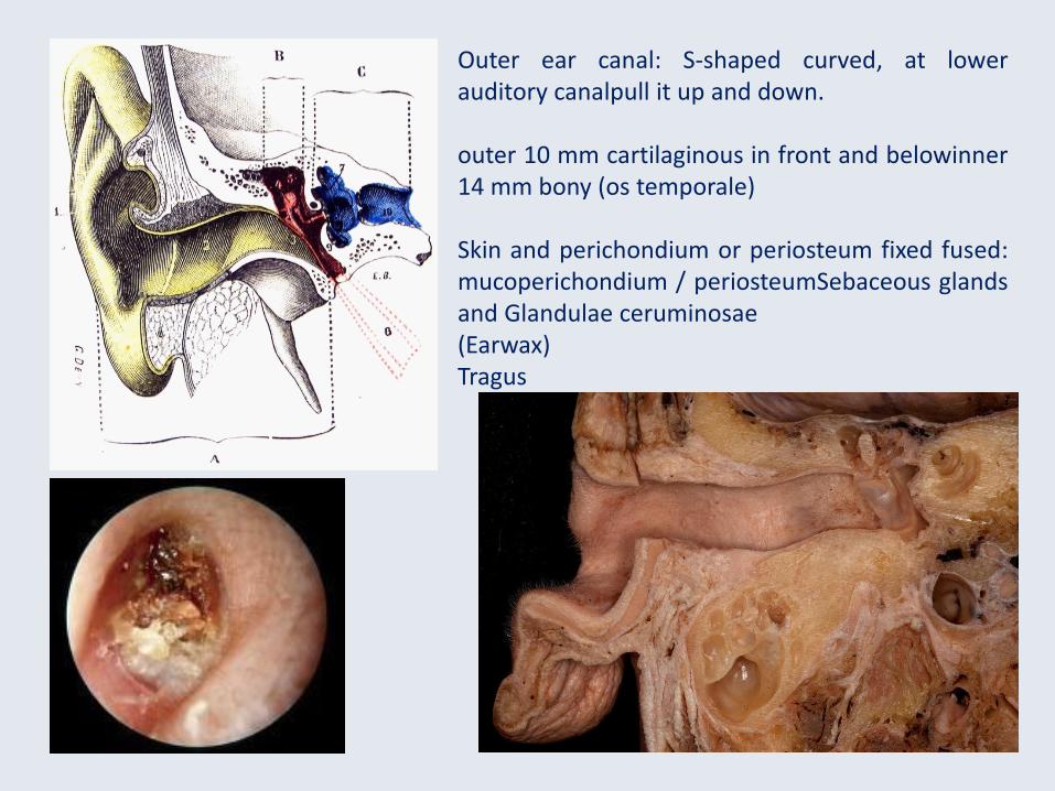
Outer ear canal: S-shaped curved, at lowerauditory canalpull it up and down.
outer 10 mm cartilaginous in front and belowinner14 mm bony (os temporale)
Skin and perichondium or periosteum fixed fused:mucoperichondium / periosteumSebaceous glandsand Glandulae ceruminosae(Earwax)Tragus
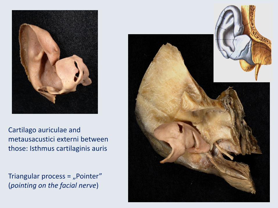
Cartilago auriculae and metausacustici externi betweenthose: Isthmus cartilaginis auris
Triangular process = „Pointer”(pointing on the facial nerve)
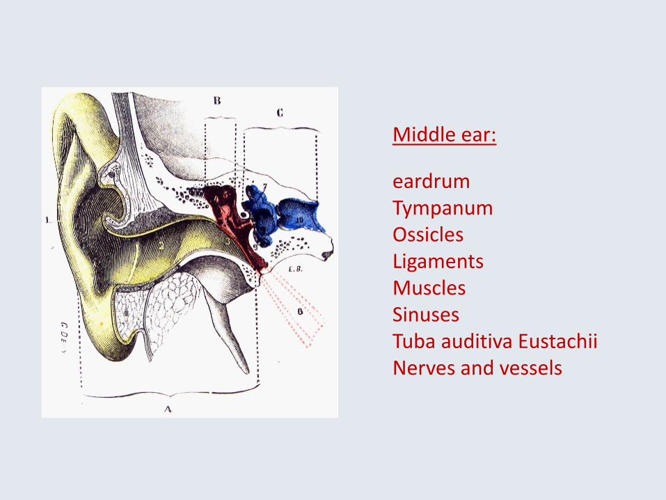
Middle ear:
eardrumTympanumOssiclesLigamentsMusclesSinusesTuba auditiva EustachiiNerves and vessels
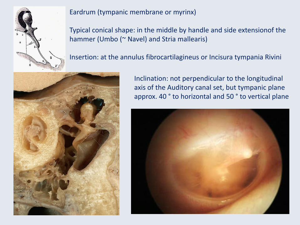
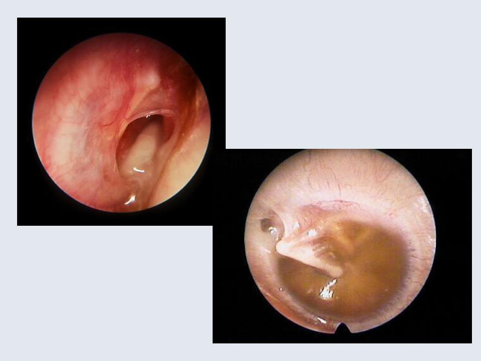
Eardrum (tympanic membrane or myrinx)
Typical conical shape: in the middle by handle and side extensionof the hammer (Umbo (~ Navel) and Stria mallearis)
Insertion: at the annulus fibrocartilagineus or Incisura tympania Rivini
Inclination: not perpendicular to the longitudinalaxis of the Auditory canal set, but tympanic planeapprox. 40 ° to horizontal and 50 ° to vertical plane
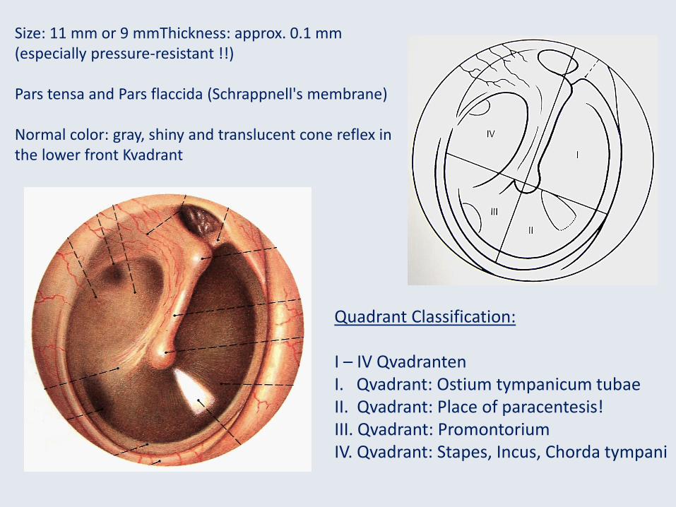
Size: 11 mm or 9 mmThickness: approx. 0.1 mm (especially pressure-resistant !!)
Pars tensa and Pars flaccida (Schrappnell's membrane)
Normal color: gray, shiny and translucent cone reflex in the lower front Kvadrant
Quadrant Classification:
I – IV QvadrantenI. Qvadrant: Ostium tympanicum tubaeII. Qvadrant: Place of paracentesis!III. Qvadrant: PromontoriumIV. Qvadrant: Stapes, Incus, Chorda tympani
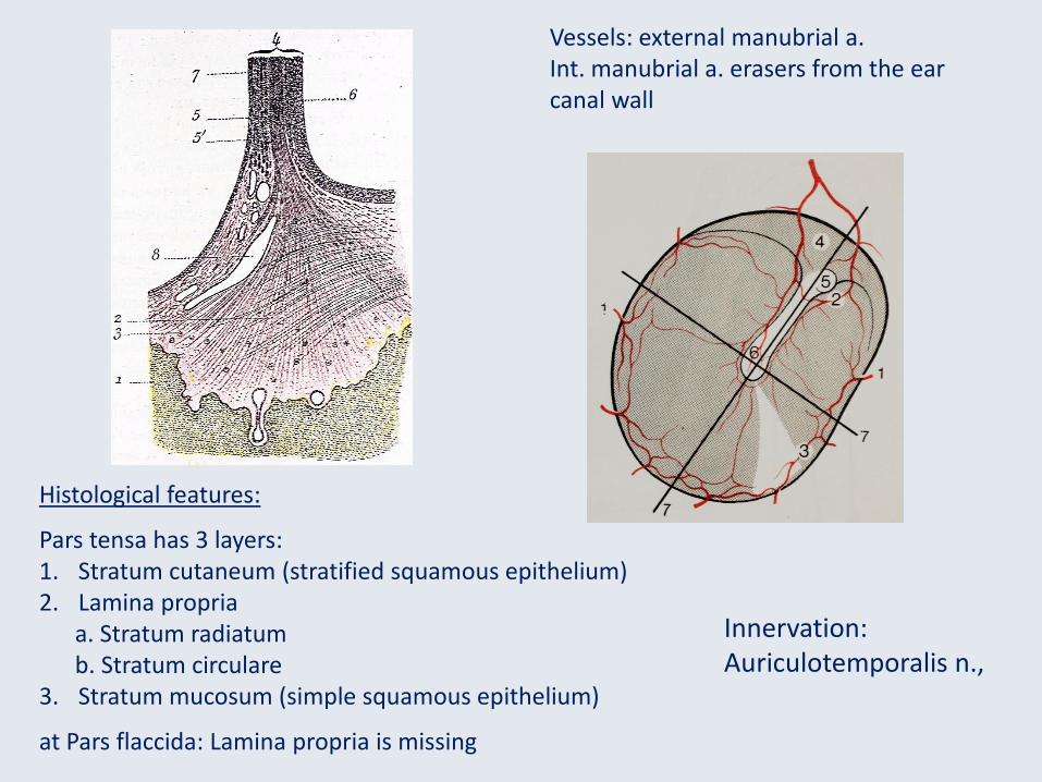
Vessels: external manubrial a.Int. manubrial a. erasers from the earcanal wall
Histological features:
Pars tensa has 3 layers:1. Stratum cutaneum (stratified squamous epithelium)2. Lamina propria
a. Stratum radiatumb. Stratum circulare
3. Stratum mucosum (simple squamous epithelium)
at Pars flaccida: Lamina propria is missing
Innervation:Auriculotemporalis n.,

tympanum (Tympanic cavity)
1. carotic part 4. membranaceus part2. labyrinthic part 5. tegmental part3. mastoideus part 6. jugular part
1
3
4
5
6
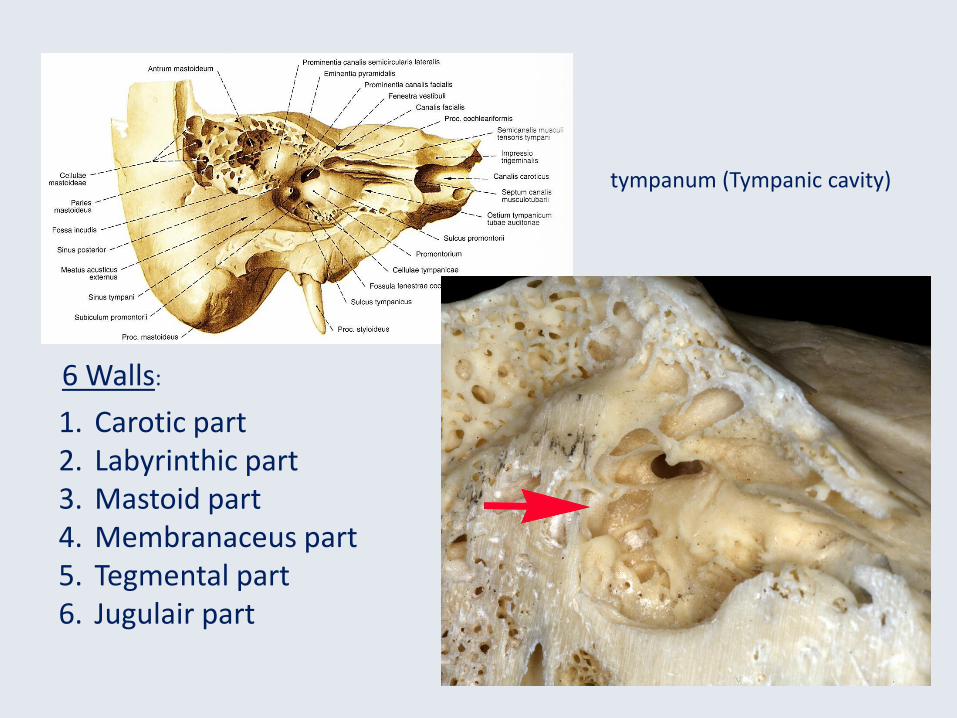
1. Carotic part2. Labyrinthic part3. Mastoid part4. Membranaceus part5. Tegmental part6. Jugulair part
6 Walls:
tympanum (Tympanic cavity)
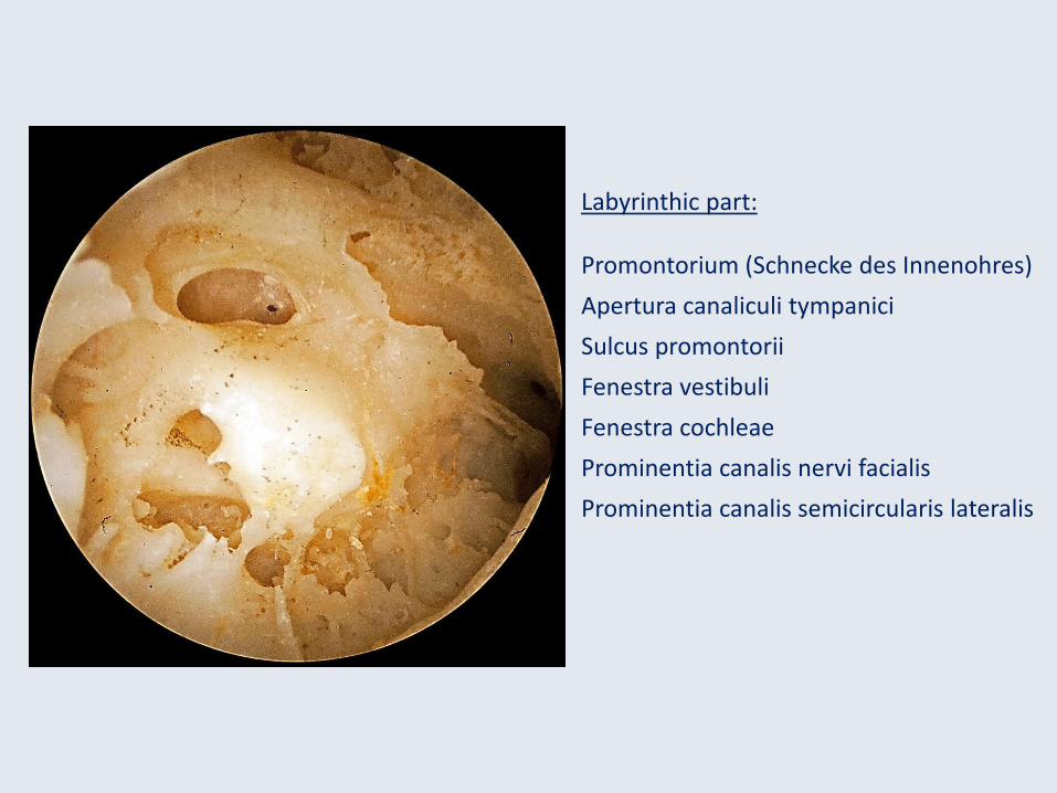
Labyrinthic part:
Promontorium (Schnecke des Innenohres)
Apertura canaliculi tympanici
Sulcus promontorii
Fenestra vestibuli
Fenestra cochleae
Prominentia canalis nervi facialis
Prominentia canalis semicircularis lateralis

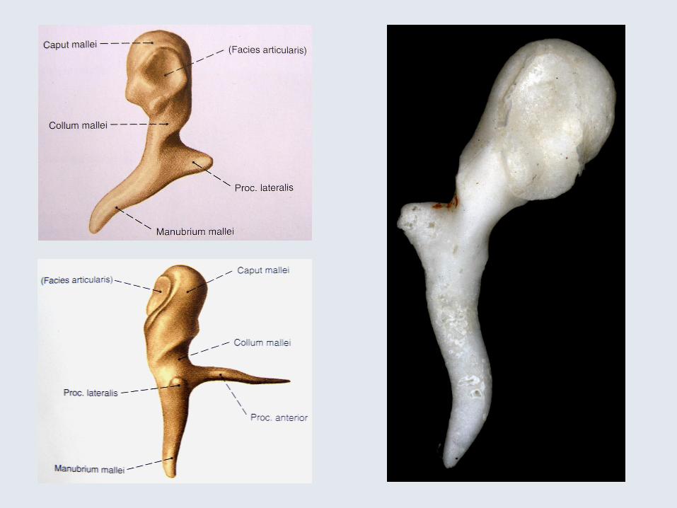
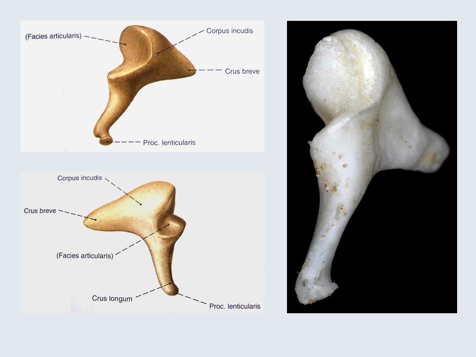

Incudo-mallear articulation (between 2 saddle forms)Movement about 5 ° tight capsule + "ratchet teeth"
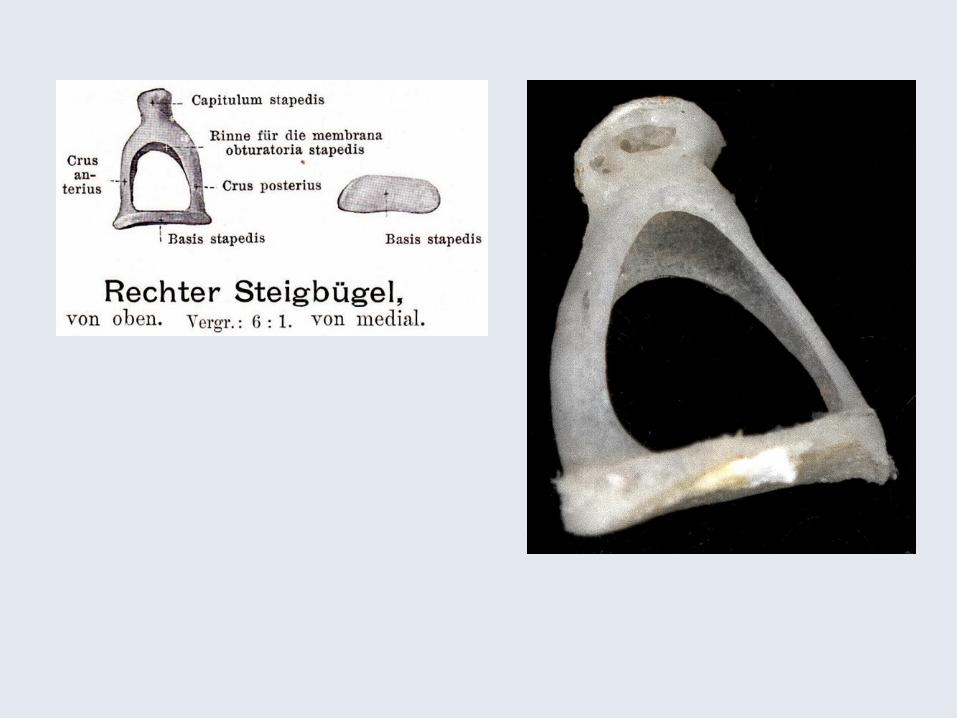
Incudo-stapedialbetween Proc. lenticularis incudisand Capitulum stapedis
Syndesmosis tympano-stapedialbetween Fenestra vestibuli (ovalwindow) and base of the stapesAnulare (baseos) lig. stapedis
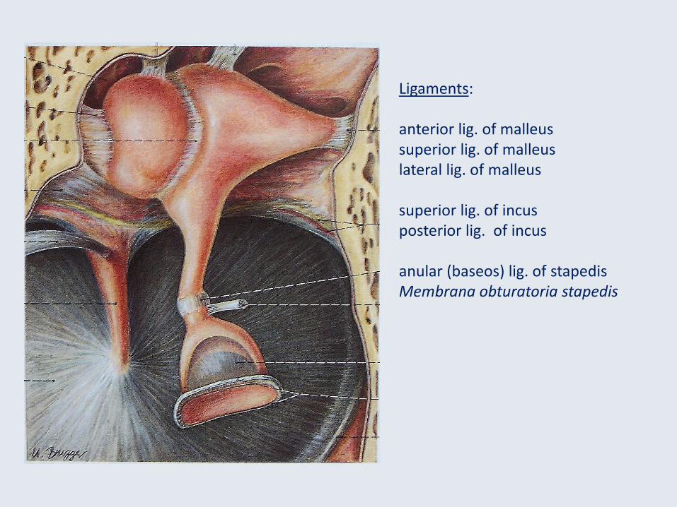
Ligaments:
anterior lig. of malleussuperior lig. of malleuslateral lig. of malleus
superior lig. of incusposterior lig. of incus
anular (baseos) lig. of stapedisMembrana obturatoria stapedis
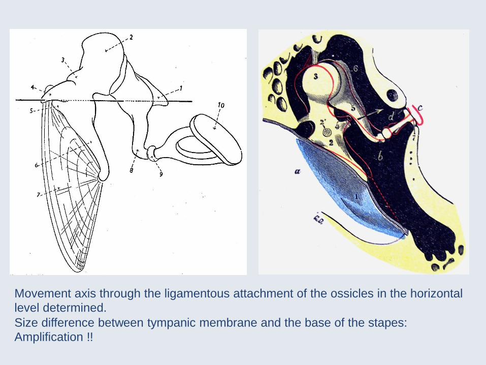
Movement axis through the ligamentous attachment of the ossicles in the horizontal
level determined.
Size difference between tympanic membrane and the base of the stapes:
Amplification !!
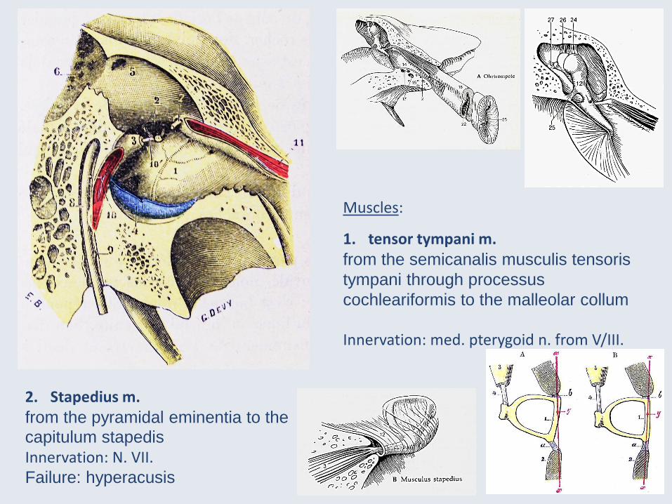
Muscles:
1. tensor tympani m.from the semicanalis musculis tensoris
tympani through processus
cochleariformis to the malleolar collum
Innervation: med. pterygoid n. from V/III.
2. Stapedius m.from the pyramidal eminentia to the
capitulum stapedis
Innervation: N. VII.Failure: hyperacusis

Folds and recesses:
anterior and posterior malleolar fold(Recessus membranae tympani anterior etposterior or Tröltsch)
Plica incudis
Plica stapedis
Sup. tympanic membrane recess membraneor Prussak’s-space: Collum and Caput mallei, lateralis malleolar proc., lateralemalleolar proc., Pars flaccida
Epitympanic recess (Atticus)
Round window (Fenestra cochleae):Secondary tympanic membrane (Scarpae)

Eustachian tube: bony and cartilaginous part (between them: Isthmus tubae auditivae)Bony part: Semicanalis tubae auditivae (Mucoperiosteum)Cartilaginous part: medial and lateral lamina of the tubae auditivae , in which below and
lateral the tuba is replaced by the so-called lamina membranacea (origin of the tensor veli palatini m.) (+ Lamina propria with mucous glands and lymphoid tissue –Tonsilla tubaria)Ostium pharyngeum and Ostium tympanicumSimple columnar ciliates epithel.
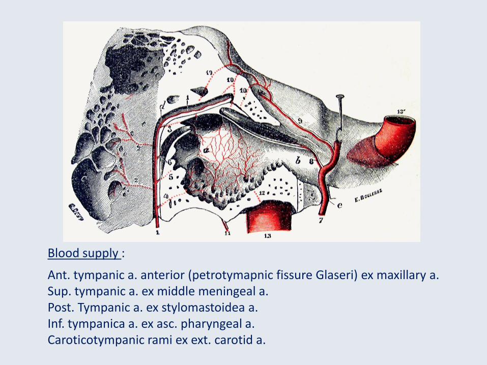
Blood supply :
Ant. tympanic a. anterior (petrotymapnic fissure Glaseri) ex maxillary a.Sup. tympanic a. ex middle meningeal a.Post. Tympanic a. ex stylomastoidea a.Inf. tympanica a. ex asc. pharyngeal a.Caroticotympanic rami ex ext. carotid a.