Embed Size (px)
Citation preview
54
POJ 7(1):54-62 (2014) ISSN:1836-3644
Overexpression of a new cellulose synthase gene (PuCesA6) from Ussuri poplar (Populus
ussuriensis) exhibited a dwarf phenotype in transgenic tobacco
Lei Xu*
Key Laboratory of Bio-Based Material Science and Technology, Northeast Forestry University, Harbin, 150040,
China
Corresponding author: [email protected]
Abstract
Cellulose is synthesized in plant cell walls by cellulose synthase (CesA) genes. The characterization of a new cellulose synthase gene
(PuCesA6) (GenBank Accession No. HQ686077) from an economically important tree, Ussuri Poplar (Populus ussuriensis) is
reported here. The predicted PuCesA6 protein is highly similar to Populus tremuloides PtrCesA6 (99%) expressing in all expanding
cells depositing primary cell wall. The domain structures of PuCesA6 was predicted by multiple alignment analysis, which contained
a N-terminal cysteine rich zinc binding domain, 8 putative transmembrane helices (TMH), a signature D, D, D, QxxRW motif, 5
alternating conserved regions (CR-P) and 2 hypervariable regions (HVR). Subcellular localization analysis showed that PuCesA6
protein was localized in the cytomembrane. Ectopic expression of PuCesA6 in tobacco significantly exhibited "dwarf" phenotype,
with final aerial height less than 35 cm (approximately two-thirds height of wild type). The phenomenon could be explained by
post-transcriptional gene silencing (PTGS) of the expression of the homologous genes in tobacco. Thus, identification of new CesA
genes from poplar tree genomes is essential for enhancing knowledge of cellulose biosynthesis in trees that has many fundamental
and commercial implications.
Keywords: "dwarf" phenotype; PuCesA6; Populus ussuriensis; Tobacco.
Abbreviations: BA_6-benzylaminopurine; NAA_naphthaleneacetic acid; TMH_transmembrane helices; CR-P_conserved region;
HVR_hypervariable regions; PTGS_post-transcriptional gene silencing; ORF_open reading frame; Km- kanamycin.
Introduction
As an integral component of plant cell walls, cellulose is of
great economic value due to its abundance and structural
characteristics. In primary cell walls of trees, cellulose
contributes to about 20–30% in dry weight. Cellulose
microfibrils of primary walls control the size and shape of plant
cells. The secondary cell walls with greater cellulose
crystallinity, higher degree of polymerization and better
organized cellulose microfibrils contain about 40–50%
cellulose, and provide mechanical strength and rigidity to the
entire plant (Mellerowicz et al., 2001; Green, 1994). Our
current understanding of the molecular mechanism of cellulose
biosynthesis in higher plants is mainly derived from studies in
model herbaceous plants and fiber crops. Paradoxically, in
contrast to cellulose abundance and its plethora of uses, we
know little about the molecular processes involved in cellulose
biosynthesis in tree plants.
The major wood component, cellulose, a linear polymer of
β-1,4-glucan residues, is formed from UDP-glucose and this
reaction is catalyzed by the enzyme cellulose synthase (CesA)
(Delmer and Amor, 1995; Saxena etal., 1990). The first CesA
gene was cloned from bacteria that produce extracelluar
cellulose (Matthysse etal., 1995; Wong etal., 1990), followed
by the identification of two putative CesA genes in cotton (Pear
et al., 1996). Today, a large number of CesA as well as
CesA-like (Csl) genes, forming a large superfamily, have been
identified and sequenced from many different plants, such as
Arabidopsis, rice and maize (Richmond and Somerville, 2000;
Holland et al., 2000; Hazen et al., 2002; Tanaka et al., 2003).
Several lines of evidence implicate the plant CesA genes in the
process of cellulose synthesis. At least 10 CesA genes in the
model plant were identified based on the molecular approaches
coupled with availability of genome sequence information,
which can be classified into six orthologous groups (Somerville,
2006). The mutant complementation analyses show that these
six groups of isoforms have nonredundant functions in
cellulose synthesis (Doblin et al., 2002). The first
cellulose-deficient mutant, rsw1 (AtCesA1) caused a
temperature-sensitive radial cell expansion defect with reduced
amounts of crystalline cellulose in Arabidopsis (Arioli et al.,
1998). It is generally accepted that in Arabidopsis the CESA1,
CESA3, and CESA6 or CESA6-like proteins are required for
functional primary cell wall complexes (Arioli et al., 1998;
Scheible et al., 2001; Fagard et al., 2000), whereas CESA4,
CESA7, and CESA8 are required for functional secondary cell
wall complexes (Taylor et al., 1999, 2000, 2003). Null mutants
for CESA1 and CESA3 are gametophytic lethal (Persson et al.,
2007), indicating the essential nature of the genes. In contrast,
CESA6 null mutants show a relatively mild phenotype, which
might be explained by the existence of CESA2, CESA5, and
CESA9, which are closely related to CESA6 (Joshi, 2003). So
far, similar direct functional evidence through mutant
identification and complementation with wild type copy of
defective CesA gene is not available for AtCesA2, AtCesA5,
AtCesA9 and AtCesA10 genes in Arabidopsis, most probably
due to their functional redundancy or overall lower level of
gene expression and tissue-specificity.
Arabidopsis is a widely accepted model system for the study
of plant biology as well as many aspects of tree biology.
However, a major difference between trees and other plants is
the wood forming capacity of trees. Since the process of wood
formation is unique for trees, more specific model systems are
needed for its detailed analysis (Taylor, 2002). Identification of
55
new and distinct tree CesA genes, defining the site and pattern
of their expression during tree development and further
exploring their functions will finally lead to their utilization
towards improving cellulose production in economically
important trees. In this study, we used a Reverse
Transcription-Polymerase Chain Reaction (RT-PCR) with gene
specific primers designed based on the conserved regions of 10
complete coding sequences of CesA from NCBI database to
isolate and characterise a full-length CesA cDNA isolated from
Populus ussuriensis Kom., an ortholog of PtrCesA6 from
Populus tremuloides, as CesA genes involved in primary cell
wall development in aspen trees.
Results
Cloning and analysis of the PuCesA6
Full-length cDNA of the gene encoding PuCesA6 was cloned,
the sequence was deposited in GenBank with the accession
number of HQ686077. The open reading frame (ORF) of
PuCesA6 is 3264 bp in length from the ATG start codon to the
TAA stop codon, encoding a predicted polypeptide of 1087
amino acids with a molecular weight of 122.51 kDa and a pI of
6.57. The predicted PuCesA6 polypeptide exhibited extremely
high identity values at the amino acid level (99%) with CesA6
in Populus tremuloides (Fig. 1).
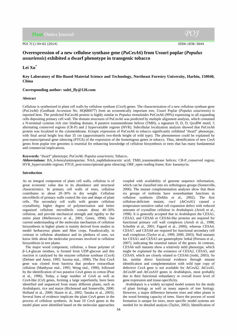
The predicted amino acid sequence of PuCesA6 contained
highly conservative features of plant cellulose synthase. Fig. 1.
shows the schematic diagram of PuCesA6 protein. A
N-terminal zinc finger domain was found to be highly
conserved in all CesA proteins known to date (Joshi, 2003).
The lacks of zinc binding domain assembled the CesA proteins
as linear terminal complexes and obstructed the cell microfibril
structures (Delmer, 1999). PuCesA6 also contained 8 putative
transmembrane helices (TMH, the first two TMH region of
PuCesA6 was towards the N-terminal of amino acid sequence,
the other six TMH regions were toward the C-terminal of
PuCesA6 sequence) (Fig. 1, Fig. 2). This phenomenon
suggested that the first two associate as the integral membrane
protein (Richmond and Somerville 2000). Holland et al. (2000)
reported many of the glycosyltransferases, including the plant
and bacterial CesA proteins predicted to be anchored in the cell
plasma membrane by transmembrane helices. The cytoplasmic
loop between the second and third TMH regions of PuCesA6
contained a conserved QxxRW motif (D, D, D, QxxRW)
sequence (Fig. 1) that was predicted to be involved in substrate
binding and catalytic activities of CesA enzymes (Vergara and
Carpita, 2001; Beeckman et al., 2002). The presence of D, D, D,
QxxRW motif in PuCesA6 suggested that the PuCesA6
associates with glycosyltransferases in catalyzing the
biosynthesis of long-chain polysaccharides (Samuga and Joshi
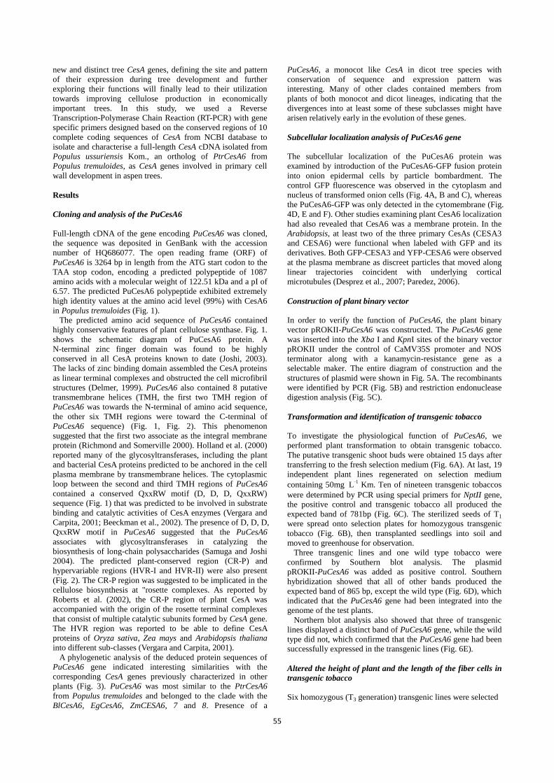
2004). The predicted plant-conserved region (CR-P) and
hypervariable regions (HVR-I and HVR-II) were also present
(Fig. 2). The CR-P region was suggested to be implicated in the
cellulose biosynthesis at "rosette complexes. As reported by
Roberts et al. (2002), the CR-P region of plant CesA was
accompanied with the origin of the rosette terminal complexes
that consist of multiple catalytic subunits formed by CesA gene.
The HVR region was reported to be able to define CesA
proteins of Oryza sativa, Zea mays and Arabidopsis thaliana
into different sub-classes (Vergara and Carpita, 2001).
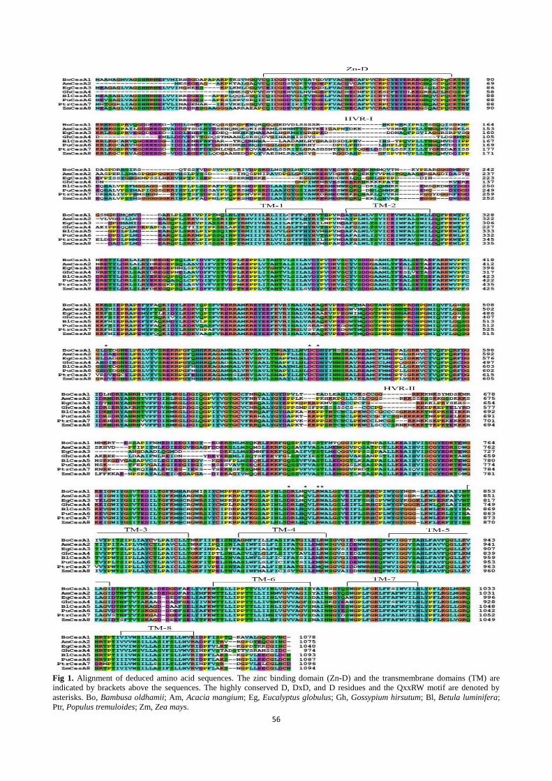
A phylogenetic analysis of the deduced protein sequences of
PuCesA6 gene indicated interesting similarities with the
corresponding CesA genes previously characterized in other
plants (Fig. 3). PuCesA6 was most similar to the PtrCesA6
from Populus tremuloides and belonged to the clade with the
BlCesA6, EgCesA6, ZmCESA6, 7 and 8. Presence of a
PuCesA6, a monocot like CesA in dicot tree species with
conservation of sequence and expression pattern was
interesting. Many of other clades contained members from
plants of both monocot and dicot lineages, indicating that the
divergences into at least some of these subclasses might have
arisen relatively early in the evolution of these genes.
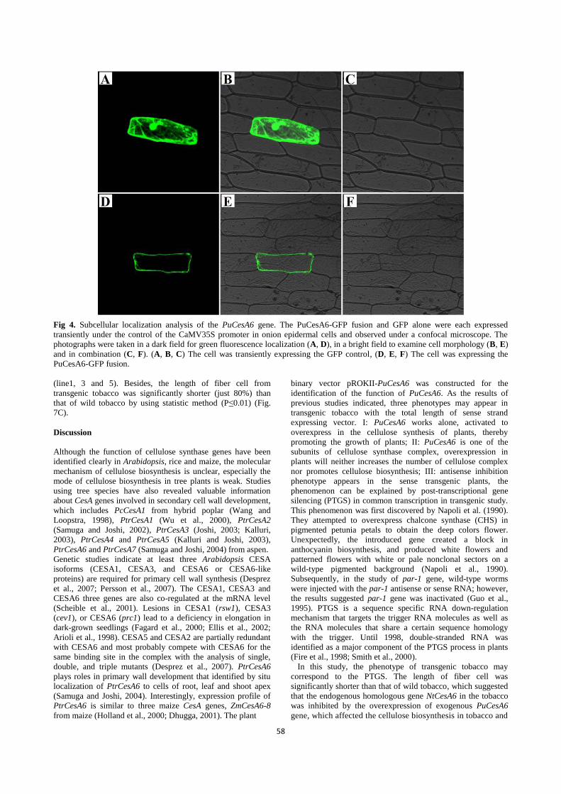
Subcellular localization analysis of PuCesA6 gene
The subcellular localization of the PuCesA6 protein was
examined by introduction of the PuCesA6-GFP fusion protein
into onion epidermal cells by particle bombardment. The
control GFP fluorescence was observed in the cytoplasm and
nucleus of transformed onion cells (Fig. 4A, B and C), whereas
the PuCesA6-GFP was only detected in the cytomembrane (Fig.
4D, E and F). Other studies examining plant CesA6 localization
had also revealed that CesA6 was a membrane protein. In the
Arabidopsis, at least two of the three primary CesAs (CESA3
and CESA6) were functional when labeled with GFP and its
derivatives. Both GFP-CESA3 and YFP-CESA6 were observed
at the plasma membrane as discreet particles that moved along
linear trajectories coincident with underlying cortical
microtubules (Desprez et al., 2007; Paredez, 2006).
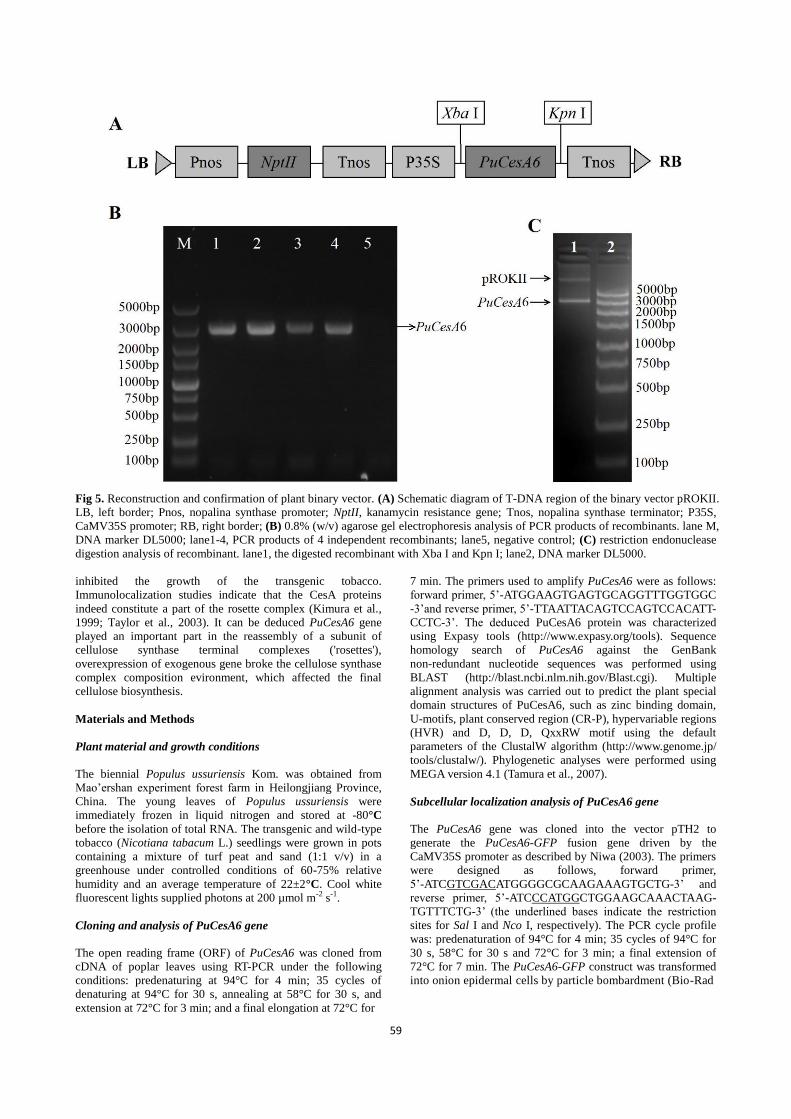
Construction of plant binary vector
In order to verify the function of PuCesA6, the plant binary
vector pROKII-PuCesA6 was constructed. The PuCesA6 gene
was inserted into the Xba I and KpnI sites of the binary vector
pROKII under the control of CaMV35S promoter and NOS
terminator along with a kanamycin-resistance gene as a
selectable maker. The entire diagram of construction and the
structures of plasmid were shown in Fig. 5A. The recombinants
were identified by PCR (Fig. 5B) and restriction endonuclease
digestion analysis (Fig. 5C).
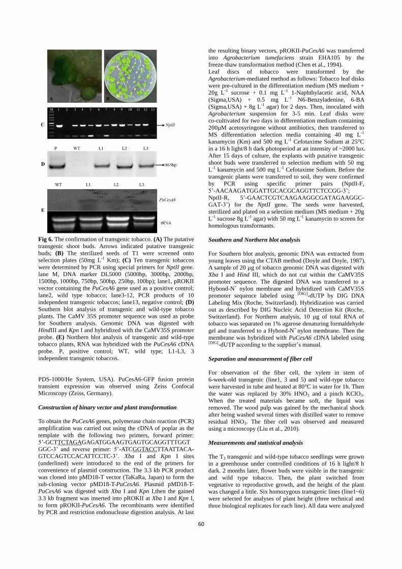
Transformation and identification of transgenic tobacco
To investigate the physiological function of PuCesA6, we
performed plant transformation to obtain transgenic tobacco.
The putative transgenic shoot buds were obtained 15 days after
transferring to the fresh selection medium (Fig. 6A). At last, 19
independent plant lines regenerated on selection medium
containing 50mg L-1
Km. Ten of nineteen transgenic tobaccos
were determined by PCR using special primers for NptII gene,
the positive control and transgenic tobacco all produced the
expected band of 781bp (Fig. 6C). The sterilized seeds of T1
were spread onto selection plates for homozygous transgenic
tobacco (Fig. 6B), then transplanted seedlings into soil and
moved to greenhouse for observation.
Three transgenic lines and one wild type tobacco were
confirmed by Southern blot analysis. The plasmid
pROKII-PuCesA6 was added as positive control. Southern
hybridization showed that all of other bands produced the
expected band of 865 bp, except the wild type (Fig. 6D), which
indicated that the PuCesA6 gene had been integrated into the
genome of the test plants.
Northern blot analysis also showed that three of transgenic
lines displayed a distinct band of PuCesA6 gene, while the wild
type did not, which confirmed that the PuCesA6 gene had been
successfully expressed in the transgenic lines (Fig. 6E).
Altered the height of plant and the length of the fiber cells in
transgenic tobacco
Six homozygous (T3 generation) transgenic lines were selected
56
Fig 1. Alignment of deduced amino acid sequences. The zinc binding domain (Zn-D) and the transmembrane domains (TM) are
indicated by brackets above the sequences. The highly conserved D, DxD, and D residues and the QxxRW motif are denoted by
asterisks. Bo, Bambusa oldhamii; Am, Acacia mangium; Eg, Eucalyptus globulus; Gh, Gossypium hirsutum; Bl, Betula luminifera;
Ptr, Populus tremuloides; Zm, Zea mays.
57
Fig 2. Domain structure of PuCesA6 protein showing various conseved regions.
Fig 3. Cladogram of the amino acid sequences of the predicted protein encoded by PuCesA6 and full-length CesA sequences from
other species. Am, Acacia mangium; At, Arabidopsis thaliana; Bl, Betula luminifera; Bn, Brassica napus; Bo, Bambusa oldhamii;
Bp, Betula platyphylla; Br, Brassica rapa; Eg, Eucalyptus globulus; Gh, Gossypium hirsutum; Hv, Hordeum vulgare; Mc,
Mesotaenium caldariorum; Na, Nicotiana alata; Pp, Physcomitrella patens subsp. Patens; Pr, Pinus radiata; Pt, Pinus taeda; Ptr,
Populus tremuloides; Rc, Ricinus communis; Sm, Selaginella moellendorffii; St, Solanum tuberosum; Zm, Zea mays; Zv, Zinnia
violacea.
for the phenotype analyses. Under long-day conditions (with a
16 h light/8 h dark cycle), all plant flowered 65 days after
sowing. All the transgenic tobacco plants showed "dwarf"
phenotype, with average aerial height of 39.5 cm, especially the
line 1 with final aerial height of 35 cm, while more than 9 wild
type plants showed average aerial height over 55 cm (Fig. 7 A).
The wild type plants were approximately 1.2- to 1.5-fold higher
than those in the plants in the control group (P≤0.01) (Fig. 7 A,
B). There was no significant variation in either flower or fruit
morphology, or fertility levels, between transgenic and wild
type plants except that the length between stem nodes of the
transgenic plants, which was shorter than that of wildtype.
HNO3 maceration was used to observe the fibers of plant
stems. Here, the length of fiber cells was measured under a
microscope. In statistical significance testing, it showed that the
length of the fiber cell had no significant differences between
transgenic individuals and wild-type individuals (P>0.05). The
length of the wild type fiber cells was 2.86mm on average,
whereas the average was 2.42 mm in transgenic fiber cells
58
Fig 4. Subcellular localization analysis of the PuCesA6 gene. The PuCesA6-GFP fusion and GFP alone were each expressed
transiently under the control of the CaMV35S promoter in onion epidermal cells and observed under a confocal microscope. The
photographs were taken in a dark field for green fluorescence localization (A, D), in a bright field to examine cell morphology (B, E)
and in combination (C, F). (A, B, C) The cell was transiently expressing the GFP control, (D, E, F) The cell was expressing the
PuCesA6-GFP fusion.
(line1, 3 and 5). Besides, the length of fiber cell from
transgenic tobacco was significantly shorter (just 80%) than
that of wild tobacco by using statistic method (P≤0.01) (Fig.
7C).
Discussion
Although the function of cellulose synthase genes have been
identified clearly in Arabidopsis, rice and maize, the molecular
mechanism of cellulose biosynthesis is unclear, especially the
mode of cellulose biosynthesis in tree plants is weak. Studies
using tree species have also revealed valuable information
about CesA genes involved in secondary cell wall development,
which includes PcCesA1 from hybrid poplar (Wang and
Loopstra, 1998), PtrCesA1 (Wu et al., 2000), PtrCesA2
(Samuga and Joshi, 2002), PtrCesA3 (Joshi, 2003; Kalluri,
2003), PtrCesA4 and PtrCesA5 (Kalluri and Joshi, 2003),
PtrCesA6 and PtrCesA7 (Samuga and Joshi, 2004) from aspen.
Genetic studies indicate at least three Arabidopsis CESA
isoforms (CESA1, CESA3, and CESA6 or CESA6-like
proteins) are required for primary cell wall synthesis (Desprez
et al., 2007; Persson et al., 2007). The CESA1, CESA3 and
CESA6 three genes are also co-regulated at the mRNA level
(Scheible et al., 2001). Lesions in CESA1 (rsw1), CESA3
(cev1), or CESA6 (prc1) lead to a deficiency in elongation in
dark-grown seedlings (Fagard et al., 2000; Ellis et al., 2002;
Arioli et al., 1998). CESA5 and CESA2 are partially redundant
with CESA6 and most probably compete with CESA6 for the
same binding site in the complex with the analysis of single,
double, and triple mutants (Desprez et al., 2007). PtrCesA6
plays roles in primary wall development that identified by situ
localization of PtrCesA6 to cells of root, leaf and shoot apex
(Samuga and Joshi, 2004). Interestingly, expression profile of
PtrCesA6 is similar to three maize CesA genes, ZmCesA6-8
from maize (Holland et al., 2000; Dhugga, 2001). The plant
binary vector pROKII-PuCesA6 was constructed for the
identification of the function of PuCesA6. As the results of
previous studies indicated, three phenotypes may appear in
transgenic tobacco with the total length of sense strand
expressing vector. I: PuCesA6 works alone, activated to
overexpress in the cellulose synthesis of plants, thereby
promoting the growth of plants; II: PuCesA6 is one of the
subunits of cellulose synthase complex, overexpression in
plants will neither increases the number of cellulose complex
nor promotes cellulose biosynthesis; III: antisense inhibition
phenotype appears in the sense transgenic plants, the
phenomenon can be explained by post-transcriptional gene
silencing (PTGS) in common transcription in transgenic study.
This phenomenon was first discovered by Napoli et al. (1990).
They attempted to overexpress chalcone synthase (CHS) in
pigmented petunia petals to obtain the deep colors flower.
Unexpectedly, the introduced gene created a block in
anthocyanin biosynthesis, and produced white flowers and
patterned flowers with white or pale nonclonal sectors on a
wild-type pigmented background (Napoli et al., 1990).
Subsequently, in the study of par-1 gene, wild-type worms
were injected with the par-1 antisense or sense RNA; however,
the results suggested par-1 gene was inactivated (Guo et al.,
1995). PTGS is a sequence specific RNA down-regulation
mechanism that targets the trigger RNA molecules as well as
the RNA molecules that share a certain sequence homology
with the trigger. Until 1998, double-stranded RNA was
identified as a major component of the PTGS process in plants
(Fire et al., 1998; Smith et al., 2000).
In this study, the phenotype of transgenic tobacco may
correspond to the PTGS. The length of fiber cell was
significantly shorter than that of wild tobacco, which suggested
that the endogenous homologous gene NtCesA6 in the tobacco
was inhibited by the overexpression of exogenous PuCesA6
gene, which affected the cellulose biosynthesis in tobacco and
59
Fig 5. Reconstruction and confirmation of plant binary vector. (A) Schematic diagram of T-DNA region of the binary vector pROKII.
LB, left border; Pnos, nopalina synthase promoter; NptII, kanamycin resistance gene; Tnos, nopalina synthase terminator; P35S,
CaMV35S promoter; RB, right border; (B) 0.8% (w/v) agarose gel electrophoresis analysis of PCR products of recombinants. lane M,
DNA marker DL5000; lane1-4, PCR products of 4 independent recombinants; lane5, negative control; (C) restriction endonuclease
digestion analysis of recombinant. lane1, the digested recombinant with Xba I and Kpn I; lane2, DNA marker DL5000.
inhibited the growth of the transgenic tobacco.
Immunolocalization studies indicate that the CesA proteins
indeed constitute a part of the rosette complex (Kimura et al.,
1999; Taylor et al., 2003). It can be deduced PuCesA6 gene
played an important part in the reassembly of a subunit of
cellulose synthase terminal complexes ('rosettes'),
overexpression of exogenous gene broke the cellulose synthase
complex composition evironment, which affected the final
cellulose biosynthesis.
Materials and Methods
Plant material and growth conditions
The biennial Populus ussuriensis Kom. was obtained from
Mao’ershan experiment forest farm in Heilongjiang Province,
China. The young leaves of Populus ussuriensis were
immediately frozen in liquid nitrogen and stored at -80°C
before the isolation of total RNA. The transgenic and wild-type
tobacco (Nicotiana tabacum L.) seedlings were grown in pots
containing a mixture of turf peat and sand (1:1 v/v) in a
greenhouse under controlled conditions of 60-75% relative
humidity and an average temperature of 22±2°C. Cool white
fluorescent lights supplied photons at 200 µmol m-2 s-1.
Cloning and analysis of PuCesA6 gene
The open reading frame (ORF) of PuCesA6 was cloned from
cDNA of poplar leaves using RT-PCR under the following
conditions: predenaturing at 94°C for 4 min; 35 cycles of
denaturing at 94°C for 30 s, annealing at 58°C for 30 s, and
extension at 72°C for 3 min; and a final elongation at 72°C for
7 min. The primers used to amplify PuCesA6 were as follows:
forward primer, 5’-ATGGAAGTGAGTGCAGGTTTGGTGGC
-3’and reverse primer, 5’-TTAATTACAGTCCAGTCCACATT-
CCTC-3’. The deduced PuCesA6 protein was characterized
using Expasy tools (http://www.expasy.org/tools). Sequence
homology search of PuCesA6 against the GenBank
non-redundant nucleotide sequences was performed using
BLAST (http://blast.ncbi.nlm.nih.gov/Blast.cgi). Multiple
alignment analysis was carried out to predict the plant special
domain structures of PuCesA6, such as zinc binding domain,
U-motifs, plant conserved region (CR-P), hypervariable regions
(HVR) and D, D, D, QxxRW motif using the default
parameters of the ClustalW algorithm (http://www.genome.jp/
tools/clustalw/). Phylogenetic analyses were performed using
MEGA version 4.1 (Tamura et al., 2007).
Subcellular localization analysis of PuCesA6 gene
The PuCesA6 gene was cloned into the vector pTH2 to
generate the PuCesA6-GFP fusion gene driven by the
CaMV35S promoter as described by Niwa (2003). The primers
were designed as follows, forward primer,
5’-ATCGTCGACATGGGGCGCAAGAAAGTGCTG-3’ and
reverse primer, 5’-ATCCCATGGCTGGAAGCAAACTAAG-
TGTTTCTG-3’ (the underlined bases indicate the restriction
sites for Sal I and Nco I, respectively). The PCR cycle profile
was: predenaturation of 94°C for 4 min; 35 cycles of 94°C for
30 s, 58°C for 30 s and 72°C for 3 min; a final extension of
72°C for 7 min. The PuCesA6-GFP construct was transformed
into onion epidermal cells by particle bombardment (Bio-Rad
60
Fig 6. The confirmation of transgenic tobacco. (A) The putative
transgenic shoot buds. Arrows indicated putative transgenic
buds; (B) The sterilized seeds of T1 were screened onto
selection plates (50mg L-1 Km); (C) Ten transgenic tobaccos
were determined by PCR using special primers for NptII gene.
lane M, DNA marker DL5000 (5000bp, 3000bp, 2000bp,
1500bp, 1000bp, 750bp, 500bp, 250bp, 100bp); lane1, pROKII
vector containing the PuCesA6 gene used as a positive control;
lane2, wild type tobacco; lane3-12, PCR products of 10
independent transgenic tobaccos; lane13, negative control; (D)
Southern blot analysis of transgenic and wild-type tobacco
plants. The CaMV 35S promoter sequence was used as probe
for Southern analysis. Genomic DNA was digested with
HindIII and Kpn I and hybridized with the CaMV35S promoter
probe. (E) Northern blot analysis of transgenic and wild-type
tobacco plants, RNA was hybridized with the PuCesA6 cDNA
probe. P, positive control; WT, wild type; L1-L3, 3
independent transgenic tobaccos.
PDS-1000/He System, USA). PuCesA6-GFP fusion protein
transient expression was observed using Zeiss Confocal
Microscopy (Zeiss, Germany).
Construction of binary vector and plant transformation
To obtain the PuCesA6 genes, polymerase chain reaction (PCR)
amplification was carried out using the cDNA of poplar as the
template with the following two primers, forward primer:
5’-GCTTCTAGAGAGATGGAAGTGAGTGCAGGTTTGGT
GGC-3’ and reverse primer: 5’-ATCGGTACCTTAATTACA-
GTCCAGTCCACATTCCTC-3’. Xba I and Kpn I sites
(underlined) were introduced to the end of the primers for
convenience of plasmid construction. The 3.3 kb PCR product
was cloned into pMD18-T vector (TaKaRa, Japan) to form the
sub-cloning vector pMD18-T-PuCesA6. Plasmid pMD18-T-
PuCesA6 was digested with Xba I and Kpn I,then the gained
3.3 kb fragment was inserted into pROKII at Xba I and Kpn I,
to form pROKII-PuCesA6. The recombinants were identified
by PCR and restriction endonuclease digestion analysis. At last
the resulting binary vectors, pROKII-PuCesA6 was transferred
into Agrobacterium tumefaciens strain EHA105 by the
freeze-thaw transformation method (Chen et al., 1994).
Leaf discs of tobacco were transformed by the
Agrobacterium-mediated method as follows: Tobacco leaf disks
were pre-cultured in the differentiation medium (MS medium +
20g L-1 sucrose + 0.1 mg L-1 1-Naphthylacetic acid, NAA
(Sigma,USA) + 0.5 mg L-1 N6-Benzyladenine, 6-BA
(Sigma,USA) + 8g L-1 agar) for 2 days. Then, inoculated with
Agrobacterium suspension for 3-5 min. Leaf disks were
co-cultivated for two days in differentiation medium containing
200µM acetosyringone without antibiotics, then transferred to
MS differentiation selection media containing 40 mg L-1
kanamycin (Km) and 500 mg L-1 Cefotaxime Sodium at 25°C
in a 16 h light/8 h dark photoperiod at an intensity of ~2000 lux.
After 15 days of culture, the explants with putative transgenic
shoot buds were transferred to selection medium with 50 mg
L-1 kanamycin and 500 mg L-1 Cefotaxime Sodium. Before the
transgenic plants were transferred to soil, they were confirmed
by PCR using specific primer pairs (NptII-F,
5’-AACAAGATGGATTGCACGCAGGTTCTCCGG-3’;
NptII-R, 5’-GAACTCGTCAAGAAGGCGATAGAAGGC-
GAT-3’) for the NptII gene. The seeds were harvested,
sterilized and plated on a selection medium (MS medium + 20g
L-1 sucrose 8g L-1 agar) with 50 mg L-1 kanamycin to screen for
homologous transformants.
Southern and Northern blot analysis
For Southern blot analysis, genomic DNA was extracted from
young leaves using the CTAB method (Doyle and Doyle, 1987).
A sample of 20 µg of tobacco genomic DNA was digested with
Xba I and Hind III, which do not cut within the CaMV35S
promoter sequence. The digested DNA was transferred to a
Hybond-N+ nylon membrane and hybridized with CaMV35S
promoter sequence labeled using [DIG]-dUTP by DIG DNA
Labeling Mix (Roche, Switzerland). Hybridization was carried
out as described by DIG Nucleic Acid Detection Kit (Roche,
Switzerland). For Northern analysis, 10 µg of total RNA of
tobacco was separated on 1% agarose denaturing formaldehyde
gel and transferred to a Hybond-N+ nylon membrane. Then the
membrane was hybridized with PuCesA6 cDNA labeled using [DIG]-dUTP according to the supplier’s manual.
Separation and measurement of fiber cell
For observation of the fiber cell, the xylem in stem of
6-week-old transgenic (line1, 3 and 5) and wild-type tobacco
were harvested in tube and heated at 80°C in water for 1h. Then
the water was replaced by 30% HNO3 and a pinch KClO3.
When the treated materials became soft, the liquid was
removed. The wood pulp was gained by the mechanical shock
after being washed several times with distilled water to remove
residual HNO3. The fiber cell was observed and measured
using a microscopy (Liu et al., 2010).
Measurements and statistical analysis
The T3 transgenic and wild-type tobacco seedlings were grown
in a greenhouse under controlled conditions of 16 h light/8 h
dark. 2 months later, flower buds were visible in the transgenic
and wild type tobacco. Then, the plant switched from
vegetative to reproductive growth, and the height of the plant
was changed a little. Six homozygous transgenic lines (line1~6)
were selected for analyses of plant height (three technical and
three biological replicates for each line). All data were analyzed
61
Fig 7. The morphological observation and measurement of wild type and transgenic tobacco. (A) The height difference between wild
type and transgenic tobacco. WT, wild type; L1, one independent transgenic tobacco; (B) Aerial height of wild type and transgenic
tobaccos; (C) The length of fiber cell from wild type and transgenic tobaccos. Each transgenic line showed significant differences
compared with wildtype by SPSS 11.5 analysis (Student's t-test, p≤0.05 and 0.01); Values are expressed as means (n=3 samples for
each test); error bars denote SD. ** p ≤0.01 for t test; * p ≤0.05 for t test. Different letters above columns indicate significance
differences (P ≤0.05) between the mean values, which were determined based on LSD test. WT, wildtype; L1-L6, independent
transgenic tobaccos.
using LSD test by SPSS 11.5, and statistical difference was
compared based on Student's t test, taking P ≤0.05 (*), P ≤0.01
(**), as significant.
Conclusions
Cellulose is of great economic value as an integral component
of plant cell walls. Cellulose synthase (CesA) represents
enzymes involved in cellulose biosynthesis. Here, we cloned
and summarized the poplar CesA6 gene (No. HQ686077) from
Populus ussuriensis. The domain structures of PuCesA6 were
predicted by multiple alignment analysis, which contained
many plant special domains. PuCesA6 protein was localized in
the cytomembrane by particle bombardment. Ectopic
expression of PuCesA6 in tobacco significantly exhibited
“dwarf” phenotype. Finally, identification of new CesA genes
from poplar tree genomes may contribute to a better
understanding of cellulose biosynthesis in trees.
Acknowledgements
This work was supported by the Fundamental Research Funds
for the Central Universities (DL12BB18) and the Programme
of Introducing Talents of Discipline to Universities of China
(B08016 )
References
Arioli T, Peng L, Betzner AS, Burn J, Wittke W, Herth W,
Camilleri C, Hofte H, Plazinski J, Birch R, Cork A ,Glover J,
Redmond J, Williamson RE (1998) Molecular analysis of
cellulose biosynthesis in Arabidopsis. Science. 279: 717-720
Beeckman T, Przemeck GK, Stamatiou G, Lau R, Terryn N,De
Rycke R, Inzé D, Berleth T (2002) Genetic complexity of
cellulose synthase a gene function in Arabidopsis
embryogenesis. Plant Physiol. 130: 1883-1893
Chen H, Nelson RS, Sherwood JL (1994) Enhanced recovery
of transformants of Agrobacterium tumifaciens after
freeze-thaw transformation and drug selection. Biotechniques.
16: 664-668, 670
Delmer DP, Amor, Y (1995) Cellulose biosynthesis. Plant Cell.
7: 987-1000
Delmer DP (1999) Cellulose biosynthesis: exciting times for a
difficult field of study. Annu Rev Plant Physiol Plant Mol
Biol. 50: 245-276
Desprez T, Juraniec M, Crowell EF, Jouy H, Pochylova Z,
Parcy F, Höfte H, Gonneau M, Vernhettes S (2007)
Organization of cellulose synthase complexes involved in
primary cell wall synthesis in Arabidopsis thaliana. Proc Natl
Acad Sci USA. 104: 15572-15577
Dhugga KS (2001) Building the wall: genes and enzyme
complexes for polysaccharide synthesis. Curr Opin Plant Biol.
4: 488-493
62
Doblin MS, Kurek I, Jacob-Wilk D, Delmer DP (2002)
Cellulose biosynthesis in plants: from genes to rosettes. Plant
Cell Physiol. 43: 1407-1420
Doyle JJ, Doyle JL (1987) A rapid DNA isolation procedure for
small quantities of fresh leaf tissue. Phytochem Bull. 19:
11-15
Fagard M, Desnos T, Desprez T, Goubet F, Refregier G,
Mouille G, McCann M, Rayon C, Vernhettes S, Höfte H
(2000) PROCUSTE1 encodes a cellulose synthase required
for normal cell elongation specificity in roots and dark-grown
hypocotyls of Arabidopsis. Plant Cell. 12: 2409-2433
Fire A, Xu S, Montgomery MK, Kostas SA, Driver SE, Mello
CC (1998) Potent and specific genetic interference by
double-stranded RNA in Caenorhabditis elegans. Nature. 391:
806-811
Green PB (1994) Connecting gene and hormone action to form
pattern organogenesis: biophysical transductions. J Exp Bot
Suppl. 45: 1775-1778
Guo S, Kemphues KJ (1995) par-1, a gene required for
establishing polarity in C. elegans embryos, encodesa
putative Ser/Thr kinase that is asymmetrically distributed.
Cell. 81: 611-620
Hazen SP, Scott-Craig JS, Walton JD (2002) Cellulose
synthase-like genes of rice. Plant Physiol. 128: 336-340
Holland D, Helentjaris T, Dhugga K, Xoconostle-Cazares B,
Delmer DP (2000) A comparative analysis of the cellulose
synthase (CesA) gene family in plants. Plant Physiol. 123:
1313-1323
Joshi CP (2003) Xylem-specific and tension stress responsive
expression of cellulose synthase genes from aspen trees. Appl
Biochem Biotechnol. 105-108: 17-26
Kimura S, Laosinchai W, Itoh T, Cui X, Linder CR, Brown
RMJr (1999) Immunogold labeling of rosette terminal
cellulose-synthesizing complexes in the vascular plant Vigna
angularis. Plant Cell. 11: 2075-2086
Liu D, Zhong T, Chang PR Li K, Wu Q (2010) Starch
composites reinforced by bamboo cellulosic crystals.
Bioresour Technol. 101: 2529-2536.
Matthysse AG, White S, Lightfoot R (1995) Gene required for
cellulose synthesis in Agrobacterium tumefaciens. J
Bacteriol. 177: 1069-1075
Mellerowicz EJ, Baucher M, Sundberg B, Boerjan W (2001)
Unravelling cell wall formation in the woody dicot stem.
Plant Mol Biol. 47: 239-274
Napoli C, Lemieux C, Jorgensen R (1990) Introduction of a
chimeric chalcone synthase gene into petunia results in
reversible co-suppression of homologous genes in trans. Plant
Cell. 2: 279-289
Niwa Y (2003) A synthetic green fluorescent protein gene for
plant biotechnology. Plant Biotechnol (Japanese journal). 20:
1-11
Paredez AR, Somerville CR, Ehrhardt DW (2006) Visualization
of cellulose synthase demonstrates functional association
with microtubules. Science. 312: 1491-1495
Pear JR, Kawagoe Y, Schreckengost WE, Delmer DP, Stalker
DM (1996) Higher plants contain homologs of the bacterial
celA genes encoding the catalytic subunit of cellulose
synthase. Proc Natl Acad Sci USA. 93: 12637-12642
Persson S, Paredez A, Carroll A, Palsdottir H, Doblin M,
Pointdexter P, Khitrov N, Auer M, Somerville CR (2007)
Genetic evidence for three unique components in primary
cell-wall cellulose synthase complexes in Arabidopsis. Proc
Natl Acad Sci USA.104: 15566-15571
Richmond TA, Somerville CR (2000) The cellulose synthase
superfamily. Plant Physiol. 124: 495-498
Roberts AW, Roberts EM, Delmer DP (2002) Cellulose
synthase (CesA) genes in the green alga Mesotaenium
caldariorum. Eukaryotic Cell. 1: 847-855
Samuga A, Joshi CP (2002) A new cellulose synthase gene
(PtrCesA2) from aspen xylem is orthologous to Arabidopsis
AtCesA7 (irx3) gene associated with secondary cell wall
synthesis. Gene. 296: 37-44
Samuga A, Joshi CP (2004) Differential expression patterns of
two new primary cell wall-related cellulose synthase cDNAs,
PtrCesA6 and PtrCesA7 from aspen trees. Gene. 334: 73-82
Saxena IM, Lin FC, Brown RM (1990) Cloning and sequencing
of the cellulose synthase catalytic subunit gene of
Acetobacter xylinum. Plant Mol Biol. 15: 673-683
Scheible WR, Eshed R, Richmond T, Delmer D, Somerville C
(2001) Modifications of cellulose synthase confer resistance
to isoxaben and thiazolidinone herbicides in Arabidopsis ixr1
mutants. Proc Natl Acad Sci USA. 98: 10079-10084
Smith NA, Singh SP, Wang MB, Stoutjesdijk PA, Green AG,
Waterhouse PM (2000) Total silencing by intron spliced
hairpin RNAs. Nature. 407: 319-320
Somerville C (2006) Cellulose synthesis in higher plants. Ann
Rev Cell Dev Biol. 22: 53-78
Tamura K, Dudley J, Ne M, Kumar S (2007) MEGA4:
molecular evolutionary genetics analysis (MEGA) software
version 4.0. Mol Biol Evol. 24: 1596-1599
Tanaka K, Murata K, Yamazaki M, Onosato K, Miyao A,
Hirochika H (2003) Three distinct rice cellulose synthase
catalytic subunit genes required for cellulose synthesis in the
secondary wall. Plant Physiol. 133: 73-83
Taylor NG, Scheible W, Cutler S, Somerville CR, Turner SR
(1999) The irregular xylem3 locus of Arabidopsis encodes a
cellulose synthase required for secondary wall synthesis.
Plant Cell. 11: 769-779
Taylor NG, Laurie S, Turner SR (2000) Multiple cellulose
synthase catalytic subunits are required for cellulose
synthesis in Arabidopsis. Plant Cell. 12: 2529-2540
Taylor G (2002) Populus: arabidopsis for forestry. Do we need
a model tree? Ann Bot. 90: 681-689
Taylor NG, Howells RM, Hutty AK, Vickers K, Turner SR
(2003) Interactions among three distinct CesA proteins
essential for cellulose synthesis. Proc Natl Acad Sci USA.
100: 1450-1455
Vergara CE, Carpita NC (2001) β-D-Glycan synthases and the
CesA gene family: lessons to be learned from the
mixed-linkage (1→3), (1→4) β-D-glucan synthase. Plant
Mol Biol Rep. 47: 145-160
Wang H, Loopstra C (1998) Cloning and characterization of a
cellulose synthase cDNA (accession no. AF081534) from
xylem of hybrid poplar (Populus tremula×Populus alba)
(PGR98-179). Plant Physiol. 118: 1101
Wong HC, Fear AL, Calhoon RD, Eichinger GH, Mayer R,
Amikam D, Benziman M, Gelfand DH, Meade JH, Emerick
AW, Bruner R, Ben-Bassat A, Tal R. (1990) Genetic
organization of the cellulose synthase operon in Acetobacter
xylinum. Proc Natl Acad Sci USA. 87: 8130-8134
Wu L, Joshi CP, Chiang VL (2000) A xylem-specific cellulose
synthase gene from aspen (Populus tremuloides) is
responsive to mechanical stress. Plant J. 22: 495-502