Embed Size (px)
Citation preview

The Plant Cell, Vol. 15, 47–62, January 2003, www.plantcell.org © 2002 American Society of Plant Biologists
Oxidative Remodeling of Chromoplast Carotenoids: Identification of the Carotenoid Dioxygenase
CsCCD
and
CsZCD
Genes Involved in Crocus SecondaryMetabolite Biogenesis
Florence Bouvier,
a
Claude Suire,
b
Jérôme Mutterer,
a
and Bilal Camara
a,1
a
Institut de Biologie Moléculaire des Plantes, Centre National de la Recherche Scientifique and Université Louis Pasteur, 67084 Strasbourg Cedex, France
b
Institut de Biochimie et Génétique Cellulaires, Centre National de la Recherche Scientifique, 33077 Bordeaux Cedex, France
The accumulation of three major carotenoid derivatives—crocetin glycosides, picrocrocin, and safranal—is in largepart responsible for the color, bitter taste, and aroma of saffron, which is obtained from the dried styles of Crocus. We
have identified and functionally characterized the Crocus zeaxanthin 7,8(7
�
,8
�
)-cleavage dioxygenase gene (
CsZCD
),which codes for a chromoplast enzyme that initiates the biogenesis of these derivatives. The Crocus carotenoid9,10(9
�
,10
�
)-cleavage dioxygenase gene (
CsCCD
) also has been cloned, and the comparison of substrate specificitiesbetween these two enzymes has shown that the CsCCD enzyme acts on a broader range of precursors.
CsZCD
ex-pression is restricted to the style branch tissues and is enhanced under dehydration stress, whereas
CsCCD
is ex-pressed constitutively in flower and leaf tissues irrespective of dehydration stress. Electron microscopy revealed thatthe accumulation of saffron metabolites is accompanied by the differentiation of amyloplasts and chromoplasts and byinteractions between chromoplasts and the vacuole. Our data suggest that a stepwise sequence exists that involvesthe oxidative cleavage of zeaxanthin in chromoplasts followed by the sequestration of modified water-soluble deriva-tives into the central vacuole.
INTRODUCTION
Carotenoids are isoprenoid pigments that have key biologi-cal functions in organisms of all major taxa. The oxidation ofthe rigid backbone of carotenoids by specific dioxygenasesleads to the formation of diverse bioactive derivatives suchas vitamin A (von Lintig and Vogt, 2000; Wyss et al., 2000),the plant hormone abscisic acid (Schwartz et al., 1997), andseveral aroma compounds (Enzell, 1985; Buttery et al.,1988; Sefton et al., 1989; Winterhalter and Rouseff, 2002)and apocarotenoid pigments (Winterhalter and Rouseff,2002). In plants, this metabolic process generally is stimu-lated during flowering (Eugster and Märki-Fischer, 1991),fruit ripening (Gross and Eckhardt, 1981; Lutz and Winterhalter,1992; Maoka et al., 2001; Fleischmann et al., 2002), indus-trial curing of tobacco (Wahlberg et al., 1977), and tea fer-mentation (Kawakami and Kobayashi, 2002).
Crocus, a cultivated sterile plant, offers a convenient
model with which to further our understanding of this caro-tenoid catabolic pathway. Upon flowering in autumn, Cro-cus displays red style branches (Figures 1A and 1B), whichonce dry constitute the spice saffron (Mathew, 1983). Saf-fron is the most expensive spice known and also is a valu-able herbal medicine (Gainer and Brumgard, 1982; Sampathuet al., 1984; Holloway and Gainer, 1988). The accumulationof three major carotenoid derivatives—crocetin glycosides,picrocrocin, and safranal—is in large part responsible for theunique color, bitter taste, and aroma of saffron (Figure 1C)(Winterhalter and Rouseff, 2002). Stereochemical configura-tions (Buchecker and Eugster, 1973) and a highly reducedcarotenoid level in saffron suggest that these secondary me-tabolites are formed by an unusual sequence that involves thecleavage of zeaxanthin (Pfander and Schurtenberger, 1982)followed by oxidative modifications and glycosylations (Fig-ure 1C). Although these compounds generate much inter-est, enzymes that initiate the reaction sequence have notbeen identified.
The limited occurrence of crocetin and related apocaro-tenoids in nature (Eugster et al., 1969; Tandon et al., 1979;Pfister et al., 1996; Liao et al., 1999) argues against their
1
To whom correspondence should be addressed. E-mail [email protected]; fax 33-03-8861-4442.Article, publication date, and citation information can be found atwww.plantcell.org/cgi/doi/10.1105/tpc.006536.
48 The Plant Cell
synthesis via an enzymatic cooxidation mechanism involv-ing ubiquitous lipoxygenases (Wu et al., 1999) or xanthineoxidases (Bosser and Belin, 1994). Two types of carotenoidcleavage dioxygenases have been identified in plants. Thefirst is the maize Vp14 (Schwartz et al., 1997), which cata-lyzes the conversion of epoxy-xanthophylls to the abscisicacid precursor xanthoxin. The second is the Arabidopsisbroad-substrate-specificity carotenoid dioxygenase (AtCCD1),which cleaves the 9,10 and 9
�
,10
�
double carbon-carbonbonds of carotenoid chromophores (Schwartz et al., 2001).However, none of these enzymes cleaves the carotenoidchromophore at the 7,8 and 7
�
,8
�
positions (Figure 1C).These two positions have the highest electron density in thecarotenoid chromophore (El-Tinay and Chichester, 1970)and represent potential targets of Crocus dioxygenase,which leads to the formation of secondary saffron metabo-lites.
Here, we report the discovery and the functional charac-terization of two Crocus carotenoid cleavage dioxygenases,CsCCD (carotenoid cleavage dioxygenase) and CsZCD (ze-axanthin cleavage dioxygenase). We demonstrate that CsCCDis a member of the broad-substrate-specificity 9,10(9
�
,10
�
)-carotenoid cleavage dioxygenase involved in the synthesisof several carotenoid-derived metabolites (Schwartz et al.,2001; Winterhalter and Rouseff, 2002), whereas, CsZCDspecifically catalyzes the synthesis of crocetin dialdehydeand hydroxy-
�
-cyclocitral from zeaxanthin. Thus, CsZCD isthe Crocus 7,8(7
�
,8
�
)-zeaxanthin cleavage dioxygenase thatinitiates the synthesis of saffron pigment and aroma (Figure1C). Further immunological and gene expression analysesrevealed that unlike CsCCD, CsZCD is expressed specifi-cally in the chromoplasts of style cells during the active pe-riod of zeaxanthin cleavage. We also investigated the ultra-structural changes in the style during the synthesis ofsecondary saffron metabolites. Our results have led to thehypothesis that in Crocus styles, the carotenoid-derivedmetabolites are sequestered in the vacuole, consistent withtheir water-soluble nature.
RESULTS
Carotenoid Analysis of Crocus Style Branches
We analyzed the carotenoid content in red Crocus stylebranches to evaluate the oxidative pathway that leads to thesynthesis of carotenoid-derived metabolites in saffron(Pfander and Schurtenberger, 1982) (Figure 1C). This analy-sis was performed by subjecting the ethanol extract pre-pared from the style branches to HPLC separation and thenevaluating the structural identities of the separated metabo-lites based on their diode-array spectra. This revealed thepresence of picrocrocin (Figures 2A and 2B) and three majorcrocetin glycosides (Figures 2C and 2D). The HPLC gradient (II)was used to profile the full spectrum of C
40
carotenoids, butthe presence of
�
-carotene and zeaxanthin, which were iso-lated previously from Crocus (Pfander and Schurtenberger,1982), was not detected. Therefore, the C
20
apocarotenoidcrocetin, which might be derived from zeaxanthin, is re-sponsible for the brilliant red color of the style branches ofCrocus.
Cloning of the Cleavage Dioxygenase Genes
CsCCD
and
CsZCD
Based on the peptide sequence of Vp14 and the relatedpeptide sequence of pepper and
Synechocystis
sp PC6803,we prepared forward and reverse oligonucleotides, whichare complementary to the conserved AHPKVDP and HD-
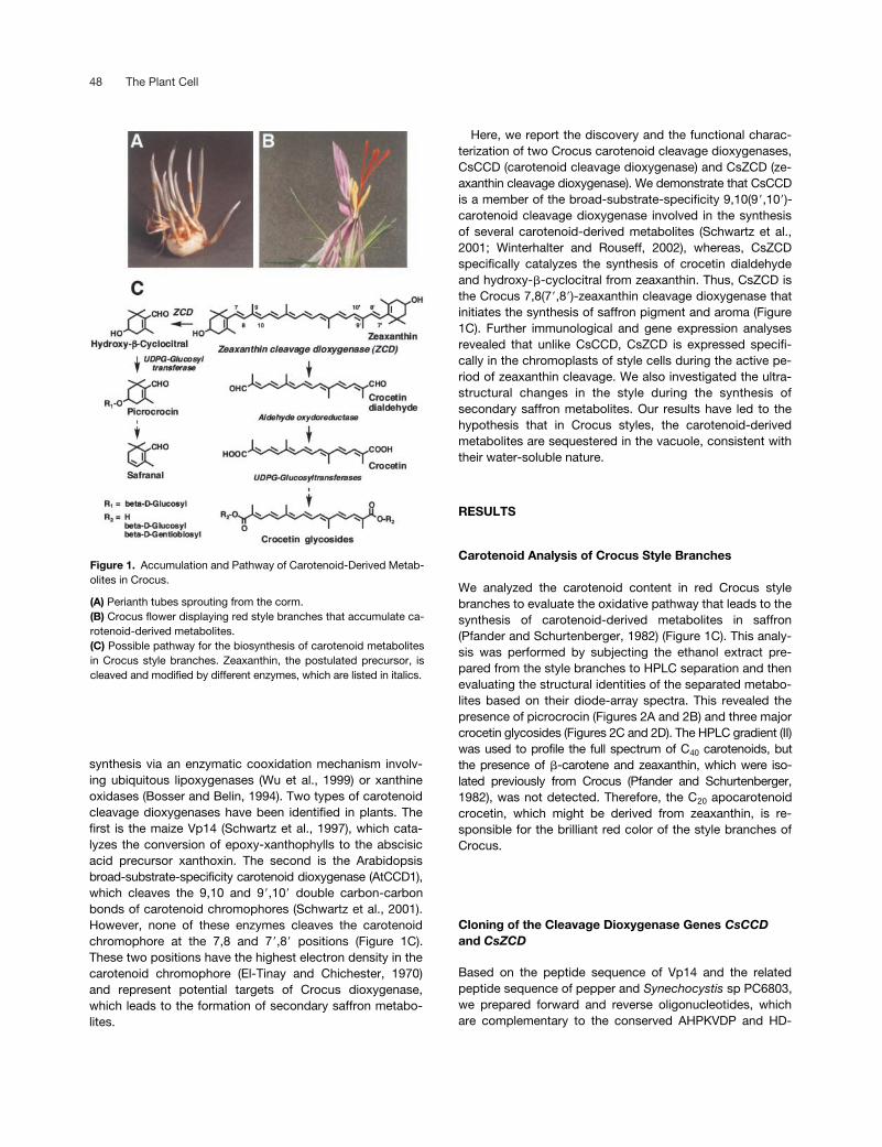
Figure 1. Accumulation and Pathway of Carotenoid-Derived Metab-olites in Crocus.
(A) Perianth tubes sprouting from the corm.(B) Crocus flower displaying red style branches that accumulate ca-rotenoid-derived metabolites.(C) Possible pathway for the biosynthesis of carotenoid metabolitesin Crocus style branches. Zeaxanthin, the postulated precursor, iscleaved and modified by different enzymes, which are listed in italics.

Chromoplast Carotenoid Cleavage Dioxygenase 49
FAITE regions, to reverse transcribe and amplify the cDNAfragments from the mRNA of Crocus style branches. Aftersequence analysis, two specific 171- and 177-bp DNAprobes were isolated and used to screen a cDNA libraryfrom Crocus style branches, as described in Methods. Thisled to the isolation of two clones (1200 and 750 bp) devoidof the 5
�
and 3
�
regions. The missing regions were obtainedby rapidly amplifying the cDNA ends. Two full-length cDNAswere identified and tentatively named
CsCCD
and
CsZCD
.The cDNA sequence of
CsCCD
is 1686 bp long with anopen reading frame coding for a protein of 546 amino acidsand has an estimated molecular mass of 62 kD, whereas
CsZCD
is 1186 bp long, codes for a protein of 369 aminoacids, and has a molecular mass of 41 kD (Figure 3). Se-quence comparison revealed that 27% of the deducedamino acid sequence of
CsCCD
is identical to that ofCsZCD (Figure 3). CsCCD is 77% identical to a partial EST
from Crocus. The same level of sequence identity is ob-served with the pepper CaCCD and Arabidopsis carotenoiddioxygenase AtCCD1 (Schwartz et al., 2001). CsZCD showsstrong identity (97%) to a partial Crocus EST and low butsignificant identity (44%) to an apple flower protein of un-known function (Watillon et al., 1998).
The deduced peptide sequences of CsCCD and CsZCDcDNAs did not reveal characteristic plastid transit peptidepatterns (Gavel and von Heijne, 1990). To determine whereCsCCD and CsZCD localize in situ, we analyzed the tissuesections of Crocus style branches by immunocytochemicalstaining and confocal microscopy using antibodies devel-oped against synthetic peptide sequences specific forCsCCD and CsZCD. Confocal microscopy using anti-CsCCD antibodies revealed a staining pattern consistentwith a cytosolic location (Figure 4A). In the case of the con-trol sample treated with the preimmune serum, no specific
Figure 2. HPLC Analysis of Carotenoid-Derived Metabolites from Crocus Style Branches.
(A) HPLC analysis of Crocus style extracts. Compounds were detected by UV-light absorbance at 220 nm. Peak 1 corresponds to picrocrocin,and peaks 2 to 4 correspond to crocetin carrying different glycosyl residues.(B) Online diode-array spectrum of picrocrocin. For peaks 2 to 4, see (D).(C) HPLC analysis of carotenoid-derived metabolites from Crocus style. Compounds were detected by visible light absorbance at 440 nm.Peaks 2 to 4 correspond to crocetin carrying different glycosyl residues.(D) Typical online diode-array spectrum of crocetin glycoside (peak 2). The profiles of peaks 3 and 4 were similar.Chromatographic separation was performed using gradient I (see Methods). mAU, milliabsorbance units.

50 The Plant Cell
staining pattern was detected (Figure 4B). On the otherhand, antibodies directed against CsZCD revealed a spe-cific labeling of plastids (Figure 4C), which was not ob-served when the tissue sections were treated with the pre-immune serum (Figure 4D).
Identification of
CsCCD
as a Carotenoid9,10(9
�
,10
�
)-Cleavage Dioxygenase
To determine whether
CsCCD
cDNA codes for a carotenoidcleavage dioxygenase, CsCCD was expressed heterolo-gously in
Escherichia coli
as a fusion protein with a molecu-lar mass of 75 kD on SDS gels (Figure 5A). Purified recombi-
nant protein from bacterial cultures induced at 20
�
C (Figure5A) was assayed for its cleavage activity using zeaxanthinas a substrate. After incubation and thin layer chromatogra-phy analysis, a new product (R
F
�
0.65) was detected thatshowed intense orange fluorescence under UV light. Theproduct also showed purple to red fluorescence when theplate was sprayed with dinitrophenylhydrazine reagent, re-vealing the presence of a carbonyl group (data not shown).No conversion was observed in the incubation medium con-taining the soluble protein isolated from
E. coli
transformedwith the empty vector. The incubation mixture was analyzedfurther by reverse-phase HPLC along with diode-array de-tection (Figures 5B to 5E). This analysis showed that theproduct was adsorbed poorly on the hydrophobic reverse
Figure 3. Comparison of the Predicted Amino Acid Sequences of Crocus Carotenoid Cleavage Dioxygenases and Related Proteins.
CsZCD and CsCCD are carotenoid cleavage dioxygenases from Crocus. Md-FS2 is an apple flower protein of unknown function (Watillon et al.,1998). CaCCD is a pepper homolog of Arabidopsis cleavage dioxygenase (Schwartz et al., 2001). Identical amino acids are indicated with blackbackgrounds.

Chromoplast Carotenoid Cleavage Dioxygenase 51
phase (Figure 5B, peak 2) and displayed a chromophorestructure (Figure 5C).
The UV-visible light spectrum in hexane, with the maxi-mum wavelength (
�
max) at 356, 374, and 396 nm (Figure5F), is characteristic of a pentaene chromophore (Vetter etal., 1971). The fine structure of the spectrum disappeared inethanol (Figure 5G) and was restored after the reduction ofcarbonyl groups with sodium borohydride (Figure 5H). Thisfinding indicated the presence of carbonyl groups conju-gated with a chromophore structure (Critchley et al., 1958).The absorbance maxima of the reduced product (
�
max at324, 340, and 359 nm) (Figure 5H) were identical to thosereported for rosafluine (Märki-Fischer and Eugster, 1988).The structure was further confirmed by mass spectrometry,which revealed a prominent molecular ion of 216 mass unitscorresponding to C
14
H
16
O
2
(Figure 6) and a fragmentationpattern consistent with the structure (Enzell et al., 1969).These characteristics indicate that the new zeaxanthin de-rivative is similar to the C
14
dialdehyde 4,9-dimethyldodeca-2,4,6,8,10-pentaene-1,12-dialdehyde (Isler et al., 1956).Collectively, these results demonstrate that
CsCCD
is a ca-rotenoid 9,10(9
�
,10
�
)-cleavage dioxygenase that catalyzesthe formation of 4,9-dimethyldodeca-2,4,6,8,10-pentaene-1,12-dialdehyde and probably hydroxydihydro-
�
-iononefrom zeaxanthin.
Identification of CsZCD as the Zeaxanthin7,8(7
�
,8
�
)-Cleavage Dioxygenase
We cloned the
CsZCD
cDNA in frame with a polyhistidineaffinity tag of the pBAD/TOPO ThioFusion vector and ex-pressed the recombinant protein in
E. coli
cultured at 20
�
C,
as described above for CsCCD. The affinity-purified fusionprotein migrated on SDS gels as a polypeptide with a mo-lecular mass of 55 kD (Figure 7A). When the purified proteinwas incubated with various carotenoid substrates, we ob-served that zeaxanthin was cleaved to yield a new nonfluo-rescent product (R
F
�
0.73) that gave a positive reactionwhen the plate was sprayed with dinitrophenylhydrazine(Figure 7B). Under our incubation conditions,
cis
- and
trans
-violaxanthin apparently were not transformed into coloredproducts (Figure 7B). Soluble proteins from
E. coli
cells car-rying the empty vector failed to transform zeaxanthin (Figure7B). Like the C
14
dialdehyde mentioned above, the CsZCDproduct behaved anomalously by reverse-phase HPLC (Fig-ures 7C to 7E). The UV-visible light spectrum revealed
�
maxat 405, 430, and 458 nm in hexane (Figure 7F). The finestructure of the UV-visible light spectrum disappeared inethanol (Figure 7G) and was restored after borohydride re-duction, as shown by the
�
max at 375, 395, and 421 nm(Figure 7H). These characteristics are consistent with an8,8
�
-diapocarotene 8,8
�
-diapocarotenone structure (Vetteret al., 1971). This structure was confirmed by mass spectrom-etry analysis, which showed a molecular ion of 296 massunits and characteristic fragments (Figure 8). Such a molec-ular mass and fragmentation pattern are consistent with thestructure of crocetin dialdehyde (C
20
H
24
O
2
) (Enzell et al.,1969; Vetter et al., 1971; Jüttner and Höflatcher, 1985).
The functional activity of CsZCD was confirmed using aplasmid-based complementation procedure. We transformed
E. coli
(strain JM109) producing zeaxanthin with the
CsZCD
cDNA. After induction and overnight culture, the bacteriaexpressing the
CsZCD
cDNA developed a red color, whichwas not observed in the control strain (Figure 9A). HPLCanalysis of the total lipid extract showed that zeaxanthin re-mained untransformed in the control strain (Figures 9B and9C), whereas in bacteria expressing the
CsZCD
cDNA, ze-axanthin was converted to crocetin dialdehyde (Figures 9Dand 9E). Further UV-visible light spectrophotometry andmass spectrometry analyses revealed that the crocetin dial-dehyde produced in vivo was identical to that obtained invitro (data not shown).
Developmental and Stress Regulation of
CsCCD
and
CsZCD
Protein gel blot analysis was performed to analyze the tis-sue-specific expression patterns of
CsCCD
and
CsZCD
us-ing polyclonal antibodies prepared against purified peptidesequences specific to each enzyme (see Methods). Whenthe protein gel blot was probed with anti-CsCCD, a single60-kD band was detected in the vegetative and floral tissuesof Crocus (Figure 10A). On the other hand, anti-CsZCD re-vealed a single 40-kD protein in the style branches only (Figure10A), in accordance with its physiological role in this tissue.
The expression of
CsCCD
and
CsZCD
was studied byRNA gel blot analysis using various Crocus tissues. Figure
Figure 4. Immunohistochemical Localization of Carotenoid Cleav-age Dioxygenases in Crocus Style Branches.
(A) and (B) Style sections incubated with anti-CsCCD antibody (A)and preimmune CsCCD serum (B). Bars � 50 �m.(C) and (D) Style sections incubated with anti-CsZCD antibody (C)and preimmune CsZCD serum (D). Bars � 5 �m.The images were obtained using confocal laser-scanning micros-copy.

52 The Plant Cell
Figure 5. Functional Analysis of Recombinant CsCCD Reaction Products.
(A) SDS-PAGE analysis of affinity-purified CsCCD. Soluble protein from bacteria grown at 20�C (lane 1) and 37�C (lane 2) and insoluble proteinfrom bacteria grown at 20�C (lane 3) and 37�C (lane 4) were examined. Affinity-purified fusion protein was loaded in lane 5 (arrowhead). MW, mo-lecular mass.(B) to (E) HPLC results (monitored at 375 nm [B] and 440 nm [D]) and online diode-array spectra of CsCCD reaction products. The separation ofzeaxanthin (peak 1) and its cleavage derivative (peak 2) and online diode-array spectra of peak 2 (C) and zeaxanthin (E) are shown. mAU, milliab-sorbance units.(F) to (H) UV-visible light spectra of the reaction product (peak 2) in hexane (F) and in ethanol before (G) and after (H) reduction with sodiumborohydride.

Chromoplast Carotenoid Cleavage Dioxygenase 53
10B shows that CsCCD was expressed constitutively at alow level in all tissues, whereas CsZCD was expressed spe-cifically in the style branches. We also hybridized the sameblot to Crocus cDNA that codes for the enzyme 1-deoxy-D-xylulose 5-phosphate reductoisomerase, which catalyzesthe first specific steps of isopentenyl diphosphate synthesisin plastids. The 1-deoxy-D-xylulose 5-phosphate reducto-isomerase gene was expressed largely in the style branchesbut to a lesser extent in petals and leaves (Figure 10B). Togain insight into the potential regulation of CsZCD, we dehy-drated the style branches and analyzed the pattern ofCsZCD transcript accumulation. The data shown in Figure10C indicate that CsZCD was regulated positively by dehy-dration. To confirm that stress by dehydration influencesCsZCD expression, we analyzed the transcript levels ofgenes reported previously to be involved in drought stress.These included the alternative oxidase (Maxwell et al.,1999), ascorbate peroxidase (Mittler and Zilinskas, 1994),calcineurin B (Kudla et al., 1999), and betaine aldehydedehydrogenase (Ishitani et al., 1995). PCR primers corre-sponding to Crocus cDNA sequences (see Methods) wereused to amplify the expected amplicon from reverse-tran-scribed RNAs of dehydrated style branches. These experi-ments were performed three times, and their results areshown in Figure 10C. The expression of CsZCD and the fourstress-regulated genes was enhanced concomitantly in de-hydrated style branches.
Plastid Differentiation during Carotenoid-Derived Metabolite Biogenesis
Because the massive synthesis of carotenoids generally isaccompanied by plastid differentiation, we analyzed by
electron microscopy the ultrastructure of Crocus stylebranch cells in relation to the intense accumulation of zea-xanthin-derived metabolites. Histochemical analysis revealedplastids that stain positively for polysaccharides (Thiéry,1967) at the very beginning of Crocus style development.These polysaccharides appear as dark deposits because oftheir starch content (Figure 11A). At the ultrastructural level,numerous amylogenic proplastids were observed duringthis developmental period (Figure 11B). Subsequently, theseproplastids differentiated into tubular chromoplasts (Figure11C) and the starch was hydrolyzed, probably for the synthe-sis of zeaxanthin, which was cleaved by CsZCD. In the laterstages of development, interactions and apparent transfer ofsecretory inclusions from chromoplasts to the vacuole wereobserved (Figures 11D to 11F). Thus, at the final stage of de-velopment, the water-soluble zeaxanthin derivatives appar-ently accumulated in the vacuole (Figure 11G).
DISCUSSION
Carotenoid Remodeling by Specific Dioxygenases
We have identified CsCCD and CsZCD, two functionally re-lated enzymes involved in the production of Crocus secondarymetabolites from zeaxanthin. CsCCD specifically cleaves the9-10 and 9�-10� double bond of carotenoid chromophores.Thus, this enzyme is homologous with Arabidopsis AtCCD1(Schwartz et al., 2001). The substrate specificity of CsCCDmay explain the presence of roseoside [(4-hydroxy-4-(3�-hydroxy-1�-butenyl)-3,5,5-trimethyl-2-cyclohexen-1-one 3�-O-�-glucopyranoside] (Straubinger et al., 1998) and 4-hydroxy-�-ionol (Winterhalter and Straubinger, 2000) in the aroma
Figure 6. Full-Scan Mass Spectrometry of the Reaction Product of CsCCD.
Electron-impact mass spectrum of the reaction product (peak 2) obtained as shown in Figure 5B. m/z, mass-to-charge ratio.

54 The Plant Cell
Figure 7. Functional Analysis of Recombinant CsZCD.
(A) SDS-PAGE analysis of affinity-purified CsZCD. Soluble protein from bacteria grown at 20�C without (lane 1) or after (lane 2) induction was ex-amined. Lane 3 was loaded with affinity-purified CsZCD fusion protein (arrowhead). MW, molecular mass.(B) Thin layer chromatography analysis of CsZCD reaction products. Lane 1, incubation using soluble proteins from E. coli harboring empty vec-tor (pBAD/TOPO ThioFusion) and zeaxanthin (Zea); lanes 2 to 4, incubations using purified recombinant CsZCD and zeaxanthin, cis-violaxanthin(cis-Viol), and trans-violaxanthin (trans-Viol). Plates were sprayed with acidic dinitrophenylhydrazine to reveal the presence of carbonyl groups.The position of the reaction product is indicated by the arrowhead. As a result of the acidic conditions, 5,6-epoxy-carotenoids were converted to5,8-epoxy-carotenoids, which appear as green to blue spots. OR refers to the origin.(C) to (E) HPLC results and online diode-array spectra of CsZCD reaction products. The separation (monitored at 440 nm [C]) of zeaxanthin(peak 1) and its cleavage derivative (peak 3) and online diode-array spectra of peak 3 (D) and zeaxanthin (E) are shown. mAU, milliabsorbanceunits.(F) to (H) UV-visible light spectra of the reaction product (peak 3) in hexane (F) and in ethanol before (G) and after (H) reduction with sodiumborohydride.

Chromoplast Carotenoid Cleavage Dioxygenase 55
compounds of saffron. On the other hand, CsZCD specifi-cally catalyzed the cleavage of zeaxanthin at the 7,8 and7�,8� positions of the chromophore and thus initiated theformation of saffron secondary metabolites according to thesequential pathway shown in Figure 1C. Very similar enzymeactivity has been described using a crude enzyme prepara-tion from Microcystis (Jüttner and Höflatcher, 1985). It alsohas been observed that crocetin dialdehyde is a guanineapocarotenoid that accumulates in the flowers of Jacquiniaangustifolia (Eugster et al., 1969) and in the roots of Coleusforskohlii (Tandon et al., 1979).
Although CsZCD is devoid of typical cleavable transitpeptide (Gavel and von Heijne, 1990), our immunologicaldata suggest that this enzyme is compartmentalized to theplastid. Precedence for this situation could be inferred fromplastid enzymes remodeling membrane acyllipids, which aresynthesized without transit peptides. These include the bar-ley allene oxide synthase (Maucher et al., 2000) and the fattyacid hydroperoxide lyases of tomato (Froehlich et al., 2001)and alfalfa (Noordermer et al., 2000). In addition, these en-zymes and CsZCD share the STVxR pattern in their N-termi-nal domains, whose role is not known at present.
The comparison between the enzymes mentioned abovethat remodel membrane acyllipids and CsZCD can be ex-tended to their mode of action. Mechanistically, the ho-molytic hydroperoxide cleavage reaction catalyzed by alleneoxide synthases and hydroperoxide lyases leads to the forma-tion of an epoxide (Song et al., 1993) or is mediated by a tran-sient epoxy carbocation (Noordermer et al., 2000). Similarly, forcarotenoids, it has been observed that during vitamin A syn-thesis, the enzymatic cleavage of the central (15-15�) doublebond of �-carotene is catalyzed by a monooxygenase en-
zyme via a transient carotene epoxide (Leuenberger et al.,2001).
Finally, comparison of the CsZCD amino acid sequencewith the sequences of plant and mammalian carotenoid di-oxygenases available in public databases revealed an over-all similarity of 17 to 27%. This information can be used todefine and narrow the catalytic domains of carotenoid dioxy-genases and to identify their active residues.
Sequestration of Water-Soluble Carotenoid Metabolites
Carotenoids are lipophilic membrane components, and theiraccumulation in plants requires specialized chromoplaststructures (Camara et al., 1995). Much less is known aboutcarotenoid cleavage derivatives when they are synthesizedin excess. In Crocus style branches, direct connectionswere observed between chromoplasts and the central vacu-ole (Figures 11D to 11F). Although convincing examples ofinteractions between plastids and vacuoles are rare, thesehave been observed in the gametophyte of the fern Pterisvittata and occasionally in a very limited group of higherplants, including the leaves of tomato (Crotty and Ledbetter,1973) and Hypoestes sanguinolenta (Vaughn and Duke,1981). Although the correlation of dynamic events and staticelectron micrographs is only tentative, it is suggested that inCrocus styles, carotenoid-derived metabolites become se-questered into vacuoles. In support of this possibility, cellsuspension cultures of Crocus glycosylate exogenous cro-cetin and accumulate the resulting glycoside in vacuoles(Dufresne et al., 1999). This can be confirmed first by thepolarity of crocetin glycosides found in the Crocus style
Figure 8. Full-Scan Mass Spectrometry of the Reaction Product of CsZCD.
Electron-impact mass spectrum of the reaction product (peak 3) obtained as shown in Figure 7C. m/z, mass-to-charge ratio.

56 The Plant Cell
tissue that conforms to the essential criterion of water solu-bility reported for vacuolar solutes (Frey-Wyssling, 1942;Matile, 1990; Jones and Vogt, 2001) and second by arbus-cular mycorrhizal fungi that induce in root vacuoles the ac-cumulation of mycorradicin (Klingner et al., 1995; Walter etal., 2000), which is a 10,10�-diapocarotene derived from theC14 dialdehyde produced by AtCCD1 (Schwartz et al., 2001)and CsCCD (this work). Sequestration of plastid-derivedmetabolites also has been observed in Stevia rebaudiana,which accumulates stevioside, a diterpenoid sweetner (Hansonand De Oliveira, 1993). The initial steps of stevioside biosyn-thesis occur in plastids and involve the cyclization of gera-nylgeranyl diphosphate into kaurene (Richman et al., 1999),which subsequently is oxidized, glycosylated, and seques-
tered in the vacuole as a water-soluble constituent (Kim etal., 1996). Finally, the sequestration of plastid-derived me-tabolites in vacuoles has been documented for chlorophyllcatabolites (Hinder et al., 1996) through an ATP-bindingcassette transporter (Lu et al., 1998).
Remodeling Carotenoids for New Functions
In plants, the biological roles of carotenoid-derived metabo-lites, except for abscisic acid, are not very well known. It hasbeen shown that 3-hydroxy-�-ionone isolated from dwarfbean shoot inhibits the growth of lettuce seedlings (Kato-Noguchi et al., 1993). The sporulation and growth of the
Figure 9. Plasmid-Based Assay of CsZCD in E. coli.
(A) Pellet of E. coli harboring the pCAR25delB plasmid, which allows zeaxanthin production alone (1) and after coexpression of CsZCD cDNA (2).(B) to (E) HPLC results (monitored at 440 nm) and online diode-array spectra of the total lipid extract from E. coli harboring pCAR25delB plasmid([B] and [C]) and coexpressing CsZCD cDNA ([D] and [E]). mAU, milliabsorbance units.

Chromoplast Carotenoid Cleavage Dioxygenase 57
pathogenic fungus Peronospora tabacina are inhibited by�-ionone (Schiltz, 1974), which protects tobacco plantsfrom being infected by the same pathogen (Salt et al., 1986).Similarly, blumenin, a glycosylated cyclohexone producedfrom root carotenoids, inhibits fungal colonization and ar-buscule formation during the initial stages of mycorrhiza de-velopment in barley and wheat (Fester et al., 1999). Finally,carotenoid-derived metabolites such as �-inone, �-ionone,and their related derivatives are important components offloral scents (Azuma et al., 2002) and could favor insect pol-linization or constitute insect lures (Donaldson et al., 1990;Flath et al., 1994; McQuate and Peck, 2001). In addition to theirpotential biological functions in plants, carotenoid-derived
metabolites form important components of the aroma pro-duced during flower and fruit development (Sefton et al.,1989; Eugster and Märki-Fischer, 1991; Winterhalter andRouseff, 2002), during tea fermentation (Sanderson et al.,1971; Sefton et al., 1989), and during tobacco curing (Enzell,
Figure 10. Molecular Analysis of CsCCD and CsZCD in DifferentTissues of Crocus and during Dehydration Stress.
(A) Protein gel blot analysis of anti-CsCCD and anti-CsZCD anti-body specificity and tissue-specific expression of CsCCD andCsZCD.(B) RNA gel blot analysis and tissue-specific expression of CsCCD,CsZCD, and 1-deoxy-D-xylulose 5-phosphate reductoisomerase(CsDXR). A Crocus rRNA probe was used to check loading andtransfer efficiency.(C) RNA gel blot analysis of CsZCD and stress-regulated gene ex-pression during dehydration. Crocus style branches were isolatedfrom the perianth tubes and dehydrated on filter paper until half oftheir weight was lost. Total RNA then was used for reverse tran-scriptase–mediated PCR experiments. Shown are the ethidium bro-mide–stained gels from quantitative reverse transcriptase–mediatedPCR experiments. The �-tubulin gene was used as a loading con-trol.
Figure 11. Amyloplast and Chromoplast Differentiation and Interac-tions between Chromoplasts and the Vacuole during the Biogenesisof Carotenoid-Derived Metabolites in Crocus Style Branches.
(A) Electron micrograph of style tissue showing electron-dense de-posits after positive staining of polysaccharides. Bar � 10 �m.(B) Details of amyloplasts showing that the electron-dense depositsare attributable to starch. Bar � 1 �m.(C) Fully differentiated reticulotubular chromoplasts. Bar � 1 �m.(D) to (F) Interaction between chromoplasts and vacuoles. Bars �1 �m.(G) Differential interference contrast microscopy observation of cellsshowing the central vacuole.

58 The Plant Cell
1985). Thus, the identification of specific enzymes that cata-lyze their formation could lead to a better appreciation oftheir biological role and to new biotechnological applica-tions.
METHODS
Plant Material and Dehydration Treatments
Fresh flowers of Crocus sativus grown under field conditions inPorchères, France, were used throughout the experiment. To studythe expression of genes induced by dehydration stress, the perianthtubes containing the floral tissues were removed from the corm anddehydrated on dry Whatman 3MM filter paper until half of theirweight was lost. Control tissues were kept on hydrated Whatman3MM filter paper for the same duration.
Chemicals
9-cis-Violaxanthin was isolated from ripe orange peel (Molnar andSzabolcs, 1980). Trans-violaxanthin and zeaxanthin were preparedas described previously (Camara, 1980; Bouvier et al., 1996).
cDNA Cloning of Carotenoid Cleavage Dioxygenase
Total RNA was isolated (Bouvier et al., 1998a) from Crocus stylebranches and poly(A) mRNAs were purified using Oligotex (Qiagen,Valencia, CA) before construction of a cDNA library in the � Uni-ZAPXR vector (Stratagene) according to the manufacturer’s instructions.Degenerated forward (5�-GC/T/C/GCAT/CCCA/T/CAAG/AG/CT/CG/C/TGAT/CCC-3�) and reverse (5�-TCA/GGTG/AATA/G/T/GCG/AAAG/ATCAT-3�) primers, corresponding to the AHPKVDP and HD-FAITE regions of the maize carotenoid dioxygenase Vp14 (Schwartzet al., 1997) and related putative carotenoid dioxygenases from pep-per and Synechocystis sp PC6803, were used to prepare a screeningprobe by reverse transcriptase–mediated PCR (Bouvier et al.,1998b). Two amplified fragments, CsCCD (171 bp) and CsZCD (177bp), were sequenced and showed a certain homology with carot-enoid cleavage dioxygenases. Subsequently, 32P-labeled CsCCD andCsZCD probes were used for plaque hybridization screening of theCrocus library according to standard procedures (Sambrook et al.,1989). Sequence analysis revealed that the longest clones corre-sponding to CsCCD (750 bp) and CsZCD (1200 bp) lacked the 5� and3� regions that were obtained by rapid amplification of cDNA ends us-ing the Gibco BRL kit, as described previously (Bouvier et al., 1998b).
Sequencing was performed using ABI 373 and ABI 3100 DNA se-quencers (Applied Biosystems, Courtaboeuf, Paris, France). Se-quence comparisons were performed through the National Centerfor Biotechnology Information using Basic Local Alignment SearchTool (BLAST) programs (Altschul et al., 1997).
Preparation of Anti-CsCCD and Anti-CsZCD Antibodies and Protein Gel Blot Analysis
Antibodies were developed in rabbits against synthetic peptides en-compassing amino acids 121 to 133 (SRYVKKTSRLKQEE) of CsCCD
and amino acids 216 to 229 (MARIDLRSGSVSRT) of CsZCD, to whichC-terminal Cys residues were added. The synthetic peptides wereconjugated to the maleimide-activated carrier protein keyhole limpethemocyanin, as described previously (Nivison and Hanson, 1987).
Total protein was extracted from Crocus tissues (Van Etten et al.,1979) and quantified (Smith et al., 1985), and aliquots (50 �g) wereused for SDS-PAGE. Proteins in the gel were transferred onto nitro-cellulose membranes, which were probed with anti-CsCCD and anti-CsZCD antibodies at a concentration of 1:2500 in Tris-buffered sa-line plus Tween 20 and processed as described previously (Suire etal., 2000).
Analysis of Carotenoid-Derived Metabolites from CrocusStyle Branches
The ethanol extract (50%) of freshly cut Crocus style branches (40 g)was used for pigment analysis. An aliquot of this extract was used forreverse-phase analysis using a Zorbax ODS column (4.6 mm 25cm; Interchim, France) and a SpectraSYSTEM (ThermoFinnigan, LesUlis, France) comprising a SCM1000 solvent degasser, a P1-1000XRgradient pump, an AS3000 autosampler, and a UV6000LP diode-array detector. Data acquisition and processing were performedusing ChromQuest version 3 software (ThermoFinnigan). The carot-enoid-derived metabolites were eluted using two gradients. GradientI (for the separation of zeaxanthin cleavage products) was as follows:0 to 10 min, 25% acetonitrile in water to 100% acetonitrile at a flowrate of 1 mL/min. Gradient II (for the separation of zeaxanthin cleav-age products and intact C40 carotenoids) was as follows: 0 to 20 min,25% acetonitrile in water to 100% acetonitrile; 20 to 25 min, a lineargradient of acetonitrile to 100% dichloromethane; and 25 to 40 min,dichloromethane, at a flow rate of 1 mL/min. The different carotenoidderivatives were identified on the basis of HPLC retention times andUV-visible light spectra (Tarantilis et al., 1995; Pfister et al., 1996).
Production and Assay of Recombinant CarotenoidCleavage Dioxygenases
Full-length CsCCD and CsZCD cDNAs were amplified by PCR usingthe forward and reverse primers 5�-ATGGGAGAAGTAGCGAAG-GAGG-3� and 5�-TACGTCGGTTTGCTGCCACTGGAG-3� for CsCCDand the forward and reverse primers 5�-ATGCAGGTGGACCCA-ACCAAGG-3� and 5�-CTGCTGTGACAGCAGCTCAGCTTC-3� forCsZCD. The resulting fragments were subcloned into pBAD/TOPOThioFusion (Invitrogen, Carlsbad, CA), which allows translation of thepolypeptide in a fusion with a 16-kD His-Patch thioredoxin under thecontrol of an arabinose-inducible promoter. Transformed Escheri-chia coli (TOP10) was grown to an OD600 of 0.5 at 20 or 37�C in aLuria-Bertani medium containing ampicillin (100 �g/mL) before beinginduced with 0.02% arabinose and overnight culture. Bacterial cellswere pelleted by centrifugation at 4�C and resuspended in 50 mMTris-HCl, pH 7.5, containing 5 mM DTT before lysis by sonication(Bouvier et al., 1998b). The lysate was centrifuged at 10,000g to col-lect the supernatant containing the fusion protein. The expressedprotein tended to form insoluble inclusion bodies when the inductiontemperature was maintained at 37�C. To overcome this problem, thetemperature was decreased to 20�C. Soluble recombinant proteinproduced under these conditions was used for affinity chromatogra-phy at 4�C using ProBond resin (Invitrogen) as described by the man-ufacturer’s protocol.
Chromoplast Carotenoid Cleavage Dioxygenase 59
The purified protein (5 �g) or soluble protein (25 �g) from E. colicells carrying the empty vector was incubated in a 200-�L assaymixture containing 5 �M FeSO4, 25 to 100 �M carotenoid substrate,0.2% octyl-�-glucoside, and 1 mM DTT buffered with 50 mM Tris-HCl, pH 7.6. After incubation at 30�C for 1 h, the reaction wasstopped by adding 200 �L of acetone followed by 300 �L of dichlo-romethane:methanol (1:1 [v/v]). The mixture was centrifuged, and thehypophase was concentrated to dryness under a stream of nitrogenbefore thin layer chromatography. Silica gel plates were preparedwith hexane:acetone (60:40 [v/v]), and the position of the reactionproducts was observed under UV-visible light before and after spray-ing the plate with a solution of acidic 2,4-dinitrophenylhydrazine todetect aldehydes and ketones (Stahl and Jork, 1965). HPLC separa-tion of CsCCD reaction products was performed on a Zorbax ODScolumn (4.6 mm 25 cm) as described above. A solvent gradientwas used for elution as follows: 0 to 5 min, 80% acetonitrile in water;5 to 10 min, 100% acetonitrile; 10 to 15 min, acetonitrile to 100%dichloromethane; and 15 to 20 min dichloromethane, at a constantflow rate of 1 mL/min. UV-visible light spectra were recorded at 374and 440 nm. For CsZCD, the same column was used, and elutionwas accomplished with the following gradient: 0 to 10 min, acetoni-trile; and 10 to 20 min, acetonitrile to 100% dichloromethane. Thecarotenoid cleavage products were identified by UV-visible spectro-photometry and mass spectrometry. Direct insertion probe massspectrometry analysis was performed with a Trio 2000 mass spec-trometer (Micromass, Manchester, UK). The ionizing voltage was 70eV, and the temperature of the ion source chamber was 200�C.
E. coli Color Complementation
CsCCD and CsZCD cDNAs were amplified by PCR. The forwardprimer 5�-ATGGGAGAAGTAGCGAAGGAGG-3� and the reverseprimer 5�-TACGTCGGTTTGCTGCCACTGGAG-3� were used forCsCCD, and the forward primer 5�-ATGCAGGTGGACCCAACC-AAGG-3� and the reverse primer 5�-CTGCTGTGACAGCAGCTC-AGCTTC-3� were used for CsZCD. The resulting fragments were li-gated into the cloning site of the pBAD TOPO TA vector (Invitrogen),and the resulting plasmid was introduced into E. coli (strain JM109)carrying the zeaxanthin biosynthetic plasmid pCAR25delB (Misawaet al., 1990). Positive colonies were selected from the Luria-Bertanimedium containing ampicillin (100 �g/mL) and chloramphenicol (50�g/mL) and grown in liquid or solid Luria-Bertani medium before be-ing induced with 0.02% arabinose. Carotenoid-derived metabolitesfrom pelleted or scraped bacteria were extracted and analyzed asdescribed for the in vitro assay.
Microscopy
For electron microscopy, the style tissues were fixed in 2.5% glutar-aldehyde and postfixed in 1% OsO4 in 0.1 M phosphate buffer at pH7.2. The tissues were dehydrated in ethanol and stained with lead ci-trate and uranyl acetate after sectioning in Spurr resin. Photographswere taken with a Philips EM 300 electron microscope (Bordeaux,France). Polysaccharide visualization was performed according to apreviously published procedure (Thiéry, 1967).
For immunohistochemistry, the tissue samples were fixed in 4%paraformaldehyde and 0.25% glutaraldehyde in PBS. The fixed tis-sues then were dehydrated in graded series of ethanol (50 to 99%)and subsequently infiltrated and embedded in paraffin. Sections of10 �m were prepared and placed on microscope slides pretreated
with 3-aminopropyltriethyoxysilane. Finally, the tissue sections weredeparaffinized in xylene and rehydrated with decreasing concentra-tions of ethanol (99 to 50%) followed by water, PBS, and PBS con-taining 0.05% Triton X-100 and 1% BSA for 1 h before blocking with5% goat serum to limit nonspecific binding. Blocked sections wereincubated at 4�C overnight with primary CsCCD or CsZCD antibod-ies diluted 1:25 in PBS containing 0.01% Triton X-100 and 1% BSA.Control tissues were incubated with the appropriate preimmune se-rum under the same conditions. After washing with the same buffer,the sections were incubated for 1 h with diluted (1:200) Alexa-Fluor488 goat anti-rabbit IgG conjugate (Molecular Probes Europe,Leiden, The Netherlands). After washing the sections with PBS, thesamples were mounted in a medium containing PBS:glycerol (1v/1v)containing 1% ascorbic acid to reduce photobleaching. Labeled tis-sue sections were examined using a Zeiss LSM510 confocal laser-scanning microscope equipped with an Axiovert 100 inverted micro-scope (Jena, Germany). Excitation and emission wavelengths were488 nm and 505 to 530 nm, respectively. The images were pro-cessed using Adobe Photoshop 5 (Adobe Systems, San Jose, CA).
Gene Expression Analysis
Total RNA was isolated from various Crocus organs and tissues us-ing the NucleoSpin RNA Plant kit (Macherey-Nagel, Duren, Ger-many). RNA gel blot analysis was performed using 32P-labeled Cro-cus probes as described previously (Bouvier et al., 1998a) using 25�g of total RNA and a Crocus ribosomal 18S rRNA probe as internalcontrols for RNA loading. The gene expression induced by dehydra-tion was analyzed three times by reverse transcriptase–mediatedPCR. Total RNA (5 �g) was isolated and used for single-strand cDNAsynthesis using Superscript II RNase H� reverse transcriptase (GibcoBRL) according to the manufacturer’s protocol. An aliquot of the re-action mixture was used as a DNA template for PCR. The forwardand reverse primers corresponding to the different Crocus genesused were as follows: CsCCD, 5�-ATGGATCTTCCTTTGTATTTCC-3�
and 5�-TTCGGATTTTATACCACGTTCACG-3�; CsZCD, 5�-GCCGTC-TTCCCCGACATCCAGATC-3� and 5�-GCCTCCGCTCTTCTTCGA-TGATCG-3�; alternative oxidase, 5�-TTGGAGGAAGCGGAGAACGAG-CGG-3� and 5�-GTGATATCCAACTGGAGCTGGATG-3�; ascorbateperoxidase, 5�-GTACCTCAAGGCGGTGGAGAAGTG-3� and 5�-CCC-TGACCGATCCTTGTGACACC-3�; betaine aldehyde dehydroge-nase, 5�-GATACGCGTTATGGCTTAGGTGG-3� and 5�-AAGCTT-GGAAGGTGGGGTATACC-3�; calcineurin B, 5�-ATGGGGTGCTTT-CAGTCCAAGTCG-3� and 5�-AAATAAAGCAAGCTGAAATTCTTCC-3�;and 1-deoxy-D-xylulose 5-phosphate reductoisomerase, 5�-GGA-TGTGCCGGTTTAAAGCCTACTG-3� and 5�-AGACCCTTTATTGAA-AAGGGTAGC-3�. Crocus �-tubulin probe amplified using the for-ward and reverse primers 5�-ATGATTTCCAACTCGACCAGTGTC-3�
and 5�-ATACTCATCACCCTCGTCACCATC-3� was used as an en-dogenous control. PCR products were separated by electrophoresison 1.5% agarose gels before detection using ethidium bromide.
Upon request, all new materials described in this article will be madeavailable in a timely manner for noncommercial research purposes.
Accession Numbers
The GenBank accession numbers for the cDNAs mentioned inthis article are as follows: CsCCD, AJ132927; CsZCD, AJ489276;

60 The Plant Cell
alternative oxidase, AJ489270; ascorbate peroxidase, AJ489279; be-taine aldehyde dehydrogenase, AJ489271; calcineurin B, AJ489272;�-tubulin, AJ489275; 1-deoxy-D-xylulose 5-phosphate reductoi-somerase, AJ489274; ribosomal 18S rRNA, AJ489273; Vp14, U95953;pepper CaCCD, Y14164; Synechocystis sp PC6803, 90917, Sll1541,Sll0572, and Slr1648; partial EST from Crocus, CAC95132; partial Cro-cus EST, CAC95133; and Md-FS2, CAB07784.
ACKNOWLEDGMENTS
We thank A. Pierronnet for providing Crocus corms and flowers, P.Hamman and A. Malek for DNA sequencing, J. Schaeffer for electronmicroscopy, P. Nkeng for mass spectrometry analysis, N. Misawaand F.X. Cunningham, Jr., for bacterial plasmids, and P. Fraser forhelpful comments on the manuscript. The Inter-Institute ConfocalMicroscopy Plate-Form was cofinanced by the Centre National de laRecherche Scientifique, the Université Louis Pasteur, the Région Al-sace, and the Association pour la Recherche sur le Cancer. Thiswork was supported by European Community Grants FAIR CT96-1633 and QLK3-CT-2000-00809.
Received August 6, 2002; accepted October 11, 2002.
REFERENCES
Altschul, S.F., Madden, T.L., Schäffer, A.A., Zhang, J., Zhang, Z.,Miller, W., and Lipman, D.J. (1997). Gapped BLAST and PSI-BLAST: A new generation of protein database search programs.Nucleic Acids Res. 25, 3389–3402.
Azuma, H., Toyota, M., Asakawa, Y., Takaso, T., and Tobe, H.(2002). Floral scent chemistry of mangrove plants. J. Plant Res.115, 47–53.
Bosser, A., and Belin, J.M. (1994). Synthesis of �-ionone in analdehyde/xanthine oxidase/�-carotene system involving free radi-cal formation. Biotechnol. Prog. 10, 129–133.
Bouvier, F., Backhaus, R.A., and Camara, B. (1998a). Inductionand control of chromoplast-specific carotenoid genes by oxida-tive stress. J. Biol. Chem. 273, 30651–30659.
Bouvier, F., d’Harlingue, A., Hugueney, P., Marin, E., Marion-Poll,A., and Camara, B. (1996). Xanthophyll biosynthesis: Cloning,expression, functional reconstitution, and regulation of �-cyclo-hexenyl carotenoid epoxidase from pepper (Capsicum annuum).J. Biol. Chem. 271, 28861–28867.
Bouvier, F., d’Harlingue, A., Suire, C., Backhaus, R.A., andCamara, B. (1998b). Dedicated roles of plastid transketolasesduring the early onset of isoprenoid biogenesis in pepper fruits.Plant Physiol. 117, 1423–1431.
Buchecker, R., and Eugster, C.H. (1973). Absolute Konfigurationvon Picrocrocin. Helv. Chim. Acta 56, 1121–1124.
Buttery, R.G., Teranishi, R., Ling, L.C., Flath, R.A., and Stern, D.J.(1988). Quantitative studies on origins of fresh tomato aroma vola-tiles. J. Agric. Food Chem. 36, 1247–1250.
Camara, B. (1980). Biosynthesis of keto-carotenoids in Capsicumannuum fruits. FEBS Lett. 118, 315–318.
Camara, B., Hugueney, P., Bouvier, F., Kuntz, M., and Monéger,
R. (1995). Biochemistry and molecular biology of chromoplastdevelopment. Int. Rev. Cytol. 163, 175–247.
Critchley, J.P., Friend, J., and Swain, T. (1958). A micro-methodfor differentiating between conjugated aldehydes and ketones.Chem. Ind. 596–597.
Crotty, W.J., and Ledbetter, M.C. (1973). Membrane continuitiesinvolving chloroplast and other organelles in plant cells. Science182, 839–841.
Donaldson, J.M.I., McGovern, T.P., and Ladd, T.L.J. (1990). Floralattractants for the Cetoniinae and Rutelinae (Coleoptera: Scara-baeidae). J. Econ. Entomol. 83, 1298–1305.
Dufresne, C., Cormier, F., Dorion, S., Higgli, U.A., Pfister, S., andPfander, H. (1999). Glycosylation of encapsulated crocetin by aCrocus sativus L. cell culture. Enzyme Microb. Technol. 24, 453–462.
El-Tinay, A.H., and Chichester, C.O. (1970). Oxidation of �-caro-tene: Site of initial attack. J. Org. Chem. 35, 2290–2293.
Enzell, C. (1985). Biodegradation of carotenoids: An important routeto aroma compounds. Pure Appl. Chem. 57, 693–700.
Enzell, C.R., Francis, G.W., and Laaen-Jensen, S. (1969). Massspectrometric studies of carotenoids. 2. A survey of fragmentationreactions. Acta Chem. Scand. 23, 727–750.
Eugster, C.H., Hürlimann, H., and Leuenberger, H.J. (1969). Cro-cetindialdehyd und Crocetinhalbaldehyd als Blütenfarbstoffe vonJacquinia angustifolia. Helv. Chim. Acta 52, 89–90.
Eugster, C.H., and Märki-Fischer, E. (1991). The chemistry of rosepigments. Angew. Chem. Int. Ed. Engl. 30, 654–672.
Fester, T., Maier, W., and Strack, D. (1999). Accumulation of sec-ondary compounds in barley and wheat roots in response to inoc-ulation with arbuscular mycorrhizal fungus and co-inoculationwith rhizosphere bacteria. Mycorrhiza 8, 241–246.
Flath, R.A., Cunningham, R.T., Liquido, N.J., and McGovern, T.P.(1994). Alpha-ionol as attractant for trapping Batrocera latifrons(Diptera: Tephritidae). J. Econ. Entomol. 87, 1470–1476.
Fleischmann, P., Studer, K., and Winterhalter, P. (2002). Partialpurification and kinetic characterization of a carotenoid cleavageenzyme from quince fruit (Cydonia oblonga). J. Agric. Food Chem.50, 1677–1680.
Frey-Wyssling, A. (1942). Zur Physiologie der pflanzlichen Gluko-side. Naturwissenchaften 30, 500–503.
Froehlich, J.E., Itoh, A., and Howe, G.A. (2001). Tomato alleneoxide synthase and fatty acid hydroperoxide lyase, two cyto-chrome P450s involved in oxylipin metabolism, are targeted todifferent membranes of chloroplast envelope. Plant Physiol. 125,306–317.
Gainer, J.L., and Brumgard, F.B. (1982). Using excess volume ofmixing to correlate diffusivities in liquids. Chem. Eng. Commun.15, 323–329.
Gavel, Y., and von Heijne, G. (1990). A conserved cleavage-sitemotif in chloroplast transit peptides. FEBS Lett. 261, 455–458.
Gross, J., and Eckhardt, G. (1981). Structures of persicaxanthin,persicachrome and other apocarotenoids of various fruits. Phy-tochemistry 20, 2267–2269.
Hanson, J.R., and De Oliveira, B.H. (1993). Stevioside and relatedsweet diterpenoid glycosides. Nat. Prod. Rep. 10, 301–309.
Hinder, B., Schellenberg, M., Rodoni, S., Ginsburg, S., Vogt, E.,Martinoia, E., Matile, P., and Hörtensteiner, S. (1996). Howplants dispose of chlorophyll catabolites. J. Biol. Chem. 271,27233–27236.
Holloway, G.M., and Gainer, J.L. (1988). The carotenoid crocetinenhances pulmonary oxygenation. J. Appl. Physiol. 65, 683–686.
Ishitani, M., Nakamura, T., Han, S.Y., and Takabe, T. (1995).

Chromoplast Carotenoid Cleavage Dioxygenase 61
Expression of the betaine aldehyde dehydrogenase gene in barleyin response to osmotic stress and abscisic acid. Plant Mol. Biol.27, 307–315.
Isler, O., Gutmann, H., Lindlar, H., Montavon, M., Rüegg, R.,Ryser, G., and Zeller, P. (1956). Synthesen in der Carotinoid-Reihe. 6. Synthese von crocetindialdhyd und lycopin. Helv. Chim.Acta 39, 463–473.
Jones, P., and Vogt, T. (2001). Glycosyltransferases in secondaryplant metabolism: Tranquilizers and stimulant controllers. Planta213, 164–174.
Jüttner, F., and Höflatcher, B. (1985). Evidence of �-carotene7,8(7�,8�) oxygenase (�-cyclocitral, crocetindial generating) inMicrocystis. Arch. Microbiol. 141, 337–343.
Kato-Noguchi, H., Kosemura, S., Yamamura, S., and Hasegawa,K. (1993). A growth inhibitor, R-(�)-3-hydroxy-�-ionone, fromlight-grown shoots of a dwarf cultivar of Phaseolus vulgaris. Phy-tochemistry 33, 553–555.
Kawakami, M., and Kobayashi, A. (2002). Carotenoid-derivedaroma compounds in tea. In Carotenoid-Derived Aroma Com-pounds, P. Winterhalter and R.L. Rouseff, eds (Washington, DC:American Chemical Society), pp. 145–159.
Kim, K.K., Sawa, Y., and Shibata, H. (1996). Hydroxylation of ent-kaurenoic acid to steviol in Stevia rebaudiana Bertoni: Purificationand partial characterization of the enzyme. Arch. Biochem. Bio-phys. 332, 223–230.
Klingner, A., Bothe, H., Wray, V., and Marner, F.J. (1995). Identifi-cation of a yellow pigment formed in maize roots upon mycor-rhizal colonization. Phytochemistry 38, 53–55.
Kudla, J., Xu, Q., Harter, K., Gruissem, W., and Luan, S. (1999).Genes for calcineurin B-like proteins in Arabidopsis are differen-tially regulated by stress signals. Proc. Natl. Acad. Sci. USA 96,4718–4723.
Leuenberger, M.G., Engeloch-Jarret, C., and Woggon, W.D.(2001). The reaction mechanism of the enzyme-catalyzed centralcleavage of �-carotene to retinal. Angew. Chem. Int. Ed. 40,2614–2617.
Liao, Y.H., Houghton, P.J., and Hoult, J.R.S. (1999). Novel andknown constituents from Buddleja species and their activityagainst leukocyte eicosanoid generation. J. Nat. Prod. 62, 1241–1245.
Lu, Y.P., Li, Z.S., Drozdowicz, Y.M., Hörtensteiner, S., Martinoia,E., and Rea, P.A. (1998). AtMRP2, an Arabidopsis ATP bindingcassette transporter able to transport glutathione S-conjugatesand chlorophyll catabolites: Functional comparisons with Atmrp1.Plant Cell 10, 267–282.
Lutz, A., and Winterhalter, P. (1992). Bio-oxidative cleavage of car-otenoids: Important route to physiological active plant constitu-ents. Tetrahedron Lett. 33, 5169–5172.
Maoka, T., Fujiwara, Y., Hashimoto, K., and Akimoto, N. (2001).Isolation of a series of apocarotenoids from the fruits of the redpaprika Capsicum annuum L. J. Agric. Food Chem. 49, 1601–1606.
Märki-Fischer, E., and Eugster, C.H. (1988). Rosafluin, ein neuesDiapocarotindiol aus Rosenblüten. Helv. Chim. Acta 71, 1491–1497.
Mathew, B. (1983). The Crocus. (Portland, OR: Timber Press).Matile, P. (1990). The toxic compartment of plant cell. In Progress in
Plant Cellular and Molecular Biology, H.J.J. Nijkamp, L.H.W. VanDer Plas, and U.U.V. Van Aartrijk, eds (Dordrecht, The Nether-lands: Kluwer Academic Publishers), pp. 557–566.
Maucher, H., Hause, B., Feussner, I., Ziegler, J., and Wasternack,
C. (2000). Allene oxide synthases of barley (Hordeum vulgare cv.Salome): Tissue specific regulation in seedling development. PlantJ. 21, 199–213.
Maxwell, D.P., Wang, Y., and McIntosh, L. (1999). The alternativeoxidase lowers mitochondrial reactive oxygen production in plantcells. Proc. Natl. Acad. Sci. USA 96, 8271–8276.
McQuate, G.T., and Peck, S.L. (2001). Enhancement of attrac-tion of alpha-ionol to male Bactrocera latifrons (Diptera:Tephritidae) by addition of a synergist, cade oil. J. Econ. Ento-mol. 94, 39–46.
Misawa, N., Nakagawa, M., Kobayashi, K., Yamano, S., Izawa,K., Nakamura, K., and Harashima, K. (1990). Elucidation of theErwinia uredovora carotenoid biosynthetic pathway by functionalanalysis of gene products expressed in Escherichia coli. J. Bacte-riol. 172, 6704–6712.
Mittler, R., and Zilinskas, B.A. (1994). Regulation of pea cytosolicascorbate peroxidase and other antioxidant enzymes during theprogression of drought stress and following recovery fromdrought. Plant J. 5, 397–405.
Molnar, P., and Szabolcs, J. (1980). �-Citraurin epoxide, a new caro-tenoid from Valencia orange peel. Phytochemistry 19, 633–637.
Nivison, H.T., and Hanson, M.R. (1987). Production and purificationof synthetic peptide antibodies. Plant Mol. Biol. Rep. 5, 295–309.
Noordermer, M.A., van Dijken, A.J.H., Smeekens, S.C.M.,Veldink, G.A., and Vliegenthart, J.F.G. (2000). Characterizationof three cloned and expressed 13-hydroperoxide lyase isoen-zymes from alfalfa with unusual N-terminal sequences and differ-ent enzyme kinetics. Eur. J. Biochem. 267, 2473–2482.
Pfander, H., and Schurtenberger, H. (1982). Biosynthesis of C20-carotenoids in Crocus sativus. Phytochemistry 21, 1039–1042.
Pfister, S., Meyer, P., Steck, A., and Pfander, H. (1996). Isolationand structure elucidation of carotenoid-glycosyl esters in Garde-nia fruits (Gardenia jasminoides Ellis) and saffron (Crocus sativusLinne). J. Agric. Food Chem. 44, 2612–2615.
Richman, A.S., Gijzen, M., Starratt, A.N., Yang, Z., and Brandle,J.E. (1999). Diterpene synthesis in Stevia rebaudiana: Recruitmentand up-regulation of key enzymes from the gibberellin biosyn-thetic pathway. Plant J. 19, 411–421.
Salt, S.D., Tuzun, S., and Kuc, J. (1986). Effect of �-ionone andabscisic acid on the growth of tobacco and resistance to bluemold: Mimicry of effects of stem infection by Peronospora taba-cina Adam. Physiol. Mol. Plant Pathol. 28, 287–297.
Sambrook, J., Fritsch, E.F., and Maniatis, T. (1989). MolecularCloning: A Laboratory Manual. (Cold Spring Harbor, NY: ColdSpring Harbor Laboratory Press).
Sampathu, S.R., Shivashankar, S., and Lewis, Y.S. (1984). Saffron(Crocus sativus Linne): Cultivation, processing, chemistry andstandardization. Crit. Rev. Food Sci. Nutr. 20, 123–157.
Sanderson, G.W., Co, H., and Gonzalez, J.G. (1971). Biochemistryof tea fermentation: The role of carotenes in black tea aroma for-mation. J. Agric. Food Chem. 36, 231–236.
Schiltz, P. (1974). Action inhibitrice de la �-ionone au cours dudéveloppement de Peronospora tabacina. Ann. Tabac 11, 207–216.
Schwartz, S.H., Qin, X., and Zeevaart, J.A.D. (2001). Characteriza-tion of a novel carotenoid cleavage dioxygenase. J. Biol. Chem.276, 25208–25211.
Schwartz, S.H., Tan, B.C., Gage, D.A., Zeevaart, J.A.D., andMcCarty, D.R. (1997). Specific oxidative cleavage of carotenoidsby VP14 of maize. Science 276, 1872–1874.
Sefton, M.A., Skouroumounis, G.K., Massy-Westropp, R.A., andWilliams, P.J. (1989). Norisoprenoids in Vitis vinifera white wine

62 The Plant Cell
grapes and the identification of a precursor of damascenone inthese fruits. Aust. J. Chem. 42, 2071–2084.
Smith, P.K., Krohn, R.I., Hermanson, G.T., Mallia, A.K., Gartner,F.H., Provenzano, M.D., Fujimoto, E.K., Goeke, N.M., Olson,B.J., and Klenk, D.C. (1985). Measurement of protein using bicin-choninic acid. Anal. Biochem. 150, 76–85.
Song, W.C., Baertschi, S.W., Boeglin, W.E., Harris, T.M., andBrash, A.R. (1993). Formation of epoxyalcohols by a purifiedallene oxide synthase: Implications for the mechanism of alleneoxide synthesis. J. Biol. Chem. 268, 6293–6298.
Stahl, E., and Jork, H. (1965). Terpene derivatives, essentials oils,balsams, and resins. In Thin-Layer Chromatography: A LaboratoryHandbook, E. Stahl, ed (Berlin: Springer-Verlag), pp. 186–210.
Straubinger, M., Bau, B., Eckstein, S., Fink, M., and Winterhalter,P. (1998). Identification of novel glycosidic aroma precursors insaffron (Crocus sativus L.). J. Agric. Food Chem. 46, 3238–3242.
Suire, C., Bouvier, F., Backhaus, R.A., Bégu, D., Bonneu, M., andCamara, B. (2000). Cellular localization of isoprenoid biosyntheticenzymes in Marchantia polymorpha: Uncovering a new role of oilbodies. Plant Physiol. 124, 971–978.
Tandon, J.S., Katti, S.B., Rüedi, P., and Eugster, C.H. (1979). Cro-cetin-dialdehyde from Coleus forskolii Briq., Labiatae. Helv. Chim.Acta 274, 2706–2707.
Tarantilis, P.A., Tsoupras, G., and Polissiou, M.G. (1995). Deter-mination of saffron (Crocus sativus L.) components in crude plantextract using high-performance liquid chromatography-UV-visiblephotodiode-array detection-mass spectrometry. J. Chromatogr.699, 107–118.
Thiéry, J.P. (1967). Mise en évidence des polysaccharides sur descoupes fines en microscopie électronique. J. Microsc. 6, 987–1018.
Van Etten, J.L., Freer, S.N., and McCune, B.K. (1979). Presence ofa major (storage?) protein in dormant spores of the fungus Botry-odiplodia theobromae. J. Bacteriol. 138, 650–652.
Vaughn, K.C., and Duke, S.O. (1981). Evaginations from the plastidenvelope: A method for transfer of substances from plastid tovacuole. Cytobios 32, 89–95.
Vetter, W., Englert, G., Rigassi, N., and Schwieter, U. (1971).Spectroscopic methods. In Carotenoids, O. Isler, ed (Basel, Swit-zerland: Birkhäuser Verlag), pp. 189–266.
von Lintig, J., and Vogt, K. (2000). Filling a gap in vitamin Aresearch: Molecular identification of an enzyme cleaving �-caro-tene to retinal. J. Biol. Chem. 275, 11915–11920.
Wahlberg, I., Karlsson, K., Austin, D.J., Junker, N., Roeraade, J.,and Enzell, C.R. (1977). Effects of flue-curing and ageing on thevolatile, neutral and acidic constituents of Virginia tobacco. Phy-tochemistry 16, 1217–1231.
Walter, M.H., Fester, T., and Strack, D. (2000). Arbuscular mycor-rhizal fungi induce the non-mevalonate methylerythritol phos-phate pathway of isoprenoid biosynthesis correlated with accu-mulation of the “yellow pigment” and other apocarotenoids. PlantJ. 21, 571–578.
Watillon, B., Kettmann, R., Arredouani, A., Hecquet, J.F., Boxux,P., and Burny, A. (1998). Apple messenger RNAs related to bac-terial lignostilbene dioxygenase and plant SAUR genes are prefer-entially expressed in flowers. Plant Mol. Biol. 36, 909–915.
Winterhalter, P., and Rouseff, R.L. (2002). Carotenoid-DerivedAroma Compounds. (Washington, DC: American Chemical Society).
Winterhalter, P., and Straubinger, M. (2000). Saffron: Renewedinterest in an ancient spice. Food Rev. Int. 16, 39–59.
Wu, Z., Robinson, D.S., Hughes, R.K., Casey, R., Hardy, D., andWest, S.I. (1999). Co-oxidation of �-carotene catalyzed by soy-bean and recombinant pea lipoxygenase. J. Agric. Food Chem.47, 4899–4906.
Wyss, A., Wirtz, G., Woggon, W.D., Brugger, R., Wyss, M.,Friedlein, A., Bachmann, H., and Hunziker, W. (2000). Cloningand expression of �,�-carotene 15,15�-dioxygenase. Biochem.Biophys. Res. Commun. 271, 334–336.

DOI 10.1105/tpc.006536; originally published online December 19, 2002; 2003;15;47-62Plant Cell
Florence Bouvier, Claude Suire, Jérôme Mutterer and Bilal CamaraCsCCD and CsZCD Genes Involved in Crocus Secondary Metabolite Biogenesis
Oxidative Remodeling of Chromoplast Carotenoids: Identification of the Carotenoid Dioxygenase
This information is current as of February 10, 2020
References /content/15/1/47.full.html#ref-list-1
This article cites 77 articles, 16 of which can be accessed free at:
Permissions https://www.copyright.com/ccc/openurl.do?sid=pd_hw1532298X&issn=1532298X&WT.mc_id=pd_hw1532298X
eTOCs http://www.plantcell.org/cgi/alerts/ctmain
Sign up for eTOCs at:
CiteTrack Alerts http://www.plantcell.org/cgi/alerts/ctmain
Sign up for CiteTrack Alerts at:
Subscription Information http://www.aspb.org/publications/subscriptions.cfm
is available at:Plant Physiology and The Plant CellSubscription Information for
ADVANCING THE SCIENCE OF PLANT BIOLOGY © American Society of Plant Biologists


![A Tetratricopeptide Repeat Protein Regulates …A Tetratricopeptide Repeat Protein Regulates Carotenoid Biosynthesis and Chromoplast Development in Monkeyflowers (Mimulus)[OPEN] Lauren](https://img.pdfslide.net/doc/110x75/5fa3b98e890f7a6e4a7ca9d6/a-tetratricopeptide-repeat-protein-regulates-a-tetratricopeptide-repeat-protein.jpg)