Embed Size (px)
Citation preview
REVIEW Open Access
Oxidative stress in prostate hyperplasia andcarcinogenesisUdensi K. Udensi and Paul B. Tchounwou*
Abstract
Prostatic hyperplasia (PH) is a common urologic disease that affects mostly elderly men. PH can be classified as benignprostatic hyperplasia (BPH), or prostate cancer (PCa) based on its severity. Oxidative stress (OS) is known to influencethe activities of inflammatory mediators and other cellular processes involved in the initiation, promotion andprogression of human neoplasms including prostate cancer. Scientific evidence also suggests that micronutrientsupplementation may restore the antioxidant status and hence improve the clinical outcomes for patients with BPHand PCa. This review highlights the recent studies on prostate hyperplasia and carcinogenesis, and examines the roleof OS on the molecular pathology of prostate cancer progression and treatment.
Keywords: Prostate cancer, Oxidative stress, Antioxidants, Cancer treatment
Abbreviations: 3αATA (8,24), 3α-acetyloxy-tir-8,24-dien-21-oic acid; 5-ARIs, 5α-Reductase inhibitors; 5-ARIs, 5α-reductaseinhibitors; 8-EPI, 8-isoprostanes; 8-OHdG, 8-hydroxy-2'-deoxyguanosine; ADT, Androgen deprivation therapy; Akt, Alphaserine/threonine-protein kinase; AP1, Activator protein 1; AR, Androgen receptor; BHP, Benign prostatic hyperplasia;BRACA1, Breast cancer 1 gene; CDH13, Cadherin13; CML, Carboxy methyl lysine; COX2, Cyclooxygenase 2; CPNE2,Calcium-dependent membrane-binding protein; CPPS, Chronic pelvic pain syndrome; CRPC, Castration-resistantprostate cancer; CTA, Cancer/testis antigen; CXCR4, Chemokine receptor type 4; DNA, Deoxyribonucleic acid; DRE,Digital rectal examination; ELAC2, ElaC ribonuclease Z 2; ER, Estrogen receptor; FM, Fat mass; GLO1, Glyoxalase 1; GPx,Glutathione peroxidase; GST, Glutathione S-transferase; HFD, High-fat diet; HGIPN, High-grade intraepithelial neoplasia;HIC1, Hypermethylated in cancer 1 gene; HIF1A, Hypoxia inducible factor 1, alpha subunit; HMBA, 2-hydroxy-4-methoxybenzoic acid; HMOX1, Heme oxygenase 1; HO-1, Heme-oxygenase-1; HPC, Hereditary prostate cancer; IL6, Interleukin 6;iNOS, Inducible nitric oxide synthase; KLK3, Kallikrein-related peptidase 3; LOH, Loss of heterozygosity; LSMK2, Lemurtyrosine kinase 2; LUTS, Lower urinary tract symptoms; MDA, Malondialdehyde; MnSOD, Manganese superoxidedismutase; MSMB, Microseminoprotein, beta; mTOR, Mammalian target of rapamycin; NADPH, Nicotinamide adeninedinucleotide phosphate; NF-kB, Nuclear factor kappa B; NIH, National Institutes of Health; NIK, Nuclear factor kappa Binducing kinase; NOX, NADPH oxidase; Nrf2, Nuclear factor erythroid 2–related factor 2; OS, Oxidative stress; PAGE4,Prostate-associated gene 4; PCa, Prostate cancer; PEN, Phosphatase and tensin homolog; PH, Prostatic hyperplasia; PIA,Proliferative inflammatory atrophy; PIN, Prostate intraepithelial neoplasia; PKM2, Pyruvate kinase M2; PPARγ, Peroxisomeproliferator-activated receptor gamma; PSCA, Prostate stem cell antigen; PTGS, Prostaglandin-endoperoxide synthase 2;Rb, Retinoblastoma; RNASeq, Ribonucleic acid sequencing; RNS, Reactive nitrogen species; ROS, Reactive oxygenspecies; SDS 1, Stromal cell-derived factor 1; SNP, Single nucleotide polymorphism; SOD, Superoxide dismutase;SRD4A2, 3-oxo-5-alpha-steroid 4-dehydrogenase 2; STAMP2, Six transmembrane protein of prostate 2; STAT3, Signaltransducer and activator of transcription 3; TBARS, Thiobarbituric acid reactive substances; TNF, Tumor necrotic factor;TRAMP, Transgenic adenocarcinoma of the mouse prostate; Trx1, Thioredoxin 1; TTG, Total thiol groups; VHL, VonHippel–Lindau syndrome; WC, Waist circumference
* Correspondence: [email protected]/NIMHD RCMI Center for Environmental Health, College of Science,Engineering and Technology, Jackson State University, Jackson, MS 39217,USA
© 2016 The Author(s). Open Access This article is distributed under the terms of the Creative Commons Attribution 4.0International License (http://creativecommons.org/licenses/by/4.0/), which permits unrestricted use, distribution, andreproduction in any medium, provided you give appropriate credit to the original author(s) and the source, provide a link tothe Creative Commons license, and indicate if changes were made. The Creative Commons Public Domain Dedication waiver(http://creativecommons.org/publicdomain/zero/1.0/) applies to the data made available in this article, unless otherwise stated.
Udensi and Tchounwou Journal of Experimental & ClinicalCancer Research (2016) 35:139 DOI 10.1186/s13046-016-0418-8
BackgroundProstate cancer is the most common non-skin cancer af-fecting men and about 3 million men in the UnitedStates (U.S.) are currently living with the disease [1]. It isthe second leading cause of cancer death after lung can-cer among men in the U.S. [2]. Developing countriesshare a higher burden of PCa with higher incidence anddeath rates. It has been reported that PCa is a leadingcause of cancer-related deaths among men in Nigeria [3].PCa is classified as an adenocarcinoma and it is fur-
ther classified based on its cell of origin. About 95 % ofprostate cancers develop in the acini of prostatic ducts.The remaining 5 % are rare histopathologic types whichinclude; small cell carcinoma, mucinous carcinoma,endometrioid cancer (prostatic ductal carcinoma), tran-sitional cell cancer, squamous cell carcinoma, basal cellcarcinoma, adenoid cystic carcinoma (basaloid), signet-ring cell carcinoma and neuroendocrine cancer [4]. Riskfactors for PCa include; increasing age, family history,genetics, race (African-Americans are mostly affected),dietary factors. Some food nutrients have a level of pro-tection against prostate cancer reduced fat intake, soyprotein, lycopene, vitamin E, selenium [5]. Some plantssuch as Vernonia amygdalina have shown to have com-ponents which may complement the therapeutic effectsof some established PCa drugs such as Paclitaxel [6].Oxidative stress (OS) is considered to be one of the
mechanisms that trigger the chain of reactions involvedin the development and progression of prostatic hyper-plasia (PH). OS is a condition in the cellular environ-ment which occurs when there is an imbalance betweenthe production of reactive oxygen species (ROS) and theability of biological systems to repair oxidative damageor neutralize the effects of reactive intermediates includ-ing peroxides and free radicals. Production of high levelsof ROS causes a significant decrease in antioxidantdefense mechanisms leading to protein, lipid and DNAdamage and subsequent disruption of cellular functionsand cell death but at lower levels induce subtle changesin intracellular signaling pathways [7, 8]. The oxidativedamage can be exacerbated by a decreased efficiency ofantioxidant defense mechanisms [9]. Like many differentcancer types, OS has been linked with benign prostatichyperplasia (BPH) and prostate cancer (PCa) develop-ment, progression and the response to therapy [10–14].OS and PCa are both associated with increasing age be-cause PCa is more prevalent in older men. Hence, it hasbeen reported that age increases the prooxidant-antioxidant balance toward a more oxidative state inmany tissues [15].Several mechanisms for prostate hyperplasia develop-
ment have been suggested and these include; oxidativestress (OS) [10–14], inflammatory mediators [3, 16–20],hormones (especially androgens whose increase in
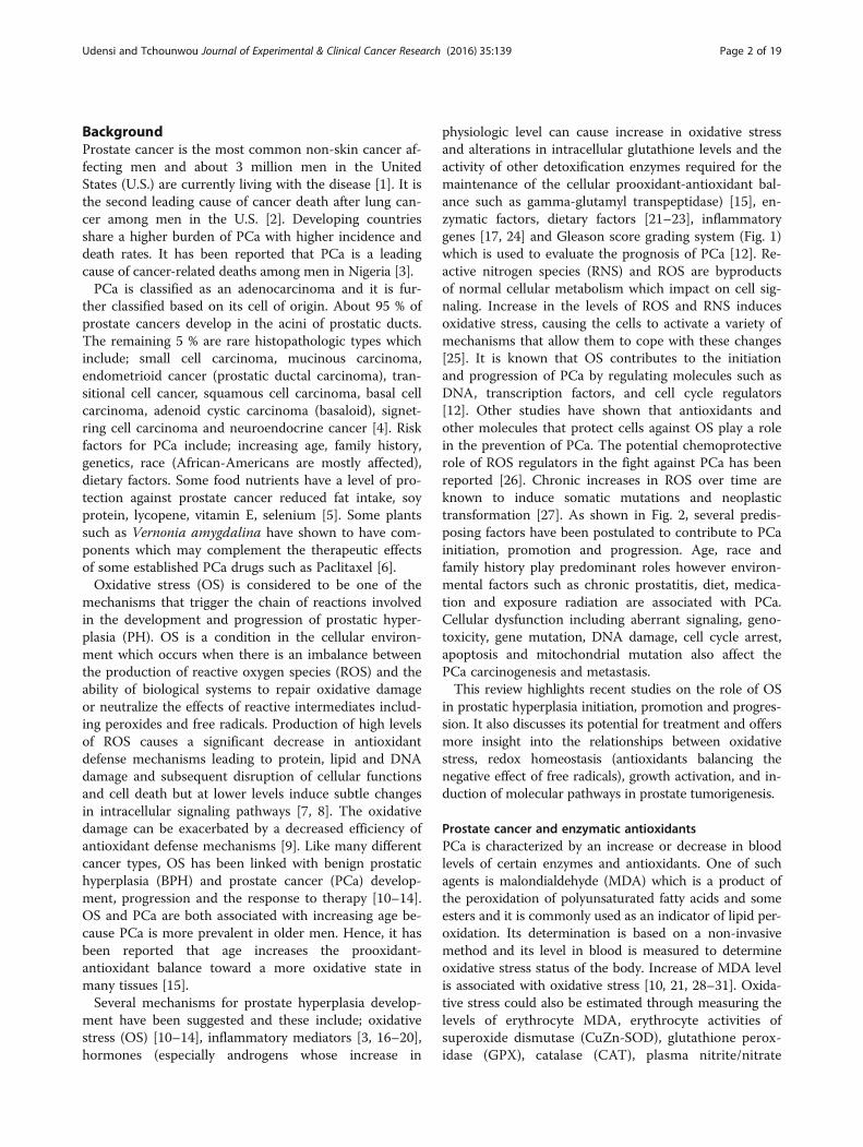
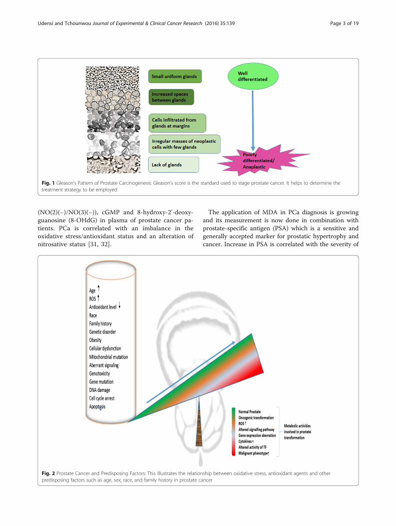
physiologic level can cause increase in oxidative stressand alterations in intracellular glutathione levels and theactivity of other detoxification enzymes required for themaintenance of the cellular prooxidant-antioxidant bal-ance such as gamma-glutamyl transpeptidase) [15], en-zymatic factors, dietary factors [21–23], inflammatorygenes [17, 24] and Gleason score grading system (Fig. 1)which is used to evaluate the prognosis of PCa [12]. Re-active nitrogen species (RNS) and ROS are byproductsof normal cellular metabolism which impact on cell sig-naling. Increase in the levels of ROS and RNS inducesoxidative stress, causing the cells to activate a variety ofmechanisms that allow them to cope with these changes[25]. It is known that OS contributes to the initiationand progression of PCa by regulating molecules such asDNA, transcription factors, and cell cycle regulators[12]. Other studies have shown that antioxidants andother molecules that protect cells against OS play a rolein the prevention of PCa. The potential chemoprotectiverole of ROS regulators in the fight against PCa has beenreported [26]. Chronic increases in ROS over time areknown to induce somatic mutations and neoplastictransformation [27]. As shown in Fig. 2, several predis-posing factors have been postulated to contribute to PCainitiation, promotion and progression. Age, race andfamily history play predominant roles however environ-mental factors such as chronic prostatitis, diet, medica-tion and exposure radiation are associated with PCa.Cellular dysfunction including aberrant signaling, geno-toxicity, gene mutation, DNA damage, cell cycle arrest,apoptosis and mitochondrial mutation also affect thePCa carcinogenesis and metastasis.This review highlights recent studies on the role of OS
in prostatic hyperplasia initiation, promotion and progres-sion. It also discusses its potential for treatment and offersmore insight into the relationships between oxidativestress, redox homeostasis (antioxidants balancing thenegative effect of free radicals), growth activation, and in-duction of molecular pathways in prostate tumorigenesis.
Prostate cancer and enzymatic antioxidantsPCa is characterized by an increase or decrease in bloodlevels of certain enzymes and antioxidants. One of suchagents is malondialdehyde (MDA) which is a product ofthe peroxidation of polyunsaturated fatty acids and someesters and it is commonly used as an indicator of lipid per-oxidation. Its determination is based on a non-invasivemethod and its level in blood is measured to determineoxidative stress status of the body. Increase of MDA levelis associated with oxidative stress [10, 21, 28–31]. Oxida-tive stress could also be estimated through measuring thelevels of erythrocyte MDA, erythrocyte activities ofsuperoxide dismutase (CuZn-SOD), glutathione perox-idase (GPX), catalase (CAT), plasma nitrite/nitrate
Udensi and Tchounwou Journal of Experimental & Clinical Cancer Research (2016) 35:139 Page 2 of 19
(NO(2)(−)/NO(3)(−)), cGMP and 8-hydroxy-2'-deoxy-guanosine (8-OHdG) in plasma of prostate cancer pa-tients. PCa is correlated with an imbalance in theoxidative stress/antioxidant status and an alteration ofnitrosative status [31, 32].
The application of MDA in PCa diagnosis is growingand its measurement is now done in combination withprostate-specific antigen (PSA) which is a sensitive andgenerally accepted marker for prostatic hypertrophy andcancer. Increase in PSA is correlated with the severity of
Fig. 2 Prostate Cancer and Predisposing Factors: This illustrates the relationship between oxidative stress, antioxidant agents and otherpredisposing factors such as age, sex, race, and family history in prostate cancer
Fig. 1 Gleason’s Pattern of Prostate Carcinogenesis: Gleason’s score is the standard used to stage prostate cancer. It helps to determine thetreatment strategy to be employed
Udensi and Tchounwou Journal of Experimental & Clinical Cancer Research (2016) 35:139 Page 3 of 19
PCa but PSA is always performed with another markersuch as MDA [21, 28, 33]. Manganese superoxide dismut-ase (MnSOD) is under consideration as potential clinicalmarker to predict the progression of PCa [34]. Superoxidedismutase-3 (SOD3) is known to protect cell surface fromoxidative stress. It has been reported that the expressionof SOD3 is reduced in PCa tissue. Also, an inhibition ofcell proliferation, migration, and invasion has been associ-ated with SOD3 overexpression in PC-3 cell line [22].Generally, PCa is accompanied with a decrease in serumlevel of anti-oxidants such as GPX, GSH-Px, SOD[14, 35], and an increase in concentrations of thiobarbitu-ric acid reactive substances (TBARS) [36] and lipid peroxi-dation byproducts [11]. Thioredoxin 1 (Trx 1), is anotherenzyme which acts as a subcellular indicator of redox sta-tus in PCa and has demonstrated that both endogenousand exogenous antioxidants are involved in determininghow PCa progresses. It can be correlated with the Gleasonscore. The level of Trx1 can be used to predict the stagesof PCa, the difference between malignant and benign tis-sue [37]. Nitric oxide generated by inducible nitric oxidesynthase (iNOS) may be involved in prostate tumorigen-esis, however the exact mechanism has not been eluci-dated, and warrants further studies [38].
Prostate cancer and non-enzymatic antioxidantsThe idea that OS is involved in prostate tumorigenesishas been supported by the observed decrease in levels ofnon-enzymatic antioxidants, such as vitamins C and E,in the plasma and erythrocytes of PCa patients com-pared to normal subjects [39, 40]. As an antioxidant,vitamin E scavenges lipid radicals and terminates oxida-tive chain reactions by interacting with the lipid peroxylradical. This prevents further generation a new radical.However, there are contrary opinions about the role ofvitamin C and vitamin E in PCa carcinogenesis. The ef-fect of vitamin C is still murky, in-vitro studies with cellline models show that vitamin C can fight against PCa[41] but in-vivo studies could not demonstrate vividlythe same result. However, different forms of vitamin Eare said to have different effects; alpha-tocopherol whichscavenges singlet oxygen potentially increases the riskwhile gamma-tocopherol potentially decreases risk ofdeveloping PCa [41, 42]. PCa patients also have lowerlevels of zinc (Zn) [11]. The reduction in antioxidants inPCa patients suggests that micronutrient supplementa-tion could be helpful in the prevention and managementof the disease [14, 35, 43, 44]. Supplementation shouldbe approached with caution as some micronutrientssuch as vitamin D may have a detrimental effect and in-crease the risk of PCa [45]. It has been reported that meta-static PCa patients have a higher Gleason score (p < 0.01)and more hormonal treatment, but lower concentrationsof PSA (p < 0.05), alpha-tocopherol (p < or = 0.05), retinol
(p < 0.01), lutein (p < 0.05) and lycopene (p < 0.01), com-pared with patients having localized disease. Lower con-centrations of carotenoids, in particular, lycopene reflectdisease progression rather than the systemic inflammatoryresponse in patients with PCa [21].
Oxidative stress, prostate cancer and dietDiet plays a pivotal role in general body wellbeing. It notonly boosts the immune system but also provides anti-oxidants that help the system to neutralize the negativeeffects of oxidative stress. Oxidative stress induced bychronic inflammation could be a cause, and dietary in-take of antioxidants such as selenium may reduce therisk of developing prostate hyperplasia by reducing thedeleterious effects of oxidative stress [46]. Selenium (Se)has been shown to prevent the development of PCa andit is highly accumulated around the prostate gland. Sele-noproteins inhibit the transformation of normal prostateepithelium into neoplasm. A reduction in blood level ofselenoproteins has been correlated with the risk of PCa[47]. Intake of some food rich in antioxidants can boostbody’s protection against disease. Pomegranate, a plantrich in antioxidants, has shown some anti-PCa promisesas it slows prostate cancer xenograft growth and pro-longs prostate-specific antigen (PSA) doubling times[48]. Extracts from Vernonia amygdalina has shownpromises a supplementary drug to taxol-resistant pros-tate adenocarcinoma cells [6].Oxidative stress and body composition contribute to
the progression of PCa. High fat diet (HFD) has beenidentified as a risk factor for PCa because HFD inducesoxidative stress and inflammation in the prostate gland.This stress triggers a cascade of activities within thegland culminating to hyperplasia. HFD induces signifi-cant increases in the levels of pro-inflammatory cyto-kines and gene products. It is also speculated that HFDcan activate signaling pathways. For example, the signaltransducer and activator of transcription (STAT)-3 andnuclear factor-kappa B (NF-kB) which are transcriptionfactors required for regulating genes involved in prolifer-ation, survival, angiogenesis, invasion and inflammation[17]. Measuring body composition can be used to pre-dict risk to developing PCa. This phenomenon has beenby using a combination of the measurement of glutathi-one, fat mass (FM) and waist circumference (WC) topredict the risk of PCa [13]. A study has suggested thatdietary fat could encourage increase in proliferation ofprostate intraepithelial neoplasia (PIN) and suppressionof glutathione peroxidase 3 (GPx3) expression [22]. PINstage is crucial in prostate carcinogenesis as shown inFig. 3. Cells have gone through neoplastic changes andhave become carcinogenic and ready to invade othercells. High-grade prostatic intraepithelial neoplasia has ahigh predictive value and is considered as a reliable
Udensi and Tchounwou Journal of Experimental & Clinical Cancer Research (2016) 35:139 Page 4 of 19
indicator of pre-invasive stage of adenocarcinoma [49]. Anantioxidant, 2-hydroxy-4-methoxy benzoic acid (HMBA)has protective effects in rats against testosterone inducedBPH and this may have a similar effect on PCa [29].
Symptoms and diagnosis of PCaPCa presents different clinical signs and symptomswhich range from asymptomatic, inactive, slow-growingtumors to aggressive, fast-growing tumors with lethalprogression. Symptoms of PCa may include; problemspassing urine, such as pain, difficulty starting or stoppingthe stream, or dribbling, low back pain and pain withejaculation. The rate at which cancer grows and the dif-ference in its appearance from surrounding tissue helpsdetermine the stage [50]. Like most epithelial cancersthe keys to survival and treatment are early diagnosisand identification of PCa type. Androgens and androgenreceptor (AR) are required by both normal prostate andprostate cancer cells for growth and survival [51]. Andro-gen receptor mutations are observed in late stage prostatecancer. Androgen ablation and antiandrogen therapycause the cancer to regress. Androgen-independent pros-tate cancer which does not respond to anti-androgen ther-apy has been observed in some patients especially inpatients whose cancer was not cured by surgery. Overex-pression of Caveolin-1 occurs in about a quarter of humanprostate cancers and is thought to induce androgen sensi-tivity in androgen-insensitive prostate cancer cells [52].However, a recent study has suggested that Metformin,commonly used for type 2 diabetes, may have promisingtherapeutic effects on both androgen-dependent andandrogen-independent PCa [40]. Most facilities use diag-nostic test kits that measure the level of prostate-specificantigen (PSA) in serum of patients to detect early stages
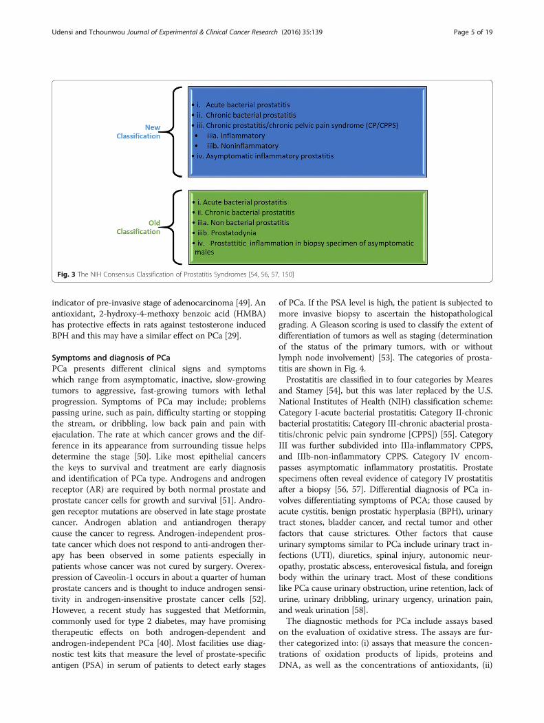
of PCa. If the PSA level is high, the patient is subjected tomore invasive biopsy to ascertain the histopathologicalgrading. A Gleason scoring is used to classify the extent ofdifferentiation of tumors as well as staging (determinationof the status of the primary tumors, with or withoutlymph node involvement) [53]. The categories of prosta-titis are shown in Fig. 4.Prostatitis are classified in to four categories by Meares
and Stamey [54], but this was later replaced by the U.S.National Institutes of Health (NIH) classification scheme:Category I-acute bacterial prostatitis; Category II-chronicbacterial prostatitis; Category III-chronic abacterial prosta-titis/chronic pelvic pain syndrome [CPPS]) [55]. CategoryIII was further subdivided into IIIa-inflammatory CPPS,and IIIb-non-inflammatory CPPS. Category IV encom-passes asymptomatic inflammatory prostatitis. Prostatespecimens often reveal evidence of category IV prostatitisafter a biopsy [56, 57]. Differential diagnosis of PCa in-volves differentiating symptoms of PCA; those caused byacute cystitis, benign prostatic hyperplasia (BPH), urinarytract stones, bladder cancer, and rectal tumor and otherfactors that cause strictures. Other factors that causeurinary symptoms similar to PCa include urinary tract in-fections (UTI), diuretics, spinal injury, autonomic neur-opathy, prostatic abscess, enterovesical fistula, and foreignbody within the urinary tract. Most of these conditionslike PCa cause urinary obstruction, urine retention, lack ofurine, urinary dribbling, urinary urgency, urination pain,and weak urination [58].The diagnostic methods for PCa include assays based
on the evaluation of oxidative stress. The assays are fur-ther categorized into: (i) assays that measure the concen-trations of oxidation products of lipids, proteins andDNA, as well as the concentrations of antioxidants, (ii)
Fig. 3 The NIH Consensus Classification of Prostatitis Syndromes [54, 56, 57, 150]
Udensi and Tchounwou Journal of Experimental & Clinical Cancer Research (2016) 35:139 Page 5 of 19

assays that determine the oxidative and reductive capacityof biological fluids, and (iii) assays that measure the exvivo susceptibility of lipids to oxidation when exposed to asource of free radicals [59]. Some of the oxidative bio-markers that are frequently measured are; lipid peroxida-tion, total antioxidant capacity and total thiol molecules[60]. Table 1 presents a list of laboratory tests that areused to diagnose and monitor the progress of PCa.These biomarkers can be assayed in different body spec-
imens such as the whole blood, plasma, serum, urine, and
tissue biopsies. Biomarkers discussed in this review arelimited to those that are linked to oxidative stress. Re-cently, F2-isoprostanes, a group of prostaglandin F(2)-likecompounds derived from the non-enzymatic oxidation ofarachidonic acid have been used as markers of lipid oxida-tion and the results showed an accurate assessment of oxi-dative stress both in vitro and in vivo in urine, blood andtissue biopsies [61]. Other biomarkers of oxidative stressare plasma fluorescent oxidation products which detectglobal oxidation, and carboxy methyl lysine (CML) which
Table 1 Laboratory Tests Used to Diagnose and Monitor PCa Biomarkers
Test Specimen Factors measured Method Reference
Oxidative stress Blood Activity changes of superoxide-dismutase(SOD), catalase (CAT), ceruloplasmin (Cp),tripeptide glutathione (GSH), glutathione-peroxidase (GSH-Px), and glutathione-reductase (GR)
Spectrophotometry [11, 12, 32]
Oxidative stress prostate tissue Thioredoxin 1 (Trx 1) Spectrophotometry [37]
Oxidative stress Tissue Inducible NOS (iNOS or NOS-2) Immunohistochemistry andreverse transcriptase-polymerasechain reaction (RT-PCR)
[152]
Oxidative stress Blood Plasma oxidized low-density lipoprotein,peroxides, and total equivalent antioxidantcapacity (TEAC)
Spectrophotometer [32]
Global oxidation Blood plasma fluorescent oxidation products Spectrophotometry [40]
Oxidative stress Blood carboxymethyllysine (CML), advancedglycation end products (AGE)
Spectrophotometry [40]
Oxidative stress Prostatebiopsy(needle biopsy)
Total thiol groups (TTG) level Spectrophotometry (2 thionitrobenzoicacid (DTNB))
[13, 153]
Lymphocyte DNA damage Blood lymphocyte DNA damage single cell alkaline gel electrophoresis,tail length migration
[15]
Lipid peroxidation Plasma Thiobarbituric acid reactive substances(TBARS), serum protein carbonylation
Spectrophotometry; Thiobarbituric acid(TBA), concentrations of TBA- MDA adduct
[12, 36, 154]
Lipid oxidation Urine, blood, tissues F2-isoprostanes Gas/liquid chromatography-massspectrometry, mass spectrometry,immunological methods
[40, 61, 155]
Body composition Air Volume of air the body displaced insidean enclosed chamber (plethysmograph)
Air plethysmography (BOD POD) [13, 156]
Fig. 4 Prostate Carcinogenesis Model: This illustrates what happens at the cellular level as prostate hyperplasia progresses from asymptomatic tometastatic stage
Udensi and Tchounwou Journal of Experimental & Clinical Cancer Research (2016) 35:139 Page 6 of 19
detects advanced glycation end products. Higher levelsof plasma CML signify increased risk of prostate cancer[40]. Lipid peroxidation contribute to cellular oxidativestress and cell death. This draws attention to the cellu-lar activities that involve the thiol groups. The level oftotal thiol groups (TTG) has been associated with agingand progression of PCa. A comparison of the levels ofTTG between BPH and PCa patients has shown thataging influences a progressive reduction of TTG in BPHpatients, while in PCa patients the glutathione concentra-tions are significantly lower [13]. The determination of thelevel of 8-isoprostanes (8-EPI) in urine using competitiveenzyme-linked immunoassay gives an idea of the oxidativestress status and the stage of the hyperplasia (NIHcategories IIIa, IIIb, and IV) [62]. The new phase ofdiagnosis is moving towards the application of genet-ics and molecular techniques such as genetic linkagestudies which is used to study the expression patternsof ncRNA transcription [63].
Prostate cancer and benign prostatic hyperplasia (BPH)These two forms of prostatic hyperplasia are leadingcause of urologic problems in older men. Although theycan co-exist in an individual, there is no evidence thatBPH is a precursor nor risk factor of PCa. There is noevidence that BPH can transform into PCa. However,both conditions share certain attributes in common.They are hormone and age dependent. Risk increaseswith age, and they are associated with certain forms ofhyperplasia [64, 65]. They present similar symptoms andfor accurate diagnosis PCa has to be differentiated fromBPH. Both PCa and BPH can cause lower urinary tractsymptoms (LUTS). Additional tests and examinationsare performed to differentiate LUTS caused by PCA andLUTS caused by BPH. For instance, PSA is performed inasymptomatic patients, while prostate biopsy is done onLUTS patients. Procedures such as bone pain are per-formed on patients with symptoms of metastasis. Evalu-ating the size of the prostate through digital rectalexamination (DRE) is essential in the management ofBPH. A combination of DRE and PSA testing can beused to differentiate clinically between PCa and BPH.The presence of a nodular abnormality puts the prob-ability of diagnosing PCa on biopsy at 50 %. Blood levelof PSA is no longer used alone for PCa diagnosis. It isnow recommended to confirm increases in serum PSAlevels with histology tests. Clinically, PCa could be ruledout if PSA and DRE density are normal [64]. Oxidativestress influences the pathophysiology of both PCa andBPH. For instance, antioxidant defense system is de-creased in the elderly patients with PCa and BPH [66].Also, systemic metabolic stress occurs in glucose andfatty acid metabolism in BPH and PCa [67]. However,their response to therapeutics agents differs. And some
drugs used in the treatment of BPH have been suspectedto worsen the prognosis of PCa. An example is 5α-Reductase inhibitors (5-ARIs) which is the commondrug of choice for BPH. It is suspected that 5-ARIs cantrigger or increase the risk of developing high-gradeprostate cancer. Studies could not associate 5-ARIs usewith an increased risk of PCa [37, 68]. Rather in their re-view, Hamilton and Parsons suggested that 5-ARIs hasprotective potential and could reduce the risk of prostatecancer in some men [69].
Prostate cancer, oxidative stress, and ageThere is a strong connection between age, OS, and PCA.PCA is predominant among older men and this makesage and family history the strongest predictors of pros-tate cancer risk. PCa is mostly seen in older men andBlack/African Americans are disproportionately affected[20]. The risk of developing PCa increases exponentiallywith age [15]. For instance, only 1 in 10,000 under age40 will be diagnosed, the rate goes up to 1 in 38 forages 40 to 59, and 1 in 14 for ages 60 to 69. Most PCaare diagnosed in men 50 years of age and older [1]. Butthe age of diagnosis is earlier among Black/AfricanAmericans and they are about four times more likely todie from PCa than White men [20, 70]. On the otherhand, according to the free radical theory of aging, oxi-dative stress is blamed for aging because of the negativeimpact the excessive free radicals or reactive oxygenspecies (ROS) have on the cells [71]. Also, antioxidantdefense system is decreased in the elderly patients withPCa [66]. ROS and age are predominant factors in PCaand other aging-related diseases, such as, diabetes, ath-erosclerosis and degenerative diseases like Parkinson’sand Alzheimer’s. This is because older cells seem to bemore susceptible to intracellular conditions that pro-duce excess ROS which trigger and accelerate tumori-genesis [72]. It has been reported that age increases theprooxidant-antioxidant balance toward a more oxida-tive state in many tissues [15].
Prostate cancer and non-coding RNA (ncRNAs)The interest in getting a better understanding of the mo-lecular mechanisms involved in PCa pathogenesis andprognosis has led to exploring the role of non-codingRNA (ncRNAs). Only about 2 % of human genome areprotein-coding sequences while the remaining 98 % arenoncoding sequences that can be transcribed inncRNAs. ncRNAs depending on the size are furthergrouped into long noncoding RNAs (lncRNAs) andsmall ncRNAs. Small ncRNAs include microRNAs (miR-NAs), piwi-interacting RNAs (piRNAs), ribosomal RNAs(rRNAs), small Cajal body-specific RNAs (scaRNAs),small interfering RNAs (siRNAs), small nuclear RNAs(snRNAs), small nucleolar RNAs (snRNAs) and transfer
Udensi and Tchounwou Journal of Experimental & Clinical Cancer Research (2016) 35:139 Page 7 of 19
RNAs (tRNAs). However, miRNAs can also be derivedfrom lncRNAs and snoRNAs [63]. Previously, no func-tions were apportioned to ncRNAs since they are non-protein-coding RNA species of the transcriptome. Recentstudies have revealed that they play special roles in thetumorigenic processes by acting sometimes as oncogenicand tumor suppressor genes. Some perform similar func-tions as the house keeping genes being involved in mRNAprocessing and protein transcription [63]. Some ncRNAsperform regulatory functions such as pre- and posttran-scriptional gene regulation and chromatin assembly. Theyare believed to be involved in carcinogenic process bypromoting tumor cell proliferation, inducing replicativeimmortality, encouraging evasion of growth suppressors,stimulating angiogenesis and promoting invasion andmetastasis [73]. Aside their role in cancer initiation andprogression, non-coding RNA can also be exploited aspossible biomarkers of PCa diagnosis, and drug develop-ment [74]. There is evidence that ncRNAs are aberrantlyexpressed in prostate cancer and carcinogenesis andtumor progression may result from series of multicellularactivities due to dysregulation of ncRNA controlledpathways [74].In PCa some lncRNAs are dysregulated and are
emerging as major biomarkers of cancer developmentand therapeutic targets. For example, PCA3, PCATs,SChLAP1, SPRY4-IT1 and TRPM2-AS are lncRNAsupregulated in PCa [75]. Other studies have demon-strated the role of lncRNAs in regulatory and cellularprocesses including chromatin modification, alternativesplicing, post-transcriptional processing and cell signalingtransduction [76]. Dysregulation of their function mayhave deleterious effects by inducing chromosomal trans-location, deletion, and nucleotide expansions. Recentstudies demonstrate that multiple prostate cancer risk lociare associated with lncRNAs and that ectopic expressionof these transcripts triggers a cascade of cellular eventsdriving tumor initiation and progression [24]. Thediscovery of the lncRNA prostate cancer antigen 3(PCA3, or DD3), which is specifically overexpressedin malignant prostate tissue supports the claim thatlncRNAs may have cancer-specific expression. Theseadditional lnRNAs AK024556, XLOC_007697, LOC100287482, XLOC_005327, XLOC_008559, and XLOC_009911 which are differentially expressed in prostaticadenocarcinoma tissue samples could be possible bio-marker targets of PCa [77].
Prostate cancer and microRNAMicroRNAs (miRNA) are small, non-coding, single-stranded, short nucleotide sequences (between 19 and25 nucleotides long) that be derived from lncRNAs andsnoRNAs [63]. miRNAs function as regulators of geneexpression and influence various physiological and
pathophysiological processes [78] by binding totargeted messenger RNA (mRNA) sequences post-transcriptionally through complementary binding andmodulate gene expression. They can control gene ex-pression by silencing or degrading targeted mRNA[79]. Deregulation of miRNA in PCa may contributeto cancer initiation and metastatic progression [69].miRNA can be good biomarker in diagnosing and predict-ing prognostic outcomes of PCa as miR-148a is differen-tially expressed in PCa [69]. Identification of dysregulatedmicroRNAs (miRNAs) in prostate cancer is critical notonly for diagnosis, but also differentiation between the ag-gressive and indolent forms of the disease. Some miRNAsmodulate the activities of mesenchymal stem cells (MSCs)especially adipose-derived stromal cell (ASC) that hastherapeutic effects on PCa. A novel miR-145 is involved inPCa cell apoptosis cell induction by mediating the in-hibitory effect of ASC on PCa [80]. Understandingthe expression patterns of circulating miRNAs andcorrelating them with disease status could offer an al-ternative minimally invasive approach to monitor theprognosis of PCa progression [81].Deregulation of miRNA expression could also have
positive outcomes; for example inhibition of miR-9 canreduce tumor growth and metastases by slowing downthe migratory and invasive potential of the M12 cell line.miR-9 modulates the expression of e-cadherin and sup-pressor of cytokine signaling 5 (SOCS5) which cancerlinked proteins [82]. Another miRNA, miR-199a-3p tar-gets stemness-related and mitogenic signaling pathwayswhich suppresses the expansion and tumorigenic cap-abilities of prostate cancer stem cells and aberrant lossof a miRNA-mediated mechanism can lead to the ex-pansion and tumorigenic activity of prostate cancer stemcells (CSCs) [83]. The use of miRNA to silence genes in-volved in PCa tumorigenesis such as STAMP2 requiredfor PCa progression could be a viable therapeutic mech-anism for PCa [40].Circulating miRNAs could be non-invasive markers of
disease and can be used as biomarkers in PCa diagnosis.For example, determining the levels of 15 miRNAs andmiR-141 could be used to differentiate metastatic PCapatients from healthy subjects [84] or between BPH andPCa [85]. But some of the miRNAs are not specific for aparticular cancer type and thus, serum miRNAs may dis-tinguish between different cancer types [86]. But miR-21appears to be elevated in CRPC patients who are resist-ant to docetaxel than in BPH. While miR-150 enhancestargeted endothelial cell migration [87]. miR-26a,miR-195, and let-7i levels were elevated in PCa com-pared to BPH samples [85]. Other studies have re-ported additional miRNAs that can be used in PCadiagnosis. Moltzahn identified 7 miRNAs differentiallyexpressed between PCa patients and healthy subjects
Udensi and Tchounwou Journal of Experimental & Clinical Cancer Research (2016) 35:139 Page 8 of 19
[88]. For instance, miR-141, and miR-375 expressionare elevated in PCa and their presence in the blood streammay serve as evidence of advanced cancer disease [89].Further, miR-141 levels have been associated with clinicalprogression and it is positively correlated with PSA [90].Both miR-21, miR-221 [91] and miR-141 levels were ele-vated in PCa patients compared to healthy controls butthe levels were higher in metastatic PCa than in localizedtumors [92]. PCa progression could be monitored bymeasuring the elevation levels of hsa-miR-141, hsa-miR-298 and hsa-miR-375 [93]. Monitoring urinary levels ofmiR-107 and miR-574-3p could also be relevant in PCadiagnosis [94]. The diagnostic reliability of miRNA in PCacould be further improved by identifying a panel ofmiRNAs instead of single miRNA. Chen and co-workers identified a panel of 5miRNAin this regard;let-7c, let-7e, miR-30c, miR-622, and miR-1285. Ac-cording to them this panel could differentiate betweenPCa patients and healthy people and between PCaand BPH [95]. miR-20a, miR-21, miR-145, and miR-221 can be associated with tumor risk scores [96]while SNORD43 may be a suitable reference gene forthe analysis of circulating miRNA in patients withurological malignancies [97].
Prostate cancer and inflammatory oxidative stressProstatic inflammation is suggested to be involved in thepathogenesis and progression PCa [98]. Inflammation isthought to incite carcinogenesis by causing cell and gen-ome damage, promoting cellular turnover [99]. Prostaticinflammation could also be caused by bacterial infec-tions, urine reflux, dietary factors, hormones, and auto-immune response [3]. Irregularities in the functioning ofgenes involved in oxidative stress have been linked to in-flammatory response in PCa. For instance the loss or ab-erration in the expression of glutathione S-transferaseP1 (GSTP1) may contribute to the transition of prolifer-ative inflammatory atrophy (PIA) into high-grade intrae-pithelial neoplasia (HGIPN) and PCa in patients withgenetic predisposition [3].Inflammation is known to trigger cytokines production
which contributes to tumorigenesis in different tissues.For example, prostaglandin endoperoxide synthase 2,also referred to as cyclooxygenase 2 (COX-2), is an en-zyme involved in the conversion of arachidonic acid toprostaglandins and other eicosanoids. The overexpres-sion of COX-2 has been reported to cause phenotypicchanges in intestinal epithelial cells that could enhancetheir tumorigenic potential [100]. COX-2 as a proinflam-matory cytokine may also create an environment that fa-vors local growth factor production and angiogenesis inthe prostatic tissue [78]. Oxidative stress ensues whenthe proinflammatory microenvironment creates a localhypoxia induced by increased oxygen demands by
proliferating cells leading to tissue injury in infiltrat-ing area [3] (Fig. 4). The literature contains numerousstudies associating PCa and inflammation but nonehas categorically established a causal relation. Intakeof anti-inflammatory drugs and antioxidants leads toa decrease in PCa risk [18]. Further understanding ofthe role of inflammation in oxidative stress inductionand PCa promotion and progression is necessary as itmay revolutionize the way PCa is treated [16].
Oxidative stress and DNA damage in prostatic hyperplasiaA genetic predisposition or acquired genetic and epi-genetic changes with effect other factors, such as ad-vanced age, race and environmental factors contributeto PCa development [53]. OS triggers metabolic re-programming responsible for malignant transform-ation and tumor development, including invasion andmetastasis [101]. The activities of antioxidant enzymesand the levels of antioxidant, reduced glutathionehave been found to be significantly decreased in pros-tatic hyperplasia. Significantly increased levels of oxi-dative stress and DNA damage suggest that oxidativedamage plays an important role in prostate tumori-genesis and timely management of oxidative stresscan be of importance in preventing the occurrence ofprostatic hyperplasia [11, 16]. Other studies have re-vealed that oxidative stress mediated pathways are in-volved in several male urologic disorders includingthe different forms of prostatitis (NIH categories IIIa,IIIb, and IV). The new insight into the role of oxida-tive stress in the pathogenesis of PCa has led to theexploitation of this mechanism as a potential strategictarget for PCa treatment [30, 62].
Molecular biology of oxidative stress and prostate cancerMore knowledge about the etiology and progressionof PCa has been provided by the advancement in mo-lecular biology and development of new techniquessuch as microarray [102] and RNAseq gene expression,genome-wide linkage analysis, and loss of heterozygosity(LOH) [103]. Recently developed methods for profilinggenome-wide occupancy of lncRNAs have allowed high-throughput identification of RNA–DNA and RNA–pro-tein interactions. Two methods called Chromatin Isolationby RNA Purification (ChIRP) [104], Native RNA immuno-precipitations sequence(RIP-seq) [105] and CHART, thatuse complementary oligonucleotides to pull downlncRNAs associated with chromatin, have been developedto determine the chromatin binding sites for lncRNAs.68Alternatively, RNA immunoprecipitation sequencing(RIP-Seq) and photoactivatable ribonucleoside-enhancedcrosslinking and immunoprecipitation (PAR-CLIP) repre-sent complementary approaches to the study of RNA–protein interactions.69,70 These new techniques represent
Udensi and Tchounwou Journal of Experimental & Clinical Cancer Research (2016) 35:139 Page 9 of 19
promising tools to explore the mechanisms that governlncRNA-chromatin interactions, as shown by the inform-ative analyses performed to date on select lncRNAs. Othertechniques that have been applied in PCa studies includemethods which identify microsatellites, single nucleotidepolymorphism (SNP) and haplotype mapping to monitorthe distribution of clusters of SNPs that segregate togetherin linkage disequilibrium [106]. These techniques have re-vealed the potential role of DNA repair genes, tumor sup-pressor genes, oncogenes, and protein expression in theprognosis, diagnosis, and possible therapeutic strategiesfor PCa. Also, linkage analyses in genome wide studieshave given clues as to why the disease runs in a family andwhy a particular race may be at higher risk of developingand dying from the disease [103, 107–111].Only about 9 % of PCa cases are linked with heredity
and studies are carried using multipoint linkage analyseswith microsatellite markers. These studies have identi-fied PCa susceptibility loci on chromosome 1, includinghereditary PCa families (HPC), HPC1 (1q24–q25), PCAP(1q42–q43), HPCX (Xq27–q28), CAPB (1p36), HPC20(20q13), HPC2/ELAC2 (17p11) and 16q23 [107, 108, 111].And based on reports 5q31–q33, 7q32 and 19q12 weredescribed as prostate cancer aggressiveness loci [103]. Al-though inconsistent results were obtained from repeatedlinkage studies of these regions, HPC1 is thought be com-mon among people with early onset disease. Nevertheless,hereditary of PCa is heterogeneous and there are multipleloci on chromosome 1 for this disease [103, 107, 108].Genetics is a major determinant of susceptibility to
PCa on a wide population [112]. Aberrations in gene ex-pression and gene mutations have been seen in prostatecancer including PTEN, KAI1, SRD5A2, and IL6 andthey are associated with PCa progression [113]. Geno-mewide studies have revealed multiple loci that can bindto PCa candidate susceptibility genes such as MSMB,LMTK2 and KLK3, CPNE3, IL16 and CDH13. For ex-ample MSMB which encodes beta-microseminoprotein,a primary constituent of semen and a potential prostatecancer biomarker, and CTBP2, a gene with antiapoptoticactivity are located on chromosome 10 [109, 110]. Genesassociated with the redox homeostasis of the cell are be-ing reported to undergo transformation that influencessusceptibility to PCa. Of particular interest is the sixtransmembrane protein of prostate 2 (STAMP2) whichis an androgen-regulated gene whose mRNA expressionis increased in PCa. The STAMP2 protein expression isincreased in human PCa. It is suggested that STAMP2also significantly increases reactive oxygen species (ROS)in PCa cells because its iron reductase activity has theability to deplete NADPH levels [40].A search for prostate cancer/molecular biology on
Cancer Index Web Resource and NCBI [50, 113] wasused to construct Table 2 which shows a list of genes
that are associated with PCa based on scientific publica-tions. The table is not exhaustive and most of genes areknown to regulate critical mechanisms in other types ofcancer. The list includes the gene symbols, gene names,and their location on the chromosome and the numberof scientific publications on each of the genes (Table 2).Genes that regulate critical mechanisms in other cancersand which are expressed in prostate cancer includeKallikrein-related peptidase 3 (KLK3), also referred to asprostate surface antigen (PSA) [50, 113]. PSA is elevatedin PCa and serum measurement of PSA is a commondiagnostic test for diagnosis and monitoring of PCa inhospital settings [114]. CD82 molecule, a metastasis sup-pressor gene product known to be downregulated intumor progression of human cancers and can be acti-vated by p53. It is co-expressed with p53 and aberrationin CD82 expression is linked with poor survival for PCapatients [115]. Another gene in Table 2 is microsemino-protein, beta (MSMB) which encodes a protein that be-longs to the immunoglobulin binding factor family. Theprotein is synthesized by the epithelial cells of the pros-tate gland and secreted into the seminal plasma. Thisprotein has inhibin-like activity and its expression de-creases in PCa [116, 117]. A tumor suppressor gene, Rasassociation (RalGDS/AF-6) domain family member 1(RASSF1) has been seen in other cancers and is believedto play a role in PCa pathogenesis [118]. Clusterin onthe other hand encodes a protein that regulates severalbasic biological events including cell death, tumor pro-gression, and neurodegenerative disorders [119]. NK3homeobox 1 (NKX3-1) encodes a homeobox-containingtranscription factor which serves as a negative regulatorof epithelial cell growth in prostate tissue. Aberrant ex-pression of this gene is associated with prostate tumorprogression [111].Prostate stem cell antigen (PSCA) encodes a
glycosylphosphatidylinositol-anchored cell membraneglycoprotein and it is secreted in the prostate as well as inother organs such as the colon and pancreas. PSCA is up-regulated in a large proportion in PCa and is also detectedin cancers of the bladder and pancreas [120]. Pten is alsoassociated with different types of malignancies and it isknown to be involved in PCa development where it coor-dinates the differentiation and proliferation of cell types[121]. ELAC ribonuclease 2 (ELAC2) functions as a tran-scription factor. It interacts with activated Smad familymember 2 (Smad2) and its nuclear partner forkhead boxH1 (FAST-1). Mutations in this gene may lead to an in-creased risk of PCa [122, 123]. PCa-associated 3 (PCA3)produces a spliced, long non-coding RNA that is highlyoverexpressed in most types of prostate cancer cells and isused as a specific biomarker for this type of cancer [83].Glutathione S-transferase mu 1 (GSTM1) is involved indetoxifying electrophilic compounds and neutralizing
Udensi and Tchounwou Journal of Experimental & Clinical Cancer Research (2016) 35:139 Page 10 of 19
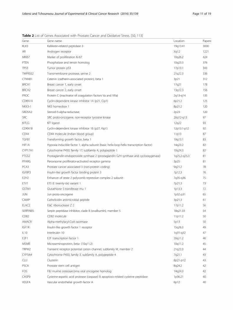
Table 2 List of Genes Associated with Prostate Cancer and Oxidative Stress. [50, 113]
Gene Gene name Location Papers
KLK3 Kallikrein-related peptidase 3 19q13.41 3000
AR Androgen receptor Xq12 1221
MKI67 Marker of proliferation Ki-67 10q26.2 424
PTEN Phosphatase and tensin homolog 10q23.3 376
TP53 Tumor protein p53 17p13.1 343
TMPRSS2 Transmembrane protease, serine 2 21q22.3 336
CTNNB1 Catenin (cadherin-associated protein), beta 1 3p21 312
BRCA1 Breast cancer 1, early onset 17q21 178
BRCA2 Breast cancer 2, early onset 13q12.3 156
PROC Protein C (inactivator of coagulation factors Va and VIIIa) 2q13-q14 135
CDKN1A Cyclin-dependent kinase inhibitor 1A (p21, Cip1) 6p21.2 125
NKX3-1 NK3 homeobox 1 8p21.2 120
SRD5A2 Steroid-5-alpha-reductase, 2p23 120
SRC SRC proto-oncogene, non-receptor tyrosine kinase 20q12-q13 97
KITLG KIT ligand 12q22 93
CDKN1B Cyclin-dependent kinase inhibitor 1B (p27, Kip1) 12p13.1-p12 92
CD44 CD44 molecule (Indian blood group) 11p13 87
TGFB1 Transforming growth factor, beta 1 19q13.1 83
HIF1A Hypoxia inducible factor 1, alpha subunit (basic helix-loop-helix transcription factor) 14q23.2 82
CYP17A1 Cytochrome P450, family 17, subfamily A, polypeptide 1 10q24.3 82
PTGS2 Prostaglandin-endoperoxide synthase 2 (prostaglandin G/H synthase and cyclooxygenase) 1q25.2-q25.3 81
PPARG Peroxisome proliferator-activated receptor gamma 3p25 81
PCA3 Prostate cancer associated 3 (non-protein coding) 9q21.2 78
IGFBP3 Insulin-like growth factor binding protein 3 7p12.3 76
EZH2 Enhancer of zeste 2 polycomb repressive complex 2 subunit 7q35-q36 75
ETV1 ETS (E twenty-six) variant 1 7p21.3 73
GSTM1 Glutathione S-transferase mu 1 1p13.3 72
JUN Jun proto-oncogene 1p32-p31 65
CAMP Cathelicidin antimicrobial peptide 3p21.3 61
ELAC2 ElaC ribonuclease Z 2 17p11.2 56
SERPINB5 Serpin peptidase inhibitor, clade B (ovalbumin), member 5 18q21.33 54
CD82 CD82 molecule 11p11.2 50
AMACR Alpha-methylacyl-CoA racemase 5p13 50
IGF1R Insulin-like growth factor 1 receptor 15q26.3 49
IL10 Interleukin 10 1q31-q32 47
E2F1 E2F transcription factor 1 20q11.2 46
MSMB Microseminoprotein, beta- (10q11.2) 10q11.2 45
TRPM2 Transient receptor potential cation channel, subfamily M, member 2 21q22.3 44
CYP3A4 Cytochrome P450, family 3, subfamily A, polypeptide 4 7q21.1 43
CLU Clusterin 8p21-p12 43
PSCA Prostate stem cell antigen 8q24.2 42
FOS FBJ murine osteosarcoma viral oncogene homolog 14q24.3 42
CASP9 Cysteine-aspartic acid protease (caspase) 9, apoptosis-related cysteine peptidase 1p36.21 40
VEGFA Vascular endothelial growth factor A 6p12 40
Udensi and Tchounwou Journal of Experimental & Clinical Cancer Research (2016) 35:139 Page 11 of 19
the effect of anti-oxidants in the cellular system,glutathione conjugates with the products of oxidativestress [124, 125]. Prostaglandin-endoperoxide synthase 2(PTGS), also known as cyclooxygenase, is the key enzymein prostaglandin biosynthesis, and acts both as a dioxygen-ase and as a peroxidase. This gene encodes inducible iso-zyme which induces prostanoid biosynthesis. Prostanoid isthought to be involved in inflammation and mitogenesis[124]. Hypoxia inducible factor 1, alpha subunit (HIF1A)encodes the alpha subunit of transcription factor hypoxia-inducible factor-1 (HIF-1), which is a heterodimer consist-ing of an alpha and a beta subunit. HIF-1 is involved inthe regulation of cellular and systemic homeostatic re-sponse to hypoxia. It activates transcription of genes in-volved in energy metabolism, angiogenesis, apoptosis, andother genes that secret protein which promote metabolicadaptation to hypoxic environment as well as increaseoxygen delivery. It may also be involved in the activationof signaling pathways. Redox-sensitive transcriptionfactors like HIF-1α has been shown to play a majorrole in progression and metastasis of the cancer cells[126]. Steroid-5-alpha-reductase (SRD5A2) gene en-codes a microsomal protein expressed at high levelsin androgen-sensitive tissues such as the prostate andmale pseudohermaphroditism, specifically pseudovagi-nal perineoscrotal hypospadias may occur if the geneis suppressed or deficient [127].Cellular dysfunction resulting from polymorphisms
and post translation modifications such as methylationin genes contributes to PCa risk. For instance, changesin the glutathione S-transferase (GST) genes have beenimplicated as risk factors for prostate cancer [128]. Lossof GSTP1 expression via promoter hypermethylation isthe most common epigenetic alteration observed in hu-man PCa. Dysfunction of GSTP1, a member of the GSTgene family, can trigger an increased production of re-active oxygen species (ROS) and DNA damage in cells.Thus, monitoring GSTP1 expression in human prostatecells may be an important target for primary preventionof PCa knowing that in its absence cells are more proneto oxidative stress induced DNA damage and cell death[129]. The GST superfamily consists of four gene classes(A, M, T, and P) encoding for enzymes which catalyzethe conjugation of electrophilic compounds to
glutathione [130]. These enzymes are also believed toplay a crucial role in the protection of DNA from oxida-tive damage [131]. The pi-class glutathione S-transferase(GSTP1) actively protect cells from carcinogens andelectrophilic compounds. Previous studies have shownthat the CpG-rich promoter region of the pi-class geneGSTP1 is methylated at single restriction sites in the mostprostate cancers [132, 133]. It has been observed that innormal prostate tissue the entire CpG island is unmethy-lated but outside the island in the body of the gene ishighly methylated. The DNA methylation of the CpG is-land in both PCa cell lines and cancer tissues occurs whenGSTP1 expression is repressed [128]. Tumor suppressorgenes and other CpG island-containing genes such as cal-citonin, p15, p16, Rb, VHL, e-cadherin, ER, and HIC1have been found in the hypermethylated region [97].There are other polymorphs of GST such as the mu
(GSTM1) and theta (GSTT1) which regulate the conju-gation of carcinogenic compounds to excretable hydro-philic metabolites making the genes more susceptible tovarious carcinogens. Changes in their structure such asdouble deletion (GSTM1-/GSTT1-) is associated withhigher oxidative stress which might exacerbate thepathogenesis of BPH and PCa [36]. In normal prostate,the transduction pathway from NIK to NF-kB seems tobe inactive. However, in BPH, it has been reported thatTNF-α/AP-1 transduction pathway is activated followedby a stimulation of the apoptotic pathway to inhibit un-controlled cell proliferation [134]. Another study hasalso demonstrated a novel link between OS and loss ofimprinting, showing that OS as measured by the in-crease in NF-kB activity, induces loss of imprinting ofinsulin-like growth factor 2 in both cancerous and non-cancerous human prostate cells (Fig. 5). This loss duringaging contributes to tumorigenesis and NF-kB modula-tion is important as it may prevent age-related alterationin the epigenome [24].PCa tumorigenesis involves a combo of genes such as
the antioxidant enzyme heme oxygenase 1 (HMOX1/HO-1) which is responsible for the maintenance of the cellularhomeostasis (See Fig. 6). HMOX1/HO-1 plays a criticalrole in oxidative stress mechanism and in the regulationof PCa development and progression. The transcriptionfactor Nrf2 and BRCA1 protein synergistically activate
Table 2 List of Genes Associated with Prostate Cancer and Oxidative Stress. [50, 113] (Continued)
FOXA1 Forkhead box A1 14q21.1 40
MET MET proto-oncogene, receptor tyrosine kinase 7q31 40
CYP3A5 Cytochrome P450, family 3, subfamily A, polypeptide 5 7q21.1 39
RASSF1 Ras association (RalGDS/AF-6) domain family member 1 3p21.3 39
PDLIM4 PDZ and LIM domain 4 5q31.1 38
MSR1 Macrophage scavenger receptor 1 8p22 38
Udensi and Tchounwou Journal of Experimental & Clinical Cancer Research (2016) 35:139 Page 12 of 19
HO-1 promoter activity forming BRCA1-Nrf2/HO-1which function in the maintenance of the cellular homeo-stasis in PCa. They exert both oxidative and genotoxicstress on HO-1 transcriptional activity [135]. ROS increasethe expression and activity of the chemokine receptor,cysteine (C)-X-C Receptor 4 (CXCR4), which enhancesmetastatic functions in prostate cancer cells. Also, CXCR4and its ligand, SDF-1α, promote ROS accumulation con-tributed by the NADPH oxidase (NOX) family of en-zymes. NOX2 expression is associated with PCa. CXCR4/SDF-1α-mediated ROS production through NOX2 en-zymes may be an emerging concept by which chemokinesignaling progresses tumorigenesis [136]. Glyoxalase 1(GLO1) is a glutathione-dependent enzyme that acts as ascavenging enzyme. It participates in ROS mechanism andis involved in the occurrence and progression of humanmalignancies. Polymorphism in Glyoxalase I A111E mayinfluence its enzymatic activity. GLO1 could be importantin PCa progression and may be a good marker for risk as-sessment and prognosis in PCa patients [137]. Anothergene, prostate-associated gene 4 (PAGE4) encodes a
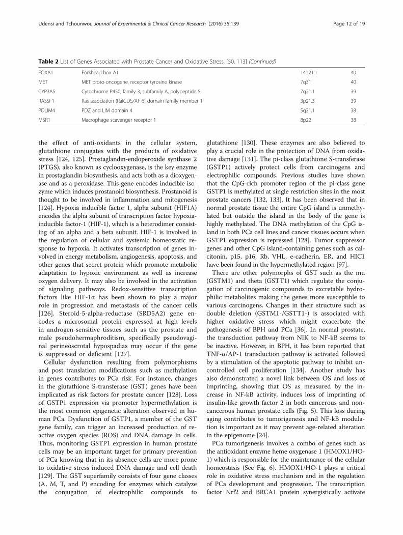
Fig. 5 Oxidative Linked Genes Involved in Prostate Cancer: Thesegenes have been linked to oxidative stress and their expression wasaberrant in prostate cancer [26, 151]
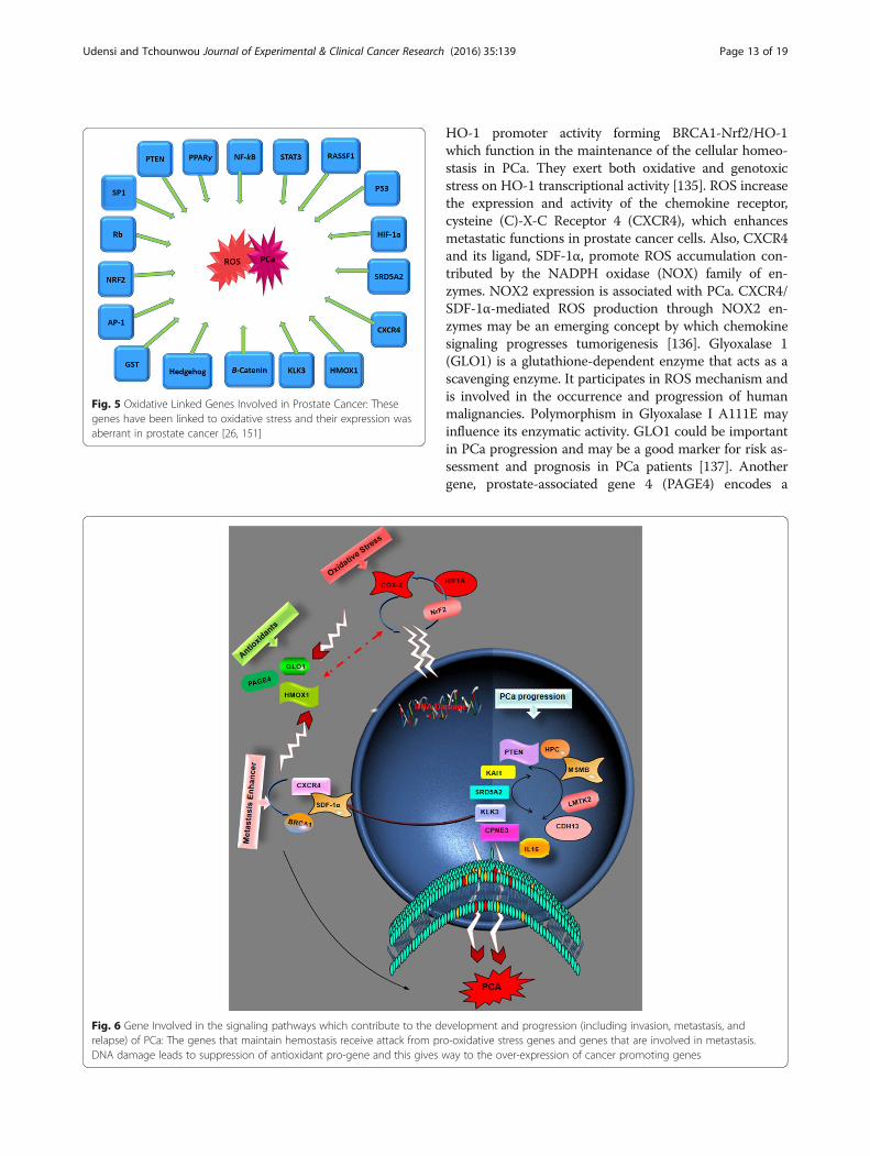
Fig. 6 Gene Involved in the signaling pathways which contribute to the development and progression (including invasion, metastasis, andrelapse) of PCa: The genes that maintain hemostasis receive attack from pro-oxidative stress genes and genes that are involved in metastasis.DNA damage leads to suppression of antioxidant pro-gene and this gives way to the over-expression of cancer promoting genes
Udensi and Tchounwou Journal of Experimental & Clinical Cancer Research (2016) 35:139 Page 13 of 19
protein which protects cells against stress by elevating p21and suppressing ROS production. PAGE4 is a cancer/testis antigen (CTA) that is up-regulated in PCa and seenin symptomatic patients. However, PAGE4 appears toprotect cells from transforming to PCa by its stress-protective and anti-apoptotic activities [138]. The roleof OS and diet in PCa mechanism was observed withthe prostate-specific ablation of PPARγ in mice. Thisaction resulted in tumorigenesis and active autophagy.Placing the mice on high-fat diet (HFD) causeddownregulation of PPARγ-regulated genes and de-creased prostate differentiation. This suggests thatsystemic metabolic stress occurs in glucose and fattyacid metabolism in benign and PCa [67].Pyruvate kinase M2 (PKM2) is essential for aerobic
glycolysis, the dominant metabolic pathway utilized bycancer cells. To determine the association of PKM2 withprostate cancer (PC). PKM2 was found to be upregu-lated and undergoes post translational modification inPCa [139]. Pten as shown in Table 2 is involved in PCacellular programing and signaling and G6PD metabol-ism. AR signaling can promote prostate cancer throughthe upregulation of G6PD and thus providing sugars viathe pentose phosphate pathway. This suggests there areother metabolic pathways apart from the glycolysiswhich prostate cancer growth can be promoted and sus-tained [140].Dietary factors are considered responsible for the geo-
graphical differences in prostate cancer incidence andmortality. Since about 50 % of all men worldwide, fromboth East and West, show evidence of microscopic can-cer by 50 years of age, growth restraint would appear tobe the pragmatic option to the possibility of preventinginitiation [141].
Preventive effects of antioxidants against prostate cancerPublished research has shown that the antioxidantdefense system is decreased in the elderly patients withPCa and BPH [66]. The 2-hydroxy-4-methoxy benzoicacid (HMBA), is an antioxidant that has been shown tohave protective potential against testosterone inducedBPH in rats [29]. Chemopreventive effects of the essen-tial trace element selenium against prostate cancer havebeen shown in preclinical models and human observa-tional studies, but results from clinical trials have beendisappointing. It appears that there is a threshold selen-ium (Se) status below which improvement will decreasePCa risk, but above which supplemental Se may be dele-terious. Different forms of selenium have different ef-fects, and genetic and other factors modify selenium'schemopreventive potential [23]. However, there has beenreduced interest in pursuing chemoprevention strategiestargeting oxidative stress because of the failure of theSelenium and Vitamin E Cancer Prevention Trial
(SELECT) [72]. Another promising therapeutic candi-date is comprised of plant-derived dietary polyphenoliccompounds, such as flavonoids that have cancer cell-specific pro-apoptotic activity and chemopreventive po-tential. Flavonoid glycosides are also found to be DNAhypomethylating agents with an ability to modulate can-cer cell epigenome leading to changes in the gene ex-pression patterns. For example, diosmin which is adietary flavonoid glycoside was found to be activeagainst DU145 cells by promoting genotoxic events thatled to apoptotic cell death [142].
Oxidative stress, antioxidants and prostate cancertreatmentThe classical treatment involves bilateral orchiectomy,or administration of diethylstilbestrol (DES). Other treat-ment strategies are based on endocrine treatment. Theprinciple for endocrine treatment of prostate cancer isto deprive the cancer cells of androgens. Androgendeprivation is an effective treatment for patients with ad-vanced prostate cancer but it is not curative and createsunwanted side effects [143]. For metastatic PCA, castra-tion is still the best choice of treatment as orchiectomy,oestrogen agonists and GnRH agonists produce equivalentclinical responses [144]. It is interesting to note that max-imum androgen blockade (MAB) is not strikingly more ef-fective than single agent GnRH agonist or orchiectomy.However, for locally advanced PCA, nonsteroidal antian-drogen monotherapy is as effective as castration [144].Although oxidative stress has been associated with sev-
eral destructive mechanisms in biological systems, induc-tion of oxidative stress can also provide a means for apotent and safe cancer treatment [145]. Oxidative stresshas been shown to promote castration resistance via an-drogen receptor (AR)-dependent pathway such as ARoverexpression, AR cofactor, and AR post-translationalmodification as well as AR-independent pathway, leadingto the emergence of castration-resistant PCa (CRPC).Thus antioxidants therapy using natural and chemicalROS scavengers and inhibitors of ROS production seemsto be a promising therapy for CRPC as well as preventingcastration resistance [146]. Androgen deprivation therapy(ADT) has been reported to lower basal ROS level inprostate cancer (PCa) and to sensitize PCa to radiation.An in vivo experiment with transgenic adenocarcinoma ofthe mouse prostate (TRAMP) androgen deprivation re-sulted in an increase in basal ROS level in PCa cells withAR expression. also, the genetic Nrf2 upregulation low-ered basal ROS similar to ADT [147].The major androgen within the prostate is dihydrotes-
tosterone (DHT). DHT and 5α-reductase are highly as-sociated with prostate cancer. It has been hypothesizedthat inhibition of 5α-reductase activity might reduce therisk of prostate cancer development, slow tumor
Udensi and Tchounwou Journal of Experimental & Clinical Cancer Research (2016) 35:139 Page 14 of 19
progression and even treat the existing disease. Thetherapeutic mechanism of some of the most recom-mended drugs for prostatic hyperplasia treatment, 5-ARIs, is based on their reductive effect on testosterone,progesterone, androstenedione, cortisol, aldosterone, anddeoxycorticosterone [148]. For example, dutasteride(Avodart) suppresses all three 5α-reductase isoenzymesand reduces dihydrotestosterone in men with benignprostatic hyperplasia [149]. Exposure of human PCa cellsto KML001 (NaAsO2, sodium metaarsenite, Kominox),an orally bioavailable arsenic compound, induces bothapoptotic and autophagic cell death via oxidative stresspathway. Also, KML001 has an antiproliferative effect onDU145 cells in xenograft mice [37]. It has been reportedthat the therapeutic silencing of STAMP2 by administra-tion of nanoliposomal siRNA profoundly inhibits tumorgrowth in two established preclinical PCa models inmice. These findings suggest that STAMP2 is requiredfor PCa progression and thus may serve as a noveltherapeutic target [40]. There is evidence that αATA(8,24) (3α-acetyloxy-tir-8,24-dien-21-oic acid) inhibitsAkt/mammalian target of rapamycin (mTOR) signaling.Compared with related tirucallic acids, αATA (8,24) isthe most potent inhibitor of the proliferation ofandrogen-insensitive PCa cells in vitro and in vivo. InPCa xenografted onto chick chorioallantoic membranesαATA (8,24) induced loss of cell membrane asymmetry,caspase-3 activation, and DNA fragmentation in vitroand in vivo. The ability of αATA (8,24) to inhibitAkt/mTOR signaling and to simultaneously induceoxidative stress could be exploited for the develop-ment of novel antitumor therapeutics with a lowerprofile of toxic side effects [70].
ConclusionsThis review has demonstrated that there are severalstudies which support the fact that oxidative stress playsa critical role in PCa. This oxidative damage is mediatedby an overproduction of oxidant molecules and con-comitant deficiency in the antioxidant system responsewhich causes an imbalance in the cell redox homeosta-sis. PCa is associated with both age, and oxidative stresswhich also contributes to the aging process. Oxidativestress affects all aspects of cellular functions and pro-cesses including DNA replication and cell division, andinflammatory responses. Both endogenous and exogen-ous antioxidants contribute to the reduction of PCa byneutralizing the damaging effect of oxidative stress.However, the oxidative mechanism can also be exploitedfor treatment of prostate related diseases including PCa.
AcknowledgementWe gratefully acknowledge all members of RCMI-Center for EnvironmentalHealth-External Advisory Committee for their support and encouragements.
FundingThis research was supported by a grant from the National Institutes ofHealth/National Institute on Minority Health and Health Disparities(G12MD007581) through the RCMI Center for Environmental Health atJackson State University.
Availability of data and supporting materialsNot applicable. Data sharing not applicable to this article as no datasetswere generated or analyzed during the current study.
Authors’ contributionsUKU and PBT conceived and designed the outline. UKU did the literaturesearch and drafted the manuscript. PBT reviewed and finalized the manuscriptfor submission.
Authors’ informationUdensi K. Udensi, PhD, MPH, MB(ASCP)cm, Research Associate The RCMICenter for Environmental Health, Jackson State University, Jackson MS 39217.Paul B. Tchounwou, Sc.D., F.A.B.I., I.O.M., Associate Dean & PresidentialDistinguished Professor, Director, NIH, RCMI-Center for Environmental Health,Jackson State University, 1400 Lynch Street, Box 18750, Jackson, MS 39217.
Competing interestsThe authors declare that they have no competing interests.
Consent for publicationNot applicable.
Ethical approval and consent to participateNot applicable.
Received: 10 May 2016 Accepted: 6 September 2016
References1. Prostate Cancer FAQS. Prostate Cancer Foundation. http://www.pcf.org/site/
c.leJRIROrEpH/b.5800851/k.645A/Prostate_Cancer_FAQs.htm. Accessed 15Apr 2016.
2. Cancer Among Men, Center For Disease Control and Prevetion. http://www.cdc.gov/cancer/dcpc/data/men.htm. Accessed 15 Apr 2016.
3. Awodele O, Adeyomoye AA, Awodele DF, Fayankinnu VB, Dolapo DC.Cancer distribution pattern in south-western Nigeria. Tanzan J Health Res.2011;13(2):125–31.
4. Theodorescu D. Prostate Cancer: Management of Localized Disease. 2004.www.emedicine.com.
5. Small E. Cecil Textbook of Medicine, Prostate Cancer. WB Saunders, anElsevier imprint. 2004.
6. Cameron KS, Howard CB, Izevbigie EB, Hill BJ, Tchounwou PB. Sensitivityand mechanisms of taxol-resistant prostate adenocarcinoma cells toVernonia amygdalina extract. Exp Toxicol Pathol. 2013;65(6):759–65.
7. Udensi UK, Tchounwou PB. Dual effect of oxidative stress on leukemiacancer induction and treatment. J Exp Clin Cancer Res. 2014;33:106.
8. Seddon M, Looi YH, Shah AM. Oxidative stress and redox signalling incardiac hypertrophy and heart failure. Heart. 2007;93(8):903–7.
9. Minciullo PL, Inferrera A, Navarra M, Calapai G, Magno C, Gangemi S.Oxidative stress in benign prostatic hyperplasia: a systematic review. UrolInt. 2015;94(3):249–54.
10. Aryal M, Pandeya A, Bas BK, Lamsal M, Majhi S, Pandit R, Agrawal CS,Gautam N, Baral N. Oxidative stress in patients with benign prostatehyperplasia. JNMAJ Nepal Med Assoc. 2007;46(167):103–6.
11. Aydin A, Arsova-Sarafinovska Z, Sayal A, Eken A, Erdem O, Erten K, Ozgok Y,Dimovski A. Oxidative stress and antioxidant status in non-metastaticprostate cancer and benign prostatic hyperplasia. Clin Biochem. 2006;39(2):176–9.
12. Battisti V, Maders LD, Bagatini MD, Reetz LG, Chiesa J, Battisti IE, GoncalvesJF, Duarte MM, Schetinger MR, Morsch VM. Oxidative stress and antioxidantstatus in prostate cancer patients: relation to Gleason score, treatment andbone metastasis. Biomed Pharmacother. 2011;65(7):516–24.
13. Cimino S, Favilla V, Russo GI, Galvano F, Li VG, Barbagallo I, Giofre SV,D'Orazio N, Di RA, Madonia M, et al. Oxidative stress and body composition
Udensi and Tchounwou Journal of Experimental & Clinical Cancer Research (2016) 35:139 Page 15 of 19
in prostate cancer and benign prostatic hyperplasia patients. Anticancer Res.2014;34(9):5051–6.
14. Duru R, Njoku O, Maduka I. Oxidative stress indicators in patients withprostate disorders in Enugu, South-East Nigeria. Biomed Res Int. 2014;2014:313015.
15. Ripple MO, Henry WF, Rago RP, Wilding G. Prooxidant-antioxidant shiftinduced by androgen treatment of human prostate carcinoma cells. J NatlCancer Inst. 1997;89(1):40–8.
16. Bostanci Y, Kazzazi A, Momtahen S, Laze J, Djavan B. Correlation betweenbenign prostatic hyperplasia and inflammation. Curr Opin Urol. 2013;23(1):5–10.
17. Shankar E, Bhaskaran N, Maclennan GT, Liu G, Daneshgari F, Gupta S.Inflammatory Signaling Involved in High-Fat Diet Induced Prostate Diseases.J Urol Res. 2015;2(1)2018.
18. Sugar LM. Inflammation and prostate cancer. Can J Urol. 2006;13 Suppl 1:46–7.19. He JH, Zhang JZ, Han ZP, Wang L, Lv YB, Li YG. Reciprocal regulation of
PCGEM1 and miR-145 promote proliferation of LNCaP prostate cancer cells.J Exp Clin Cancer Res. 2014;33:72.
20. Jones RA, Underwood SM, Rivers BM. Reducing prostate cancer morbidityand mortality in African American men: issues and challenges. Clin J OncolNurs. 2007;11(6):865–72.
21. Almushatat AS, Talwar D, McArdle PA, Williamson C, Sattar N, O'Reilly DS,Underwood MA, McMillan DC. Vitamin antioxidants, lipid peroxidation andthe systemic inflammatory response in patients with prostate cancer. Int JCancer. 2006;118(4):1051–3.
22. Chang SN, Han J, Abdelkader TS, Kim TH, Lee JM, Song J, Kim KS, Park JH,Park JH. High animal fat intake enhances prostate cancer progression andreduces glutathione peroxidase 3 expression in early stages of TRAMP mice.Prostate. 2014;74(13):1266–77.
23. Christensen MJ. Selenium and prostate cancer prevention: what next–ifanything? Cancer Prev Res (Phila). 2014;7(8):781–5.
24. Li C, Yang L, Lin C. Long noncoding RNAs in prostate cancer: mechanismsand applications. Mol Cell Oncol. 2014;1(3):e963469.
25. El GM, Buchele B, Syrovets T, Agnolet S, Schneider B, Schmidt CQ, Simmet T.An alpha-acetoxy-tirucallic acid isomer inhibits Akt/mTOR signaling andinduces oxidative stress in prostate cancer cells. J Pharmacol Exp Ther. 2015;352(1):33–42.
26. Gupta-Elera G, Garrett AR, Robison RA, O'Neill KL. The role of oxidative stressin prostate cancer. Eur J Cancer Prev. 2012;21(2):155–62.
27. Khandrika L, Kumar B, Koul S, Maroni P, Koul HK. Oxidative stress in prostatecancer. Cancer Lett. 2009;282(2):125–36.
28. Merendino RA, Salvo F, Saija A, Di PG, Tomaino A, Minciullo PL, Fraccica G,Gangemi S. Malondialdehyde in benign prostate hypertrophy: a usefulmarker? Mediators Inflamm. 2003;12(2):127–8.
29. Ahmad M, Suhail N, Mansoor T, Banu N, Ahmad S. Evaluation of oxidativestress and DNA damage in benign prostatic hyperplasia patients andcomparison with controls. Indian J Clin Biochem. 2012;27(4):385–8.
30. Srivastava DS, Mittal RD. Free radical injury and antioxidant status in patientswith benign prostate hyperplasia and prostate cancer. Indian J ClinBiochem. 2005;20(2):162–5.
31. Arsova-Sarafinovska Z, Eken A, Matevska N, Erdem O, Sayal A, Savaser A,Banev S, Petrovski D, Dzikova S, Georgiev V, et al. Increased oxidative/nitrosative stress and decreased antioxidant enzyme activities in prostatecancer. Clin Biochem. 2009;42(12):1228–35.
32. Pace G, Di MC, De AD, Corbacelli C, Di RL, Vicentini C, Miano L, TozziCiancarelli MG. Oxidative stress in benign prostatic hyperplasia and prostatecancer. Urol Int. 2010;85(3):328–33.
33. Teillac P, Peyret C, Leroy M, Najean Y, Le DA. [Prostate-specific antigen inprostatic pathology]. Ann Urol (Paris). 1988;22(3):193–6.
34. Iguchi T, Wang CY, Delongchamps NB, Kato M, Tamada S, Yamasaki T, de laRoza G, Nakatani T, Haas GP. Association of MnSOD AA Genotype with theProgression of Prostate Cancer. PLoS One. 2015;10(7):e0131325.
35. Kotrikadze N, Alibegashvili M, Zibzibadze M, Abashidze N, Chigogidze T,Managadze L, Artsivadze K. Activity and content of antioxidant enzymes inprostate tumors. Exp Oncol. 2008;30(3):244–7.
36. Kumar V, Yadav CS, Datta SK, Singh S, Ahmed RS, Goel S, Gupta S, MustafaM, Grover RK, Banerjee BD. Association of GSTM1 and GSTT1 polymorphismwith lipid peroxidation in benign prostate hyperplasia and prostate cancer:a pilot study. Dis Markers. 2011;30(4):163–9.
37. Azoulay L, Eberg M, Benayoun S, Pollak M. 5alpha-reductase inhibitors andthe risk of cancer-related mortality in men with prostate cancer. JAMAOncol. 2015;1(3):314–20.
38. Baltaci S, Orhan D, Gogus C, Turkolmez K, Tulunay O, Gogus O. Induciblenitric oxide synthase expression in benign prostatic hyperplasia, low- andhigh-grade prostatic intraepithelial neoplasia and prostatic carcinoma. BJUInt. 2001;88(1):100–3.
39. Bidoli E, Talamini R, Zucchetto A, Bosetti C, Negri E, Lenardon O, Maso LD,Polesel J, Montella M, Franceschi S, et al. Dietary vitamins E and C andprostate cancer risk. Acta Oncol. 2009;48(6):890–4.
40. Bai XY, Qu X, Jiang X, Xu Z, Yang Y, Su Q, Wang M, Wu H. Associationbetween dietary vitamin c intake and risk of prostate cancer: a meta-analysis involving 103,658 subjects. J Cancer. 2015;6(9):913–21.
41. Maramag C, Menon M, Balaji KC, Reddy PG, Laxmanan S. Effect of vitamin Con prostate cancer cells in vitro: effect on cell number, viability, and DNAsynthesis. Prostate. 1997;32(3):188–95.
42. Thieu W, Tilki D, de Vere White RW, Evans CP. The role of microRNA incastration-resistant prostate cancer. Urol Oncol. 2014;32(5):517–23.
43. Evans P, Halliwell B. Micronutrients: oxidant/antioxidant status. Br J Nutr.2001;85 Suppl 2:S67–74.
44. Manzanares W, Dhaliwal R, Jiang X, Murch L, Heyland DK. Antioxidantmicronutrients in the critically ill: a systematic review and meta-analysis. CritCare. 2012;16(2):R66.
45. Kristal AR, Till C, Song X, Tangen CM, Goodman PJ, Neuhauser ML, SchenkJM, Thompson IM, Meyskens Jr FL, Goodman GE, et al. Plasma vitamin Dand prostate cancer risk: results from the selenium and Vitamin E cancerprevention trial. Cancer Epidemiol Biomarkers Prev. 2014;23(8):1494–504.
46. Eichholzer M, Steinbrecher A, Kaaks R, Teucher B, Linseisen J, Rohrmann S.Effects of selenium status, dietary glucosinolate intake and serumglutathione S-transferase alpha activity on the risk of benign prostatichyperplasia. BJU Int. 2012;110(11 Pt C):E879–85.
47. Zachara BA, Szewczyk-Golec K, Tyloch J, Wolski Z, Szylberg T, Stepien S,Kwiatkowski S, Bloch-Boguslawska E, Wasowicz W. Blood and tissue seleniumconcentrations and glutathione peroxidase activities in patients with prostatecancer and benign prostate hyperplasia. Neoplasma. 2005;52(3):248–54.
48. Freedland SJ, Carducci M, Kroeger N, Partin A, Rao JY, Jin Y, Kerkoutian S,Wu H, Li Y, Creel P, et al. A double-blind, randomized, neoadjuvant study ofthe tissue effects of POMx pills in men with prostate cancer before radicalprostatectomy. Cancer Prev Res (Phila). 2013;6(10):1120–7.
49. Bostwick DG, Qian J. High-grade prostatic intraepithelial neoplasia. ModPathol. 2004;17(3):360–79.
50. Malignant tumor of prostate: Prostate cancer. National Center forBiotechnology Information (NCBI) http://www.ncbi.nlm.nih.gov/gtr/conditions/C0376358/. Accessed 04/26/16.
51. Feldman BJ, Feldman D. The development of androgen-independentprostate cancer. Nat Rev Cancer. 2001;1(1):34–45.
52. Yang G, Truong LD, Wheeler TM, Thompson TC. Caveolin-1 expression inclinically confined human prostate cancer: a novel prognostic marker.Cancer Res. 1999;59(22):5719–23.
53. Ziaran S, Varchulova NZ, Bohmer D, Danisovic L. Biomarkers fordetermination prostate cancer: implication for diagnosis and prognosis.Neoplasma. 2015;62(5):683–91.
54. Gurunadha Rao Tunuguntla HS, Evans CP. Management of prostatitis.Prostate Cancer Prostatic Dis. 2002;5(3):172–9.
55. Shoskes DA, Berger R, Elmi A, Landis JR, Propert KJ, Zeitlin S. Muscletenderness in men with chronic prostatitis/chronic pelvic pain syndrome:the chronic prostatitis cohort study. J Urol. 2008;179(2):556–60.
56. Krieger JN, Nyberg Jr L, Nickel JC. NIH consensus definition andclassification of prostatitis. JAMA. 1999;282(3):236–7.
57. Litwin MS, McNaughton-Collins M, Fowler Jr FJ, Nickel JC, Calhoun EA,Pontari MA, Alexander RB, Farrar JT, O'Leary MP. The National Institutes ofHealth chronic prostatitis symptom index: development and validation of anew outcome measure. Chronic Prostatitis Collaborative Research Network.J Urol. 1999;162(2):369–75.
58. Sharp VJ, Takacs EB, Powell CR. Prostatitis: diagnosis and treatment. Am FamPhysician. 2010;82(4):397–406.
59. Dotan Y, Lichtenberg D, Pinchuk I. Lipid peroxidation cannot be used as auniversal criterion of oxidative stress. Prog Lipid Res. 2004;43(3):200–27.
60. Taheri MG, Hosseini-Zijoud SM, Heidary ST, Ghasemi H, Ranjbar A.Attenuation of cisplathin-induced toxic oxidative stress by propofol. AnesthPain Med. 2014;4(4):e14221.
61. Milne GL, Yin H, Brooks JD, Sanchez S, Jackson RL, Morrow JD.Quantification of F2-isoprostanes in biological fluids and tissues as ameasure of oxidant stress. Methods Enzymol. 2007;433:113–26.
Udensi and Tchounwou Journal of Experimental & Clinical Cancer Research (2016) 35:139 Page 16 of 19
62. Kullisaar T, Turk S, Punab M, Mandar R. Oxidative stress–cause orconsequence of male genital tract disorders? Prostate. 2012;72(9):977–83.
63. Ronnau CG, Verhaegh GW, Luna-Velez MV, Schalken JA. Noncoding RNAs asnovel biomarkers in prostate cancer. Biomed Res Int. 2014;2014:591703.
64. Ali MI, Kondreddi HD, Veeresh B. Protective effect of 2-hydroxy-4-methoxybenzoic acid on testosterone induced benign prostatic hyperplasia in Wisterrats. Eur J Pharmacol. 2013;698(1–3):397–403.
65. Alcaraz A, Hammerer P, Tubaro A, Schroder FH, Castro R. Is there evidenceof a relationship between benign prostatic hyperplasia and prostate cancer?Findings of a literature review. Eur Urol. 2009;55(4):864–73.
66. Szewczyk-Golec K, Tyloch J, Czuczejko J. Antioxidant defense system inprostate adenocarcinoma and benign prostate hyperplasia of elderlypatients. Neoplasma. 2015;62(1):119–23.
67. Strand DW, Jiang M, Murphy TA, Yi Y, Konvinse KC, Franco OE, Wang Y,Young JD, Hayward SW. PPARgamma isoforms differentially regulatemetabolic networks to mediate mouse prostatic epithelial differentiation.Cell Death Dis. 2012;3:e361.
68. Figg WD, Thompson IM. Effect of 5alpha-reductase inhibitor use onmortality from prostate cancer. JAMA Oncol. 2015;1(3):321–2.
69. Hamilton MP, Rajapakshe KI, Bader DA, Cerne JZ, Smith EA, Coarfa C, HartigSM, McGuire SE. The landscape of microRNA targeting in prostate cancerdefined by AGO-PAR-CLIP. Neoplasia. 2016;18(6):356–70.
70. American Cancer Society. Cancer Facts & Figures 2013. Atlanta: AmericanCancer Society; 2013. http://www.cancer.org/research/cancerfactsfigures/cancerfactsfigures/cancer-facts-figures.
71. Desai N, Sabanegh Jr E, Kim T, Agarwal A. Free radical theory of aging:implications in male infertility. Urology. 2010;75(1):14–9.
72. Paschos A, Pandya R, Duivenvoorden WC, Pinthus JH. Oxidative stress inprostate cancer: changing research concepts towards a novel paradigm forprevention and therapeutics. Prostate Cancer Prostatic Dis. 2013;16(3):217–25.
73. Hanahan D, Weinberg RA. Hallmarks of cancer: the next generation. Cell.2011;144(5):646–74.
74. Bolton EM, Tuzova AV, Walsh AL, Lynch T, Perry AS. Noncoding RNAs inprostate cancer: the long and the short of it. Clin Cancer Res. 2014;20(1):35–43.
75. Mouraviev V, Lee B, Patel V, Albala D, Johansen TE, Partin A, Ross A, Perera RJ.Clinical prospects of long noncoding RNAs as novel biomarkers and therapeutictargets in prostate cancer. Prostate Cancer Prostatic Dis. 2016;19(1):14–20.
76. Malik B, Feng FY. Long noncoding RNAs in prostate cancer: overview andclinical implications. Asian J Androl. 2016;18(4):568–74.
77. Velonas VM, Woo HH, dos Remedios CG, Assinder SJ. Current status ofbiomarkers for prostate cancer. Int J Mol Sci. 2013;14(6):11034–60.
78. Lee RC, Feinbaum RL, Ambros V. The C. elegans heterochronic gene lin-4encodes small RNAs with antisense complementarity to lin-14. Cell. 1993;75(5):843–54.
79. Sapre N, Selth LA. Circulating microRNAs as biomarkers of prostate cancer:the state of play. Prostate Cancer. 2013;2013:539680.
80. Takahara K, Ii M, Inamoto T, Nakagawa T, Ibuki N, Yoshikawa Y, Tsujino T,Uchimoto T, Saito K, Takai T, et al. microRNA-145 mediates the inhibitoryeffect of adipose tissue-derived stromal cells on prostate cancer. Stem CellsDev. 2016;25(17):1290–8.
81. Nguyen HC, Xie W, Yang M, Hsieh CL, Drouin S, Lee GS, Kantoff PW.Expression differences of circulating microRNAs in metastatic castrationresistant prostate cancer and low-risk, localized prostate cancer. Prostate.2013;73(4):346–54.
82. Seashols-Williams SJ, Budd W, Clark GC, Wu Q, Daniel R, Dragoescu E,Zehner ZE. miR-9 Acts as an OncomiR in Prostate Cancer throughMultiple Pathways That Drive Tumour Progression and Metastasis.PLoS One. 2016;11(7):e0159601.
83. Liu R, Liu C, Zhang D, Liu B, Chen X, Rycaj K, Jeter C, Calhoun-Davis T, Li Y,Yang T, et al. miR-199a-3p targets stemness-related and mitogenic signalingpathways to suppress the expansion and tumorigenic capabilities ofprostate cancer stem cells. Oncotarget. 2016. doi:10.18632/oncotarget.10652.
84. Mitchell PS, Parkin RK, Kroh EM, Fritz BR, Wyman SK, Pogosova-AgadjanyanEL, Peterson A, Noteboom J, O'Briant KC, Allen A, et al. CirculatingmicroRNAs as stable blood-based markers for cancer detection. Proc NatlAcad Sci U S A. 2008;105(30):10513–8.
85. Mahn R, Heukamp LC, Rogenhofer S, von Ruecker A, Muller SC, Ellinger J.Urology. 2011;77(5):1265.e9–16.
86. Lodes MJ, Caraballo M, Suciu D, Munro S, Kumar A, Anderson B. Detectionof cancer with serum miRNAs on an oligonucleotide microarray. PLoS One.2009;4(7):e6229.
87. Zhang Y, Liu D, Chen X, Li J, Li L, Bian Z, Sun F, Lu J, Yin Y, Cai X, et al.Secreted monocytic miR-150 enhances targeted endothelial cell migration.Mol Cell. 2010;39(1):133–44.
88. Moltzahn F, Olshen AB, Baehner L, Peek A, Fong L, Stoppler H, Simko J,Hilton JF, Carroll P, Blelloch R. Microfluidic-based multiplex qRT-PCRidentifies diagnostic and prognostic microRNA signatures in the sera ofprostate cancer patients. Cancer Res. 2011;71(2):550–60.
89. Brase JC, Johannes M, Schlomm T, Falth M, Haese A, Steuber T, Beissbarth T,Kuner R, Sultmann H. Circulating miRNAs are correlated with tumorprogression in prostate cancer. Int J Cancer. 2011;128(3):608–16.
90. Gonzales JC, Fink LM, Goodman Jr OB, Symanowski JT, Vogelzang NJ, WardDC. Comparison of circulating MicroRNA 141 to circulating tumor cells,lactate dehydrogenase, and prostate-specific antigen for determiningtreatment response in patients with metastatic prostate cancer. ClinGenitourin Cancer. 2011;9(1):39–45.
91. Zheng C, Yinghao S, Li J. MiR-221 expression affects invasion potential ofhuman prostate carcinoma cell lines by targeting DVL2. Med Oncol. 2012;29(2):815–22.
92. Yaman Agaoglu F, Kovancilar M, Dizdar Y, Darendeliler E, Holdenrieder S,Dalay N, Gezer U. Investigation of miR-21, miR-141, and miR-221 in bloodcirculation of patients with prostate cancer. Tumour Biol. 2011;32(3):583–8.
93. Selth LA, Townley S, Gillis JL, Ochnik AM, Murti K, Macfarlane RJ, Chi KN,Marshall VR, Tilley WD, Butler LM. Discovery of circulating microRNAsassociated with human prostate cancer using a mouse model of disease. IntJ Cancer. 2012;131(3):652–61.
94. Bryant RJ, Pawlowski T, Catto JW, Marsden G, Vessella RL, Rhees B, Kuslich C,Visakorpi T, Hamdy FC. Changes in circulating microRNA levels associatedwith prostate cancer. Br J Cancer. 2012;106(4):768–74.
95. Chen ZH, Zhang GL, Li HR, Luo JD, Li ZX, Chen GM, Yang J. A panel of fivecirculating microRNAs as potential biomarkers for prostate cancer. Prostate.2012;72(13):1443–52.
96. Shen J, Hruby GW, McKiernan JM, Gurvich I, Lipsky MJ, Benson MC, SantellaRM. Dysregulation of circulating microRNAs and prediction of aggressiveprostate cancer. Prostate. 2012;72(13):1469–77.
97. Sanders I, Holdenrieder S, Walgenbach-Brunagel G, von Ruecker A,Kristiansen G, Muller SC, Ellinger J. Evaluation of reference genes for theanalysis of serum miRNA in patients with prostate cancer, bladder cancerand renal cell carcinoma. Int J Urol. 2012;19(11):1017–25.
98. Nickel JC, Roehrborn CG, O'Leary MP, Bostwick DG, Somerville MC,Rittmaster RS. The relationship between prostate inflammation and lowerurinary tract symptoms: examination of baseline data from the REDUCE trial.Eur Urol. 2008;54(6):1379–84.
99. Sciarra A, Mariotti G, Salciccia S, Autran GA, Monti S, Toscano V, Di SF. Prostategrowth and inflammation. J Steroid Biochem Mol Biol. 2008;108(3–5):254–60.
100. Tsujii M, DuBois RN. Alterations in cellular adhesion and apoptosis inepithelial cells overexpressing prostaglandin endoperoxide synthase 2. Cell.1995;83(3):493–501.
101. Yoshida GJ. Metabolic reprogramming: the emerging concept andassociated therapeutic strategies. J Exp Clin Cancer Res. 2015;34:111.
102. Udensi UK, Cohly HH, Graham-Evans BE, Ndebele K, Garcia-Reyero N,Nanduri B, Tchounwou PB, Isokpehi RD. Aberrantly expressed genes inHaCaT keratinocytes chronically exposed to arsenic trioxide. BiomarkerInsights. 2011;6:7–16.
103. Nwosu V, Carpten J, Trent JM, Sheridan R. Heterogeneity of geneticalterations in prostate cancer: evidence of the complex nature of thedisease. Hum Mol Genet. 2001;10(20):2313–8.
104. Chu C, Qu K, Zhong FL, Artandi SE, Chang HY. Genomic maps of longnoncoding RNA occupancy reveal principles of RNA-chromatin interactions.Mol Cell. 2011;44(4):667–78.
105. Zhao J, Ohsumi TK, Kung JT, Ogawa Y, Grau DJ, Sarma K, Song JJ, KingstonRE, Borowsky M, Lee JT. Genome-wide identification of polycomb-associated RNAs by RIP-seq. Mol Cell. 2010;40(6):939–53.
106. Karayi MK, Markham AF. Molecular biology of prostate cancer. ProstateCancer Prostatic Dis. 2004;7(1):6–20.
107. Gronberg H, Smith J, Emanuelsson M, Jonsson BA, Bergh A, Carpten J, IsaacsW, Xu J, Meyers D, Trent J, et al. In Swedish families with hereditary prostatecancer, linkage to the HPC1 locus on chromosome 1q24-25 is restricted tofamilies with early-onset prostate cancer. Am J Hum Genet. 1999;65(1):134–40.
108. Xu J, Meyers D, Freije D, Isaacs S, Wiley K, Nusskern D, Ewing C, Wilkens E,Bujnovszky P, Bova GS, et al. Evidence for a prostate cancer susceptibilitylocus on the X chromosome. Nat Genet. 1998;20(2):175–9.
Udensi and Tchounwou Journal of Experimental & Clinical Cancer Research (2016) 35:139 Page 17 of 19
109. Eeles RA, Kote-Jarai Z, Giles GG, Olama AA, Guy M, Jugurnauth SK,Mulholland S, Leongamornlert DA, Edwards SM, Morrison J, et al. Multiplenewly identified loci associated with prostate cancer susceptibility. NatGenet. 2008;40(3):316–21.
110. Thomas G, Jacobs KB, Yeager M, Kraft P, Wacholder S, Orr N, Yu K,Chatterjee N, Welch R, Hutchinson A, et al. Multiple loci identified in agenome-wide association study of prostate cancer. Nat Genet. 2008;40(3):310–5.
111. Song LN, Bowen C, Gelmann EP. Structural and functional interactions ofthe prostate cancer suppressor protein NKX3.1 with topoisomerase I.Biochem J. 2013;453(1):125–36.
112. Ntais C, Polycarpou A, Ioannidis JP. Association of GSTM1, GSTT1, and GSTP1gene polymorphisms with the risk of prostate cancer: a meta-analysis.Cancer Epidemiol Biomarkers Prev. 2005;14(1):176–81.
113. Prostate Cancer Molecular Biology, http://www.cancerindex.org/. Accessed15 Apr 2016.
114. Bettin A, Reyes I, Reyes N. Gene expression profiling of prostate cancer-associated genes identifies fibromodulin as potential novel biomarkerfor prostate cancer. Int J Biol Markers. 2015. doi:10.5301/jbm.5000184.
115. Lee HA, Park I, Byun HJ, Jeoung D, Kim YM, Lee H. Metastasis suppressorKAI1/CD82 attenuates the matrix adhesion of human prostate cancer cellsby suppressing fibronectin expression and beta1 integrin activation. CellPhysiol Biochem. 2011;27(5):575–86.
116. Sjoblom L, Saramaki O, Annala M, Leinonen K, Nattinen J, Tolonen T,Wahlfors T, Nykter M, Bova GS, Schleutker J, et al. Microseminoprotein-BetaExpression in Different Stages of Prostate Cancer. PLoSOne. 2016;11(3):e0150241.
117. PLOS ONE Staff. Correction: Microseminoprotein-Beta Expression in DifferentStages of Prostate Cancer. PLoS One. 2016;11(4):e0153732.
118. Ikeda M, Hirabayashi S, Fujiwara N, Mori H, Kawata A, Iida J, Bao Y, Sato Y,Iida T, Sugimura H, et al. Ras-association domain family protein 6 inducesapoptosis via both caspase-dependent and caspase-independent pathways.Exp Cell Res. 2007;313(7):1484–95.
119. Muhammad LA, Saad F. The role of clusterin in prostate cancer: treatmentresistance and potential as a therapeutic target. Expert Rev Anticancer Ther.2015;15(9):1049–61.
120. Fujimoto N, Kanno J. Increase in prostate stem cell antigen expression inprostatic hyperplasia induced by testosterone and 17beta-estradiol in C57BLmice. J Steroid Biochem Mol Biol. 2016;158:56–62.
121. Lokody IB, Francis JC, Gardiner JR, Erler JT, Swain A. Pten Regulates EpithelialCytodifferentiation during Prostate Development. PLoS One. 2015;10(6):e0129470.
122. Alvarez-Cubero MJ, Martinez-Gonzalez LJ, Saiz M, Carmona-Saez P,Alvarez JC, Pascual-Geler M, Lorente JA, Cozar JM. Prognostic role ofgenetic biomarkers in clinical progression of prostate cancer. Exp MolMed. 2015;47:e176.
123. Izmirli M, Arikan B, Bayazit Y, Alptekin D. Associations of polymorphisms inHPC2/ELAC2 and SRD5A2 genes with benign prostate hyperplasia in Turkishmen. Asian Pac J Cancer Prev. 2011;12(3):731–3.
124. Cao DL, Ye DW, Dai B, Zhang HL, Shen YJ, Zhu Y, Zhu YP, Shi GH, MaCG, Xiao WJ, et al. Association of glutathione S-transferase T1 and M1polymorphisms with prostate cancer susceptibility in populations ofAsian descent: a meta-analysis. Oncotarget. 2015;6(34):35843–50.
125. Zabaiou N, Mabed D, Lobaccaro JM, Lahouel M. Oxidative stress in benignprostate hyperplasia. Andrologia. 2015. doi:10.1111/and.12420.
126. Mak P, Li J, Samanta S, Mercurio AM. ERbeta regulation of NF-kBactivation in prostate cancer is mediated by HIF-1. Oncotarget. 2015;6(37):40247–54.
127. Poniah P, Mohamed Z, Apalasamy YD, Mohd ZS, Kuppusamy S, Razack AH.Genetic polymorphisms in the androgen metabolism pathway and risk ofprostate cancer in low incidence Malaysian ethnic groups. Int J Clin ExpMed. 2015;8(10):19232–40.
128. Millar DS, Ow KK, Paul CL, Russell PJ, Molloy PL, Clark SJ. Detailedmethylation analysis of the glutathione S-transferase pi (GSTP1) gene inprostate cancer. Oncogene. 1999;18(6):1313–24.
129. Kanwal R, Pandey M, Bhaskaran N, Maclennan GT, Fu P, Ponsky LE, Gupta S.Protection against oxidative DNA damage and stress in human prostate byglutathione S-transferase P1. Mol Carcinog. 2014;53(1):8–18.
130. Hayes JD, Pulford DJ. The glutathione S-transferase supergene family:regulation of GST and the contribution of the isoenzymes to cancerchemoprotection and drug resistance. Crit Rev Biochem Mol Biol. 1995;30(6):445–600.
131. Ryberg D, Skaug V, Hewer A, Phillips DH, Harries LW, Wolf CR, Ogreid D,Ulvik A, Vu P, Haugen A. Genotypes of glutathione transferase M1 and P1and their significance for lung DNA adduct levels and cancer risk.Carcinogenesis. 1997;18(7):1285–9.
132. Lee WH, Morton RA, Epstein JI, Brooks JD, Campbell PA, Bova GS, HsiehWS, Isaacs WB, Nelson WG. Cytidine methylation of regulatorysequences near the pi-class glutathione S-transferase gene accompanieshuman prostatic carcinogenesis. Proc Natl Acad Sci U S A. 1994;91(24):11733–7.
133. Cookson MS, Reuter VE, Linkov I, Fair WR. Glutathione S-transferase PI (GST-pi) class expression by immunohistochemistry in benign and malignantprostate tissue. J Urol. 1997;157(2):673–6.
134. Hamid AR, Umbas R, Mochtar CA. Recent role of inflammation in prostatediseases: chemoprevention development opportunity. Acta Med Indones.2011;43(1):59–65.
135. Labanca E, De LP, Gueron G, Paez A, Moiola CP, Massillo C, Porretti J,Giudice J, Zalazar F, Navone N, et al. Association of HO-1 and BRCA1 iscritical for the maintenance of cellular homeostasis in prostate cancer. MolCancer Res. 2015;13:1455–64.
136. Jones KJ, Chetram MA, Bethea DA, Bryant LK, Odero-Marah V, HintonCV. Cysteine (C)-X-C Receptor 4 Regulates NADPH Oxidase-2 DuringOxidative Stress in Prostate Cancer Cells. Cancer Microenviron. 2013[Epub ahead of print].
137. Antognelli C, Mezzasoma L, Mearini E, Talesa VN. Glyoxalase 1-419C>Avariant is associated with oxidative stress: implications in prostate cancerprogression. PLoS One. 2013;8(9):e74014.
138. Zeng Y, Gao D, Kim JJ, Shiraishi T, Terada N, Kakehi Y, Kong C, GetzenbergRH, Kulkarni P. Prostate-associated gene 4 (PAGE4) protects cells againststress by elevating p21 and suppressing reactive oxygen speciesproduction. Am J Clin Exp Urol. 2013;1(1):39–52.
139. Wong N, Yan J, Ojo D, De MJ, Cutz JC, Tang D. Changes in PKM2associate with prostate cancer progression. Cancer Invest. 2014;32(7):330–8.
140. Tsouko E, Khan AS, White MA, Han JJ, Shi Y, Merchant FA, Sharpe MA, Xin L,Frigo DE. Regulation of the pentose phosphate pathway by an androgenreceptor-mTOR-mediated mechanism and its role in prostate cancer cellgrowth. Oncogenesis. 2014;3:e103.
141. Griffiths K, Prezioso D, Turkes A, Denis LJ. The prevention of prostate cancer.Recent Results Cancer Res. 2007;175:33–63.
142. Lewinska A, Siwak J, Rzeszutek I, Wnuk M. Diosmin induces genotoxicityand apoptosis in DU145 prostate cancer cell line. Toxicol In Vitro. 2015;29(3):417–25.
143. Denis LJ, Griffiths K. Endocrine treatment in prostate cancer. Semin SurgOncol. 2000;18(1):52–74.
144. Tammela TL. Endocrine prevention and treatment of prostate cancer. MolCell Endocrinol. 2012;360(1–2):59–67.
145. Rozanov D, Cheltsov A, Sergienko E, Vasile S, Golubkov V, Aleshin AE, LevinT, Traer E, Hann B, Freimuth J, et al. TRAIL-Based High ThroughputScreening Reveals a Link between TRAIL-Mediated Apoptosis andGlutathione Reductase, a Key Component of Oxidative Stress Response.PLoS One. 2015;10(6):e0129566.
146. Shiota M, Yokomizo A, Naito S. [The development of therapeutics targetingoxidative stress in prostate cancer]. Nihon Rinsho. 2014;72(12):2131–5.
147. Guo J, Wang M, Liu X. MicroRNA-195 suppresses tumor cell proliferationand metastasis by directly targeting BCOX1 in prostate carcinoma. J ExpClin Cancer Res. 2015;34:91.
148. Azzouni F, Godoy A, Li Y, Mohler J. The 5 alpha-reductase isozyme family: areview of basic biology and their role in human diseases. Adv Urol. 2012;2012:530121.
149. Clark RV, Hermann DJ, Cunningham GR, Wilson TH, Morrill BB, Hobbs S.Marked suppression of dihydrotestosterone in men with benign prostatichyperplasia by dutasteride, a dual 5alpha-reductase inhibitor. J ClinEndocrinol Metab. 2004;89(5):2179–84.
150. Naber KG. Antibiotic treatment of chronic bacterial prostatitis. In: Nickel JC,editor. Textbook of Prostatitis. Oxford: Isis Medical Media; 1999. p. 285–92.
151. Gupta SC, Hevia D, Patchva S, Park B, Koh W, Aggarwal BB. Upsides anddownsides of reactive oxygen species for cancer: the roles of reactiveoxygen species in tumorigenesis, prevention, and therapy. Antioxid RedoxSignal. 2012;16(11):1295–322.
152. Gradini R, Realacci M, Ginepri A, Naso G, Santangelo C, Cela O, Sale P,Berardi A, Petrangeli E, Gallucci M, et al. Nitric oxide synthases in normal
Udensi and Tchounwou Journal of Experimental & Clinical Cancer Research (2016) 35:139 Page 18 of 19

and benign hyperplastic human prostate: immunohistochemistry andmolecular biology. J Pathol. 1999;189(2):224–9.
153. Hu M, Dillard C. Plasma SH and GSH measurement. Methods Enzymol. 1994;233(385):87.
154. Moore K, Roberts LJ. Measurement of lipid peroxidation. Free Radic Res.1998;28(6):659–71.
155. Grune T. Protein Oxidation Products as Biomarkers. Free Radic Biol Med.2014;75 Suppl 1:S7.
156. Fields DA, Goran MI, McCrory MA. Body-composition assessment via air-displacement plethysmography in adults and children: a review. Am J ClinNutr. 2002;75(3):453–67.
• We accept pre-submission inquiries
• Our selector tool helps you to find the most relevant journal
• We provide round the clock customer support
• Convenient online submission
• Thorough peer review
• Inclusion in PubMed and all major indexing services
• Maximum visibility for your research
Submit your manuscript atwww.biomedcentral.com/submit
Submit your next manuscript to BioMed Central and we will help you at every step:
Udensi and Tchounwou Journal of Experimental & Clinical Cancer Research (2016) 35:139 Page 19 of 19


![Endometrium presentation - Dr Wright[1] · Endometrial Hyperplasia Simple hyperplasia Complex hyperplasia (adenomatous) Simple atypical hyperplasia ... Progression of Hyperplasia](https://img.pdfslide.net/doc/110x75/5b8a421e7f8b9a50388bc13d/endometrium-presentation-dr-wright1-endometrial-hyperplasia-simple-hyperplasia.jpg)