Embed Size (px)
Citation preview
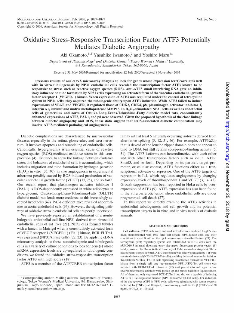
MOLECULAR AND CELLULAR BIOLOGY, Feb. 2006, p. 1087–1097 Vol. 26, No. 30270-7306/06/$08.00�0 doi:10.1128/MCB.26.3.1087–1097.2006Copyright © 2006, American Society for Microbiology. All Rights Reserved.
Oxidative Stress-Responsive Transcription Factor ATF3 PotentiallyMediates Diabetic Angiopathy
Aki Okamoto,1,2 Yasuhiko Iwamoto,2 and Yoshiro Maru1*Department of Pharmacology1 and Diabetes Center,2 Tokyo Women’s Medical University,
8-1 Kawada-cho, Shinjuku-ku, Tokyo 162-8666, Japan
Received 31 May 2005/Returned for modification 12 July 2005/Accepted 8 November 2005
Previous results of our cDNA microarray analysis to look for genes whose expression level correlates wellwith in vitro tubulogenesis by NP31 endothelial cells revealed the transcription factor ATF3 known to beresponsive to stress such as reactive oxygen species (ROS). Anti-ATF3 small interfering RNA gave an inhib-itory influence on tube formation by NP31 cells expressing an activated form of the vascular endothelial growthfactor receptor 1 (VEGFR-1) kinase. When expression of ATF3 was regulated under the control of tetracyclinesystem in NP31 cells, they acquired the tubulogenic ability upon ATF3 induction. While ATF3 failed to induceexpressions of VEGF and VEGFR, it regulated those of CDK2, CDK4, p8, plasminogen activator inhibitor 1,integrin �1, subunit and matrix metalloprotease MMP13. In H2O2-stimulated NP31 cells as well as endothelialcells of glomerulus and aorta of Otsuka-Long-Evans-Tokushima-Fatty diabetic model rats, concomitantlyenhanced expressions of ATF3, PAI-1, and p8 were observed. Given the proposed hypothesis of the close linkagebetween diabetic angiopathy and ROS, those data suggest that ROS-associated diabetic complication mayinvolve ATF3-mediated pathological angiogenesis.
Diabetic complications are characterized by microvasculardiseases especially in the retina, glomerulus, and vasa nervo-rum. It involves apoptosis and remodeling of endothelial cells.Canonically, hyperglycemia is an essential cause of reactiveoxygen species (ROS)-mediated oxidative stress in this com-plication (4). Evidence to show the linkage between oxidativestress and behaviors of endothelial cells is accumulating, whichincludes migration and tube formation by hydrogen peroxide(H2O2) in vitro (35, 40), in vivo angiogenesis in experimentalatheroma possibly caused by ROS-induced production of vas-cular endothelial growth factor (VEGF) (17, 25), and so forth.Our recent report that plasminogen activator inhibitor 1(PAI-1) is ROS-dependently expressed in white adipocytes inhyperglycemic Otsuka-Long-Evans-Tokushima-Fatty (OLETF)diabetic model rats lends more credence to this increasingly ac-cepted hypothesis (42). PAI-1-deficient mice revealed abnormal-ities in aortic endothelial cells (34). However, the signaling path-ways of oxidative stress in endothelial cells are poorly understood.
We have previously reported an establishment of a nontu-bulogenic endothelial cell line NP31 derived from sinusoidalendothelial cells of rat liver (21). NP31 cells formed tubuleswith a lumen in Matrigel when a constitutively activated formof VEGF receptor 1 (VEGFR-1) (Flt-1) kinase, BCR-FLTm1,was expressed (NP31/kinase cells) (22, 23). By applying cDNAmicroarray analysis to those nontubulogenic and tubulogeniccells in a variety of culture conditions to look for gene(s) whosemRNA expression levels are up-regulated in tubulogenic con-ditions, we found the oxidative stress-responsive transcriptionfactor ATF3 with high scores (18).
ATF3 is a member of the ATF/CREB transcription factor
family with at least 5 naturally occurring isoforms derived fromalternative splicing (5, 12, 31, 46). For example, ATF3�Zipthat is devoid of the leucine zipper domain does not appear tobind to DNA but still retains corepressor-binding activity (5,31). The ATF3 isoforms can heterodimerize with each otherand with other transcription factors such as c-Jun, ATF2,Smad3, and so forth. Depending on its partner, target pro-moter, or cellular context, ATF3 functions either as a tran-scriptional activator or repressor. One of the ATF3 targets ofrepression is Id1, which regulates angiogenesis by changingexpression levels of thrombospondin 1 and VEGF (3, 14, 45).Growth suppression has been reported in HeLa cells by over-expression of ATF3 (9). ATF3 expression has also been foundin the atherosclerotic lesion where endothelial cells are underprogrammed cell death (27).
In this report we directly examine the ATF3 activities inendothelial tubulogenesis and cell growth and its potentialtranscription targets in in vitro and in vivo models of diabeticanimals.
MATERIALS AND METHODS
Cell cultures. COS7 cells were cultured in Dulbecco’s modified Eagle’s me-dium supplemented with 10% fetal calf serum. NP31/kinase cells and theirconditions in usual liquid or Matrigel cultures were described before (23). Thetetracycline (Tet) regulatory system was established in NP31 cells with thepUHD10-3 internal ribosome entry site green fluorescent protein vector (8)kindly provided by Owen Witte (University of California—Los Angeles). Threeindependent clones in which ATF3 expression was clearly regulated by Tet wereeventually isolated (NP31/ATF3-Tet cells), and they behaved in a similar fashion.To establish NP31/ATF3-Tet cells expressing an activated form of the VEGFR-1kinase from a single cell, one representative NP31/ATF3-Tet cell clone wasinfected with BCR-FLTm1 retrovirus (22) and plated into soft agar beforeseveral macroscopic colonies were picked up and plated back into liquid culture.All of them not only expressed BCR-FLTm1 but also were capable of inducingATF3 in a Tet-regulated manner (NP31/kinase/ATF3-Tet cells). For inductionof the endogenous ATF3 in NP31 cells, cells were stimulated with tumor necrosisfactor alpha (TNF-�) at 10 ng/ml, transforming growth factor � (TGF-�) at 20ng/ml, or H2O2 at 100 �M.
* Corresponding author. Mailing address: Department of Pharma-cology, Tokyo Women’s Medical University, 8-1 Kawada-cho, Shin-juku-ku, Tokyo 162-8666, Japan. Phone and fax: 81-3-5269-7417. E-mail: [email protected].
1087
on Novem
ber 25, 2018 by guesthttp://m
cb.asm.org/
Dow
nloaded from
In vitro angiogenesis assay in Matrigel. Quantification of the length of cordsin branching morphogenesis was performed as described previously (19). InNP31/ATF3-Tet cells, cells were plated onto Matrigel after removing Tet for theindicated period of time. Statistical results were expressed as means � standarddeviations (SD) of the results from three or four independent experiments.Statistical significance was evaluated by one-way analysis of variance, followed byBonferroni/Dunn’s test. A P value of �0.05 was accepted as significant.
RNA extraction and reverse transcriptase-mediated PCR (RT-PCR) analysis.Total cellular RNA was extracted by Isogen (Invitrogen), followed by the cDNAsynthesis by SuperScript (Invitrogen). Approximately 5- by 5- by 5-mm pieces ofeach organ in Isogen solution were homogenized in a glass homogenizer beforethe addition of chloroform. For preparation of glomeruli, the kidney cortex cutinto small pieces was subjected to 3 rounds of filtration through stainless steel
mesh screens with 3 different pore sizes as described by Karnovsky and Ryan(15). Primers were �-CAACATCCAGGCCAGGTCTGT-3� (forward) and 5�-GCGGCCGCCTCTGCAATGTTCCTTCTTTT-3� (reverse) for rat ATF3, 5�-AACAGGCAAGACTTTGGAG-3� (forward) and 5�-GTTGTACAGTTTATTGTTACTG-3� (reverse) for rat p8, 5�-ATGAACTTTCTGCTCTCTTGGGT-3�
(forward) and 5�-GAACATTTACACGTCTGCGGATC-3� (reverse) for ratVEGF-A, 5�-CCAAGATGCTATGCCATTCA (forward) and 5�-ACTTGTTGTTCTGAGCCATC-3� (reverse) for rat PAI-1, 5�-CAAGAAATGCAGCAAGACCA-3� (forward) and 5�-CCGGGCTGGAGTACTATGAA-3� (reverse) for ratCYR61, and 5�-GCCAAGGTCATCCATGACAA-3� (forward) and 5�-GTTTCTTACTCCTTGGAGGC-3� (reverse) for rodent glyceraldehyde-3-phosphatedehydrogenase (GAPDH).
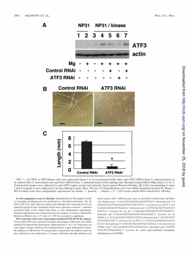
FIG. 1. (A) NP31 or NP31/kinase cells were untreated (lanes 1 to 4) or pretreated with either anti-ATF3 siRNA (lane 6; antisense/sense) orits control (lane 5; sense/sense) and anti-Nox1 siRNA (lane 7; antisense/sense) before plating onto Matrigel-coated dishes (Mg) (lanes 2, 4 to 7).Total protein lysates were subjected to anti-ATF3 (upper panel) and antiactin (lower panel) Western blotting. (B) Cells corresponding to lanes5 and 6 in panel A were subjected to in vitro Matrigel assays. Bars, 100 �m. (C) Quantification of in vitro tubule formation in panel B. Means �SD of results from three independent experiments are shown. �, present; , absent; �, P � 0.05 versus control RNA interference (RNAi).
1088 OKAMOTO ET AL. MOL. CELL. BIOL.
on Novem
ber 25, 2018 by guesthttp://m
cb.asm.org/
Dow
nloaded from
To ensure normalization of the cDNA amounts, we also used anotherGAPDH primer pair (forward, 5�-CATTGTCTACTATGACGGAC-3�; reverse,5�-GCAATTGTCATCTTCTCCCA-3�) and a �-actin primer pair (forward, 5�-ATGGATGATGATATCGCCGCG-3�; reverse, 5�-GGGCACAGTGTGGGTGACCCC-3�), which we have previously utilized for NP31 cells (21). All sets ofprimers were initially tested with cDNA samples that were synthesized with orwithout reverse transcriptase, and resulting RT-PCR products were examined byDNA sequencing. For semiquantitative PCR, the linear range of measurementwas established by trying different numbers of cycles (usually 15, 20, 25, 30, and35). Results before saturation were shown for each gene.
siRNA-mediated knockdown experiments. Based on the rat ATF3 sequence,sense primer (S) (5�-GCACCUUUGCCAUGGGAUGTT-3�) and antisenseprimer (A) (5�-CAUCCGAUGGCAAAGGUGCTT-3�) were designed. A smallinterfering RNA (siRNA) for Nox1 has been described previously (19). An-nealed primers were added to NP31/kinase cells by utilizing transfection reagent(TransIT-TKO; Mirus) and Opti-MEM (GIBCO). After incubation for 24 h,cells were plated onto Matrigel.
Plasmid construction, transfection, and reporter assay. V5-tagged constructs,V5-ATF3 (amino acids [aa] 1 to 181) and V5-ATF3�Zip (aa 1 to 115) weregenerated by PCR and cloned in frame into the NotI and BamHI site of pcDNA4containing V5 (Invitrogen). PCR primers, 5�-CCGCTCGAGGTCCCCAGTTAGGAGTCCCG-3� and 5�-CCCAAGCTTGTGTTCTTCCCTCCAGCAA-3�,were used to amplify the human CDK2 promoter region originally described byStennett et al. (39). The PCR product was subcloned into XhoI/HindIII sites ofthe pGL3 basic vector (Promega) to generate pCDK2-Luc. PCR primers, 5�-CGGGGTACCATGGGAAGTTGTGACTAATCCT-3� and 5�-CCCAAGCTTGGGTGGCCATTATGCCTAGTCTGCT-3�, were used to amplify the 974 to�43 region of the mouse p8 promoter (GenBank accession no. AF131195) (43).The PCR product was subcloned into KpnI/HindIII sites of the same vector togenerate pp8-Luc. All PCR products were verified by DNA sequencing. ThePAI-1 promoter-luciferase fusion reporter construct pGL-3-PAI-1/full (829 to�36) was kindly provided by S. Fujii at Hokkaido University (7).
Cos7 cells were seeded at 2 105 per well 20 h before transfection usingSuperfect reagent (QIAGEN). The reporter plasmid was cotransfected with
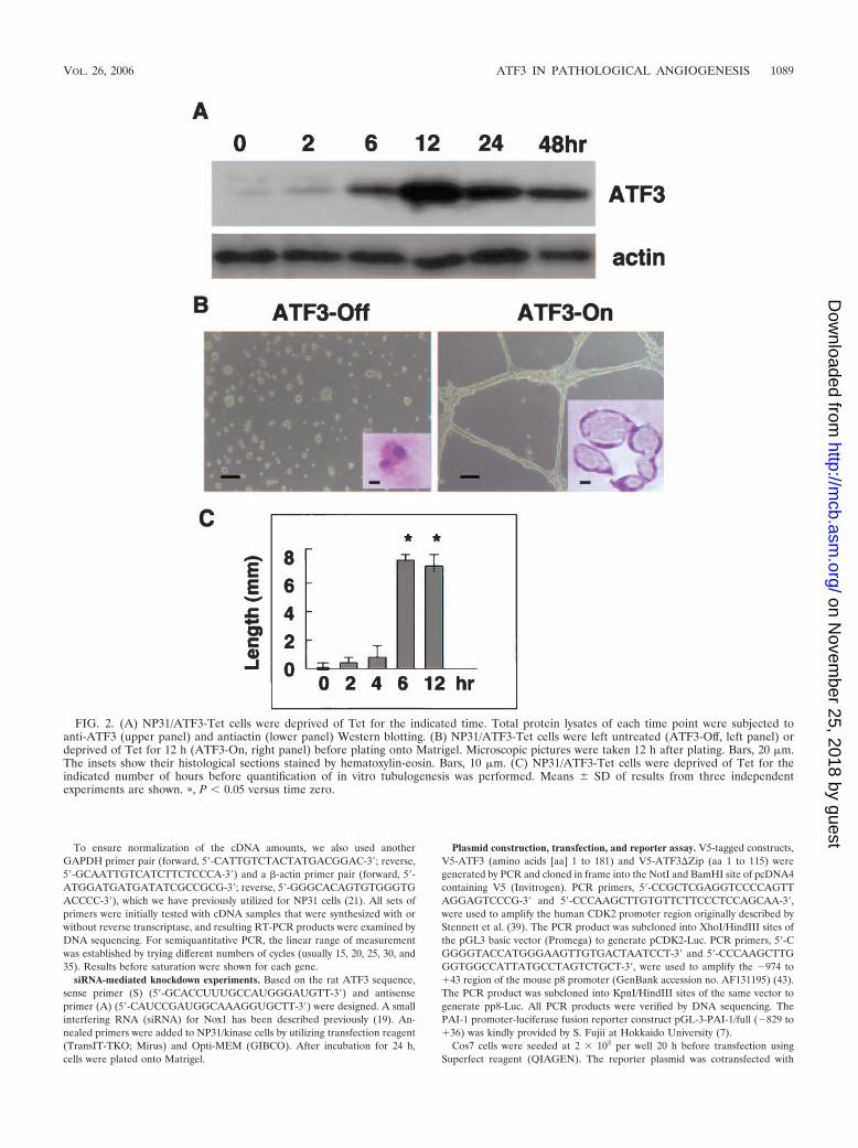
FIG. 2. (A) NP31/ATF3-Tet cells were deprived of Tet for the indicated time. Total protein lysates of each time point were subjected toanti-ATF3 (upper panel) and antiactin (lower panel) Western blotting. (B) NP31/ATF3-Tet cells were left untreated (ATF3-Off, left panel) ordeprived of Tet for 12 h (ATF3-On, right panel) before plating onto Matrigel. Microscopic pictures were taken 12 h after plating. Bars, 20 �m.The insets show their histological sections stained by hematoxylin-eosin. Bars, 10 �m. (C) NP31/ATF3-Tet cells were deprived of Tet for theindicated number of hours before quantification of in vitro tubulogenesis was performed. Means � SD of results from three independentexperiments are shown. �, P � 0.05 versus time zero.
VOL. 26, 2006 ATF3 IN PATHOLOGICAL ANGIOGENESIS 1089
on Novem
ber 25, 2018 by guesthttp://m
cb.asm.org/
Dow
nloaded from
either the V5-tagged full-length ATF3 or �Zip in pcDNA4 (0.5 �g each).NP31/ATF3-Tet cells at 2 105 per well were also transfected with the reporterplasmid before and 24 h after ATF3 induction. In every case, 50 ng of pRL-SV40(Promega) was added as an internal control. Cells were harvested 24 h aftertransfection, and luciferase activities were determined with the dual luciferasereporter assay system (Promega). Means of the results from six independentexperiments were calculated. A statistical value of P � 0.05 is accepted assignificant.
Antibodies and Western blotting. Polyclonal antibodies to ATF3, integrin �1,integrin �1, p53, Id1, p16, p21, p27, cyclinD1, CDK2, CDK4, MMP2, MMP9, andMMP10 were from Santa Cruz Biotechnology. Rabbit anti-rat PAI-1 antibodywas from American Diagnostica. Monoclonal antibody against V5 was fromInvitrogen, CD31 was from Pharmingen, and those against MMP13, RAGE, andactin were from Chemicon International. Anti-von Willebrand factor antibodywas from Dako. Western blotting was performed as described previously (23).Briefly, cells were collected by cell scrapers, washed twice with phosphate-buffered saline (pH 7.4), and lysed with lysis buffers (50 mM HEPES, pH 7.4, 150mM NaCl, 1% Triton X-100, 2 mM Na3VO3, 10 mM NaF, 10 mM pyrophos-phate, 1 mM EDTA, 1 mM phenylmethylsulfonyl fluoride, 20 �g/ml leupeptin,0.02% sodium dodecyl sulfate). Total lysates (approximately 20 �g each) wererun on sodium dodecyl sulfate-polyacrylamide gel electrophoresis slab gels. The
gel was transblotted on a nitrocellulose membrane (Amersham) and incubatedwith 5% bovine serum albumin for blocking. After incubation with appropriateantibodies, filters were subjected to washing and enhanced chemiluminescenceWestern blotting detection procedures (Amersham). The intensity of bands wasquantified by NIH image. To compare the ATF3 protein levels, the ratios of theintensity of ATF3 bands relative to that of actin were calculated.
Animals. Male OLETF rats (body weight, 650 to 680 g), a model animal oftype II diabetes mellitus, and male Long-Evans-Tokushima-Fatty (LETO) rats(450 to 500 g) for control (n � 5 each) were kindly provided by K. Kawano atTokushima Research Institute, Otsuka Pharmaceutical Co., Japan (26). Meanvalues of plasma glucose concentration were 350 mg/dl and 127 mg/dl forOLETF and LETO rats, respectively, at the age of 25 weeks, when they weresacrificed. A dramatic elevation of PAI-1 expression was also observed only inOLETF rats at that age (42).
Histological analysis. Paraffin sections were dried for 24 h at 4°C, dewaxedwith xylene twice (5 min), and passed through 96% ethanol twice (1 min) andTris-buffered saline–Ca three times (5 min) before staining. These sections werepreincubated in 0.1% bovine serum albumin for 1 h at 22°C and then incubatedwith an individual primary antibody at 4°C for 12 h, followed by incubation witha biotinylated secondary antibody (Vector) at 22°C for 1 h, a streptavidin-alkaline phosphatase (Dako) at 37°C for 15 min, and 5-bromo-4-chloro-3-in-dolylphosphate/nitroblue tetrazolium (Vector) at 22°C for 20 min. Triple stainingof glomeruli was performed with mouse anti-rat CD31, rabbit anti-rat ATF3 (orrabbit anti-rat PAI-1), and Vectashield medium with propidium iodide (PI) fornuclei. Secondary antibodies were Cy5-conjugated goat anti-rabbit immunoglob-ulin G (for ATF3) and fluorescein isothiocyanate-conjugated goat anti-mouseimmunoglobulin G (for CD31) (Jackson ImmunoResearch). Samples were an-alyzed under a Leica confocal laser-scanning microscope. Fluorescence imageswere presented by pseudocolors.
RESULTS
Inhibition of in vitro angiogenesis by anti-ATF3 siRNA. Wehave previously reported cDNA microarray analysis betweennontubulogenic NP31 endothelial cells and tubulogenic NP31cells expressing a constitutively activated form of theVEGFR-1 (Flt-1) kinase (BCR-FLTm1) (NP31/kinase cells) inMatrigel cultures. We found that expression of ATF3 mRNAswas induced by Matrigel in both of them, but the level ofinduction was much higher in NP31/kinase than NP31 cells asjudged by semiquantitative RT-PCR analysis (18). This wassupported by anti-ATF3 Western blotting shown in Fig. 1A(lanes 1 to 4). The induced expression of ATF3 at proteinlevels was prominent in NP31/kinase cells but not in NP31cells. Among the ATF3 isoforms, the anti-ATF3 antibody usedin our experiments can recognize only the full length (181 aa)and �Zip2a,b (135 aa) but not others (5, 12, 31, 46). Consid-ering the size of the detected band, we suppose that Matrigelinduced at least the full-length ATF3 protein.
In usual cultures on collagen type I dishes, expression ofBCR-FLTm1 did not result in enhanced expression of ATF3 inNP31 cells at protein levels. Nor could we find ATF3 inductionin human umbilical vein endothelial cells that were stimulatedby VEGF (data not shown). However, the fact that tubulogen-esis is always accompanied by induced expression of ATF3urged us to directly test its biological significance by siRNA-mediated gene knockdown.
The ATF3 induction by Matrigel was successfully inhibitedby anti-ATF3 siRNA as shown in Fig. 1A (lane 4 versus 6) intubulogenic NP31/kinase cells. To ensure the specificity ofsiRNAs, we have utilized two different controls: one is sense/sense primers for ATF3 (lane 5) and the other is antisense/sense primers for Nox1 (irrelevant siRNA) (lane 7). To oursurprise, anti-ATF3 siRNA-treated cells no longer formed tu-bules in Matrigel, while cells with control RNAs did so (Fig.
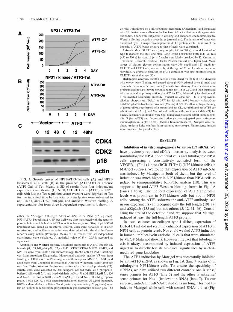
FIG. 3. Growth curves of NP31/ATF3-Tet cells (A) and NP31/kinase/ATF3-Tet cells (B) in the presence (ATF3-Off) or absence(ATF3-On) of Tet. Means � SD of results from four independentexperiments are shown. (C) NP31/ATF3-Tet cells (ATF3) or NP31cells with just the Tet regulation vector (vector) were deprived of Tetfor the indicated time before total protein lysates were subjected toanti-CDK4, anti-CDK2, anti-p16, and antiactin Western blotting. Arepresentative blot from three independent experiments is shown.
1090 OKAMOTO ET AL. MOL. CELL. BIOL.
on Novem
ber 25, 2018 by guesthttp://m
cb.asm.org/
Dow
nloaded from
1B, C). Anti-Nox1 siRNAs did reduce the expression of Nox1but not ATF3 (Fig. 1A, lane 7) without affecting tubulogenesis,as we described previously (19). This indicates that ATF3 in-duction is required for Matrigel-stimulated tubulogenesis inNP31/kinase cells that are programmed to form tubules.
Regulated expression of ATF3 in NP31 cells. We then triedto address the issue of whether or not ATF3 alone is capableof inducing angiogenesis. The full-length rat ATF3 cDNA wasisolated from Matrigel-stimulated NP31/kinase cells, and wasexpressed under the control of a tetracycline (Tet)-regulatedelement (NP31/ATF3-Tet cells). Tet-regulated expression ofthe ATF3 protein on usual collagen plates was successful andachieved a maximum level of expression 12 h after induction,which was approximately 10-fold over that of the baseline asjudged by densitometric analysis (Fig. 2A). Those cells showedno apparent morphological changes after ATF3 induction onusual culture dishes. However, when they were plated ontoMatrigel 12 h after induction, they exhibited network forma-
tion with a lumen as shown in Fig. 2B and C. The tubulogenicability of NP31/ATF3-Tet cells was evident when ATF3 wasinduced for at least 6 h before plating onto Matrigel, suggest-ing that a certain threshold in terms of both time and expres-sion level may exist for NP31 cells to be ready for tubuleformation in Matrigel.
Growth suppression by the induced expression of ATF3. Theeffect of ATF3 on cell growth has been under debate andappears to depend on cell types. This prompted us to test it inthe background of endothelial cells. We found that cell growthwas suppressed when ATF3 was induced in NP31/ATF3-Tetcells (Fig. 3A). The suppressive effect was also prominent whenTet-regulated ATF3 expression was achieved in the trans-formed NP31/kinase cells (NP31/kinase/ATF3-Tet cells) (Fig.3B). Expression levels of cyclin-dependent protein kinase 4(CDK4) and CDK2 were found to be decreased (Fig. 3C).However, those of CDK inhibitors remained unchanged, whichinclude p27, p21 (data not shown), and p16 (Fig. 3C).
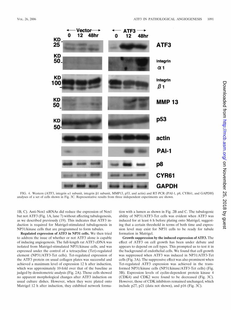
FIG. 4. Western (ATF3, integrin �1 subunit, integrin �1 subunit, MMP13, p53, and actin) and RT-PCR (PAI-1, p8, CYR61, and GAPDH)analyses of a set of cells shown in Fig. 3C. Representative results from three independent experiments are shown.
VOL. 26, 2006 ATF3 IN PATHOLOGICAL ANGIOGENESIS 1091
on Novem
ber 25, 2018 by guesthttp://m
cb.asm.org/
Dow
nloaded from
Direct analysis of gene expression in NP31/ATF3-Tet cells.We tried to identify a set of genes whose expression is directlyregulated by ATF3 in this in vitro angiogenesis system. Weinitially examined the genes whose expression was alreadyshown to be either up- or down-regulated when ATF3 wasup-regulated by Matrigel in NP31/kinase cells in our previousresults of microarray analysis (18). The up-regulated genesincluded integrin subunit �1, colony-stimulating factor 3 (csf3),p8, matrix Gla protein, and metallothionein 1, and down-reg-ulated genes included p38 mitogen-activated protein kinase,heat shock protein 70, hsp90, and CYR61 (24). We also ana-lyzed genes which are thought to be involved in angiogenesis or
diabetic complications, including VEGF, matrix metallopro-tease 2 (MMP2), MMP9, MMP10, MMP13, KDR (VEGFR-2), CD31, p53, PAI-1, receptor for advanced glycation endproducts (28), and Id1 (14). The expression level of none of thedown-regulated genes in the microarray was altered. Integrin�1 subunit, p8, PAI-1, and MMP13 were the only genes thatwere found to be up-regulated in an ATF3-dependent manner(Fig. 4). The peak of expression of integrin �1 subunit andMMP13 was at 48 h after ATF3 induction, and expressionlevels decreased thereafter.
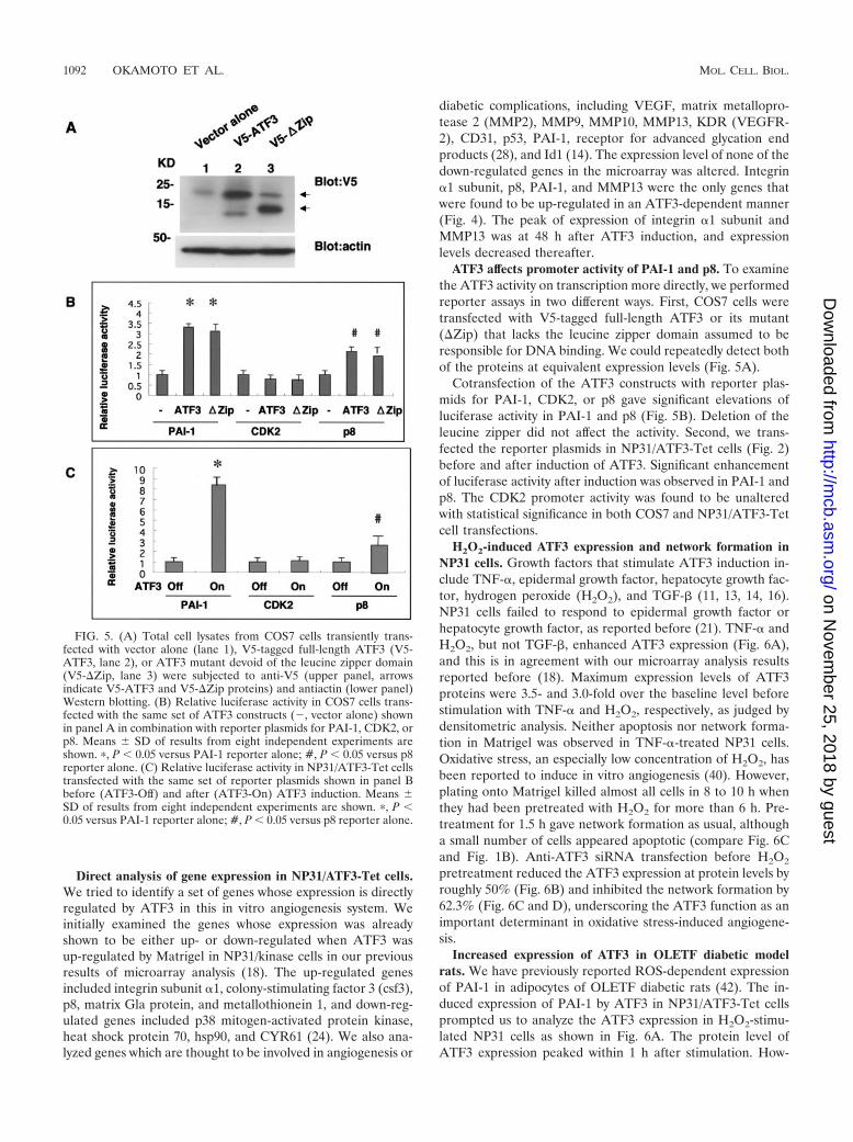
ATF3 affects promoter activity of PAI-1 and p8. To examinethe ATF3 activity on transcription more directly, we performedreporter assays in two different ways. First, COS7 cells weretransfected with V5-tagged full-length ATF3 or its mutant(�Zip) that lacks the leucine zipper domain assumed to beresponsible for DNA binding. We could repeatedly detect bothof the proteins at equivalent expression levels (Fig. 5A).
Cotransfection of the ATF3 constructs with reporter plas-mids for PAI-1, CDK2, or p8 gave significant elevations ofluciferase activity in PAI-1 and p8 (Fig. 5B). Deletion of theleucine zipper did not affect the activity. Second, we trans-fected the reporter plasmids in NP31/ATF3-Tet cells (Fig. 2)before and after induction of ATF3. Significant enhancementof luciferase activity after induction was observed in PAI-1 andp8. The CDK2 promoter activity was found to be unalteredwith statistical significance in both COS7 and NP31/ATF3-Tetcell transfections.
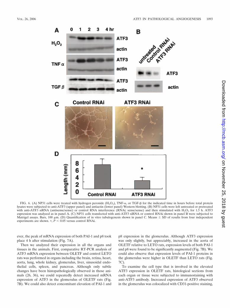
H2O2-induced ATF3 expression and network formation inNP31 cells. Growth factors that stimulate ATF3 induction in-clude TNF-�, epidermal growth factor, hepatocyte growth fac-tor, hydrogen peroxide (H2O2), and TGF-� (11, 13, 14, 16).NP31 cells failed to respond to epidermal growth factor orhepatocyte growth factor, as reported before (21). TNF-� andH2O2, but not TGF-�, enhanced ATF3 expression (Fig. 6A),and this is in agreement with our microarray analysis resultsreported before (18). Maximum expression levels of ATF3proteins were 3.5- and 3.0-fold over the baseline level beforestimulation with TNF-� and H2O2, respectively, as judged bydensitometric analysis. Neither apoptosis nor network forma-tion in Matrigel was observed in TNF-�-treated NP31 cells.Oxidative stress, an especially low concentration of H2O2, hasbeen reported to induce in vitro angiogenesis (40). However,plating onto Matrigel killed almost all cells in 8 to 10 h whenthey had been pretreated with H2O2 for more than 6 h. Pre-treatment for 1.5 h gave network formation as usual, althougha small number of cells appeared apoptotic (compare Fig. 6Cand Fig. 1B). Anti-ATF3 siRNA transfection before H2O2
pretreatment reduced the ATF3 expression at protein levels byroughly 50% (Fig. 6B) and inhibited the network formation by62.3% (Fig. 6C and D), underscoring the ATF3 function as animportant determinant in oxidative stress-induced angiogene-sis.
Increased expression of ATF3 in OLETF diabetic modelrats. We have previously reported ROS-dependent expressionof PAI-1 in adipocytes of OLETF diabetic rats (42). The in-duced expression of PAI-1 by ATF3 in NP31/ATF3-Tet cellsprompted us to analyze the ATF3 expression in H2O2-stimu-lated NP31 cells as shown in Fig. 6A. The protein level ofATF3 expression peaked within 1 h after stimulation. How-
FIG. 5. (A) Total cell lysates from COS7 cells transiently trans-fected with vector alone (lane 1), V5-tagged full-length ATF3 (V5-ATF3, lane 2), or ATF3 mutant devoid of the leucine zipper domain(V5-�Zip, lane 3) were subjected to anti-V5 (upper panel, arrowsindicate V5-ATF3 and V5-�Zip proteins) and antiactin (lower panel)Western blotting. (B) Relative luciferase activity in COS7 cells trans-fected with the same set of ATF3 constructs (, vector alone) shownin panel A in combination with reporter plasmids for PAI-1, CDK2, orp8. Means � SD of results from eight independent experiments areshown. �, P � 0.05 versus PAI-1 reporter alone; #, P � 0.05 versus p8reporter alone. (C) Relative luciferase activity in NP31/ATF3-Tet cellstransfected with the same set of reporter plasmids shown in panel Bbefore (ATF3-Off) and after (ATF3-On) ATF3 induction. Means �SD of results from eight independent experiments are shown. �, P �0.05 versus PAI-1 reporter alone; #, P � 0.05 versus p8 reporter alone.
1092 OKAMOTO ET AL. MOL. CELL. BIOL.
on Novem
ber 25, 2018 by guesthttp://m
cb.asm.org/
Dow
nloaded from
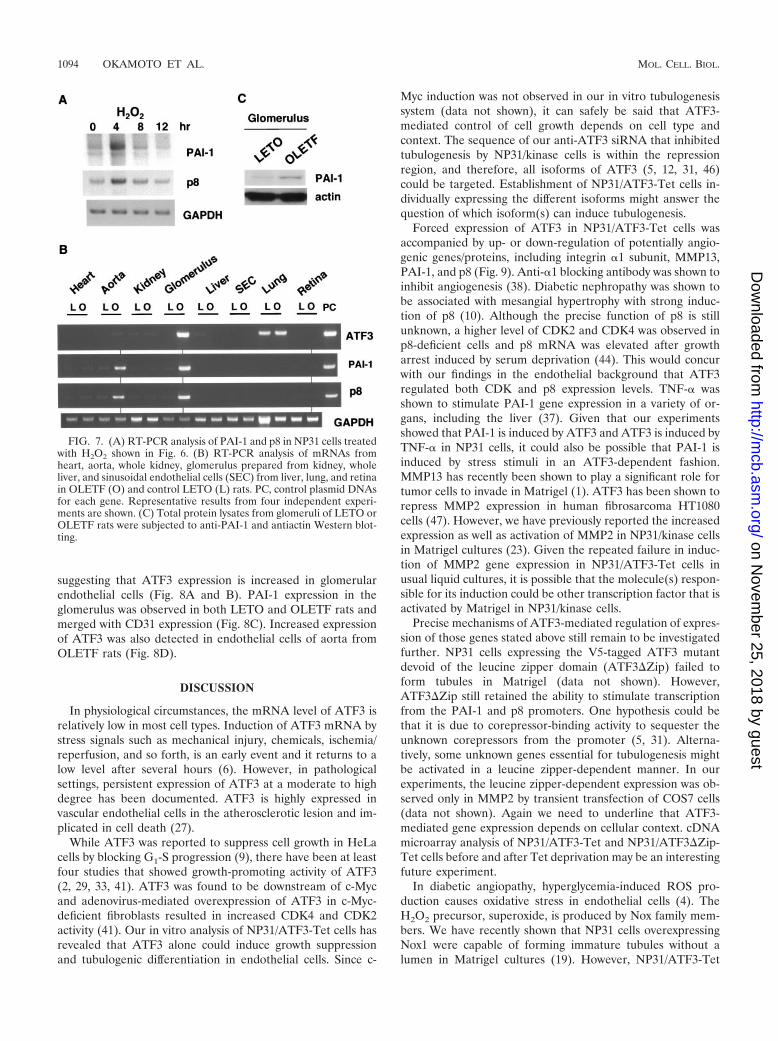
ever, the peak of mRNA expression of both PAI-1 and p8 tookplace 4 h after stimulation (Fig. 7A).
Then we analyzed their expression in all the organs andtissues in the animals. First, comparative RT-PCR analysis ofATF3 mRNA expression between OLETF and control LETOrats was performed in organs including the brain, retina, heart,aorta, lung, whole kidney, glomerulus, liver, sinusoidal endo-thelial cells, spleen, and pancreas. Although only subtlechanges have been histopathologically observed in those ani-mals (26, 36), we could repeatedly detect increased mRNAexpression of ATF3 in the glomerulus of OLETF rats (Fig.7B). We could also detect concomitant elevation of PAI-1 and
p8 expression in the glomerulus. Although ATF3 expressionwas only slightly, but appreciably, increased in the aorta ofOLETF relative to LETO rats, expression levels of both PAI-1and p8 were found to be significantly augmented (Fig. 7B). Wecould also observe that expression levels of PAI-1 proteins inthe glomerulus were higher in OLETF than LETO rats (Fig.7C).
To examine the cell type that is involved in the elevatedATF3 expression in OLETF rats, histological sections fromeach organ or tissue were subjected to immunostaining withanti-ATF3 antibody. Increased expression of ATF3 observedin the glomerulus was colocalized with CD31-positive staining,
FIG. 6. (A) NP31 cells were treated with hydrogen peroxide (H2O2), TNF-�, or TGF-� for the indicated time in hours before total proteinlysates were subjected to anti-ATF3 (upper panel) and antiactin (lower panel) Western blotting. (B) NP31 cells were left untreated or pretreatedwith anti-ATF3 siRNA (antisense/sense) or control RNA interference (RNAi; sense/sense) and then stimulated with H2O2 for 1.5 h. ATF3expression was analyzed as in panel A. (C) NP31 cells transfected with anti-ATF3 siRNA or control RNAi shown in panel B were subjected toMatrigel assays. Bars, 100 �m. (D) Quantification of in vitro tubulogenesis shown in panel C. Means � SD of results from four independentexperiments are shown. �, P � 0.05 versus control RNAi.
VOL. 26, 2006 ATF3 IN PATHOLOGICAL ANGIOGENESIS 1093
on Novem
ber 25, 2018 by guesthttp://m
cb.asm.org/
Dow
nloaded from
suggesting that ATF3 expression is increased in glomerularendothelial cells (Fig. 8A and B). PAI-1 expression in theglomerulus was observed in both LETO and OLETF rats andmerged with CD31 expression (Fig. 8C). Increased expressionof ATF3 was also detected in endothelial cells of aorta fromOLETF rats (Fig. 8D).
DISCUSSION
In physiological circumstances, the mRNA level of ATF3 isrelatively low in most cell types. Induction of ATF3 mRNA bystress signals such as mechanical injury, chemicals, ischemia/reperfusion, and so forth, is an early event and it returns to alow level after several hours (6). However, in pathologicalsettings, persistent expression of ATF3 at a moderate to highdegree has been documented. ATF3 is highly expressed invascular endothelial cells in the atherosclerotic lesion and im-plicated in cell death (27).
While ATF3 was reported to suppress cell growth in HeLacells by blocking G1-S progression (9), there have been at leastfour studies that showed growth-promoting activity of ATF3(2, 29, 33, 41). ATF3 was found to be downstream of c-Mycand adenovirus-mediated overexpression of ATF3 in c-Myc-deficient fibroblasts resulted in increased CDK4 and CDK2activity (41). Our in vitro analysis of NP31/ATF3-Tet cells hasrevealed that ATF3 alone could induce growth suppressionand tubulogenic differentiation in endothelial cells. Since c-
Myc induction was not observed in our in vitro tubulogenesissystem (data not shown), it can safely be said that ATF3-mediated control of cell growth depends on cell type andcontext. The sequence of our anti-ATF3 siRNA that inhibitedtubulogenesis by NP31/kinase cells is within the repressionregion, and therefore, all isoforms of ATF3 (5, 12, 31, 46)could be targeted. Establishment of NP31/ATF3-Tet cells in-dividually expressing the different isoforms might answer thequestion of which isoform(s) can induce tubulogenesis.
Forced expression of ATF3 in NP31/ATF3-Tet cells wasaccompanied by up- or down-regulation of potentially angio-genic genes/proteins, including integrin �1 subunit, MMP13,PAI-1, and p8 (Fig. 9). Anti-�1 blocking antibody was shown toinhibit angiogenesis (38). Diabetic nephropathy was shown tobe associated with mesangial hypertrophy with strong induc-tion of p8 (10). Although the precise function of p8 is stillunknown, a higher level of CDK2 and CDK4 was observed inp8-deficient cells and p8 mRNA was elevated after growtharrest induced by serum deprivation (44). This would concurwith our findings in the endothelial background that ATF3regulated both CDK and p8 expression levels. TNF-� wasshown to stimulate PAI-1 gene expression in a variety of or-gans, including the liver (37). Given that our experimentsshowed that PAI-1 is induced by ATF3 and ATF3 is induced byTNF-� in NP31 cells, it could also be possible that PAI-1 isinduced by stress stimuli in an ATF3-dependent fashion.MMP13 has recently been shown to play a significant role fortumor cells to invade in Matrigel (1). ATF3 has been shown torepress MMP2 expression in human fibrosarcoma HT1080cells (47). However, we have previously reported the increasedexpression as well as activation of MMP2 in NP31/kinase cellsin Matrigel cultures (23). Given the repeated failure in induc-tion of MMP2 gene expression in NP31/ATF3-Tet cells inusual liquid cultures, it is possible that the molecule(s) respon-sible for its induction could be other transcription factor that isactivated by Matrigel in NP31/kinase cells.
Precise mechanisms of ATF3-mediated regulation of expres-sion of those genes stated above still remain to be investigatedfurther. NP31 cells expressing the V5-tagged ATF3 mutantdevoid of the leucine zipper domain (ATF3�Zip) failed toform tubules in Matrigel (data not shown). However,ATF3�Zip still retained the ability to stimulate transcriptionfrom the PAI-1 and p8 promoters. One hypothesis could bethat it is due to corepressor-binding activity to sequester theunknown corepressors from the promoter (5, 31). Alterna-tively, some unknown genes essential for tubulogenesis mightbe activated in a leucine zipper-dependent manner. In ourexperiments, the leucine zipper-dependent expression was ob-served only in MMP2 by transient transfection of COS7 cells(data not shown). Again we need to underline that ATF3-mediated gene expression depends on cellular context. cDNAmicroarray analysis of NP31/ATF3-Tet and NP31/ATF3�Zip-Tet cells before and after Tet deprivation may be an interestingfuture experiment.
In diabetic angiopathy, hyperglycemia-induced ROS pro-duction causes oxidative stress in endothelial cells (4). TheH2O2 precursor, superoxide, is produced by Nox family mem-bers. We have recently shown that NP31 cells overexpressingNox1 were capable of forming immature tubules without alumen in Matrigel cultures (19). However, NP31/ATF3-Tet
FIG. 7. (A) RT-PCR analysis of PAI-1 and p8 in NP31 cells treatedwith H2O2 shown in Fig. 6. (B) RT-PCR analysis of mRNAs fromheart, aorta, whole kidney, glomerulus prepared from kidney, wholeliver, and sinusoidal endothelial cells (SEC) from liver, lung, and retinain OLETF (O) and control LETO (L) rats. PC, control plasmid DNAsfor each gene. Representative results from four independent experi-ments are shown. (C) Total protein lysates from glomeruli of LETO orOLETF rats were subjected to anti-PAI-1 and antiactin Western blot-ting.
1094 OKAMOTO ET AL. MOL. CELL. BIOL.
on Novem
ber 25, 2018 by guesthttp://m
cb.asm.org/
Dow
nloaded from
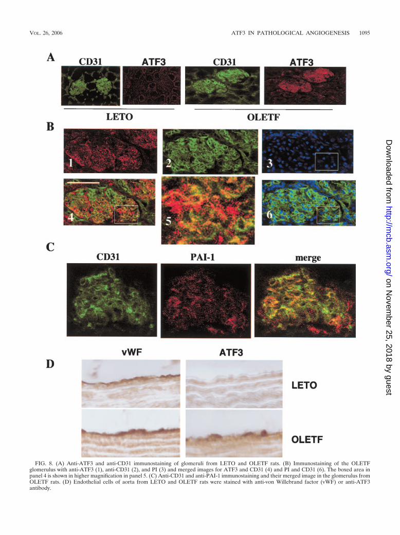
FIG. 8. (A) Anti-ATF3 and anti-CD31 immunostaining of glomeruli from LETO and OLETF rats. (B) Immunostaining of the OLETFglomerulus with anti-ATF3 (1), anti-CD31 (2), and PI (3) and merged images for ATF3 and CD31 (4) and PI and CD31 (6). The boxed area inpanel 4 is shown in higher magnification in panel 5. (C) Anti-CD31 and anti-PAI-1 immunostaining and their merged image in the glomerulus fromOLETF rats. (D) Endothelial cells of aorta from LETO and OLETF rats were stained with anti-von Willebrand factor (vWF) or anti-ATF3antibody.
VOL. 26, 2006 ATF3 IN PATHOLOGICAL ANGIOGENESIS 1095
on Novem
ber 25, 2018 by guesthttp://m
cb.asm.org/
Dow
nloaded from
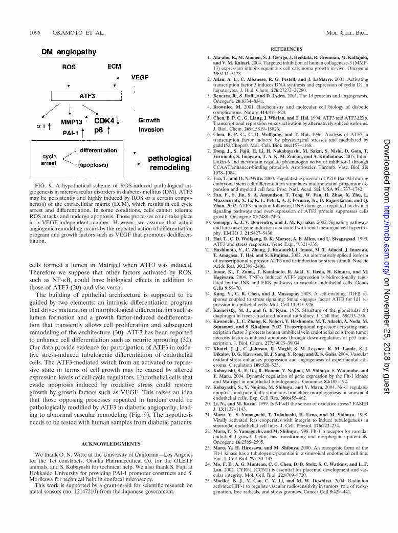
cells formed a lumen in Matrigel when ATF3 was induced.Therefore we suppose that other factors activated by ROS,such as NF-�B, could have biological effects in addition tothose of ATF3 (20) and vise versa.
The building of epithelial architecture is supposed to beguided by two elements: an intrinsic differentiation programthat drives maturation of morphological differentiation such aslumen formation and a growth factor-induced dedifferentia-tion that transiently allows cell proliferation and subsequentremodeling of the architecture (30). ATF3 has been reportedto enhance cell differentiation such as neurite sprouting (32).Our data provide evidence for participation of ATF3 in oxida-tive stress-induced tubulogenic differentiation of endothelialcells. The ATF3-mediated switch from an activated to repres-sive state in terms of cell growth may be caused by alteredexpression levels of cell cycle regulators. Endothelial cells thatevade apoptosis induced by oxidative stress could restoregrowth by growth factors such as VEGF. This raises an ideathat those opposing processes repeated in tandem could bepathologically modified by ATF3 in diabetic angiopathy, lead-ing to abnormal vascular remodeling (Fig. 9). The hypothesisneeds to be tested with human samples from diabetic patients.
ACKNOWLEDGMENTS
We thank O. N. Witte at the University of California—Los Angelesfor the Tet constructs, Otsuka Pharmaceutical Co. for the OLETFanimals, and S. Kobayashi for technical help. We also thank S. Fujii atHokkaido University for providing PAI-1 promoter constructs and S.Morikawa for technical help in confocal microscopy.
This work is supported by a grant-in-aid for scientific research onmetal sensors (no. 12147210) from the Japanese government.
REFERENCES
1. Ala-aho, R., M. Ahonen, S. J. George, J. Heikkila, R. Grenman, M. Kallajoki,and V. M. Kahari. 2004. Targeted inhibition of human collagenase-3 (MMP-13) expression inhibits squamous cell carcinoma growth in vivo. Oncogene23:5111–5123.
2. Allan, A. L., C. Albanese, R. G. Pestell, and J. LaMarre. 2001. Activatingtranscription factor 3 induces DNA synthesis and expression of cyclin D1 inhepatocytes. J. Biol. Chem. 276:27272–27280.
3. Benezra, R., S. Rafil, and D. Lyden. 2001. The Id proteins and angiogenesis.Oncogene 20:8334–8341.
4. Brownlee, M. 2001. Biochemistry and molecular cell biology of diabeticcomplications. Nature 414:813–820.
5. Chen, B. P. C., G. Liang, J. Whelan, and T. Hai. 1994. ATF3 and ATF3�Zip.Transcriptional repression versus activation by alternatively spliced isoforms.J. Biol. Chem. 269:15819–15826.
6. Chen, B. P. C., C. D. Wolfgang, and T. Hai. 1996. Analysis of ATF3, atranscription factor induced by physiological stresses and modulated bygadd153/Chop10. Mol. Cell. Biol. 16:1157–1168.
7. Dong, J., S. Fujii, H. Li, H. Nakabayashi, M. Sakai, S. Nishi, D. Goto, T.Furumoto, S. Imagawa, T. A. K. M. Zaman, and A. Kitabatake. 2005. Inter-leukin-6 and mevastatin regulate plasminogen activator inhibitor-1 throughCCAAT/enhancer-binding protein- . Arterioscler. Thromb. Vasc. Biol. 25:1078–1084.
8. Era, T., and O. N. Witte. 2000. Regulated expression of P210 Bcr-Abl duringembryonic stem cell differentiation stimulates multipotential progenitor ex-pansion and myeloid cell fate. Proc. Natl. Acad. Sci. USA 97:1737–1742.
9. Fan, F., S. Jin, S. A. Amundson, T. Tong, W. Fan, H. Zhao, X. Zhu, L.Mazzacurati, X. Li, K. L. Petrik, A. J. Fornace, Jr., B. Rajasekaran, and Q.Zhan. 2002. ATF3 induction following DNA damage is regulated by distinctsignaling pathways and over-expression of ATF3 protein suppresses cellsgrowth. Oncogene 21:7488–7496.
10. Goruppi, S., J. V. Bonventre, and J. M. Kyriakis. 2002. Signaling pathwaysand late-onset gene induction associated with renal mesangial cell hypertro-phy. EMBO J. 21:5427–5436.
11. Hai, T., C. D. Wolfgang, D. K. Marsee, A. E. Allen, and U. Sivaprasad. 1999.ATF3 and stress responses. Gene Expr. 7:321–335.
12. Hashimoto, Y., C. Zhang, J. Kawauchi, I. Imoto, M. T. Adachi, J. Inazawa,T. Amagasa, T. Hai, and S. Kitajima. 2002. An alternatively spliced isoformof transcriptional repressor ATF3 and its induction by stress stimuli. NucleicAcids Res. 30:2398–2406.
13. Inoue, K., T. Zama, T. Kamimoto, R. Aoki, Y. Ikeda, H. Kimura, and M.Hagiwara. 2004. TNF-� induced ATF3 expression is bidirectionally regu-lated by the JNK and ERK pathways in vascular endothelial cells. GenesCells 9:59–70.
14. Kang, Y., C. R. Chen, and J. Massague. 2003. A self-enabling TGF� re-sponse coupled to stress signaling: Smad engages factor ATF3 for Id1 re-pression in epithelial cells. Mol. Cell 11:915–926.
15. Karnovsky, M. J., and G. B. Ryan. 1975. Structure of the glomerular slitdiaphragm in freeze-fractured normal rat kidney. J. Cell Biol. 65:233–236.
16. Kawauchi, J., C. Zhang, K. Nobori, Y. Hashimoto, M. T. Adachi, A. Noda, M.Sunamori, and S. Kitajima. 2002. Transcriptional repressor activating tran-scription factor 3 protects human umbilical vein endothelial cells from tumornecrosis factor-�-induced apoptosis through down-regulation of p53 tran-scription. J. Biol. Chem. 277:39025–39034.
17. Khatri, J. J., C. Johnson, R. Magid, S. M. Lessner, K. M. Laude, S. I.Dikalov, D. G. Harrison, H. J. Sung, Y. Rong, and Z. S. Galis. 2004. Vascularoxidant stress enhances progression and angiogenesis of experimental ath-eroma. Circulation 109:520–525.
18. Kobayashi, S., E. Ito, R. Honma, Y. Nojima, M. Shibuya, S. Watanabe, andY. Maru. 2004. Dynamic regulation of gene expression by the Flt-1 kinaseand Matrigel in endothelial tubulogenesis. Genomics 84:185–192.
19. Kobayashi, S., Y. Nojima, M. Shibuya, and Y. Maru. 2004. Nox1 regulatesapoptosis and potentially stimulates branching morphogenesis in sinusoidalendothelial cells. Exp. Cell Res. 300:455–462.
20. Li, N., and M. Karin. 1999. Is NF-�B the sensor of oxidative stress? FASEBJ. 13:1137–1143.
21. Maru, Y., S. Yamaguchi, T. Takahashi, H. Ueno, and M. Shibuya. 1998.Virally activated Ras cooperates with integrin to induce tubulogenesis insinusoidal endothelial cell lines. J. Cell. Physiol. 176:223–234.
22. Maru, Y., S. Yamaguchi, and M. Shibuya. 1998. Flt-1, a receptor for vascularendothelial growth factor, has transforming and morphogenic potentials.Oncogene 16:2585–2595.
23. Maru, Y., H. Hirosawa, and M. Shibuya. 2000. An oncogenic form of theFlt-1 kinase has a tubulogenic potential in a sinusoidal endothelial cell line.Eur. J. Cell Biol. 79:130–143.
24. Mo, F. E., A. G. Muntean, C. C. Chen, D. B. Stolz, S. C. Watkins, and L. F.Lau. 2002. CYR61 (CCN1) is essential for placental development and vas-cular integrity. Mol. Cell. Biol. 22:8709–8720.
25. Moeller, B. J., Y. Cao, C. Y. Li, and M. W. Dewhirst. 2004. Radiationactivates HIF-1 to regulate vascular radiosensitivity in tumors: role of reoxy-genation, free radicals, and stress granules. Cancer Cell 5:429–441.
FIG. 9. A hypothetical scheme of ROS-induced pathological an-giogenesis in microvascular disorders in diabetes mellitus (DM). ATF3may be persistently and highly induced by ROS or a certain compo-nent(s) of the extracellular matrix (ECM), which results in cell cyclearrest and differentiation. In some conditions, cells cannot tolerateROS attacks and undergo apoptosis. Those processes could take placein a VEGF-independent manner. However, we assume that actualangiogenic remodeling occurs by the repeated action of differentiationprogram and growth factors such as VEGF that promotes dedifferen-tiation.
1096 OKAMOTO ET AL. MOL. CELL. BIOL.
on Novem
ber 25, 2018 by guesthttp://m
cb.asm.org/
Dow
nloaded from
26. Mori, S., K. Kawano, T. Hirashima, and T. Natori. 1996. Relationshipsbetween diet control and the development of spontaneous type II diabetesand diabetic nephropathy in OLETF rats. Diabetes Res. Clin. Pract. 33:145–152.
27. Nawa, T., M. T. Nawa, M. T. Adachi, I. Uchimura, R. Shimokawa, K.Fujisawa, A. Tanaka, F. Numano, and S. Kitajima. 2002. Expression oftranscriptional repressor ATF3/LRF1 in human atherosclerosis colocaliza-tion and possible involvement in cell death of vascular endothelial cells.Atherosclerosis 16:281–291.
28. Neeper, M., A. M. Schmidt, J. Brett, S. D. Yan, F. Wang, Y. C. Pan, K.Elliston, D. Stern, and A. Shaw. 1992. Cloning and expression of a cellsurface receptor for advanced glycosylation end products of proteins. J. Biol.Chem. 267:14998–15004.
29. Nilssen, L. S., J. Ødegård, G. H. Thoresen, A. Molven, D. Sandnes, and T.Christoffersen. 2004. G protein-coupled receptor agonist-stimulated expres-sion of ATF3/LRF-1 and c-myc and comitogenic effects in hepatocytes donot require EGF receptor transactivation. J. Cell. Physiol. 201:349–358.
30. O’Brien, L. E., M. M. Zegers, and K. E. Mostov. 2002. Building epithelialarchitecture: insights from three-dimensional culture models. Nat. Rev. Mol.Cell Biol. 3:531–537.
31. Pan, Y., H. Chen, F. Siu, and M. S. Kilberg. 2003. Amino acid deprivationand endoplasmic reticulum stress induce expression of multiple activatingtranscription factor-3 mRNA species that, when overexpressed in HepG2cells, modulate transcription by the human asparagine synthetase promoter.J. Biol. Chem. 278:38402–38412.
32. Pearson, A. G., C. W. Gray, J. F. Pearson, J. M. Greenwood, M. J. During,and M. Dragunow. 2003. ATF3 enhances c-Jun mediated neurite sprouting.Mol. Brain Res. 120:38–45.
33. Perez, S., E. Vial, V. H. Dam, and M. Castellazzi. 2001. Transcription factorATF3 partially transforms chick embryo fibroblasts by promoting growthfactor-independent proliferation. Oncogene 20:1135–1141.
34. Ploplis, V. A., R. Balsara, M. J. Sandoval-Cooper, Z. J. Yin, J. Betten, N.Modi, D. Gadoua, D. Donahue, J. A. Martin, and F. J. Castellino. 2003.Enhanced in vitro proliferation of aortic endothelial cells from plasminogenactivator inhibitor-1-deficient mice. J. Biol. Chem. 279:6143–6151.
35. Qian, Y., J. Luo, S. S. Leonard, G. K. Harris, L. Millecchia, D. C. Flynn, andX. Shi. 2003. Hydrogen peroxide formation and actin filament reorganizationby Cdc42 are essential for ethanol-induced in vitro angiogenesis. J. Biol.Chem. 278:16189–16197.
36. Saito, F., M. Kawaguchi, J. Izumida, T. Asakura, K. Maehara, and Y.Maruyama. 2003. Alteration in haemodynamics and pathological changes inthe cardiovascular system during the development of type 2 diabetes mellitusin OLETF rats. Diabetologia 46:1161–1169.
37. Sawdey, M. S., and D. J. Loskutoff. 1991. Regulation of murine type 1plasminogen activator inhibitor gene expression in vivo. Tissue specificityand induction by lipopolysaccharide, tumor necrosis factor-alpha, and trans-forming growth factor-beta. J. Clin. Investig. 88:1346–1353.
38. Senger, A. P., K. P. Claffey, J. E. Benes, C. A. Perruzzi, A. P. Sergiou, and M.Detmar. 1997. Angiogenesis promoted by vascular endothelial growth factor:regulation through �1�1 and �2�1 integrins. Proc. Natl. Acad. Sci. USA94:13612–13617.
39. Stennett, L. S., A. I. Riker, T. M. Kroll, J. Chamberlin, T. Miki, B. J.Nickoloff, and I. C. Le Poole. 2004. Expression of gp100 and CDK2 inmelanoma cells is not coregulated by a shared promoter region. Pigment CellRes. 17:525–532.
40. Stone, J. R., and J. Collins. 2002. The role of hydrogen peroxide in endo-thelial proliferative responses. Endothelium 9:231–238.
41. Tamura, K., B. Hua, S. Adachi, I. Guney, J. Kawauchi, M. Morioka, M.Tamamori-Adachi, Y. Tanaka, Y. Nakabeppu, M. Sunamori, J. M. Sedivy,and S. Kitajima. 2005. Stress response gene ATF3 is a target of c-myc inserum-induced cell proliferation. EMBO J. 24:2590–2601.
42. Uchida, Y., K. Ohba, T. Yoshioka, K. Irie, T. Muraki, and Y. Maru. 2004.Cellular carbonyl stress enhances the expression of plasminogen activatorinhibitor-1 in rat white adipocytes via reactive oxygen species-dependentpathway. J. Biol. Chem. 279:4075–4083.
43. Vasseur, S., G. V. Mallo, A. Garcia-Montero, E. M. Ortiz, F. Fiedler, E.Canepa, S. Moreno, and J. L. Iovanna. 1999. Structural and functionalcharacterization of the mouse p8 gene: promotion of transcription by theCAAT-enhancer binding protein � (C/EBP�) and C/EBP� trans-acting fac-tors involves a C/EBP cis-acting element and other regions of the promoter.Biochem. J. 343:377–383.
44. Vasseur, S., A. Hoffmeister, A. Garcia-Montero, G. V. Mallo, R. Feil, S.Kuhbandner, J. C. Dagorn, and J. L. Iovanna. 2002. p8-deficient fibroblastsgrow more rapidly and are more resistant to adriamycin-induced apoptosis.Oncogene 21:1685–1694.
45. Volpert, O. V., R. Pili, H. A. Sikder, T. Nelius, T. Zaichuk, C. Morris, C. B.Shiflett, M. K. Devlin, K. Conant, and R. M. Alani. 2002. Id1 regulatesangiogenesis through transcriptional repression of thrombospondin-1. Can-cer Cell 2:473–483.
46. Wang, J., Y. Cao, and D. F. Steiner. 2003. Regulation of proglucagon tran-scription by activated transcription factor (ATF)3 and a novel isoform,ATF3b, through the cAMP-response element/ATF site of the proglucagongene promoter. J. Biol. Chem. 278:32899–32904.
47. Yan, C., H. Wang, and D. D. Boyd. 2002. ATF3 represses 72-kDa type IVcollagenase (MMP-2) expression by antagonizing p53-dependent trans-acti-vation of the collagenase promoter. J. Biol. Chem. 277:10804–10812.
VOL. 26, 2006 ATF3 IN PATHOLOGICAL ANGIOGENESIS 1097
on Novem
ber 25, 2018 by guesthttp://m
cb.asm.org/
Dow
nloaded from