Embed Size (px)
Citation preview
Molecular Cell, Vol. 11, 577–590, March, 2003, Copyright 2003 by Cell Press
p53 Has a Direct Apoptogenic Role at the Mitochondria
shows that the type, strength, and kinetics of the targetMotohiro Mihara,1,4 Susan Erster,1,4
gene profiles depend on p53 levels, stress type, andAlexander Zaika,1 Oleksi Petrenko,1
cell type (Zhao et al., 2000). This indicates that onlyThomas Chittenden,2 Petr Pancoska,3
individual genes will be chosen from the complex spec-and Ute M. Moll1,*trum of potentially inducible genes to mediate a specific1Department of Pathologyp53 response in a given physiological situation.Stony Brook University
In addition, evidence for transcription-independentStony Brook, New York 11794p53-mediated apoptosis has been accumulating. In2 ImmunoGen, Inc.some cell types, p53-dependent apoptosis occurs in128 Sidney Streetthe absence of any gene transcription or translationCambridge, Massachusetts 02139(Caelles et al., 1994; Wagner et al., 1994; Gao and Tsu-3 Department of Chemistrychida, 1999). Also, inhibitors of protein phosphatasesUniversity of Illinois at Chicagoinduce p53-dependent death in the absence of trans-Chicago, Illinois 60607activation (Yan et al., 1997). Moreover, the transcription-ally inactive p53 mutants (1–214) and p53 22/23QS actas potent death inducers in tumor cells (Haupt et al.,Summary1995; Chen et al., 1996; Kokontis et al., 2001). In (mito-chondria-containing) cell-free S100 extracts that modelp53 induces apoptosis by target gene regulation andp53-dependent but transcription-independent apopto-transcription-independent signaling. However, a mecha-sis, p53 protein directly mediates caspase-3 activation,nism for the latter was unknown. We recently reportedand immunodepletion of p53 completely blocks this ac-that a fraction of induced p53 translocates to the mito-tivity. This pathway requires caspase 8 activation (Dingchondria of apoptosing tumor cells. Targeting p53 toet al., 2000). Also, in cell-free cytoplasts, activation ofmitochondria is sufficient to launch apoptosis. Here,cytosolic p53 can induce mitochondrial cytochrome cwe provide evidence that p53 translocation to the mi-release (Schuler and Green, 2001). Together, these datatochondria occurs in vivo in irradiated thymocytes. Fur-indicate the coexistence of a transcription-independentther, we show that the p53 protein can directly inducepathway of p53-mediated apoptosis. However, the un-permeabilization of the outer mitochondrial membranederlying mechanism of action remained unknown.by forming complexes with the protective BclXL and
Mitochondria are central death regulators in responseBcl2 proteins, resulting in cytochrome c release. p53to DNA damage, growth factor withdrawal, hypoxia, andbinds to BclXL via its DNA binding domain. We probeoncogene deregulation and are critical for p53-depen-the significance of mitochondrial p53 and show thatdent death (Wang, 2001). When mitochondria receive atumor-derived transactivation-deficient mutants ofdeath signal, the outer mitochondrial membrane (OMM)p53 concomitantly lose the ability to interact withundergoes permeabilization, which causes the releaseBclXL and promote cytochrome c release. This opensof potent death factors from the intermembraneousthe possibility that mutations might represent “double-space into the cytosol (Green and Evan, 2002). Thesehits” by abrogating the transcriptional and mitochon-apoptogenic factors activate caspase-9 (cytochrome c),drial apoptotic activity of p53.inhibit cytosolic IAPs (Smac, Htra2), induce chromatincondensation (AIF), or degrade DNA (Endonuclease G).IntroductionOMM permeabilization is regulated by the opposing ac-tions of pro- and antiapoptotic Bcl2 proteins, although
The basis for p53’s striking apoptotic and tumor sup-the exact mechanism of how the Bcl2 family controls
pressive potency lies in its pleiotropism, which includes OMM permeability is unclear. The antiapoptotic mem-transcription-dependent and -independent functions. bers, typified by Bcl2 and BclXL, constitutively residep53 kills cells predominantly via the mitochondrial death at the OMM and mediate a critical pro-survival functionpathway (reviewed in Johnstone et al., 2002). p53 can by stabilizing the OMM and preventing the release ofmediate apoptosis by transcriptional activation of pro- death factors. Overexpressed Bcl2 and BclXL suppressapoptotic genes like the BH3-only proteins Noxa and p53-dependent and -independent cell death. The pro-Puma, Bax, p53 AIP1, Apaf-1, and PERP and by tran- apoptotic members consist of the BH3-only class, whichscriptional repression of Bcl2 and IAPs (references in regulates the protective Bcl2/XL proteins, and the multi-Johnstone et al., 2002). For Noxa, Puma, and PIDD, domain BH123 class. The type II BH3-only proteinsdownregulation decreases—but does not abolish—the Noxa, Puma, Bik, Bim, and Bad couple death signals toextent of death after stress. Of note, induction of these mitochondria and in healthy cells are sequestered totarget gene products shows variable kinetics, with some cytosolic sites other than the OMM. Upon sensing deathbeing delayed in their response (over 24 hr), e.g., Bax stimuli, BH3-only proteins undergo posttranslationaland p53AIP1 (Nakano and Vousden, 2001; Attardi et al., modifications and mitochondrial translocation (re-2000). Analysis of p53-regulated global gene expression viewed in Huang and Strasser, 2000). Translocated BH3-
only proteins then bind to Bcl2/XL via their BH3 domain,thereby inactivating their protective function (Cheng et*Correspondence: [email protected]
4 These authors contributed equally to this work. al., 2001). In resting cells, BH123 proteins exist as inac-
Molecular Cell578
tive monomers in the cytosol (Bax) or at mitochondria(Bak) (Wolter et al., 1997) and can be induced to homo-oligomerize and insert into the OMM by tBid after deathstimuli, leading to cytochrome c release (Wei et al., 2000;Eskes et al., 2000). BH3-only proteins are upstream ofBH123 proteins since Bax/Bak double null cells are re-sistant to Bim- and Bad-induced apoptosis (Zong et al.,2001).
In search of the basis for transcription-independentp53-mediated death, we recently found that a fractionof induced wild-type p53 (wt) rapidly translocates to themitochondrial surface of cultured tumor cells. Bypassingthe nucleus by targeting p53 to mitochondria is sufficientto induce marked death in p53-deficient tumor cells,indicating that p53 can launch apoptosis directly frommitochondria (Marchenko et al., 2000). Here, we showthat wtp53 protein directly induces mitochondrial per-meabilization and cytochrome c release by forming in-hibitory complexes with the protective BclXL and Bcl2
Figure 1. p53 Rapidly Accumulates at Mitochondria during �IR-proteins. p53 binds to BclXL via its DNA binding domain.Induced Apoptosis in Primary ThymocytesTumor-derived p53 mutants are incapable or severelyMouse thymocytes were either mock treated (�) or irradiated withimpaired in forming BclXL complexes and releasing cy-10 Gy (�).tochrome c. These data argue for an in vivo role of(A) p53 immunoblot from whole-cell lysates.
mitochondrial p53 and suggest that missense mutations (B) p53 immunoblot of crude cell or mitochondrial lysates. Mem-in tumors might select against both the transcriptional brane was reblotted with �-PCNA as nuclear contamination markerand the mitochondrial apoptotic activity of p53. and �-mt hsp70 as mitochondrial marker (5 �g total protein per lane
in all).
Results
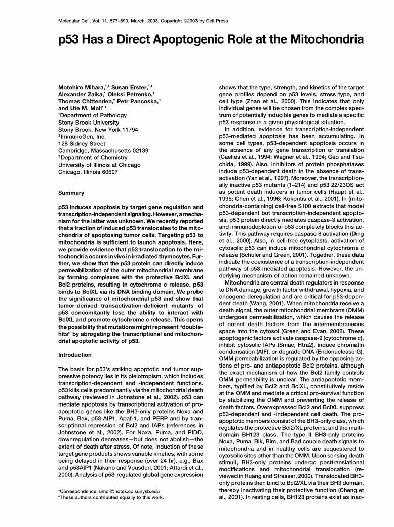
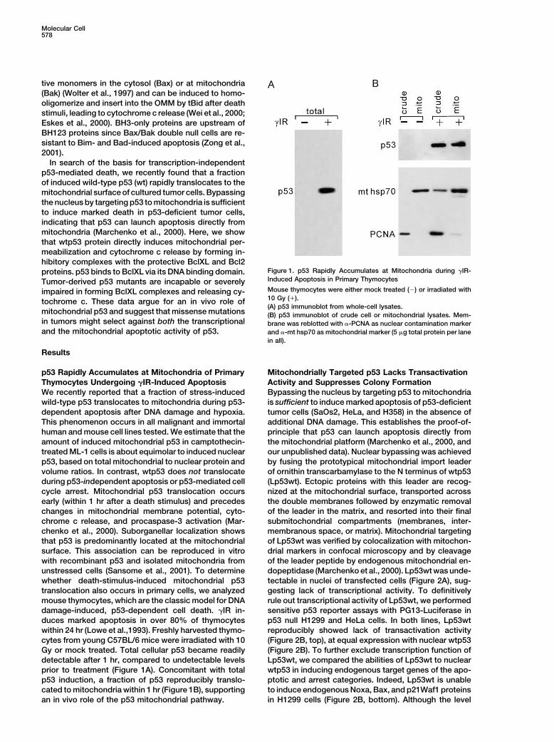
p53 Rapidly Accumulates at Mitochondria of Primary Mitochondrially Targeted p53 Lacks TransactivationActivity and Suppresses Colony FormationThymocytes Undergoing �IR-Induced Apoptosis
We recently reported that a fraction of stress-induced Bypassing the nucleus by targeting p53 to mitochondriais sufficient to induce marked apoptosis of p53-deficientwild-type p53 translocates to mitochondria during p53-
dependent apoptosis after DNA damage and hypoxia. tumor cells (SaOs2, HeLa, and H358) in the absence ofadditional DNA damage. This establishes the proof-of-This phenomenon occurs in all malignant and immortal
human and mouse cell lines tested. We estimate that the principle that p53 can launch apoptosis directly fromthe mitochondrial platform (Marchenko et al., 2000, andamount of induced mitochondrial p53 in camptothecin-
treated ML-1 cells is about equimolar to induced nuclear our unpublished data). Nuclear bypassing was achievedby fusing the prototypical mitochondrial import leaderp53, based on total mitochondrial to nuclear protein and
volume ratios. In contrast, wtp53 does not translocate of ornithin transcarbamylase to the N terminus of wtp53(Lp53wt). Ectopic proteins with this leader are recog-during p53-independent apoptosis or p53-mediated cell
cycle arrest. Mitochondrial p53 translocation occurs nized at the mitochondrial surface, transported acrossthe double membranes followed by enzymatic removalearly (within 1 hr after a death stimulus) and precedes
changes in mitochondrial membrane potential, cyto- of the leader in the matrix, and resorted into their finalsubmitochondrial compartments (membranes, inter-chrome c release, and procaspase-3 activation (Mar-
chenko et al., 2000). Suborganellar localization shows membranous space, or matrix). Mitochondrial targetingof Lp53wt was verified by colocalization with mitochon-that p53 is predominantly located at the mitochondrial
surface. This association can be reproduced in vitro drial markers in confocal microscopy and by cleavageof the leader peptide by endogenous mitochondrial en-with recombinant p53 and isolated mitochondria from
unstressed cells (Sansome et al., 2001). To determine dopeptidase (Marchenko et al., 2000). Lp53wt was unde-tectable in nuclei of transfected cells (Figure 2A), sug-whether death-stimulus-induced mitochondrial p53
translocation also occurs in primary cells, we analyzed gesting lack of transcriptional activity. To definitivelyrule out transcriptional activity of Lp53wt, we performedmouse thymocytes, which are the classic model for DNA
damage-induced, p53-dependent cell death. �IR in- sensitive p53 reporter assays with PG13-Luciferase inp53 null H1299 and HeLa cells. In both lines, Lp53wtduces marked apoptosis in over 80% of thymocytes
within 24 hr (Lowe et al.,1993). Freshly harvested thymo- reproducibly showed lack of transactivation activity(Figure 2B, top), at equal expression with nuclear wtp53cytes from young C57BL/6 mice were irradiated with 10
Gy or mock treated. Total cellular p53 became readily (Figure 2B). To further exclude transcription function ofLp53wt, we compared the abilities of Lp53wt to nucleardetectable after 1 hr, compared to undetectable levels
prior to treatment (Figure 1A). Concomitant with total wtp53 in inducing endogenous target genes of the apo-ptotic and arrest categories. Indeed, Lp53wt is unablep53 induction, a fraction of p53 reproducibly translo-
cated to mitochondria within 1 hr (Figure 1B), supporting to induce endogenous Noxa, Bax, and p21Waf1 proteinsin H1299 cells (Figure 2B, bottom). Although the levelan in vivo role of the p53 mitochondrial pathway.
p53, Apoptosis, Mitochondria579
Figure 2. Mitochondrially Targeted p53 Lacks Transactivation Activity and Suppresses Colony Formation
(A) HeLa cells transfected with Flag-tagged Lp53wt and stained with Flag antibody (epifluorescence). Exclusive mitochondrial localizationwith nuclear sparing is apparent.(B) Mitochondrially targeted Lp53wt lacks transactivation activity. (Top) p53 reporter assay in H1299 cells using the PG13 Luc reporter and500 ng of each plasmid. (Bottom) Lp53wt fails to transactivate endogenous p53 targets such as p21, Bax, and Noxa. Western blots comparinginduction by nuclear wtp53, Lp53wt, and empty vector in H1299 cells 10 and 20 hr after transfection (25 �g protein per lane). The Baxmembrane was reblotted for vimentin (loading control) and for p53 expression. The upper band of Lp53wt is the fusion protein.(C) Colony suppression assay of SaOs2 cells transfected with the indicated plasmids. After G418 selection for 18 days, cells were fixed andstained.(D) Colony suppression assays from three independent experiments �/� SD.(E) Mitochondrial translocation of nuclear wtp53 in transiently transfected H1299 cells. Whole-cell or mitochondrial lysates were blotted with�-p53, �-PCNA, and �-mthsp70 antibodies (10 �g per lane).
Molecular Cell580
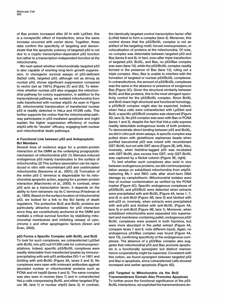
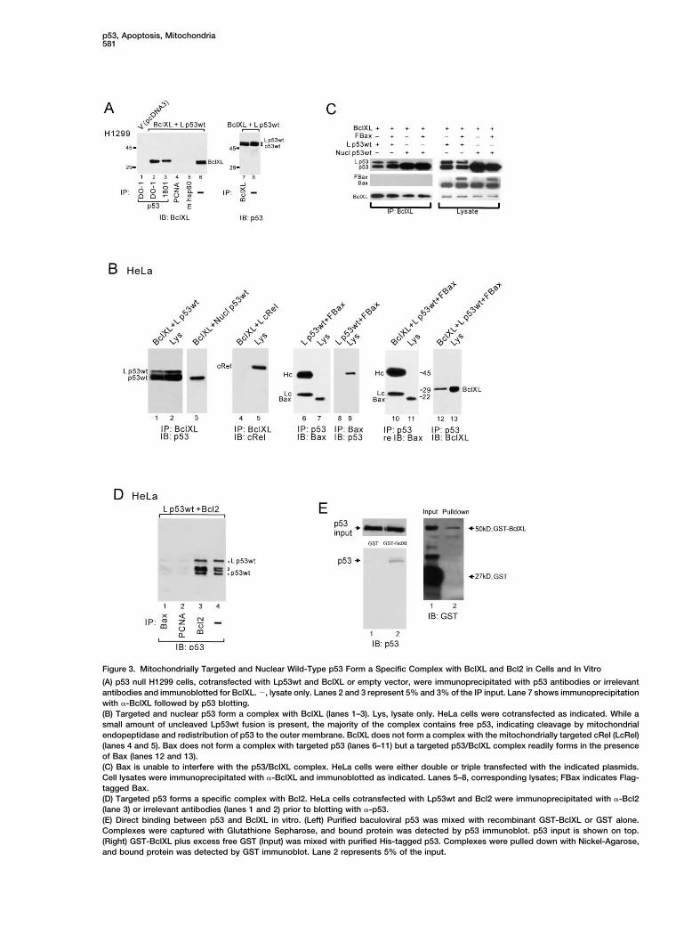
of Bax protein increased after 20 hr with Lp53wt, this the identically targeted control transcription factor cRel(LcRel) failed to form a complex (lane 4). Moreover, thisis a nonspecific effect of transfection, since the samecontrol shows that the p53/BclXL complex is not anincrease occurred with vector alone. Together, theseartifact of the targeting motif, forced overexpression, ordata confirm the specificity of targeting and demon-colocalization of proteins at the mitochondria. Of note,strate that the apoptotic potency of targeted p53 is notno complex was detectable between targeted p53 anddue to a cryptic transcription-dependent p53 functionBax (lanes 6 and 8). In fact, even after triple transfectionbut rather to a transcription-independent function at theof targeted p53, BclXL, and Bax, no p53/Bax complexmitochondria.was seen (lane 10), while the p53/BclXL complex readilyWe next asked whether mitochondrially targeted p53formed in the presence of Bax (lane 12), ruling out ais also capable of exerting long-term growth suppres-triple complex. Also, Bax is unable to interfere with thesion. In clonogenic survival assays of p53-deficientformation of targeted or nuclear p53/BclXL complexes.SaOs2 cells, targeted p53, although not as strong asIn cotransfections, the amount of p53/BclXL complexesnuclear p53, shows significant suppression comparedwas the same in the absence or presence of exogenousto vector (set as 100%) (Figures 2C and 2D). To deter-Bax (Figure 3C). Given the structural similarity betweenmine whether nuclear p53 also engages the mitochon-BclXL and Bax proteins, this is the most stringent speci-drial pathway for colony suppression, in addition to theficity control for the p53/BclXL complex. Since BclXLtranscriptional pathway, we isolated mitochondria fromand Bcl2 share high structural and functional homology,cells transfected with nuclear wtp53. As seen in Figurea p53/Bcl2 complex might also be expected. Indeed,2E, mitochondrial translocation of transfected nuclearwhen HeLa cells were cotransfected with Lp53wt andp53 is readily detected in HeLa and H1299 cells. ThisBcl2, a specific p53/Bcl2 complex was observed (Figurefurther supports the notion that the mitochondrial path-3D, lane 3). No p53 complex was seen with Bax or PCNAway participates in p53-mediated apoptosis and might(lanes 1 and 2), despite the fact that HeLa cells expressexplain the higher suppression efficiency of nuclearreadily detectable endogenous levels of both proteins.wtp53 with its “double” efficacy, engaging both nuclearTo demonstrate direct binding between p53 and BclXL,and mitochondrial death pathways.we did in vitro pull-down assays. A specific complex waspulled down with glutathione sepharose beads whenA Functional Link between p53 and Antiapoptoticpurified baculoviral p53 was mixed with recombinantBcl MembersGST-BclXL but not with GST alone (Figure 3E, left). Also,Several lines of evidence argue for a protein-proteininversely, when histidine-tagged p53 was incubatedinteraction at the OMM as the underlying proapoptoticwith GST-BclXL plus excess free GST, only GST-BclXLmechanism of mitochondrial p53: (1) Stimulus-inducedwas captured by a Nickel column (Figure 3E, right).endogenous p53 mainly translocates to the surface of
To test whether such complexes also exist in vivomitochondria; (2) This surface association can be repro-between endogenous proteins, we did coimmunprecipi-duced in vitro with recombinant p53 and unstimulatedtation assays on solubilized mitochondria from wtp53-mitochondria (Sansome et al., 2001); (3) Truncation ofharboring ML-1 and RKO cells after short-term DNAthe entire p53 C terminus is dispensable for its mito-damage by camptothecin. Mitochondrial isolates werechondrial apoptotic action, arguing for a protein-proteinfree of nuclear contamination as verified by the PCNAinteraction (Marchenko et al., 2000). In contrast, whenmarker (Figure 4C). Specific endogenous complexes ofp53 acts as a transcription factor, it depends on thep53/BclXL and p53/Bcl2 were detected when extracts
ability to form tetramers via its C terminus (Friedman etwere precipitated with anti-BclXL (Figure 4A, lanes 1, 7,
al., 1993). Based on the surface location of mitochondrialand 8) or anti-Bcl2 (Figure 4B, lane 5) and blotted with
p53, we looked for a link to the Bcl family of death anti-p53 or, inversely, when extracts were precipitatedregulators. The protective Bcl2 and BclXL proteins are with anti-p53 and blotted with anti-BclXL (Figure 4A,particularly attractive candidates for p53 interaction lane 3) or anti-Bcl2 (Figure 4B, lane 1). Moreover, whensince they are constitutively anchored at the OMM and solubilized mitochondria were separated into superna-mediate a critical survival function by stabilizing mito- tant and membrane-containing pellet, endogenous p53/chondrial membranes and inhibiting release of cyto- BclXL complexes were present in both fractions, butchrome c and other apoptogenic factors (Green and were more abundant in the pellet extract (Figure 4A,Evan, 2002). compare lanes 7 and 8, note different input). Again, no
endogenous p53/Bax complex was found (Figure 4A,p53 Forms a Specific Complex with BclXL and Bcl2 lane 10), confirming specificity of the endogenous com-To look for such complexes, we cotransfected Lp53wt plexes. The absence of a p53/Bax complex also sug-with BclXL into p53 null H1299 cells for coimmunopreci- gests that mitochondrial p53 and Bax promote apopto-pitations. Indeed, specific p53/BclXL complexes were sis in a functionally synergistic but distinct manner;readily detectable from transfected lysates by immuno- hence cooperativity might be expected. Consistent withprecipitating with anti-p53 antibodies DO-1 or 1801 and this notion, we found synergism between targeted p53blotting with anti-BclXL (Figure 3A, lanes 2 and 3). No and Bax in apoptosis, since cotransfected cells showedcomplexes were seen with irrelevant antibodies against increased and earlier apoptosis (Figure 4D).abundant nuclear or mitochondrial proteins such asPCNA and mt hsp60 (lanes 4 and 5). The same complex p53 Targeted to Mitochondria via the Bcl2was also seen in reverse (lane 7) and in cotransfected Transmembrane Domain Also Promotes ApoptosisHeLa cells coexpressing BclXL and either targeted (Fig- To further prove the functional significance of the p53-
BclXL interactions, we exploited the transmembrane do-ure 3B, lane 1) or nuclear wtp53 (lane 3). In contrast,
p53, Apoptosis, Mitochondria581
Figure 3. Mitochondrially Targeted and Nuclear Wild-Type p53 Form a Specific Complex with BclXL and Bcl2 in Cells and In Vitro
(A) p53 null H1299 cells, cotransfected with Lp53wt and BclXL or empty vector, were immunoprecipitated with p53 antibodies or irrelevantantibodies and immunoblotted for BclXL. �, lysate only. Lanes 2 and 3 represent 5% and 3% of the IP input. Lane 7 shows immunoprecipitationwith �-BclXL followed by p53 blotting.(B) Targeted and nuclear p53 form a complex with BclXL (lanes 1–3). Lys, lysate only. HeLa cells were cotransfected as indicated. While asmall amount of uncleaved Lp53wt fusion is present, the majority of the complex contains free p53, indicating cleavage by mitochondrialendopeptidase and redistribution of p53 to the outer membrane. BclXL does not form a complex with the mitochondrially targeted cRel (LcRel)(lanes 4 and 5). Bax does not form a complex with targeted p53 (lanes 6–11) but a targeted p53/BclXL complex readily forms in the presenceof Bax (lanes 12 and 13).(C) Bax is unable to interfere with the p53/BclXL complex. HeLa cells were either double or triple transfected with the indicated plasmids.Cell lysates were immunoprecipitated with �-BclXL and immunoblotted as indicated. Lanes 5–8, corresponding lysates; FBax indicates Flag-tagged Bax.(D) Targeted p53 forms a specific complex with Bcl2. HeLa cells cotransfected with Lp53wt and Bcl2 were immunoprecipitated with �-Bcl2(lane 3) or irrelevant antibodies (lanes 1 and 2) prior to blotting with �-p53.(E) Direct binding between p53 and BclXL in vitro. (Left) Purified baculoviral p53 was mixed with recombinant GST-BclXL or GST alone.Complexes were captured with Glutathione Sepharose, and bound protein was detected by p53 immunoblot. p53 input is shown on top.(Right) GST-BclXL plus excess free GST (Input) was mixed with purified His-tagged p53. Complexes were pulled down with Nickel-Agarose,and bound protein was detected by GST immunoblot. Lane 2 represents 5% of the input.
Molecular Cell582
Figure 4. Endogenous Complexes betweenwtp53 and BclXL or wtp53 and Bcl2 Form inMitochondria of Stressed Cells
(A and B) wtp53-containing RKO and ML-1cells were subject to short-term DNA damage(5 �M camptothecin for 5 hr). Extracts fromsonicated and solubilized mitochondria wereimmunoprecipitated followed by blotting withthe indicated antibodies. Lys, lysate only. P,mitochondrial pellet; SN, supernatant; mock,contains no extract. * denotes a variable non-specific band.(C) Mitochondria used in (A) and (B) were freeof nuclear contamination. PCNA, p53, andmthsp70 immunoblots (5 �g per lane).(D) Targeted wtp53 and Bax cooperate in pro-moting cell death. HeLa cells were single ordouble transfected with 300 ng each of theindicated plasmids. After 24 hr, apoptosis inexpressing cells was quantitated by Hoechststaining, counting 20 random microscopicfields. Two representative experiments froma total of four are shown.
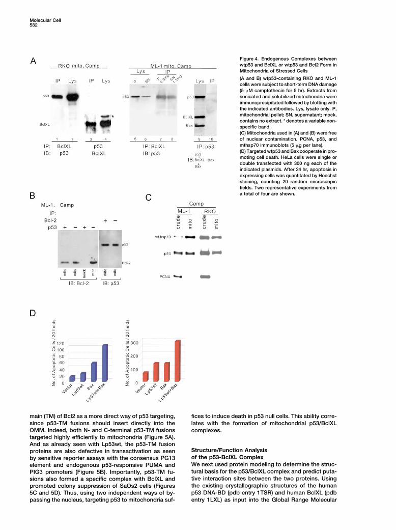
main (TM) of Bcl2 as a more direct way of p53 targeting, fices to induce death in p53 null cells. This ability corre-since p53-TM fusions should insert directly into the lates with the formation of mitochondrial p53/BclXLOMM. Indeed, both N- and C-terminal p53-TM fusions complexes.targeted highly efficiently to mitochondria (Figure 5A).And as already seen with Lp53wt, the p53-TM fusion
Structure/Function Analysisproteins are also defective in transactivation as seenof the p53-BclXL Complexby sensitive reporter assays with the consensus PG13We next used protein modeling to determine the struc-element and endogenous p53-responsive PUMA andtural basis for the p53/BclXL complex and predict puta-PIG3 promoters (Figure 5B). Importantly, p53-TM fu-tive interaction sites between the two proteins. Usingsions also formed a specific complex with BclXL andthe existing crystallographic structures of the humanpromoted colony suppression of SaOs2 cells (Figuresp53 DNA-BD (pdb entry 1TSR) and human BclXL (pdb5C and 5D). Thus, using two independent ways of by-
passing the nucleus, targeting p53 to mitochondria suf- entry 1LXL) as input into the Global Range Molecular
p53, Apoptosis, Mitochondria583
Figure 5. wtp53 Fused to the Transmembrane Domain of Bcl2 Targets to Mitochondria, Lacks Transactivation Function, Forms a Complexwith BclXL, and Promotes Growth Suppression
(A) N- and C-terminal fusions between p53 and the transmembrane domain of human Bcl2 (aa 211–239), called NTM-p53 and p53-CTM,colocalize with the mitochondrial marker mt hsp70, without nuclear staining. SaOs2 cells transfected with p53-CTM are shown here.(B) p53-TM fusions lack transactivation activity. Luciferase reporter assays driven by PG13 consensus response element (top) or the endogenousPIG3 and PUMA promoters (bottom). SaOs2 cells were transfected with 500 ng of each plasmid.(C) SaOs2 cells transfected with the indicated plasmids were immunoprecipitated for p53 followed by BclXL blotting (top). Equal amounts ofimmunoprecipitated p53 were loaded (bottom).(D) Saos2 colony suppression assay after transfection with indicated plasmids. Crystal violet staining after 12 days of G418 selection.
Molecular Cell584
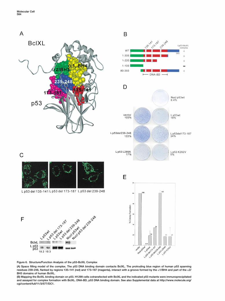
Figure 6. Structure/Function Analysis of the p53-BclXL Complex
(A) Space filling model of the complex. The p53 DNA binding domain contacts BclXL. The protruding blue region of human p53 spanningresidues 239–248, flanked by regions 135–141 (red) and 173–187 (magenta), interact with a groove formed by the �1/BH4 and part of the �2/BH3 domains of human BclXL.(B) Mapping the BclXL binding domain on p53. H1299 cells cotransfected with BclXL and the indicated p53 mutants were immunoprecipitatedand assayed for complex formation with BclXL. DNA-BD, p53 DNA binding domain. See also Supplemental data at http://www.molecule.org/cgi/content/full/11/3/577/DC1.
p53, Apoptosis, Mitochondria585
Matching Program (Vakser and Nikiforovich, 1995), we correlation between the ability of p53 to form BclXLcomplexes and its ability to induce cell death via theemployed a classic protein folding algorithm that we
expanded by integrating quantitative local context mitochondrial pathway.descriptors for each amino acid position within the poly-peptide sequence of each protein. This allows the com- Naturally Occurring Mutations in Human Tumors
Select against the Transcriptional andputation of contextual similarity surfaces of two interac-tion partners (detailed description of the method will Mitochondrial Apoptotic Activity of p53
Our model implies that the DNA binding domain of p53be published elsewhere). For validation, our algorithmcorrectly predicted the structure and interaction sur- is a dual-function domain, mediating both the transacti-
vation and the mitochondrial proapoptotic function. Thisfaces of a crystallographically solved complex betweenthe p53 DNA binding domain and 53BP2 (data not leads to the prediction that missense mutations in this
region abrogate both functions. During malignant trans-shown) (pdb entry 1YCS; Gorina and Pavletich, 1996),while it did not predict the existence of a p53/Bax com- formation of human tumors, p53 mutations are selected
for loss of their apoptotic and tumor suppressor ability,plex, consistent with our experimental data. We com-puted about 500 possible structures for a p53/BclXL which coincides with loss of their transactivation func-
tion (Hollstein et al., 1999). Do tumor-derived p53 mu-complex, but only the structure in Figure 6A satisfiesthe constraints given by the contextual similarity analy- tants concomitantly lose the ability to bind to BclXL? We
examined four randomly chosen tumor-derived breastsis (see also Supplemental Figure S1 at http://www.molecule.org/cgi/content/full/11/3/577/DC1). The contact cancer cell lines with mutant p53 and LOH of the wt
alleles. SKBr3 harbors R175H, T47D harbors L194F,surface involves the p53 DNA binding domain, inter-acting with the �1/BH4 and partial �2/BH3 domains of MDA 468 harbors R273H, and MDA 231 harbors R280K.
Together, these four mutations represent 13% of all p53BclXL. The major contact site on p53, comprised ofresidues 239–248 (part of the L3 loop), is the most pro- mutations found in over 16,000 human tumors (IARC
p53 database [http://www.iarc.fr/p53/Index.html]). Thetruding and centrally located. This domain is flankedby two lateral domains on either side, contributed by R175H mutant is the prototype of a structural mutant
due to unfolding of the � sandwich scaffold of the DNA-residues 135–141 (L1 loop) and residues 173–187 (part ofthe L2 loop and H1 helix). Using coimmunoprecipitation BD. The R273H mutant is a classic DNA contact mutant
and both are hotspots in human cancers. Arg280 is partassays on a series of N- and C-terminal truncations ofLp53wt, we mapped the BclXL binding domain of p53 of the H2 helix and makes direct contact with the major
groove. Leu194 is immediately adjacent to the hy-to its DNA binding domain (Figure 6B). Lp53(1–305),Lp53(1–220), and Lp53(80–393) were able to bind to drophobic core of the � sandwich (Cho et al., 1994).
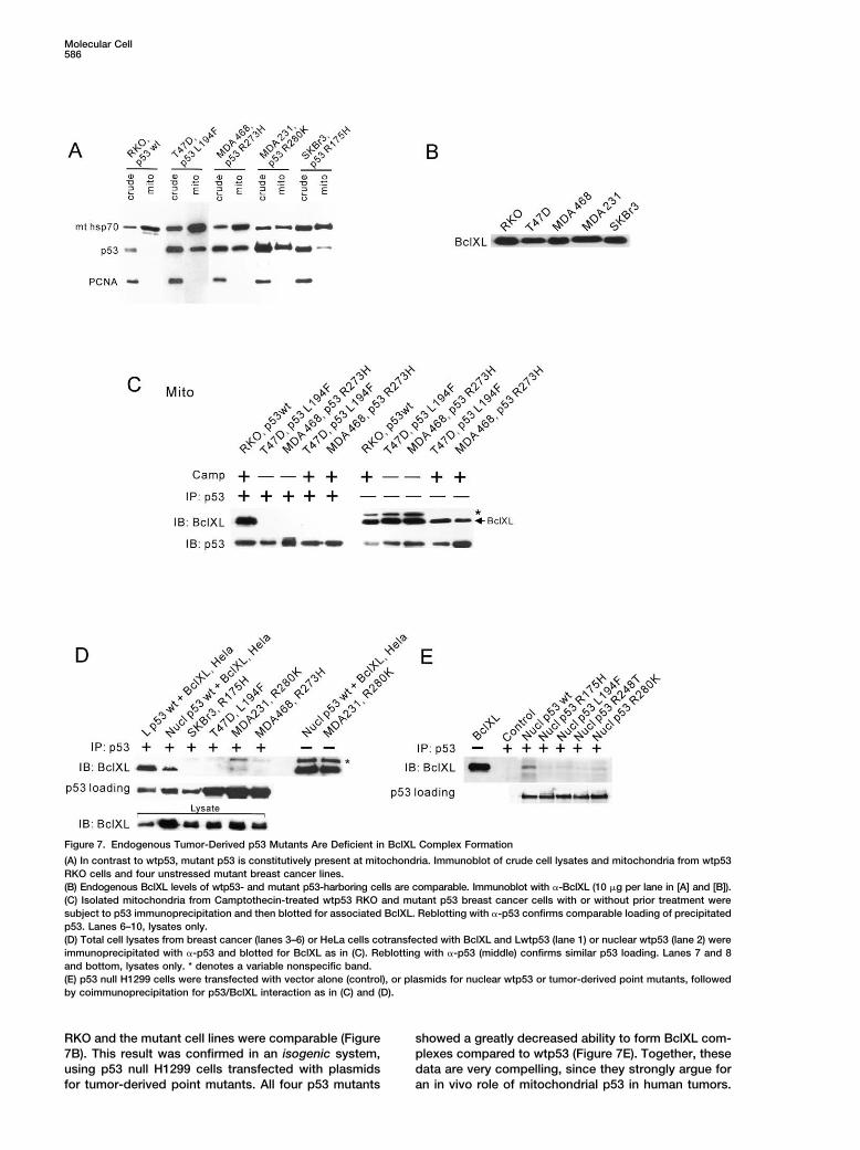
Using careful quantitative coimmunoprecipitations,BclXL, while Lp53(1–100), which is missing the contactsites, had lost its binding. Together, these data implicate we compared the amount of endogenous p53/BclXL
complexes in wtp53 harboring RKO cells with those ofthe p53 core domain to interact with BclXL. To determinethe importance of BclXL binding for mitochondrial p53 mutant p53 cells. Due to lack of HDM2 induction, mutant
cells have high total levels of p53. Of note, proportionalfunction, we deleted each of the contact regions fromLp53wt and tested them for loss of mitochondrial killing to their abnormally stabilized p53, all four lines constitu-
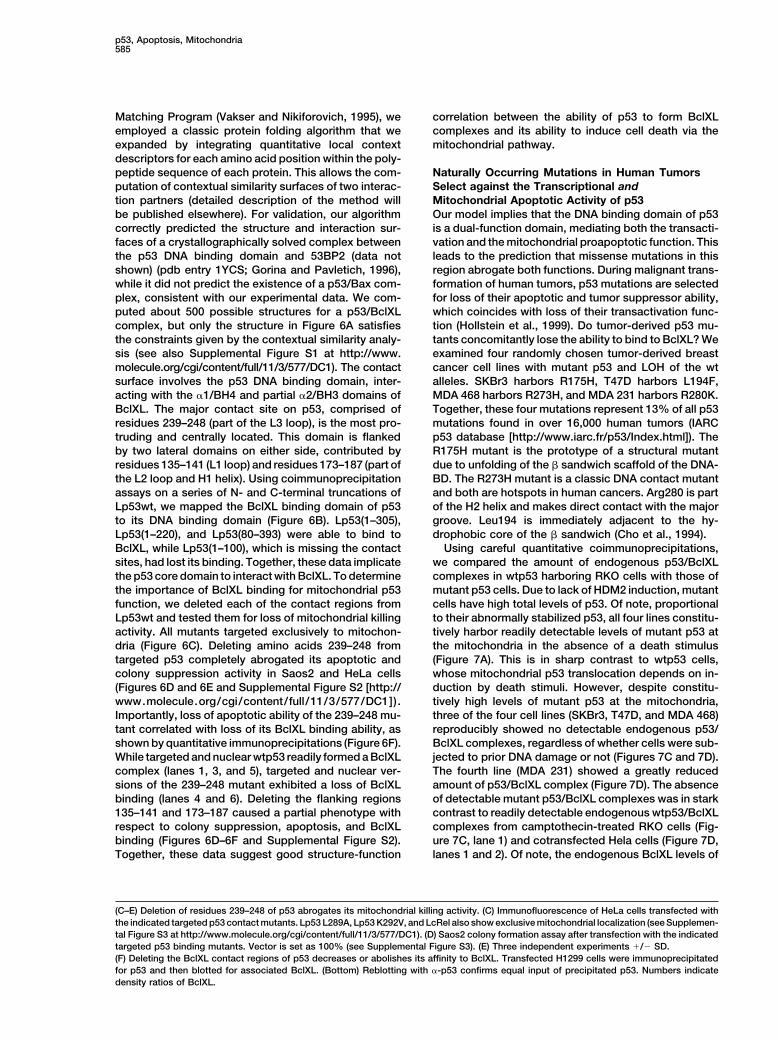
tively harbor readily detectable levels of mutant p53 atactivity. All mutants targeted exclusively to mitochon-dria (Figure 6C). Deleting amino acids 239–248 from the mitochondria in the absence of a death stimulus
(Figure 7A). This is in sharp contrast to wtp53 cells,targeted p53 completely abrogated its apoptotic andcolony suppression activity in Saos2 and HeLa cells whose mitochondrial p53 translocation depends on in-
duction by death stimuli. However, despite constitu-(Figures 6D and 6E and Supplemental Figure S2 [http://www.molecule.org/cgi/content/full/11/3/577/DC1]). tively high levels of mutant p53 at the mitochondria,
three of the four cell lines (SKBr3, T47D, and MDA 468)Importantly, loss of apoptotic ability of the 239–248 mu-tant correlated with loss of its BclXL binding ability, as reproducibly showed no detectable endogenous p53/
BclXL complexes, regardless of whether cells were sub-shown by quantitative immunoprecipitations (Figure 6F).While targeted and nuclear wtp53 readily formed a BclXL jected to prior DNA damage or not (Figures 7C and 7D).
The fourth line (MDA 231) showed a greatly reducedcomplex (lanes 1, 3, and 5), targeted and nuclear ver-sions of the 239–248 mutant exhibited a loss of BclXL amount of p53/BclXL complex (Figure 7D). The absence
of detectable mutant p53/BclXL complexes was in starkbinding (lanes 4 and 6). Deleting the flanking regions135–141 and 173–187 caused a partial phenotype with contrast to readily detectable endogenous wtp53/BclXL
complexes from camptothecin-treated RKO cells (Fig-respect to colony suppression, apoptosis, and BclXLbinding (Figures 6D–6F and Supplemental Figure S2). ure 7C, lane 1) and cotransfected Hela cells (Figure 7D,
lanes 1 and 2). Of note, the endogenous BclXL levels ofTogether, these data suggest good structure-function
(C–E) Deletion of residues 239–248 of p53 abrogates its mitochondrial killing activity. (C) Immunofluorescence of HeLa cells transfected withthe indicated targeted p53 contact mutants. Lp53 L289A, Lp53 K292V, and LcRel also show exclusive mitochondrial localization (see Supplemen-tal Figure S3 at http://www.molecule.org/cgi/content/full/11/3/577/DC1). (D) Saos2 colony formation assay after transfection with the indicatedtargeted p53 binding mutants. Vector is set as 100% (see Supplemental Figure S3). (E) Three independent experiments �/� SD.(F) Deleting the BclXL contact regions of p53 decreases or abolishes its affinity to BclXL. Transfected H1299 cells were immunoprecipitatedfor p53 and then blotted for associated BclXL. (Bottom) Reblotting with �-p53 confirms equal input of precipitated p53. Numbers indicatedensity ratios of BclXL.
Molecular Cell586
Figure 7. Endogenous Tumor-Derived p53 Mutants Are Deficient in BclXL Complex Formation
(A) In contrast to wtp53, mutant p53 is constitutively present at mitochondria. Immunoblot of crude cell lysates and mitochondria from wtp53RKO cells and four unstressed mutant breast cancer lines.(B) Endogenous BclXL levels of wtp53- and mutant p53-harboring cells are comparable. Immunoblot with �-BclXL (10 �g per lane in [A] and [B]).(C) Isolated mitochondria from Camptothecin-treated wtp53 RKO and mutant p53 breast cancer cells with or without prior treatment weresubject to p53 immunoprecipitation and then blotted for associated BclXL. Reblotting with �-p53 confirms comparable loading of precipitatedp53. Lanes 6–10, lysates only.(D) Total cell lysates from breast cancer (lanes 3–6) or HeLa cells cotransfected with BclXL and Lwtp53 (lane 1) or nuclear wtp53 (lane 2) wereimmunoprecipitated with �-p53 and blotted for BclXL as in (C). Reblotting with �-p53 (middle) confirms similar p53 loading. Lanes 7 and 8and bottom, lysates only. * denotes a variable nonspecific band.(E) p53 null H1299 cells were transfected with vector alone (control), or plasmids for nuclear wtp53 or tumor-derived point mutants, followedby coimmunoprecipitation for p53/BclXL interaction as in (C) and (D).
RKO and the mutant cell lines were comparable (Figure showed a greatly decreased ability to form BclXL com-plexes compared to wtp53 (Figure 7E). Together, these7B). This result was confirmed in an isogenic system,
using p53 null H1299 cells transfected with plasmids data are very compelling, since they strongly argue foran in vivo role of mitochondrial p53 in human tumors.for tumor-derived point mutants. All four p53 mutants
p53, Apoptosis, Mitochondria587
It further suggests that tumorigenic mutations that are only protein tBID which translocates to mitochondriaupon a death stimulus to induce Bak oligomerizationselected during human tumor formation might represent
double-hit mutations by abrogating both the transcrip- (Figure 8F, lane 1). Moreover, since overexpression ofBclXL and Bcl2 completely block p53-dependent apo-tional and the mitochondrial apoptotic activity of p53.ptosis in vivo (Schuler and Green, 2001), it predicts thatexcess BclXL also prevents p53-induced cytochrome cPurified p53 Protein Triggers the Rapid Releaserelease in vitro. To address this, we performed BclXLof Cytochrome C from Isolated Mitochondriacompetition experiments. Indeed, the effectiveness ofWhat is the functional basis of the proapoptotic actionp53 in inducing cytochrome c release is inhibited in aof mitochondrial p53? Of note, death-stimulus-induceddose-dependent fashion by adding increasing amountsmitochondrial translocation of p53 precedes the releaseof excess GST-BclXL (Figure 8G), indicating that p53of cytochrome c (Marchenko et al., 2000). To testand antiapoptotic BclXL proteins are in a rheostat-likewhether cytochrome c release is a direct effect of wtp53relationship. Together, these data demonstrate that p53translocation, we asked whether baculoviral wtp53itself is able to directly permeabilize the OMM and triggeralone was sufficient to trigger cytochrome c release fromthe release of proapoptotic activators. It does so byfreshly isolated mouse liver mitochondria. This classicengaging in inhibitory complexes with protective Bcl2/assay was used to establish the membrane-permeabiliz-XL, thereby indirectly activating proapoptotic BH123ing effect of Bax and Bak proteins (Jurgensmeier etproteins like Bak.al., 1998; Eskes et al., 1998). The purity of all our p53
The most persuasive evidence for the physiologic im-preparations was verified by silver gels showing a singleportance of the mitochondrial p53 pathway would comeband of 53 kDa (Figure 8A). Strikingly, incubation withfrom the behavior of mutant p53 proteins. Is there amouse wtp53 triggered the release of over 90% of mito-direct functional link between the failure of p53 mutantschondrial cytochrome c within 30 min, similar to CaCl2,to form BclXL complexes (Figures 7C–7E) and a failurea known permeabilizer and PTP pore opener (Eskes etto release cytochrome c in vitro? Indeed, in contrast toal., 1998), while buffer alone released �5% (Figure 8B).wtp53 that readily induces release, tumor-derived mu-This effect was due to added p53, since endogenoustant p53 proteins are defective in permeabilizing thep53 levels of unstressed mitochondria were undetect-outer membrane. The hotspot mutant R175H has com-able (Figure 8D, lane 1), and it was dose dependent withpletely lost the ability to release cytochrome c, whilesignificant release with 400 nM and maximum releasethe hotspot mutant R273H is severely suppressed inwith 1 �M of wtp53 (Figure 8C, lanes 1–4). In contrast, athis activity (Figure 8H). Thus, this direct link supportsC-terminal fragment of p53 (aa 315–390) failed to inducethe hypothesis that tumors select against both the nu-release (Figure 8C, lanes 5–8). Moreover, p53-inducedclear and mitochondrial apoptotic function of p53. More-cytochrome c release is fast. Within 5 min after addingover, it shows that the interaction with BclXL is requiredp53, 69% of the total cytochrome c release has alreadyfor p53-mediated cytochrome c release.occurred and is almost complete at 30 min (Figure 8D,
lanes 4, 6, 8, and 10). Importantly, the amount of mito-chondria-associated p53 remained constant between 0 Discussionand 60 min, suggesting that p53 binding to mitochondriais a very rapid, saturable process. Identical results were The present data reveal an extranuclear death function
of p53 and show that this function is critically involvedobtained with human wtp53 from baculoviral and bacte-rial sources. in mediating the proapoptotic action of p53 at the mito-
chondria. We show that p53 protein can directly induceWhat is the mechanism by which p53 triggers cyto-chrome c release? Is there a direct link between p53/ permeabilization of the outer mitochondrial membrane
by forming inhibitory complexes with protective Bcl pro-BclXL complex formation and cytochrome c release?To test this notion, we looked for complexes in pellets of teins, resulting in cytochrome c release. p53 binds to
BclXL via its DNA binding domain. This mitochondrialmitochondria after cytochrome c release had occurred(after 30 min incubation with 400 nM wtp53). Indeed, in action might be derailed in tumors since at least some
p53 mutants lose the ability to interact with BclXL andthese post-release mitochondria, p53/BclXL complexeswere clearly detected (Figure 8E, lane 1). This supports a promote cytochrome c release. Thus, tumor-derived
mutations might represent double-hits by concomitantlymechanism where p53 directly induces permeabilizationof the outer mitochondrial membrane by forming inhibi- abrogating the transcriptional and mitochondrial apo-
ptotic activity of p53.tory complexes with BclXL. Furthermore, since p53 is adirect inducer of cytochrome c release and of mitochon- Functional parallels between p53 and other proapo-
ptotic proteins appear to be emerging. p53, Noxa, Puma,drial dysfunction, p53 might also indirectly activate theultimate effectors for cytochrome c release—Bak and Bim, and Bad all rapidly translocate to mitochondria
upon a death stimulus, bind and inhibit the protectiveBax proteins—by inducing their conformational changeand intramembraneous oligomerization. To test this, we BclXL and Bcl2 proteins, and induce OMM permeabiliza-
tion, leading to cytochrome c release and caspase acti-examined whether p53 induces Bak oligomerization(mouse liver mitochondria are poor in Bax). Indeed, us- vation. For both p53 and the BH3 proteins, complex
formation with BclXL/2 is critical for apoptogenicity,ing the chemical crosslinker BS3, purified wtp53, whenadded to freshly isolated nascent mitochondria, readily since interference with binding blocks their mitochon-
drial killing activity (our data; Cheng et al., 2001; Oda etinduced crosslinked Bak multimers, consistent with tri-mers (72 kDa) and tetramers (96 kDa) (Figure 8F, lane al., 2000; Nakano and Vousden, 2001; Yu et al., 2001).
Other proapoptotic proteins such as Siva-1 and2). This p53 behavior is similar to the prototypical BH3-
Molecular Cell588
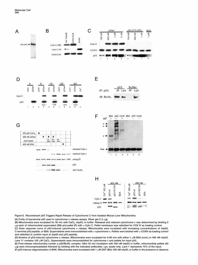
Figure 8. Recombinant p53 Triggers Rapid Release of Cytochrome C from Isolated Mouse Liver Mitochondria
(A) Purity of baculoviral p53 used in cytochrome c release assays. Silver gel (1.2 �g).(B) Mitochondria were incubated for 30 min with CaCl2, wtp53, or buffer. Released and retained cytochrome c was determined by blotting 5�g each of mitochondrial supernatant (SN) and pellet (P) with �-Cyto C. Pellet membrane was reblotted for COX IV as loading control.(C) Dose response curve of p53-induced cytochrome c release. Mitochondria were incubated with increasing concentrations of wtp53,C-terminal p53 peptide, or BSA. Supernatants were immunoblotted with �-cytochrome c. Pellets were blotted with �-COXIV as loading controland reblotted to confirm input of wtp53 and p53 peptide.(D) Kinetics of p53-induced cytochrome c release. Mitochondria were incubated for 0–60 min with either 1 �M BSA (cont.) or 400 nM wtp53.Lane 11 contains 100 �M CaCl2. Supernatants were immunoblotted for cytochrome c and pellets for input p53.(E) Post-release mitochondria contain a p53/BclXL complex. After 30 min incubation with 400 nM wtp53 or buffer, mitochondrial pellets (50�g) were immunoprecipitated followed by blotting with the indicated antibodies. Lys, lysate only. Lane 1 represents 15% of the input.(F) p53 induces oligomerization of BAK. Mitochondria were incubated with 1 �M GST-tBid, 400 nM wtp53, or buffer in the presence or absence
p53, Apoptosis, Mitochondria589
(Sigma). For endogenous complexes, mitochondrial lysates (1 mg)p53AIP1, which like p53 lack a BH3 domain, also bindfrom Camp-treated ML-1 or RKO cells were immunoprecipitatedand inactivate BclXL (Xue et al., 2002; Matsuda et al.,with 1 �g antibody and immunoblotted. For exogenous complexes,2002). While BH3-only proteins bind BclXL/2 throughwhole-cell lysates (1 mg) of transfected cells were used. For quanti-
their BH3 domain, SIVA-1 binds through a unique amphi- tative immunoprecipitations in Figure 7, 200 �g of mitochondrial (C)pathic helix (Xue et al., 2002) and p53 binds through its or 1 mg of whole-cell (D) lysates were incubated with 20 �l of DO-1
plus 1801 Sepharose beads for 2 hr. Beads were washed three timescentral core effector domain (our data). Our finding ofwith SNNTE plus 2 � RIPA (50 mM Tris, 150 mM NaCl, 1% Tritonthe ability of excess BclXL to block p53-mediated cyto-X-100, 0.1% SDS, 1% NaDeoxycholate [pH 7.4]) and subjected tochrome c release in vitro parallels that seen with Baxan initial Western blot for quantitation. Loading of the definitive blotand tBid (Jurgensmeier et al., 1998; Desagher et al.,was then normalized for equal amounts of immuneprecipitated p53
1999). Also, Puma- and Siva-1-mediated apoptosis is and blotted for BclXL. For pull-down experiments, purified humansuppressed by excess BclXL (Nakano and Vousden, full-length GST-BclXL (Chittenden et al., 1995) and/or free GST were
incubated with purified baculoviral His-tagged wtp53, washed, and2001; Xue et al., 2002).pulled down with either glutathione sepharose or Ni-NTA AgaroseIn summary, our study provides a mechanistic basisbeads. Luciferase reporter assays were performed with PG13-Luc,for transcription-independent apoptosis by p53 andPUMA-Luc (Yu et al., 2001), and PIG3-Luc (Contente et al., 2002)opens a new avenue toward comprehensive under-reporters. All values were normalized for Renilla activity.
standing of p53 function. We propose that p53 exertsa rapid and direct proapoptotic role at the mitochondria, Apoptosis and Colony Suppression Assaysthereby jump-starting and amplifying its transcription- TUNEL assays were performed as described (Marchenko et al.,
2000). For colony suppression, SaOs2 cells (1 � 103 ) in 100 mmbased apoptotic action that requires more time to takeplates were transfected with 300 ng of the indicated plasmids. 48effect. Given its implication in a broad spectrum of cellhr later, cells were placed under G418 selection (500 �g/ml) for 12–18types and death signals and the central role of mitochon-days, fixed, and stained with crystal violet. Foci were quantitated via
dria in p53-mediated apoptosis, this pathway could en- integral optical plate density (BioRad, Laser Pix Software), settinghance many types of p53-mediated killing. A complete vector as 100%.analysis with respect to tissue types and gamut of stresssignals will further delineate the scope of this pathway Plasmid Constructions
All deletion and point mutants were generated by PCR into ain p53 stress responses. Another future question is thepcDNA3-based plasmid encoding a mitochondrial import leader andrelative contributions of the transcriptional and mito-cleavage site for mitochondrial endopeptidase, fused to the N termi-chondrial pathways to tumor suppression in vivo. Basednus of wt p53 (Marchenko et al., 2000). For mitochondrial p53 tar-
on the pleiotropic p53 function, it might well be that geting via the transmembrane (TM) domain of Bcl2, N- and C-ter-individual effector pathways are necessary but not suffi- minal fusions of wtp53 and TM domain (aa 211–239) of human Bcl2cient to execute the full scale of the p53 death and were generated by PCR: NTMp53wt (TMp53(3–393)) and p53wtCTM
(p53(1–392)TM). Expression plasmids of human Bcl2, BclXL, andsuppressor program.Bax (Chittenden et al., 1995) and LcRel were described (Marchenkoet al., 2000). All constructs were sequence confirmed and shown toExperimental Procedurestarget to mitochondria by colocalization by immunofluorescence.
Cell CultureCells were grown in RPMI 1640 (ML-1) or DMEM (all others), plus Cytochrome C Release
Freshly harvested livers of 10-week-old C57BL/6 mice were used for10% FBS. Camptothecin (Sigma) stress was always 5 �M for 5 hr.mitochondrial isolation (Eskes et al., 1998). His-tagged baculoviralhuman and mouse mutant and wild-type p53 or C-terminal fragmentMitochondrial Fractionation
Mitochondria were prepared by sucrose density gradients (Mar- were purified over Ni-NTA agarose, followed by heparin and MonoQ/FPLC columns. Immunoaffinity-purified baculoviral p53 R175H pro-chenko et al., 2000). Thymocytes of 4- to 6-week-old C57BL/6 fe-
males were isolated within 5 min of sacrifice, pulse treated with 10 tein was a gift of C. Prives. p53 and BSA (Sigma) were dialyzedagainst KCl buffer (15 mM HEPES/NaOH, 125 mM KCl, 4 mM MgCl2,Gy, and incubated for 1 hr at 37C prior to mitochondrial isolation.5 mM Na2HPO4, 0.5 mM EGTA, 5 �M Rotenon (Sigma), 5 mM succi-nate [pH 7.4]). Mitochondria (70 �g protein) were incubated withImmunoblots and Coimmunoprecipitation
Equal total protein of mitochondrial and crude cell lysates were proteins for 30 min at 30C in 200 �l KCl buffer, then centrifugedat 13,000 g for 10 min at 4C. Mitochondrial pellets (5 �g) andimmunoblotted with the following antibodies: monoclonal 1801, 240
and DO-1 (Santa Cruz Biotechnology), or polyclonal CM-1 (Vector) corresponding supernatants were immunoblotted for cytochrome cand cytochrome oxidase IV (COXIV) antibodies to verify equal load-for human p53; UM-1 for mouse p53 (raised against recombinant
mouse wtp53); mt hsp60 and mt hsp70 (Affinity Bioreagents); PCNA, ing. For crosslinking experiments, 70 �g of freshly isolated livermitochondria was incubated with 400 nM wtp53 or 1 �M purifiedGST, and cRel (Santa Cruz), cytochrome c (Pharmingen), and cyto-
chrome oxidase IV (Molecular Probes); polyclonal BclXL (S18 and GST-tBid or buffer (mock) in the presence or absence of 2 mMcrosslinker BS3 (Bissulfosuccinitidyl suberate; Pierce) for 30 min atL19; Santa Cruz), Bcl2 (Transduction Labs), Bax (Neomarker), Noxa
(Imgenex), Bak (Upstate Biotechnology), and Flag and rabbit IgG room temperature.
of 2 mM crosslinker BS3 for 30 min. Crosslinked Bak species were detected by Bak immunoblot. * indicates Bak complexes consistent withdimers, trimers, and tetramers. While endogenous tBid and Bim induce trimeric and tetrameric Bak complexes, recombinant GST-tBid mainlygenerates Bak dimers (Wei et al., 2000; Cheng et al., 2001).(G) p53-induced cytochrome c release in vitro is inhibited by excess BclXL. Cytochrome c release assay as in (C), but where indicated 10-to 20-fold molar excess of GST-BclXL was added at the start of the reaction. Released and retained cytochrome c was determined by blotting5 �g of mitochondrial supernatant and pellet each with �-Cyto C. Pellet membrane was reblotted for mthsp70 (loading control) and for p53and GST-BclXL to show input.(H) Tumor-derived hotspot mutant p53 proteins are defective in inducing cytochrome c release. Cytochrome c release assay as above withbaculoviral wt or mutant p53 (400 nM). Calcium and BSA (400 nM) are used as controls. Equal input of p53 and mitochondria is shown byreblotting the post-release pellets with �-p53 and �-mthsp70 antibodies.
Molecular Cell590
Acknowledgments Hollstein, M., Hergenhahn, M., Yang, Q., Bartsch, H., Wang, Z.Q.,and Hainaut, P. (1999). New approaches to understanding p53 genetumor mutation spectra. Mutat. Res. 43, 199–209.We thank A. Sidorenko for technical assistance. This work was
supported by grants from the NIH/NCI and the American Cancer Johnstone, R.W., Ruefli, A.A., and Lowe, S.W. (2002). Apoptosis: aSociety. link between cancer genetics and chemotherapy. Cell 108, 153–164.
Jurgensmeier, J.M., Xie, Z., Deveraux, Q., Ellerby, L., Bredesen, D.,Received: June 6, 2002 and Reed, J.C. (1998). Bax directly induces release of cytochromeRevised: January 13, 2003 c from isolated mitochondria. Proc. Natl. Acad. Sci. USA 95, 4997–Published online: January 27, 2003 5002.
Kokontis, J.M., Wagner, A.J., O’Leary, M., Liao, S., and Hay, N.References (2001). A transcriptional activation function of p53 is dispensable
for and inhibitory of its apoptotic function. Oncogene 20, 659–668.Attardi, L.D., Reczek, E.E., Cosmas, C., Demicco, E.G., McCurrach,
Lowe, S.W., Schmitt, E.M., Smith, S.W., Osborne, B.A., and Jacks,M.E., Lowe, S.W., and Jacks, T. (2000). PERP, an apoptosis-associ-
T. (1993). p53 is required for radiation-induced apoptosis in mouseated target of p53, is a novel member of the PMP-22/gas3 family.
thymocytes. Nature 362, 847–849.Genes Dev. 14, 704–718.
Marchenko, N.D., Zaika, A., and Moll, U.M. (2000). Death signal-Caelles, C., Helmberg, A., and Karin, M. (1994). P53-dependent apo- induced localization of p53 protein to mitochondria. A potential roleptosis in the absence of transcriptional activation of p53-target in apoptotic signaling. J. Biol. Chem. 275, 16202–16212.genes. Nature 370, 220–223.
Matsuda, K., Yoshida, K., Taya, Y., Nakamura, K., Nakamura, Y., andChen, X., Ko, L.J., Jayaraman, L., and Prives, C. (1996). P53 levels, Arakawa, H. (2002). p53AIP1 regulates the mitochondrial apoptoticfunctional domains, and DNA damage determine the extent of the pathway. Cancer Res. 62, 2883–2889.apoptotic response of tumor cells. Genes Dev. 10, 2438–2451.
Nakano, K., and Vousden, K.H. (2001). PUMA, a novel proapoptoticCheng, E.H., Wei, M.C., Weiler, S., Flavell, R.A., Mak, T.W., Lindsten, gene, is induced by p53. Mol. Cell 7, 683–694.T., and Korsmeyer, S.J. (2001). BCL-2, BCL-X(L) sequester BH3
Oda, E., Ohki, R., Murasawa, H., Nemoto, J., Shibue, T., Yamashita,domain-only molecules preventing BAX- and BAK-mediated mito-T., Tokino, T., Taniguchi, T., and Tanaka, N. (2000). Noxa, a BH3-chondrial apoptosis. Mol. Cell 8, 705–711.only member of the Bcl-2 family and candidate mediator of p53-
Chittenden, T., Flemington, C., Houghton, A.B., Ebb, R.G., Gallo, induced apoptosis. Science 288, 1053–1058.G.J., Elangovan, B., Chinnadurai, G., and Lutz, R.J. (1995). A con-
Sansome, C., Zaika, A., Marchenko, N.D., and Moll, U.M. (2001).served domain in Bak, distinct from BH1 and BH2, mediates cell
Hypoxia death stimulus induces translocation of p53 protein to mito-death and protein binding functions. EMBO J. 14, 5589–5596.
chondria. Detection by immunofluorescence on whole cells. FEBSCho, Y., Gorina, S., Jeffrey, P.D., and Pavletich, N.P. (1994). Crystal Lett. 488, 110–115.structure of a p53 tumor suppressor-DNA complex: understanding
Schuler, M., and Green, D.R. (2001). Mechanisms of p53-dependenttumorigenic mutations. Science 265, 346–355.
apoptosis. Biochem. Soc. Trans. 29, 684–688.Contente, A., Dittmer, A., Koch, M.C., Roth, J., and Dobbelstein, M. Vakser, I.A., and Nikiforovich, G.V. (1995). Global range molecular(2002). A polymorphic microsatellite that mediates induction of PIG3 matching program protein docking in the absence of detailed molec-by p53. Nat. Genet. 30, 315–320. ular structures. In Methods in Protein Structure Analysis, M.Z. AtassiDesagher, S., Osen-Sand, A., Nichols, A., Eskes, R., Montessuit, S., and E. Appella, eds. (New York: Plenum Press), pp. 505–514.Lauper, S., Maundrell, K., Antonsson, B., Martinou, J.C. (1999). Bid- Wagner, A.J., Kokontis, J.M., and Hay, N. (1994). Myc-mediatedinduced conformational change of Bax is responsible for mitochon- apoptosis requires wild-type p53 in a manner independent of celldrial cytochrome c release during apoptosis. J. Cell Biol. 144, cycle arrest and the ability of p53 to induce p21waf1/cip1. Genes891–901. Dev. 8, 2817–2830.Ding, H.F., Lin, Y.L., McGill, G., Juo, P., Zhu, H., Blenis, J., Yuan, J., Wang, X. (2001). The expanding role of mitochondria in apoptosis.and Fisher, D.E. (2000). Essential role for caspase-8 in transcription- Genes Dev. 15, 2922–2933.independent apoptosis triggered by p53. J. Biol. Chem. 275, 38905–
Wei, M.C., Lindsten, T., Mootha, V.K., Weiler, S., Gross, A., Ashiya,38911.M., Thompson, C.B., and Korsmeyer, S.J. (2000). tBID, a membrane-
Eskes, R., Antonsson, B., Osen-Sand, A., Montessuit, S., Richter, targeted death ligand, oligomerizes BAK to release cytochrome c.C., Sadoul, R., Mazzei, G., Nichols, A., and Martinou, J.C. (1998). Genes Dev. 14, 2060–2071.Bax-induced cytochrome C release from mitochondria is indepen-
Wolter, K.G., Hsu, Y.T., Smith, C.L., Nechushtan, A., Xi, X.G., anddent of the permeability transition pore but highly dependent on
Youle, R.J. (1997). Movement of Bax from the cytosol to mitochon-Mg2� ions. J. Cell Biol. 143, 217–224.
dria during apoptosis. J. Cell Biol. 139, 1281–1292.Eskes, R., Desagher, S., Antonsson, B., and Martinou, J.C. (2000).
Xue, L., Chu, F., Cheng, Y., Sun, X., Borthakur, A., Ramarao, M.,Bid induces the oligomerization and insertion of Bax into the outer
Pandey, P., Wu, M., Schlossman, S.F., and Prasad, K.V. (2002). Siva-1mitochondrial membrane. Mol. Cell. Biol. 20, 929–935.
binds to and inhibits BCL-X(L)-mediated protection against UV radi-Friedman, P.N., Chen, X., Bargonetti, J., and Prives, C. (1993). The ation-induced apoptosis. Proc. Natl. Acad. Sci. USA 99, 6925–6930.p53 protein is an unusually shaped tetramer that binds directly to Yan, Y., Shay, J.W., Wright, W.E., and Mumby, M.C. (1997). InhibitionDNA. Proc. Natl. Acad. Sci. USA 90, 3319–3323. of protein phosphatase activity induces p53-dependent apoptosis inGao, C., and Tsuchida, N. (1999). Activation of caspases in p53- the absence of p53 transactivation. J. Biol. Chem. 272, 15220–15226.induced transactivation-independent apoptosis. Jpn. J. Cancer Res. Yu, J., Zhang, L., Hwang, P.M., Kinzler, K.W., and Vogelstein, B.90, 180–187. (2001). PUMA induces the rapid apoptosis of colorectal cancer cells.Gorina, S., and Pavletich, N.P. (1996). Structure of the p53 tumor Mol. Cell 7, 673–682.suppressor bound to the ankyrin and SH3 domains of 53BP2. Sci- Zhao, R., Gish, K., Murphy, M., Yin, Y., Notterman, D., Hoffman,ence 274, 1001–1005. W.H., Tom, E., Mack, D.H., and Levine, A.J. (2000). Analysis of p53-Green, D.R., and Evan, G.I. (2002). A matter of life and death. Cancer regulated gene expression patterns using oligonucleotide arrays.Cell 1, 19–30. Genes Dev. 14, 981–993.
Haupt, Y., Rowan, S., Shaulian, E., Vousden, K.H., and Oren, M. Zong, W.X., Lindsten, T., Ross, A.J., MacGregor, G.R., and Thomp-(1995). Induction of apoptosis in HeLa cells by trans-activation- son, C.B. (2001). BH3-only proteins that bind pro-survival Bcl-2 fam-deficient p53. Genes Dev. 9, 2170–2183. ily members fail to induce apoptosis in the absence of Bax and Bak.
Genes Dev. 15,1481–1486.Huang, D.C., and Strasser, A. (2000). BH3-only proteins-essentialinitiators of apoptotic cell death. Cell 103, 839–842.