Embed Size (px)
Citation preview
10
Respiration and DivingPhysiology
10.1. Introduction
A prominent feature of the normal behavior of marine mammals is diving. The detailsof diving behavior often are difficult to observe and interpret, for they usually occur wellbelow the sea surface. Whether diving below the surface to forage for food, to increaseswimming efficiency by diving below the high drag conditions found at the surface, tosave energy by reducing metabolic costs, or to sleep while minimizing the risk of preda-tion, most marine mammals spend a large portion of their lives below the surface of thewater (e.g., Le Boeuf, 1994; Thorson and Le Boeuf, 1994; Le Boeuf and Crocker, 1996;Andrews et al., 1997).
Measured either as maximal achieved depth or maximal duration of a dive, the divingcapabilities of marine mammals vary immensely. Some species of marine mammals arelittle better than the best human free-divers, whereas some whales and pinnipeds arecapable of astounding feats that include diving for periods of hours to depths of kilo-meters (Table 10.1). At either end of the scale of ability, the diving capabilities of marinemammals allow them to explore and exploit the oceans of the world. Although consid-erable progress has been made in documenting the diving behavior of marine mammalsin recent years, knowledge regarding diving ability is limited to very few species and ourunderstanding regarding the physiology of diving is still elementary. Much of the infor-mation gathered to date regarding how marine mammals dive has come from studies ofWeddell seals and elephant seals, two of the most extreme pinniped divers. Additionalexperimentation with other seals and small cetaceans has lead to the conclusion thatthere is no generic marine mammal and that extrapolations from one species to anothermust be done with caution. However, some basic patterns are seen among marine mam-mals with respect to diving. These are the focus of much of the following discussion.
10.2. Problems of Deep and Prolonged Dives for Breath-Holders
Aristotle recognized over 20 centuries ago that dolphins are air-breathing mammals (seeChapter 1). However, it was not until early in the 20th century that the physiological basis
237
P885522-10.qxd 10/17/05 11:19 PM Page 237

Table 10.1. Maximum Dive Depths and Breath-Holding Capabilities of Marine Mammals(depths and durations do not necessary correspond to same dive or the same animal). Datafrom original sources listed in Schreer and Kovacs (1997) unless otherwise indicated.
Species Maximal Maximal duration depth (m) of breath-hold (min.) Source
PinnipedsPhocids
Northern elephant sealMale 1530 77Female 1273 62
Southern elephant sealFemale 1430 120 Slip et al. (1994)Male 1282 88.5 Slip et al. (1994)
Weddell seal 626 82 Castellini et al. (1992)Crabeater seal 528 10.8Harbor seal 508 7Harp seal 370 16Grey seal 268 32Hawaiian monk seal 12 Hochachka and
Mottishaw (1998)Ross seal 9.8 Hochachka and Mottishaw
Otariids (1998)California sea lion 482 15Hooker’s sea lion 474 12Steller sea lion 424 6New Zealand fur seal 474 11 Costa et al. (1998)Northern fur seal 207 8South African fur seal 204 8Antarctic fur seal 181 10South American fur seal 170 7Galapagos fur seal 115 8Southern sea lion 112 6Australian sea lion 105 6 Costa and Gales (2003)Guadalupe fur seal 82 18Walrus 300 12.7 Lydersen, personal
communicationCetaceans
OdontocetesSperm whale 3000 138Bottlenose whale 1453 120 Hooker and Baird (1999)Narwhal 1400 20 Laidre et al. (2003)Blainsville beaked whale 890 23 Baird et al. (2004)Beluga 647 20Pilot whale 610 20Bottlenose dolphin 535 12Killer whale 260 15Pacific white sided dolphin 214 6Dall’s porpoise 180 7
MysticetesFin whale 500 30Bowhead whale 352 80 Krutikowsky and Mate
(2000)Right whale 184 50Gray whale 170 26Humpback 148 21Blue whale 153 50 Lagerquist et al. (2000)
SireniansWest Indian manatee 600 6Dugong 400 8
Sea otter 100 Bodkin et al. (2004)
238
P885522-10.qxd 10/17/05 11:19 PM Page 238
for the deep and prolonged dives of marine mammals was explored. Unlike humanSCUBA divers who carry air for breathing underwater, marine mammals must ceasebreathing during dives, and that leads to several worsening, or at least conflicting,physiological conditions during those apneic conditions (Castellini, 1985a, 1991;Castellini et al., 1985). First, oxygen stores begin to deplete when the intensity of activ-ity is increasing and the demand for oxygen is highest. Second, without ventilation, CO2and lactate (a metabolic end product produced in vertebrates when oxygen stores aredepleted) increase in both blood and muscle tissues, making the blood serum and cellfluid more acidic. During these periods of hypoxia, continued muscle activity is main-tained anaerobically, which results in an even greater accumulation of lactate. Decadesago, when relatively little was known about the extent of the diving abilities of marinemammals, the anaerobic aspect of marine mammal physiology received considerableresearch attention. However, the focus more recently has turned to how marine mam-mals manage to remain within their aerobic limits so much of the time while performingremarkable diving feats. Documentation of at-sea behavior in a variety of marine mam-mal species has shown that they routinely dive in sequence over many hours; aerobicmetabolism is the only practical option for this behavior.
In addition to dealing with the challenges of limited available oxygen, when mammalsdive below the sea surface, they must also tolerate an increase in water pressure; increas-ing pressures of 1 atmosphere (atm) for each 10 m of water depth must be dealt with atdepth and the consequence of having been under pressure must be dealt with on surfac-ing (300 atm for a sperm whale at 3000 m, Table 10.1). Increasing water pressure on theoutside of an animal squeezes air-filled spaces inside the animal, causing the spaces todistort or collapse as the air they contain is compressed, which can damage membranesor rupture tissues. Absorbing gases from air at high pressures poses some potentiallyserious problems for diving mammals, because oxygen can be toxic at high concentra-tions, nitrogen can have a narcotic effect on the central nervous system, and both canform damaging bubbles in tissues and blood vessels during and immediately followingascent (Moon et al., 1995). The nervous systems of mammals also are generally sensitiveto exposure to high pressure.
10.3. Pulmonary and Circulatory Adaptations to Diving
10.3.1. Anatomy and Physiology of the Cardiovascular System
10.3.1.1. Heart and Blood Vessels
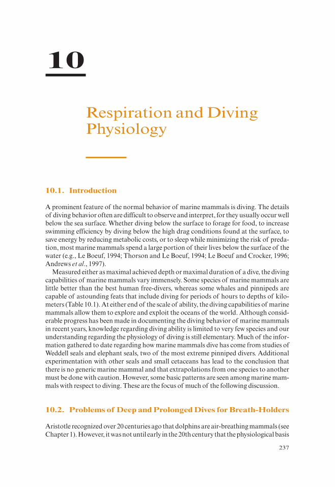
Marine mammals undergo important circulatory changes during diving. In general, thestructure of the heart of cetaceans and pinnipeds closely resembles that of other mam-mals (Figure 10.1).At the ultrastructural level, several distinctive differences are observedin the hearts of ringed and harp seals, such as enlarged stores of glycogen, which stronglysuggest that the cardiac tissues of theses animals are capable of a greater anaerobic capac-ity that those of terrestrial mammals (Pfeiffer, 1990; Pfeiffer and Viers, 1995). Ridgwayand Johnston (1966) suggested that the relatively larger heart size of the Dall’s porpoise,when compared to the less active inshore Atlantic bottlenose dolphin was related to itspelagic deep-diving habit. However, neither is heart size fundamentally different inmarine versus terrestrial mammals nor would it be expected to be as marine mammalhearts do not work particularly hard to support circulation when diving (see later).
10.3. Pulmonary and Circulatory Adaptations to Diving 239
P885522-10.qxd 10/17/05 11:19 PM Page 239

The ascending aorta in pinnipeds increases in diameter by 30–40% immediately out-side the heart to form an expanded elastic aortic bulb (aortic arch). After all the great ves-sels (i.e., brachiocephalic, left common carotid and left subclavian arteries) havebranched from the aorta, at about the level of the ductus arteriosus, there is a suddendecrease in the diameter of the aorta by about 50%, and it then continues posteriorly asa relatively slender abdominal aorta. The aortic bulb and smaller arteries at the base ofthe brain serve to dampen blood pressure pulses at each heart beat.
Although the enlarged aortic bulb is characteristic of all pinnipeds (Drabek, 1975,1977; King, 1977), there is thought to be some correlation between the size of the bulband the diving habits of the species. The shallow water leopard seal, for example, has asmaller bulb than the deep-diving Weddell seal. The proportionately larger heart andaortic bulb of deep-diving hooded seals likely function to increase lung perfusion duringsurface recovery and to assist in maintaining high blood pressures throughout the car-diac cycle during dives. The absence of age-related differences in heart morphology ofhooded seals suggests that these adaptations are important in the development of divingbehavior (Drabek and Burns, 2002).
A bulbous expansion of the aortic arch similar to that seen in pinnipeds has also beendescribed in some cetaceans (Shadwick and Gosline, 1994; Gosline and Shadwick, 1996;Melnikov, 1997). Further examination of the structural properties of the aorta and asso-ciated vessels (Gosline and Shadwick, 1996) indicates that most of the arterial compli-ance in a whale’s circulation results from volumetric expansion in the aortic arch anddifferences in the mechanical properties of its walls such as the thickness and organiza-tion of elastic tissues.
In addition to having a heart with a deep interventricular cleft (double ventricularapex) extending almost the full length of the ventricles, sirenians are distinguished fromother marine mammals in having a dorsally located left atrium (see Figure 10.1). In mostrespects the manatee heart is very similar to the dugong heart. Among the differences arethe quadrate shape of the manatee left ventricle, as opposed to its conical shape in thedugong, and the bulbous swelling of the aortic arch in the manatee, similar to that seenin pinnipeds (Drabek, 1977) and fin whales (Shadwick and Gosline, 1994). It seemsunlikely that expansion of the aortic arch in manatees can be attributed to differences in
240 10. Respiration and Diving Physiology
(a) (b) (c)Deep interventricular cleft
Figure 10.1. Ventral view of heart of (a) pinniped (Weddell seal) (modified from Drabek, 1977), (b)cetacean (porpoise) (modified from Slijper, 1979), and (c) sirenian (dugong) (modified fromRowlatt and Marsh, 1985).
P885522-10.qxd 10/17/05 11:19 PM Page 240
diving performance because all sirenians are modest divers compared to most othermarine mammals (Rowlatt and Marsh, 1985, and see Table 10.1).
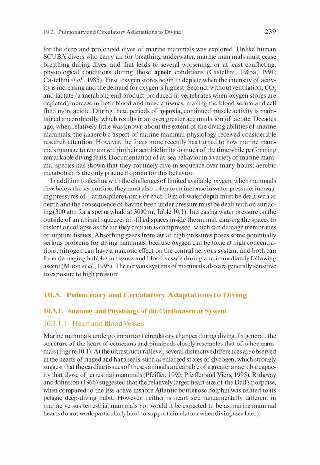
The circulatory systems of marine mammals are characterized by groups of bloodvessels (retia mirabilia). Retia mirabilia are tissue masses containing extensive contortedspirals of blood vessels, mainly arteries but with thin-walled veins among them, that usu-ally form blocks of tissue on the inner dorsal wall of the thoracic cavity and extremitiesor periphery of the body (Figure 10.2). The sperm whale has been described as having themost extensively developed thoracic retia among cetaceans (Melnikov, 1997). Thesestructures serve as blood reservoirs to increase oxygen stores for use during diving (e.g.,Pfeiffer and Kinkead, 1990).
The main changes in the venous system of pinnipeds are the enlargement and increasedcomplexity of veins to enhance their capacity. Most of our knowledge of these adaptationscomes from work done on the venous system of phocid seals (Harrison and Kooyman,1968; Ronald et al., 1977; Figure 10.3). In phocids, the posterior vena cava is frequently isdeveloped as a pair of vessels each capable of considerable distension of their thin, elasticwalls.Each branch of the posterior vena cava drains extensive plexi from the veins in the flip-pers, pelvis, and lateral abdominal wall. Each branch also receives several tributaries ofvarying sizes from the stellate plexus that encloses the kidneys (see Figure 10.3). Just poste-rior to the diaphragm and covered by the lobes of the liver, lies the hepatic sinus, which isformed from enlarged hepatic veins. It also receives blood from the posterior vena cava andconveys it to the heart. Immediately anterior to the diaphragm, the vena cava has a muscu-lar caval sphincter surrounding it (see Figure 10.3). Anterior to the sphincter the veins fromthe pericardial plexus enter the vena cava. This convoluted mass of interconnected veins(which are often encased in brown adipose tissue) forms a ring around the base of the peri-cardium and sends out leaf-like projections into the pleural cavities containing the lungs.
10.3. Pulmonary and Circulatory Adaptations to Diving 241
(a) (b)
Figure 10.2. Retia mirabilia, their anatomical position relative to the ribs (a) (adapted from Slijper, 1979),and the right thoracic retia of a spotted dolphin (b).
P885522-10.qxd 10/17/05 11:19 PM Page 241
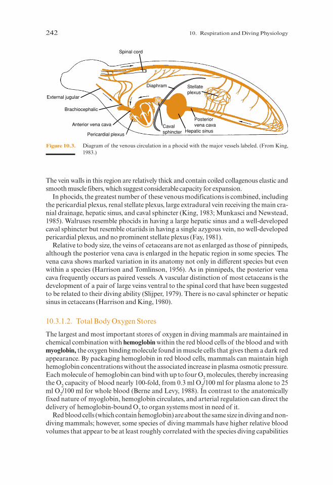
The vein walls in this region are relatively thick and contain coiled collagenous elastic andsmooth muscle fibers, which suggest considerable capacity for expansion.
In phocids, the greatest number of these venous modifications is combined, includingthe pericardial plexus, renal stellate plexus, large extradural vein receiving the main cra-nial drainage, hepatic sinus, and caval sphincter (King, 1983; Munkasci and Newstead,1985). Walruses resemble phocids in having a large hepatic sinus and a well-developedcaval sphincter but resemble otariids in having a single azygous vein, no well-developedpericardial plexus, and no prominent stellate plexus (Fay, 1981).
Relative to body size, the veins of cetaceans are not as enlarged as those of pinnipeds,although the posterior vena cava is enlarged in the hepatic region in some species. Thevena cava shows marked variation in its anatomy not only in different species but evenwithin a species (Harrison and Tomlinson, 1956). As in pinnipeds, the posterior venacava frequently occurs as paired vessels. A vascular distinction of most cetaceans is thedevelopment of a pair of large veins ventral to the spinal cord that have been suggestedto be related to their diving ability (Slijper, 1979). There is no caval sphincter or hepaticsinus in cetaceans (Harrison and King, 1980).
10.3.1.2. Total Body Oxygen Stores
The largest and most important stores of oxygen in diving mammals are maintained inchemical combination with hemoglobin within the red blood cells of the blood and withmyoglobin, the oxygen binding molecule found in muscle cells that gives them a dark redappearance. By packaging hemoglobin in red blood cells, mammals can maintain highhemoglobin concentrations without the associated increase in plasma osmotic pressure.Each molecule of hemoglobin can bind with up to four O2 molecules, thereby increasingthe O2 capacity of blood nearly 100-fold, from 0.3 ml O2/100 ml for plasma alone to 25ml O2/100 ml for whole blood (Berne and Levy, 1988). In contrast to the anatomicallyfixed nature of myoglobin, hemoglobin circulates, and arterial regulation can direct thedelivery of hemoglobin-bound O2 to organ systems most in need of it.
Red blood cells (which contain hemoglobin) are about the same size in diving and non-diving mammals; however, some species of diving mammals have higher relative bloodvolumes that appear to be at least roughly correlated with the species diving capabilities
242 10. Respiration and Diving Physiology
Spinal cord
External jugular
Brachiocephalic
Anterior vena cava
Pericardial plexus
Cavalsphincter Hepatic sinus
Posteriorvena cava
Stellateplexus
Diaphram
Figure 10.3. Diagram of the venous circulation in a phocid with the major vessels labeled. (From King,1983.)
P885522-10.qxd 10/17/05 11:19 PM Page 242
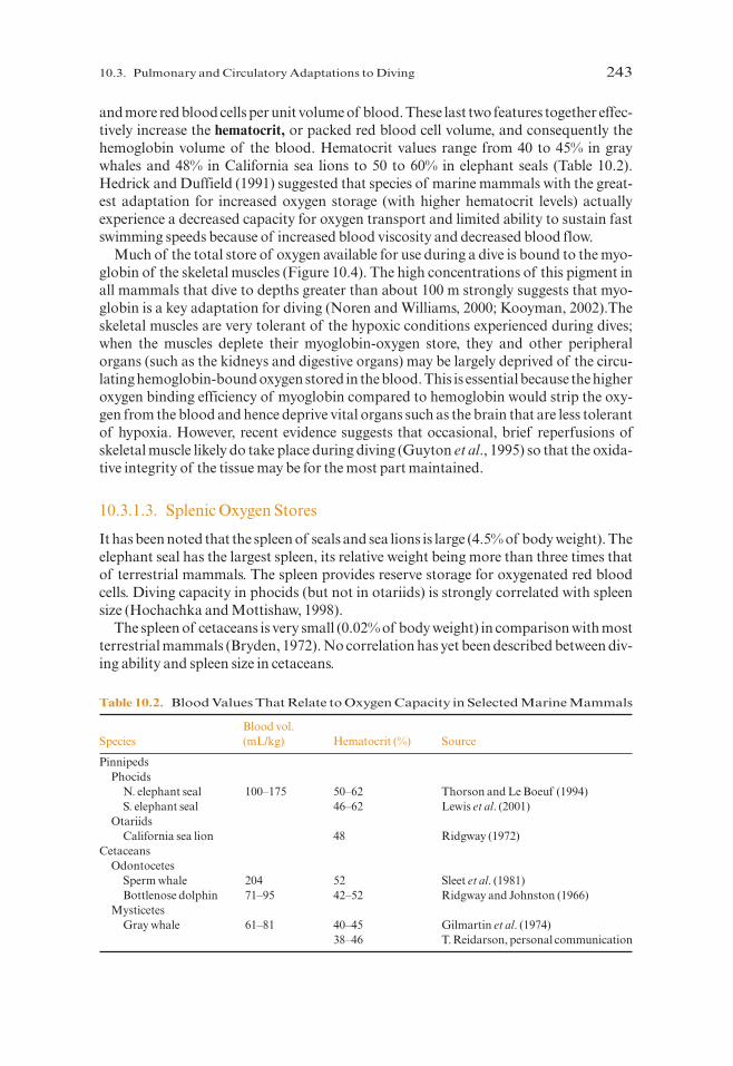
and more red blood cells per unit volume of blood. These last two features together effec-tively increase the hematocrit, or packed red blood cell volume, and consequently thehemoglobin volume of the blood. Hematocrit values range from 40 to 45% in graywhales and 48% in California sea lions to 50 to 60% in elephant seals (Table 10.2).Hedrick and Duffield (1991) suggested that species of marine mammals with the great-est adaptation for increased oxygen storage (with higher hematocrit levels) actuallyexperience a decreased capacity for oxygen transport and limited ability to sustain fastswimming speeds because of increased blood viscosity and decreased blood flow.
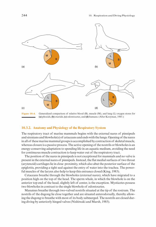
Much of the total store of oxygen available for use during a dive is bound to the myo-globin of the skeletal muscles (Figure 10.4). The high concentrations of this pigment inall mammals that dive to depths greater than about 100 m strongly suggests that myo-globin is a key adaptation for diving (Noren and Williams, 2000; Kooyman, 2002).Theskeletal muscles are very tolerant of the hypoxic conditions experienced during dives;when the muscles deplete their myoglobin-oxygen store, they and other peripheralorgans (such as the kidneys and digestive organs) may be largely deprived of the circu-lating hemoglobin-bound oxygen stored in the blood. This is essential because the higheroxygen binding efficiency of myoglobin compared to hemoglobin would strip the oxy-gen from the blood and hence deprive vital organs such as the brain that are less tolerantof hypoxia. However, recent evidence suggests that occasional, brief reperfusions ofskeletal muscle likely do take place during diving (Guyton et al., 1995) so that the oxida-tive integrity of the tissue may be for the most part maintained.
10.3.1.3. Splenic Oxygen Stores
It has been noted that the spleen of seals and sea lions is large (4.5% of body weight). Theelephant seal has the largest spleen, its relative weight being more than three times thatof terrestrial mammals. The spleen provides reserve storage for oxygenated red bloodcells. Diving capacity in phocids (but not in otariids) is strongly correlated with spleensize (Hochachka and Mottishaw, 1998).
The spleen of cetaceans is very small (0.02% of body weight) in comparison with mostterrestrial mammals (Bryden, 1972). No correlation has yet been described between div-ing ability and spleen size in cetaceans.
10.3. Pulmonary and Circulatory Adaptations to Diving 243
Table 10.2. Blood Values That Relate to Oxygen Capacity in Selected Marine Mammals
Blood vol.Species (mL/kg) Hematocrit (%) Source
PinnipedsPhocids
N. elephant seal 100–175 50–62 Thorson and Le Boeuf (1994)S. elephant seal 46–62 Lewis et al. (2001)
OtariidsCalifornia sea lion 48 Ridgway (1972)
CetaceansOdontocetes
Sperm whale 204 52 Sleet et al. (1981)Bottlenose dolphin 71–95 42–52 Ridgway and Johnston (1966)
MysticetesGray whale 61–81 40–45 Gilmartin et al. (1974)
38–46 T. Reidarson, personal communication
P885522-10.qxd 10/17/05 11:19 PM Page 243
10.3.2. Anatomy and Physiology of the Respiratory System
The respiratory tract of marine mammals begins with the external nares of pinnipedsand sirenians and blowhole(s) of cetaceans and ends with the lungs. Opening of the naresin all of these marine mammal groups is accomplished by contraction of skeletal muscle,whereas closure is a passive process. The active opening of the nostrils or blowholes is anenergy-conserving adaptation to spending life in an aquatic medium, avoiding the needfor continuous muscle contraction to keep water out of the respiratory tract.
The position of the nares in pinnipeds is not exceptional for mammals and no valve ispresent in the external nares of pinnipeds. Instead, the flat medial surfaces of two throat(arytenoid) cartilages lie in close proximity, which also abut the posterior surface of theepiglottis, providing a tight seal against the entry of water into the trachea. The power-ful muscles of the larynx also help to keep this entrance closed (King, 1983).
Cetaceans breathe through the blowholes (external nares), which have migrated to aposition high on the top of the head. The sperm whale, in which the blowhole is on theanterior top end of the head, slightly left of center, is the exception. Mysticetes possesstwo blowholes in contrast to the single blowhole of odontocetes.
Manatees breathe through two-valved nostrils situated at the tip of the rostrum. Thenostrils of the dugong lie close together and are situated anterodorsally, thereby allow-ing the dugong to breathe with most of its body submerged. The nostrils are closed dur-ing diving by anteriorly hinged valves (Nishiwaki and Marsh, 1985).
244 10. Respiration and Diving Physiology
LL
L
L
MM
M
M
B
B
BB
(a) (b)
(c) (d)
Figure 10.4. Generalized comparison of relative blood (B), muscle (M), and lung (L) oxygen stores for(a) phocids, (b) otariids, (c) odontocetes, and (d) humans. (After Kooyman, 1985.)
P885522-10.qxd 10/17/05 11:19 PM Page 244

Deep-diving marine mammals have flexible chest walls and other structures thatare capable of sufficient collapse to render the lungs virtually airless. The trachea inmost pinnipeds is supported by cartilaginous rings that either form complete circlesor are incomplete and overlap dorsally. This morphology permits the alveoli tocollapse while the proximal airways are still open, ensuring that pulmonary gasexchange is eliminated and pulmonary entrapment of air is avoided when the animalis at depth.
The tracheal rings are reduced to ventral bars in Ross, Weddell, and leopard seals(King, 1983). In the ribbon seal, a longitudinal slit occurs between the ends of the incom-plete cartilage rings on the dorsal surface of the trachea just before it bifurcates to formthe bronchi. A membranous sac of unknown function extends both anteriorly and pos-teriorly from the trachea on the right side of the body. Male ribbon seals possess this sac,and it increases in size with age suggesting that it may have a role in sound productionassociated with mating behavior. The tracheal slit is present in females, but the sac issmall or absent (King, 1983). In phocids and the walrus, the trachea divides into two pri-mary bronchi immediately outside the substance of the lung. In otariids this bifurcationis located more anteriorly, at approximately the level of the first rib, and the two elon-gated bronchi run parallel until they diverge to enter the lungs dorsal to the heart (seeFigure 3.11).
The cetacean larynx is composed of a cartilaginous framework held together by aseries of muscles. To keep inhaled air separate from food, the larynx of odontocetes (butnot mysticetes) has two elongate cartilages (Figure 10.5; Lawrence and Schevill, 1965;Slijper, 1979; Reidenberg and Laitman, 1987). This provides a more direct connectionbetween the trachea and blowhole than is found in mysticetes.
In cetaceans the trachea is short and broad and consists of several cartilaginous rings(varying from 5 to 7 rings in the beluga and sperm whale to 13 to 15 in the fin whale) thatare interconnected with each other. Unlike baleen whales, the tracheal rings of toothedwhales are closed and form a noncollapsing tube (Yablokov et al., 1972).
The dugong trachea is short (only four cartilage rings) and it is deeply divided by amedial septum (Hill, 1945; Harrison and King, 1980). The manatee trachea is longer andis supported by 8–12 tracheal rings (Harrison and King, 1980).
10.3. Pulmonary and Circulatory Adaptations to Diving 245
Figure 10.5. Lateral view of cetacean larynx in (a) mysticete (fin whale) and (b) odontocete (narwhal).Note the elongated arytenoid and thyroid cartilages in the odontocete. (From Slijper, 1979.)
P885522-10.qxd 10/17/05 11:19 PM Page 245

10.3.2.1. Lungs
The lungs of marine mammals are not larger than those of terrestrial mammals, butsome important differences exist between the lungs of marine mammals and those of ter-restrial mammals. The left and right lungs of pinnipeds are approximately equal in sizeand are lobulated as in terrestrial carnivores; both lungs have three main lobes, but theright lung has an additional small intermediate lobe. There appears to be a tendency toreduce lobulation in the lungs of some pinnipeds, such as the walrus and ribbon, harp,and spotted seals. The latter species has been reported as having little or no lobulation(King, 1983). The bronchi subdivide within the lungs to form bronchioles and eventuallyend in alveoli, but the details of this transition vary among the three families (Figure10.6; see King, 1983).
The lungs of cetaceans are distinct from those of all other mammals in their overallsacculate shape and lack of lobes (Figure 10.7). Only occasionally is the apical part of theright lung somewhat prominent, resembling the apical lobe of the lungs of other mam-mals. The right lung is usually larger, longer, and heavier than the left. Such asymmetryof lung size, which is related in whales as in other mammals to the somewhat asymmet-ric position of the heart in the chest cavity, is seen in virtually all studied cetaceans:rorquals, various dolphins, and sperm and beluga whales.
The lungs of cetaceans, compared to those of terrestrial mammals, show greater rigid-ity and elasticity because of increased cartilaginous support. In baleen, sperm, and bot-tlenose whales, the septa projecting into the proximal portion of the air sacs contain heavymyoelastic bundles. In the smaller toothed whales these bundles are atrophied, but thereare a series of myoelastic sphincters in the smallest bronchioles. In both the bundles andthe sphincters, the muscular part may act to close the air sacs, whereas the elastic part mayfacilitate rapid expiration.Airway closure should delay alveolar collapse during dives andallow additional pulmonary gas exchange to occur for those odontocete families knownto possess myoelastic terminal airway sphincters (Drabek and Kooyman, 1983).
246 10. Respiration and Diving Physiology
Figure 10.6. Diagram of the structure of alveoli and associated cartilage and muscle in pinnipeds. (a)Phocid. (b) Otariid. (c) Odobenid. (From King, 1983.)
P885522-10.qxd 10/17/05 11:19 PM Page 246
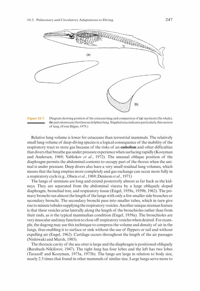
Relative lung volume is lower for cetaceans than terrestrial mammals. The relativelysmall lung volume of deep-diving species is a logical consequence of the inability of therespiratory tract to store gas because of the risks of an embolism and other difficultiesthat divers that breathe gas under pressure experience when surfacing rapidly (Kooymanand Andersen, 1969; Yablokov et al., 1972). The unusual oblique position of thediaphragm permits the abdominal contents to occupy part of the thorax when the ani-mal is under pressure. Deep divers also have a very small residual lung volumes, whichmeans that the lung empties more completely and gas exchange can occur more fully ina respiratory cycle (e.g., Olsen et al., 1969; Denison et al., 1971)
The lungs of sirenians are long and extend posteriorly almost as far back as the kid-neys. They are separated from the abdominal viscera by a large obliquely slopeddiaphragm, bronchial tree, and respiratory tissue (Engel, 1959a, 1959b, 1962). The pri-mary bronchi run almost the length of the lungs with only a few smaller side branches orsecondary bronchi. The secondary bronchi pass into smaller tubes, which in turn giverise to minute tubules supplying the respiratory vesicles. Another unique sirenian featureis that these vesicles arise laterally along the length of the bronchioles rather than fromtheir ends, as is the typical mammalian condition (Engel, 1959a). The bronchioles arevery muscular and may function to close off respiratory vesicles when desired. For exam-ple, the dugong may use this technique to compress the volume and density of air in thelungs, thus enabling it to surface or sink without the use of flippers or tail and withoutexpelling air (Engel, 1962). Cartilage occurs throughout the length of the air passages(Nishiwaki and Marsh, 1985).
The thoracic cavity of the sea otter is large and the diaphragm is positioned obliquely(Barabash-Nikiforov, 1947). The right lung has four lobes and the left has two lobes(Tarasoff and Kooyman, 1973a, 1973b). The lungs are large in relation to body size,nearly 2.5 times that found in other mammals of similar size. Large lungs serve more to
10.3. Pulmonary and Circulatory Adaptations to Diving 247
Figure 10.7. Diagram showing position of the cetacean lung and comparison of (a) mysticete (fin whale),(b) and odontocete (bottlenose dolphin) lung. Stippled area indicates particularly thin sectionof lung. (From Slijper, 1979.)
P885522-10.qxd 10/17/05 11:19 PM Page 247

regulate buoyancy than to store oxygen (Lenfant et al., 1970; Kooyman, 1973; Leith,1976; Costa and Kooyman, 1982). The polar bear respiratory system is not unlike that ofother bears. Although they are powerful swimmers they are not known to have any spe-cial physiological adaptations specific to diving.
10.3.2.2. Breathing
Breathing patterns of marine mammals vary. Pinnipeds breathe vigorously and frequentlyduring the recovery phase after prolonged diving, but many species are typically periodicbreathers under other circumstances. Particularly when resting or sleeping it is normal forseals to perform quite long apneas with short periods of rapid respiration between thesenonbreathing periods (e.g., Huntley et al., 1984). Although pinnipeds commonly exhaleprior to diving, Hooker et al. (2005) found that Antarctic fur seals consistently dive withfull lungs and exhale during the latter stage of the ascent portion of a dive.
Cetaceans exhale and inhale singly but very rapidly on surfacing. Whale blows repre-sent the rapid emptying or expiration of whales’ lungs through their blowholes in prepa-ration for the next inspiration. A blow is one of the most visible behaviors of whaleswhen they are observed at the sea surface. A particularly large amount of water maybe spouted in baleen whales, whose blowholes are located in rather deep folds. Thevisibility of a blow is due to a mixture of vapor and seawater entrained into the exhaledcolumn of air at the sea surface (Figure 10.8). When a blow occurs below the sea surface,
248 10. Respiration and Diving Physiology
Figure 10.8. Towering blow of a blue whale. (Courtesy of P. Colla.)
P885522-10.qxd 10/17/05 11:19 PM Page 248

as it sometimes does as bubble-blasts in gray whales or bubble trains in humpbackwhales, it may be intended as an audible or visual signal to other nearby whales. The size,shape, and orientation of a blow can help to identify some species of whales from adistance.
Vapor formed by contact between air warmed in the lungs and the cold external airsometimes enhances the visibility of the blow. The expired air of a blow also contains sur-factant from the lungs. Surfactant is a complex a mixture of lipoproteins that reduce sur-face tension in pulmonary fluids and facilitate easy reinflation of collapsed lungs onsurfacing. Pulmonary surfactant is secreted by alveolar type II cells and is necessary fornormal mammalian lung function (Miller et al., 2004). Spragg et al. (2004) have founddifferences in surfactant of nondiving and diving mammals and suggest that these dif-ferences are associated with the repetitive collapse and reinflation of the lungs of divers.
A complete breathing cycle typically consists of a very rapid expiration (the blow)immediately followed by a slightly longer and much less obvious inspiration, then anextended yet variable period of breath-holding, or apnea. The rapid expiration of a typ-ical whale blow provides more time to complete the next inspiration as the blowholes ofa swimming animal breaks through the sea surface and results in little delay before sub-merging again (Kooyman and Cornell, 1981).
The rapidity of the blow is accomplished by maintaining high flow rates throughoutalmost the entire expiration (Figure 10.9), which is in strong contrast to humans andother land mammals. The high expiratory flow rates of cetaceans are enhanced by veryflexible chest walls and by cartilage reinforcement of the smallest terminal air passagesof the lungs to prevent them from collapsing until the lungs are almost completely emp-tied. Small dolphins, for instance, expire and inspire in about 0.1 s, then hold their breathfor 20 to 30 s before taking another breath. Even adult blue whales can empty their lungsof 1500 liters of air and refill them in as little as 2 s.
10.3. Pulmonary and Circulatory Adaptations to Diving 249
2.000.00 1.00
0
−100
100
TE
TI
Time (sec)
Flo
w r
ate
(l/se
c)
Figure 10.9. Typical ventilatory flow rates of a single expiratory (TE)/inspiratory (TI) event from a rehabil-itating gray whale calf.
P885522-10.qxd 10/17/05 11:19 PM Page 249
Blow patterns of whales vary, depending on their behaviors. In small cetaceans, swim-ming at low speeds, blowhole exposure during a blow is minimal and gradually changesto porpoising above the sea surface at higher swimming speeds (see Chapter 9). Whenmigrating or feeding, larger baleen whales typically surface to blow several times in rapidsuccession, then make an extended dive of several minutes duration.
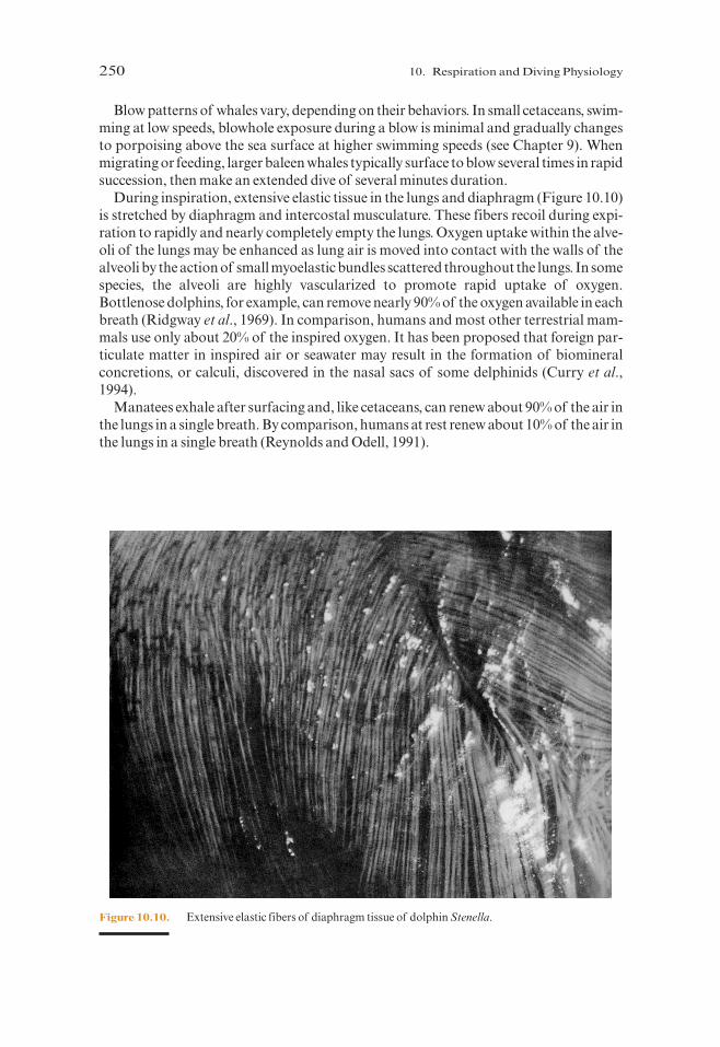
During inspiration, extensive elastic tissue in the lungs and diaphragm (Figure 10.10)is stretched by diaphragm and intercostal musculature. These fibers recoil during expi-ration to rapidly and nearly completely empty the lungs. Oxygen uptake within the alve-oli of the lungs may be enhanced as lung air is moved into contact with the walls of thealveoli by the action of small myoelastic bundles scattered throughout the lungs. In somespecies, the alveoli are highly vascularized to promote rapid uptake of oxygen.Bottlenose dolphins, for example, can remove nearly 90% of the oxygen available in eachbreath (Ridgway et al., 1969). In comparison, humans and most other terrestrial mam-mals use only about 20% of the inspired oxygen. It has been proposed that foreign par-ticulate matter in inspired air or seawater may result in the formation of biomineralconcretions, or calculi, discovered in the nasal sacs of some delphinids (Curry et al.,1994).
Manatees exhale after surfacing and, like cetaceans, can renew about 90% of the air inthe lungs in a single breath. By comparison, humans at rest renew about 10% of the air inthe lungs in a single breath (Reynolds and Odell, 1991).
250 10. Respiration and Diving Physiology
Figure 10.10. Extensive elastic fibers of diaphragm tissue of dolphin Stenella.
P885522-10.qxd 10/17/05 11:19 PM Page 250
10.3.2.3. Respiratory Systems and Diving
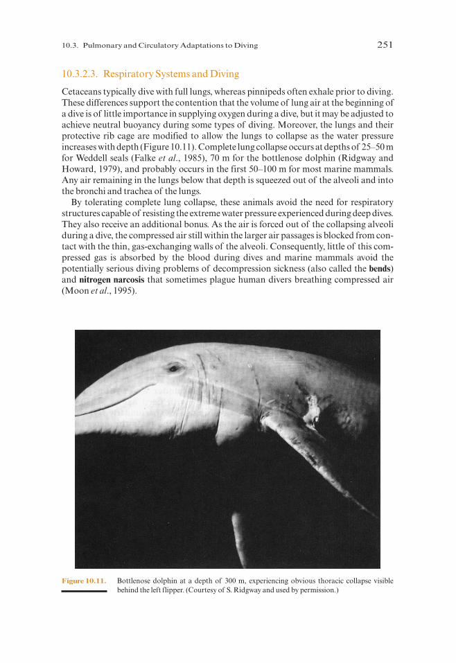
Cetaceans typically dive with full lungs, whereas pinnipeds often exhale prior to diving.These differences support the contention that the volume of lung air at the beginning ofa dive is of little importance in supplying oxygen during a dive, but it may be adjusted toachieve neutral buoyancy during some types of diving. Moreover, the lungs and theirprotective rib cage are modified to allow the lungs to collapse as the water pressureincreases with depth (Figure 10.11). Complete lung collapse occurs at depths of 25–50 mfor Weddell seals (Falke et al., 1985), 70 m for the bottlenose dolphin (Ridgway andHoward, 1979), and probably occurs in the first 50–100 m for most marine mammals.Any air remaining in the lungs below that depth is squeezed out of the alveoli and intothe bronchi and trachea of the lungs.
By tolerating complete lung collapse, these animals avoid the need for respiratorystructures capable of resisting the extreme water pressure experienced during deep dives.They also receive an additional bonus. As the air is forced out of the collapsing alveoliduring a dive, the compressed air still within the larger air passages is blocked from con-tact with the thin, gas-exchanging walls of the alveoli. Consequently, little of this com-pressed gas is absorbed by the blood during dives and marine mammals avoid thepotentially serious diving problems of decompression sickness (also called the bends)and nitrogen narcosis that sometimes plague human divers breathing compressed air(Moon et al., 1995).
10.3. Pulmonary and Circulatory Adaptations to Diving 251
Figure 10.11. Bottlenose dolphin at a depth of 300 m, experiencing obvious thoracic collapse visiblebehind the left flipper. (Courtesy of S. Ridgway and used by permission.)
P885522-10.qxd 10/17/05 11:19 PM Page 251

10.4. Diving Response
When a marine mammal leaves the surface its on-board oxygen stores (described previ-ously) must satisfy its needs throughout submergence. As a dive proceeds there is asteady decline in the amount of available oxygen (hypoxia) and an increase in carbondioxide (hypercapnia), which together create a condition known as asphyxia. Eventually,if the dive continues beyond a time that can be serviced by aerobic metabolism, byprod-ucts of anaerobic metabolism such as lactic acid and hydrogen ions also begin to accu-mulate. However, marine mammals and other animals that have become adapted todealing with periods of asphyxia have a complex array of physiological responses thatextend the time that a given oxygen supply can service their bodies. These responsesinclude a pronounced decline in heart rate (bradycardia) accompanied by regional vaso-constriction (selective ischemia) that entails a preferential distribution of circulatingblood to oxygen-sensitive organs as well as a drop in core body temperature and likelymetabolic rate in regions that receive reduced blood supplies.
Bradycardia and its implied reduction in metabolic costs have been recognized asbeing a diving response since the late 1800s (Bert, 1870; Richet, 1894, 1899). However,it was not until the 1930s that Irving’s (e.g., Irving et al., 1935; Irving, 1939) experi-mental laboratory studies provided evidence that oxygen was conserved during div-ing by selective circulatory adjustments during periods of bradycardia. A series ofelaborate physiological experiments conducted by Irving and Scholander in the labo-ratory with forced-dived marine mammals, demonstrated the fundamental factorsemployed by diving animals to conserve oxygen and deal with the products of anaer-obic metabolism postdiving (e.g., Irving and Scholander, 1941a, 1941b; Irving et al.,1942; Scholander, 1940, 1960, 1964; Scholander et al., 1942a, 1942b). These experi-ments attracted criticism for a time, because of the unnatural conditions under whichthe animals were forced to dive. However, as technology has advanced and studieshave been performed in the wild on unrestrained animals, it has become clear that theearly experiments did evoke natural dive responses although they tended to beextreme, presumably because the animals did not know when they were going to beable to surface.
The range of heart-rate responses to diving is variable between species and within aspecies under different circumstances. However, in general, short dives evoke only slightresponses, whereas long dives promote more intense levels of bradycardia. The mostextreme levels of heart rate reduction, down to 5% of predive levels, have been recordedduring free diving by phocid seals (e.g., Jones et al., 1973; Elsner et al., 1989; Thompsonand Fedak, 1993; Andrews et al., 1995). Variable responses are at least in part due to whatappears to be a remarkable level of voluntary control over the cardiovascular system inat least some species. Experiments with free-diving seals of several species have shownthat seals have heart rates at the start of dives that are correlated with the subsequentduration of the dive, strongly suggesting that the animal prepares for a dive of a cer-tain duration on leaving the surface, and anticipatory elevations in heart rate take placeprior to the end of dives as well (e.g., Fedak, 1986; Hill et al., 1987; Elsner et al., 1989;Wartzok et al., 1992). There is increasing evidence that fine adjustments can be madeduring a dive in some species (Andrews et al., 1997). Although much less data are avail-able for cetaceans, it is clear that they also experience bradycardia when diving (e.g.,Elsner et al., 1966; Spencer et al., 1967). Diving bradycardia in manatees and dugongs ismodest, as is their diving ability (Elsner, 1999).
252 10. Respiration and Diving Physiology
P885522-10.qxd 10/17/05 11:20 PM Page 252
Despite the marked decline in cardiac output that accompanies the drop in heart rateduring diving, core body arterial blood pressure remains relatively constant so that per-fusion of vital organs (brain, heart, placenta, etc.) is maintained. This is achieved in partby the elastic recoil of the aortic bulb in the hearts of marine mammals but primarilythrough ischemia that restricts blood flow to the visceral organs, skin, and muscles.Tissues such as those in the liver and kidney that regularly experience drastic reductionsin blood flow during diving show extreme tolerance of these conditions and their conse-quences. Deprivation of arterial blood flow to selected organs produces a gradual reduc-tion in body temperature (Scholander et al., 1942b; Hammel et al., 1977; Hill et al., 1987;Andrews et al., 1995); in the extreme, perhaps even brain cooling occurs (Odden et al.,1997). There is evidence that even normally sensitive tissues such as the brain and heartare adapted to dealing with low oxygen conditions in some marine mammal species(Ridgway et al., 1969; Kjukshus et al., 1982; Elsner and Gooden, 1983; White et al.,1990).
Hypometabolism almost certainly occurs during diving, because the metabolic cost ofdiving is so low (Kooyman et al., 1973; Castellini et al., 1992; Costa, 1993; Andrews et al.,1995), but direct evidence is difficult to obtain in the wild. Depressed metabolic rateshave been documented directly during voluntary diving in captive grey seals (Sparlingand Fedak, 2004). In addition to reduction in temperature, another mechanism thatmight result in metabolic inhibition during diving is increasing tissue acidity (Harken,1976). Although the details of how an animal that is hypometabolic is able to retain theability to actively swim remains unclear, it is clear that compromises must be madebetween diving time, exertion, and oxygen economy (Castellini, 1985a, 1985b). Marinemammals have relatively low aerobic scope for activity, but their anaerobic capabilitiesare well beyond those of terrestrial mammals (e.g., Elsner, 1987; Ponganis et al., 1990;Williams et al., 1991, 1993).
The biochemical manifestations of cellular resistance to conditions of apnea remainelusive and continue to be the subject of some debate (Blix, 1976; Castellini et al., 1981;Kooyman et al., 1981; Hochachka et al., 1988; Hochachka, 1992), but it is conclusivelyestablished that marine mammals can tolerate anaerobic cellular conditions with ele-vated lactic acid and declining pH that terrestrial mammals would find disruptive oreven lethal. In an extensive comparison of diving and nondiving mammals, the most sig-nificant differences in tissue biochemistry were found in the levels of myoglobin andmuscle buffering capacity (Castellini and Somero, 1981; Castellini et al., 1981).Buffering capacity is the ability to hold tissue pH constant in the face of an increasingamount of acidic end products created by anaerobic metabolism. Marine mammals havea greater buffering ability than terrestrial mammals, and phocids have higher non-carbonate plasma buffering capacities than either otariids or most cetaceans (Boutilieret al., 1993). This probably reflects their general patterns of breath-hold diving and rela-tive potentials for tolerating low oxygen and metabolic acidosis. Several aspects of thebiochemistry of marine mammals suggest that their lifestyles generally do not requiresustained endurance of intense exercise, but rather they seem to support burst activitylevels that can switch over to anaerobic sources of energy for short periods (Costa andWilliams, 1999).
The ability to dive in all species of marine mammals studied to date shows an ontoge-netic pattern of development; one that is very rapid in some species (e.g., Le Boeuf et al.,1996; Horning and Trillmich, 1997; Baker and Donohue, 1999; Lydersen and Kovacs,1999; Jørgensen et al., 2001). Of course, marine mammals are “experienced”divers when
10.4. Diving Response 253
P885522-10.qxd 10/17/05 11:20 PM Page 253

they are born; fetuses show heart rate declines when their mothers dive (Elsner et al.,1970; Liggins et al., 1980; Hill et al., 1987), although hypoxia is likely first experiencedpostbirth because uterine arterial blood flow is maintained during dives undertaken dur-ing pregnancy (Elsner et al., 1970). Although neonatal dolphins and seals lack the myo-globin concentrations required for prolonged dive durations, myoglobin content inskeletal muscles increases significantly during subsequent development, (Noren andWilliams, 2000). Noren et al. (2001) also demonstrated an age-related capability ofTursiops to decrease heart rate during dives.
Many questions remain to be resolved regarding how marine mammals are protectedagainst the adverse effects of their frequent exposures to high pressure in deep dives, aswell as questions such as how those species without sonar manage to find food at greatdepths (see Chapter 7, Sensory Systems). However, general patterns about their divingperformances and patterns are beginning to emerge.
10.5. Diving Behavior and Phylogenetic Patterns
Simple observations and incidental catches of marine mammals in gear set at depth pro-vided evidence that some marine mammals dive for extended period of time to greatdepths. However, it was not until the development of time-depth recorders (TDRs) ofvarious types that systematic data on the diving behavior of marine mammals started toaccumulate. Pioneering studies beginning in the late 1960s with mechanical devicesdeveloped by Kooyman and his colleagues that were deployed on Weddell seals in theAntarctic (e.g., Kooyman, 1966, 1985; Kooyman and Campbell, 1972). The excitingresults stimulated rapid technological advances in the development of smaller electronicinstruments with increasing complex sensors. TDRs, which had to be recovered to down-load data, and later independently reporting satellite-linked platform transmitter termi-nals (PTTs) provided new opportunities for deployments of instruments on a widevariety of marine mammal species. Although data on many species are still lacking, oravailable for only some age or sex classes during part of their annual cycle, some excitingdata sets are available for several species (most notably Weddell seals and elephant seals)and at least fragmented patterns are emerging among marine mammals regarding theirdiving behavior.
Diving ability is of course intimately linked with physiological capabilities, but bodysize, ecological niche, and life-history strategy also play a role in the type of diving thatdominates a species repertoire (Boyd and Croxall, 1996; Boyd, 1997; Schreer andKovacs, 1997; Schreer et al., 1998; Costa and Williams, 1999; Costa et al., 2001). Fortheir body size, phocid seals are the most capable divers among all of the marine mam-mals. They utilize a strategy that has been referred to as “energy conserving”(Hochachka et al., 1997; Mottishaw et al., 1999), and all phocids exhibit deep and longdives compared to otariids or whales of similar size. Phocid seals tend to be large whichmeans that they have a low mass specific metabolic rate and they can carry a largeamount of oxygen in their tissues. They also have higher blood oxygen storage capacitiesin their blood because they have elevated hematocrits (Lenfant et al., 1970). Phocids alsoperform the most profound bradycardia responses and the greatest degree of vaso-constriction among pinnipeds resulting in extremely low metabolism during diving (e.g.,Castellini et al., 1992; Costa, 1993). They have slow swimming speeds that minimizes thecost of locomotion.
254 10. Respiration and Diving Physiology
P885522-10.qxd 10/17/05 11:20 PM Page 254

The Weddell seal has been the subject of the most extensive and comprehensive exam-inations of diving physiology in the wild (Figure 10.12). Kooyman and others have foundthat Weddell seals perform dives up to about 20 minutes in length without adjusting theirheart rate or circulatory patterns. These results suggest that Weddell seals have sufficientstored oxygen to last about 20 minutes (Figure 10.13). Only during dives lasting longer
10.5. Diving Behavior and Phylogenetic Patterns 255
Dep
th, m
Time, min.
200
400
600
800
0
0 10 20 30 40
type 2
type 6type 1
type 5D
epth
, m
100
200
300
400
0
Time, min.0 10 15 20 255
type 1
type 3
type 4
(a)
(b)
Figure 10.12. (a) Time-depth profiles of four Weddell seal dive types characterized by Schreer and Testa(1996). (b) Three Weddell seal dive profiles, although similar in shape to type 2 dives in (a),their 3-D maps emphasize large differences in horizontal as well as vertical movements(from Davis et al., 2003). Three-D maps scaled in 100-m increments.
P885522-10.qxd 10/17/05 11:20 PM Page 255
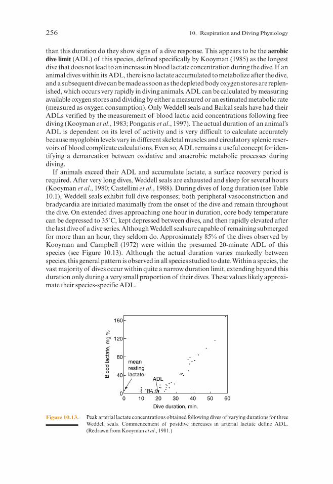
than this duration do they show signs of a dive response. This appears to be the aerobicdive limit (ADL) of this species, defined specifically by Kooyman (1985) as the longestdive that does not lead to an increase in blood lactate concentration during the dive. If ananimal dives within its ADL, there is no lactate accumulated to metabolize after the dive,and a subsequent dive can be made as soon as the depleted body oxygen stores are replen-ished, which occurs very rapidly in diving animals. ADL can be calculated by measuringavailable oxygen stores and dividing by either a measured or an estimated metabolic rate(measured as oxygen consumption). Only Weddell seals and Baikal seals have had theirADLs verified by the measurement of blood lactic acid concentrations following freediving (Kooyman et al., 1983; Ponganis et al., 1997). The actual duration of an animal’sADL is dependent on its level of activity and is very difficult to calculate accuratelybecause myoglobin levels vary in different skeletal muscles and circulatory splenic reser-voirs of blood complicate calculations. Even so, ADL remains a useful concept for iden-tifying a demarcation between oxidative and anaerobic metabolic processes duringdiving.
If animals exceed their ADL and accumulate lactate, a surface recovery period isrequired. After very long dives, Weddell seals are exhausted and sleep for several hours(Kooyman et al., 1980; Castellini et al., 1988). During dives of long duration (see Table10.1), Weddell seals exhibit full dive responses; both peripheral vasoconstriction andbradycardia are initiated maximally from the onset of the dive and remain throughoutthe dive. On extended dives approaching one hour in duration, core body temperaturecan be depressed to 35˚C, kept depressed between dives, and then rapidly elevated afterthe last dive of a dive series. Although Weddell seals are capable of remaining submergedfor more than an hour, they seldom do. Approximately 85% of the dives observed byKooyman and Campbell (1972) were within the presumed 20-minute ADL of thisspecies (see Figure 10.13). Although the actual duration varies markedly betweenspecies, this general pattern is observed in all species studied to date. Within a species, thevast majority of dives occur within quite a narrow duration limit, extending beyond thisduration only during a very small proportion of their dives. These values likely approxi-mate their species-specific ADL.
256 10. Respiration and Diving Physiology
Blo
od la
ctat
e, m
g %
Dive duration, min.
120
80
40
0
160
0 10 20 30 40 50 60
ADL
meanrestinglactate
Figure 10.13. Peak arterial lactate concentrations obtained following dives of varying durations for threeWeddell seals. Commencement of postdive increases in arterial lactate define ADL.(Redrawn from Kooyman et al., 1981.)
P885522-10.qxd 10/17/05 11:20 PM Page 256
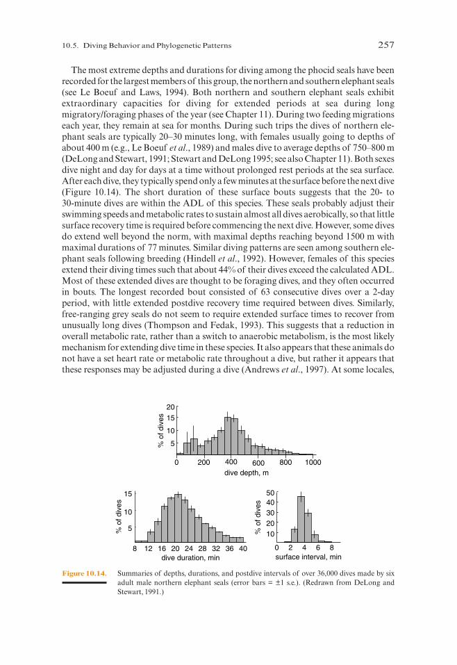
The most extreme depths and durations for diving among the phocid seals have beenrecorded for the largest members of this group, the northern and southern elephant seals(see Le Boeuf and Laws, 1994). Both northern and southern elephant seals exhibitextraordinary capacities for diving for extended periods at sea during longmigratory/foraging phases of the year (see Chapter 11). During two feeding migrationseach year, they remain at sea for months. During such trips the dives of northern ele-phant seals are typically 20–30 minutes long, with females usually going to depths ofabout 400 m (e.g., Le Boeuf et al., 1989) and males dive to average depths of 750–800 m(DeLong and Stewart, 1991; Stewart and DeLong 1995; see also Chapter 11). Both sexesdive night and day for days at a time without prolonged rest periods at the sea surface.After each dive, they typically spend only a few minutes at the surface before the next dive(Figure 10.14). The short duration of these surface bouts suggests that the 20- to 30-minute dives are within the ADL of this species. These seals probably adjust theirswimming speeds and metabolic rates to sustain almost all dives aerobically, so that littlesurface recovery time is required before commencing the next dive. However, some divesdo extend well beyond the norm, with maximal depths reaching beyond 1500 m withmaximal durations of 77 minutes. Similar diving patterns are seen among southern ele-phant seals following breeding (Hindell et al., 1992). However, females of this speciesextend their diving times such that about 44% of their dives exceed the calculated ADL.Most of these extended dives are thought to be foraging dives, and they often occurredin bouts. The longest recorded bout consisted of 63 consecutive dives over a 2-dayperiod, with little extended postdive recovery time required between dives. Similarly,free-ranging grey seals do not seem to require extended surface times to recover fromunusually long dives (Thompson and Fedak, 1993). This suggests that a reduction inoverall metabolic rate, rather than a switch to anaerobic metabolism, is the most likelymechanism for extending dive time in these species. It also appears that these animals donot have a set heart rate or metabolic rate throughout a dive, but rather it appears thatthese responses may be adjusted during a dive (Andrews et al., 1997). At some locales,
10.5. Diving Behavior and Phylogenetic Patterns 257
0 2 4 6 8surface interval, min
% o
f div
es
% o
f div
es
dive duration, min
dive depth, m
5
10
15
1020304050
0 200 1000400 600 800
208 12 16 24 28 32 36 40
10
20
% o
f div
es 15
5
Figure 10.14. Summaries of depths, durations, and postdive intervals of over 36,000 dives made by sixadult male northern elephant seals (error bars = ±1 s.e.). (Redrawn from DeLong andStewart, 1991.)
P885522-10.qxd 10/17/05 11:20 PM Page 257

hooded seals, another large, deep-diving phocid seal, seem to specialize on deep dwellingfish and can dive to depths of 1000 m for periods of 52 minutes or more. However, mostdives in this species are much shorter and occur to depths of 100–600 m (Folkow andBlix, 1995).
Phocid seals of intermediate size tend to display diving behavior that is concomitantwith their body sizes. Species such as grey seals, harp seals, Ross seals, and crabeater sealsdo most of their diving to depths of about 100 m for less than 10 minutes, although max-imal values can be significantly longer and deeper (e.g., Lydersen and Kovacs, 1993;Bengston and Stewart, 1992, 1997; Lydersen et al., 1994; Folkow et al., 2004). Small pho-cids tend to be the most conservative divers in this group with Baikal, Saimaa, ringed,and harbor seals usually diving for periods of only a few minutes to relatively shallowdepths. However, even these small species do on occasion display deep and long dives.Harbor seals have been documented to dive to over 450 m during dives that last morethan 30 minutes (Bowen et al., 1999; Gjertz et al., 2001), and juvenile ringed seals weigh-ing less than 40 kg dive to over 500 m and remain submerged for longer than 30 minutes(Lydersen, Kovacs, and Fedak, unpublished data).
Hawaiian monk seals and bearded seals are both relatively large phocids, but bothspecies display quite shallow and short duration diving patterns, because these twospecies usually forage in shallow coastal waters. However, adult monk seals do some-times dive outside lagoon areas, where adult males have been recorded as deep as 550 mand bearded seals are also clearly capable of more extreme diving than is performed incoastal areas. Bearded seals pups of only a few months of age hold the dive records forthis species. During their early wanderings, 7 of 7 postmolting pups dove to depths ofover 400 m (Gjertz et al., 2000), whereas adults of this species normally dive for only afew minutes (2–4) to depths of 20 m (Gjertz et al., 2000; Krafft et al., 2000). These twospecies illustrate the strong influence of foraging preference on behavioral patterns indiving.
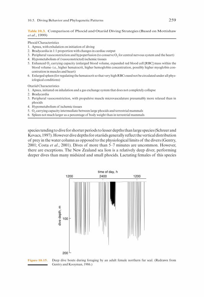
Otariids do not spend as much time diving as phocids and they usually dive for only afew minutes to relatively shallow depths. They are also at sea for relatively short periodsof time compared to many phocids that spend a lot of time pelagically. The otariid strat-egy has been described as being energy dissipative (Hochachka et al., 1997; Mottishawet al., 1999; Table 10.3). Otariids have a relatively small and hydrodynamically sleek bodyconsistent with a high speed predator lifestyle. They appear to sacrifice extended forag-ing time for energy needed for higher speed swimming (Costa, 1991). This pinnipedgroup shows the same basic patterns with respect to ADL as do the phocids, in that theyremain within their aerobic limits during most of their diving, although patterns are per-haps somewhat more variable. Data from Antarctic fur seals indicate that less than 6% oftheir dives exceed the estimated ADL but some dive bouts were of significantly longerduration and are followed by longer surface intervals (Boyd and Croxall, 1996).Although some individual dives exceeded their ADL, these specific dives did not appearto have any immediate effect on their subsequent diving behavior. Relatively deep forag-ing dives (>75 m) are common for northern fur seals (Gentry et al., 1986; Figure 10.15),and in one study 92% of dives exceeded the calculated ADL for this species, whereas only8% of shallow foraging dives (<40 m) exceeded the ADL (Ponganis et al., 1992). Thissuggests that at least some otariids may also employ energy conservation/metabolicstrategies in addition to anaerobic metabolism for extended diving duration.
Among otariid species, allometric patterns are apparent intra- and interspecificallywith males of sexually dimorphic species tending to dive deeper than females and small
258 10. Respiration and Diving Physiology
P885522-10.qxd 10/17/05 11:20 PM Page 258
species tending to dive for shorter periods to lesser depths than large species (Schreer andKovacs, 1997). However dive depths for otariids generally reflect the vertical distributionof prey in the water column as opposed to the physiological limits of the divers (Gentry,2001; Costa et al., 2001). Dives of more than 5–7 minutes are uncommon. However,there are exceptions. The New Zealand sea lion is a relatively deep diver, performingdeeper dives than many midsized and small phocids. Lactating females of this species
10.5. Diving Behavior and Phylogenetic Patterns 259
Table 10.3. Comparison of Phocid and Otariid Diving Strategies (Based on Mottishawet al., 1999)
Phocid Characteristics1. Apnea, with exhalation on initiation of diving2. Bradycardia in 1:1 proportion with changes in cardiac output3. Peripheral vasoconstriction and hypoperfusion (to conserve O2 for central nervous system and the heart)4. Hypometabolism of (vasoconstricted) ischemic tissues5. Enhanced O2 carrying capacity (enlarged blood volume, expanded red blood cell [RBC] mass within the
blood volume–i.e., higher hematocrit, higher hemoglobin concentration, possibly higher myoglobin con-centration in muscles and heart)
6. Enlarged spleen (for regulating the hematocrit so that very high RBCs need not be circulated under all phys-iological conditions)
Otariid Characteristics1. Apnea, initiated on inhalation and a gas exchange system that does not completely collapse2. Bradycardia3. Peripheral vasoconstriction, with propulsive muscle microvasculature presumably more relaxed than in
phocids4. Hypometabolism of ischemic tissues5. O2 carrying capacity intermediate between large phocids and terrestrial mammals6. Spleen not much larger as a percentage of body weight than in terrestrial mammals
dive
dep
th, m
200
100
1200time of day, h
12002400
Figure 10.15. Deep dive bouts during foraging by an adult female northern fur seal. (Redrawn fromGentry and Kooyman, 1986.)
P885522-10.qxd 10/17/05 11:20 PM Page 259
dive to average depths of 123 m during their 4- to 6-minute average dives, with maximaldive records of 474 m and 11 minutes. This otariid has the highest blood volume yetreported for an otariid, one that falls within the range of phocids (Costa et al., 1998).
Although walruses are among the largest pinnipeds they usually dive for short periodsto very shallow depths (Fay and Burns, 1988). This reflects the fact that, similar tobearded seals, they feed in shallow waters on benthic prey. However, walruses are capa-ble of diving to much greater depths than most studies report; walruses moving betweensummering areas in Svalbard and winter breeding areas in Frans Joseph Land dive toover 300 m where the bathymetry permits (Lydersen et al., unpublished data). The phys-iological limits of walruses are not known, but they almost certainly exceed divingrecords documented to date on this species.
On the basis of body size it would be expected that cetaceans should be able to divelonger and deeper than other marine mammals because they can store more oxygen andhave lower mass-specific metabolic rates. However, this is not the case and cetaceans(with the exception of the sperm whale and the northern bottlenose whale; Hooker andBaird, 1999; Watkins et al., 2002; Amano and Yoshioka, 2003) are surpassed in averagediving capacity by considerably smaller phocids (see Schreer and Kovacs, 1997; andCosta and Williams, 1999 for reviews). There are several reasons for this. First, the feed-ing ecology of many cetaceans involves exploitation of prey that is located at shallowdepths and therefore they may not need to dive as deep or for as long as species feedingon more diverse or generally deeper dwelling prey. It can also be argued that the bodysizes of mysticete whales are allometrically distorted by their extensive blubber volumesand enlarged mouths (see Chapter 12). Approximately one third of the body length ofbalaenopterids is dedicated just to the jaw and its support. Among odontocetes, allo-metric patterns are apparent with the largest species, sperm whales and northern bot-tlenose whales, performing the longest and deepest dives among all marine mammals(see Table 10.1).
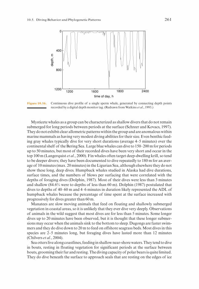
Small social groups of sperm whales spend hours together at the surface each day, theninitiate a series of long and deep foraging dives. Adult Caribbean sperm whales trackedwith acoustic transponders exhibited dive profiles like those in Figure 10.16, withextended periods of resting and socializing at the surface, especially during afternoonhours (Watkins et al., 1993). Diving was most common at night and during early morn-ing hours. Dives typically lasted more than 30 minutes to depths usually below 400 m.Like the dives of foraging elephant seals and migrating gray whales, little surface recov-ery time was apparent between dives. Sperm whales, like the other species of marinemammals discussed in this chapter, presumably dive within their ADL or are compen-sating by other mechanisms.
Some midsized odontocetes display considerable diving abilities. Narwhals andwhite whales in some parts of their range routinely dive deeply (>500 m) and they candive to over 1000 m (e.g., Heide-Jørgensen and Dietz, 1995; Martin and Smith, 1999).Their dive durations are relatively modest with most dives lasting only 5–15 minutes.Killer whales are remarkably shallow divers for their size, whereas the long-finned pilotwhales and bottlenose dolphins dive to significant depths (max. 500–600 m, 16 minutes)although durations still tend to remain under 10 minutes. Other, smaller dolphins suchas the short-beaked common dolphin and pantropical spotted dolphin routinely dive toabout 100 m although they can go to approximately twice this depth. All of the smallerdolphins studied to date usually dive for periods of only a few minutes (e.g., Ridgway,1986).
260 10. Respiration and Diving Physiology
P885522-10.qxd 10/17/05 11:20 PM Page 260
Mysticete whales as a group can be characterized as shallow divers that do not remainsubmerged for long periods between periods at the surface (Schreer and Kovacs, 1997).They do not exhibit clear allometric patterns within the group and are anomalous withinmarine mammals as having very modest diving abilities for their size. Even benthic feed-ing gray whales typically dive for very short durations (average 4–5 minutes) over thecontinental shelf of the Bering Sea. Large blue whales can dive to 150–200 m for periodsup to 50 minutes, but most of their recorded dives have been very short and occur in thetop 100 m (Langerquist et al., 2000). Fin whales often target deep-dwelling krill, so tendto be deeper divers; they have been documented to dive repeatedly to 180 m for an aver-age of 10 minutes (max. 20 minutes) in the Ligurian Sea, although elsewhere they do notshow these long, deep dives. Humpback whales studied in Alaska had dive durations,surface times, and the numbers of blows per surfacing that were correlated with thedepths of foraging dives (Dolphin, 1987). Most of their dives were less than 3 minutesand shallow (84.6% were to depths of less than 60 m). Dolphin (1987) postulated thatdives to depths of 40–60 m and 4–6 minutes in duration likely represented the ADL ofhumpback whales because the percentage of time spent at the surface increased withprogressively for dives greater than 60 m.
Manatees are slow moving animals that feed on floating and shallowly submergedvegetation in coastal areas, so it is unlikely that they ever dive very deeply. Observationsof animals in the wild suggest that most dives are for less than 5 minutes. Some longerdives up to 20 minutes have been observed, but it is thought that these longer submer-sions may occur when the animals sink to the bottom to sleep. Dugongs are faster swim-mers and they do dive down to 20 m to feed on offshore seagrass beds. Most dives in thisspecies are 2–5 minutes long, but foraging dives have lasted more than 12 minutes(Chilvers et al., 2004).
Sea otters live along coastlines, feeding in shallow near-shore waters. They tend to divein bouts, resting in floating vegetation for significant periods at the surface betweenbouts, grooming their fur and resting. The diving capacity of polar bears is quite limited.They do dive beneath the surface to approach seals that are resting on the edges of ice
10.5. Diving Behavior and Phylogenetic Patterns 261
400
0
800
12001200 1600 1800 2400
time of day, h
dept
h, m
Figure 10.16. Continuous dive profile of a single sperm whale, generated by connecting depth pointsrecorded by a digital depth monitor tag. (Redrawn from Watkins et al., 1993.)
P885522-10.qxd 10/17/05 11:20 PM Page 261

floes for periods of a minute or two following a stealthful swimming approach at the sur-face. They have also been seen diving in attempts to hunt white whales, diving in from theice edge and remaining submerged for a minute or so, but polar bears spend most of theiraquatic time swimming on the surface.
10.6. Summary and Conclusions
The effects of pressure on the diving animal involve circulatory and respiratory adapta-tions. Among circulatory changes in pinnipeds and cetaceans are enlargement andincreased complexity of blood vessels, including the development of retia mirabiliathroughout the body that serve as oxygen reservoirs during deep dives. The muscles,blood, and spleen are important for oxygen stores in marine mammals. Respiratoryadjustments that occur during diving involve modifications in the structure of the lungs,especially the bronchioles.
Bradycardia, peripheral vasoconstriction, and other circulatory adjustments areimportant components of an integrated set of diving responses. Research on the Weddellseal and other pinnipeds revealed that the majority of dives are aerobic and that onlydives exceeding an animal’s ADL elicited one or more of the diving responses. Thephylogenetic implications of the diving patterns indicate two strategies among pin-nipeds, an otariid “energy dissipative” strategy and a phocid “energy conserving” strat-egy. It is suggested that the walrus is capable of deep dives but has little reason to do sobecause of the availability of their prey in shallow water. In addition to their function inenergy conservation and foraging, the diving patterns of pinnipeds may also play a rolein the avoidance of predators. The diving and breath-hold capacities of most whales(with the exception of the sperm whale) are exceeded by considerably smaller phocidseals. Possible explanations for this include less accurate measurements of cetacean div-ing behavior and their exploitation of prey located at shallower depths.
10.7. Further Reading
For a general introduction to diving physiology and behavior in marine mammals seeKooyman (1989); popular accounts of diving in the Weddell seal can be found inKooyman (1981) and Williams (2004). A comprehensive account of elephant seal divingbehavior is summarized in an edited volume of their biology (Le Boeuf and Laws, 1994)and see Gentry and Kooyman (1986) for diving behaviors of various fur seals. Summaryaccounts of cetacean diving include Ridgway (1986) and Ridgway and Harrison (1986).
References
Amano, M., and M. Yoshioka (2003). “Sperm Whale Diving Behavior Monitored Using a Suction-Cup-Attached TDR Tag.”Mar. Ecol. Progr. Ser. 258: 291–295.
Andrews, R. D., D. R. Jones, J. D. Williams, D. E. Crocker, D. P. Costa, and B. J. Le Boeuf (1995). “Metabolicand Cardiovascular Adjustments to Diving in Northern Elephant Seals (Mirounga angustirostris).”Physiol. Zool. 68: 105.
Andrews, R. D., D. R. Jones, J. D. Williams, P. H. Thorson, G. W. Oliver, D. P. Costa, and B. J. Le Boeuf (1997).“Heart Rates of Northern Elephant Seals Diving at Sea and Resting on the Beach.” J. Exp. Biol. 200:2083–2095.
262 10. Respiration and Diving Physiology
P885522-10.qxd 10/17/05 11:20 PM Page 262

References 263
Baird, R. W., D. J. McSweeney, A. D. Ligon, and D. L. Webster (2004). Tagging Feasibility and Diving ofCuvier’s Beaked Whales (Ziphius cavirostris) and Blainville’s Beaked Whales (Mesoplodon densirostris) inHawaii. Report prepared under Order No. AB133F-03-SE-0986 to the Hawai’i Wildlife Fund, Volcano,HI.
Baker, J. D., and M. J. Donohue (1999). “Ontogeny of Swimming and Diving in Northern Fur Seal(Callorhinus ursinus) Pups.”Can. J. Zool. 78: 100–109.
Barabash-Nikiforov, I. I. (1947). “Kalan” (The Sea Otter). Soviet Ministrov RSFSR. (Translated fromRussian, Israel Program for Scientific Translations, Jerusalem, 1962).
Bengtson, J. L., and B. S. Stewart (1992). “Diving and Haulout Behavior of Crabeater Seals in the Weddell Sea,Antarctic During March 1996.”Polar Biol. 12: 635–644.
Bengtson, J. L., and B. S. Stewart (1997). “Diving Patterns of a Ross Seal (Ommatophoca rossii) near theEastern Coast of the Antarctic Peninsula.”Polar Biol. 18: 214–218.
Berne, R. M., and M. N. Levy (1988). Physiology. Mosby, St. Louis, MO.Bert, P. (1870). In “Leçons sure la Physiologie Comparéde la Respiration,”pp. 526–553. Baillière, Paris.Blix, A. S. (1976). “Metabolic Consequences of Submersion Asphyxia in Mammals and Birds.”Biochem. Soc.
Symp. 41: 169–178.Bodkin, J. L., G. G. Esslinger, and D. H. Monson. (2004). “Foraging Depths of Sea Otters and Implications to
Coastal Marine Communities.”Mar. Mamm. Sci. 20: 305–321.Boutilier, R. G., M. Nikinmaa, and B. L. Tufts (1993). “Relationship Between Blood Buffering Properties,
Erythrocyte pH and Water Content, in Gray Seals (Halichoerus grypus).” Acta. Physiol. Scand. 147:241–247.
Bowen, W. D., D. J. Boness, and S. J. Iverson (1999). “Diving Behaviour of Lactating Harbour Seals and TheirPups During Maternal Foraging Trips.”Can. J. Zool. 77: 978–988.
Boyd, I. L. (1997). “The Behavioural and Physiological Ecology of Diving.”Trends Ecol. Evol. 12: 213–217.Boyd, I. L., and J. P. Croxall (1996). “Dive Durations in Pinnipeds and Seabirds.” Can. J. Zool. 74:
1696–1705.Bryden, M. M. (1972). Growth and development of marine mammals. In “Functional Anatomy of Marine
Mammals,”Vol. 1 (R. J. Harrison, ed.), pp. 1–79. Academic Press, New York.Castellini, M. A. (1985a). “Metabolic Depression in Tissues and Organs of Marine Mammals During Diving:
Living with Less Oxygen.”Mol. Physiol. 8: 427–437.Castellini, M. A. (1985b). Closed systems: resolving potentially conflicting demands of diving and exercise in
marine mammals. In “Circulation, Respiration and Metabolism” (R. Gilles, ed.), pp. 220–226. Springer-Verlag, Berlin.
Castellini, M. A. (1991). “The Biology of Diving Mammals: Behavioral, Physiological, and BiochemicalLimits.”Adv. Comp. Physiol. 8: 105–134.
Castellini, M. A., Davis, R. W., and G. L. Kooyman (1988). “Blood Chemistry Regulation During RepetitiveDiving in Weddell Seals.”Physiol. Zool. 61: 379–386.
Castellini, M. A., G. L. Kooyman, and P. J. Ponganis. (1992). “Metabolic Rates of Freely Diving Weddell Seals:Correlations with Oxygen Stores, Swim Velocity and Diving Duration.”J. Exp. Biol. 165: 181–194.
Castellini, M. A., B. J. Murphy, M. Fedak, K. Ronald, N. Gofton, and P. W. Hochachka. (1985). “PotentiallyConflicting Metabolic Demands of Diving and Exercise in Seals.”J. Appl. Physiol. 58: 392–399.
Castellini, M. A., and G. N. Somero (1981). “Buffering Capacity of Vertebrate Muscle: Correlations withPotentials for Anaerobic Function.”J. Comp. Physiol. 143: 191–198.
Castellini, M. A., G. N. Somero, and G. L. Kooyman (1981). “Glycolytic Enzyme Activities in Tissues ofMarine and Terrestrial Mammals.”Physiol. Zool. 54: 242–252.
Chilvers, B. L., S. Delean, N. J. Gales, D. K. Holley, I. R. Lawler, H. Marsh, and A. R. Preen (2004). “DivingBehaviour of Dugongs, Dugong dugon.”J. Exp. Mar. Biol. Ecol. 304: 203–224.
Costa, D. P. (1991). Reproductive and foraging energetics of pinnipeds: Implications for life history patterns.In “The Behavior of Pinnipeds”(D. Renouf, ed.), pp. 300–344. Chapman & Hall, London.
Costa, D. P. (1993). The relationship between reproduction and foraging energetics and the evolution of thePinnipedia. In “Recent Advances in Marine Mammal Science” (I. Boyd, ed.), pp. 293–314. Symp. Zool.Soc., Lond. No. 66, Oxford University Press, London.
Costa, D. P., and N. J. Gales (2003) “Energetics of a Benthic Diver: Seasonal Foraging Ecology of theAustralian Sea Lion, Neophoca cinera.”Ecol. Monogr. 73: 27–43.
Costa, D. P, N. J. Gales, and D. E. Crocker (1998). “Blood Volume and Diving Ability of the New Zealand SeaLion, Phocarctos hookeri.”Physiol. Zool. 71: 208–213.
Costa, D. P., N. J. Gales, and M. E. Goebel (2001). “Aerobic Dive Limit: How Often Does It Occur in Nature?”Comp. Biochem. Physiol. A 129: 771–783.
P885522-10.qxd 10/17/05 11:20 PM Page 263

Costa, D. P., and G. L. Kooyman (1982). “Oxygen Consumption, Thermoregulation, and the Effect of FurOiling and Washing on the Sea Otter, Enhydra lutris.”Can. J. Zool. 60: 2761–2767.
Costa, D. P., and T. M. Williams (1999). Marine mammal energetics. In “Biology of Marine Mammals”(R. E.Reynolds and S. A. Rommel, eds.), pp. 176–217. Smithsonian Institute Press, Washington, D. C.
Curry, B. E., J. Mead, and A. P. Purgue (1994). “The Occurrence of Calculi in the Nasal Diverticula ofPorpoises (Phocoenidae).”Mar Mamm. Sci. 10: 81–86.
Davis, R. W., L. A. Fuiman, T. M. Williams, M. Horning, and W. Hagey (2003). “Classification of Weddell SealDives Based on 3 Dimensional Movements and Video-Recorded Observations.” Mar. Ecol. Prog. Ser. 264:109–122.
DeLong, R. L., and B. S. Stewart (1991). “Diving Patterns of Northern Elephant Seal Bulls.”Mar. Mamm. Sci.7: 369–384.
Denison, D. M., D. A. Warrell, and J. B. West (1971). “Airway Structure and Alveolar Emptying in the Lungsof Sea Lions and Dogs.”Respir. Physiol. 13: 253–260.
Dolphin, W. F (1987). “Dive Behavior and Estimated Energy Expenditure of Foraging Humpback Whales inSouth-East Alaska.”Can. J. Zool. 65: 354–362.
Drabek, C. M. (1975). “Some Anatomical Aspects of the Cardiovascular System of Antarctic Seals and TheirPossible Functional Significance in Diving. J. Morphol. 145(1): 85–106.
Drabek, C. M. (1977). Some anatomical and functional aspects of seal hearts and aortae. In “FunctionalAnatomy of Marine Mammals,”Vol. 3 (R. J. Harrison, ed.), pp. 217–234. Academic Press, London.
Drabek, C. M., and J. M. Burns (2002). “Heart and Aorta Morphology of the Deep-Diving Hooded Seal(Cystophora cristata).”Can. J. Zool. 80: 2030–2036.
Drabek, C. M., and G. L. Kooyman (1983). “Terminal Airway Embryology of the Delphinid Porpoises,Stenella attenuata and S. longirostris.”J. Morphol. 175: 65–72.
Elsner, R. (1987). The contribution of anaerobic metabolism to maximum exercise in seals. In “MarineMammal Energetics”(A. C. Huntley, D. P. Costa, G. A. J. Worthy, and M. A. Castellini, eds.), pp. 109–114.Soc. Mar. Mammal., Spec. Publ. No 1, Lawrence, KS.
Elsner, R. (1999). Living in water: Solutions to physiological problems. In “Biology of Marine Mammals”(R.E. Reynolds and S. A. Rommel, eds.), pp. 73–116. Smithsonian Institute Press, Washington, D. C.
Elsner, R., and B. Gooden (1983). Diving and Asphyxia: A Comparative Study of Animals and Men. CambridgeUniversity Press, New York.
Elsner, R., D. D. Hammond, and H. R. Parker (1970). “Circulatory Responses to Asphyxia in Pregnant andFetal Animals: A Comparative Study of Weddell Seals and Sheep.”Yale J. Biol. Med. 42: 202–217.
Elsner,R.,D.W.Kenney,and K.Burgess (1966).“Diving Bradycardia in the Trained Dolphin.”Nature 212:407–408.Elsner, R., D. Wartzok, N. B. Sonafrank, and B. P. Kelly (1989). “Behavioural and Physiological Reactions of
Arctic Seals During Under-Ice Pilotage.”Can. J. Zool. 67: 2506–2513.Engel, S. (1959a). “The Respiratory Tissue of Dugong (Halicore dugong).”Anat. Anz. 106: 90–100.Engel, S. (1959b). “Rudimentary Mammalian Lungs.”Gegenbaurs Morphol. Jahrb. 106: 95–114.Engel, S. (1962). “The Air Passages of the Dugong Lung.”Acta Anat. 48: 95–107.Falke, K. J., R. D. Hill, J. Qvist, R. C. Schneider, M. Guppy, G. C. Liggins, P. W.Hochachka, R. E. Elliott, and W. M. Zapol (1985). “Seal Lungs Collapse During Free Diving: Evidence From
Arterial Nitrogen Tensions.”Science 229: 556–557.Fay, F. (1981). Walrus-Odobenus rosmarus. In “Handbook of Marine Mammals” (S. H. Ridgway and R.
J. Harrison, eds.), pp. 1–23. Academic Press, London.Fay, F H., and J. J. Burns (1988). “Maximum Feeding Depth of Walruses.”Arctic 41: 239–240.Fedak, M. A. (1986). Diving and exercise in seals a benthic perspective. In “Diving in Animals and Man”
(A. Brubakk, J. W. Kanwisher, and G. Sundnes, eds.), pp. 11–32. Tapir Publ., Trondheim, Norway.Folkow, L. P., and A. S. Blix (1995). Distribution and diving behaviour of hooded seals. In “Whales, Seals, Fish
and Man”(A. S. Blix, L. Walløe, and Ø. Ulltang, eds.), pp. 193–202. Elsevier, Amsterdam.Folkow, L. P., E. S. Nordøy, and A. S. Blix (2004). “Distribution and Diving Behaviour of Harp Seals
(Pagophilus groenlandicus) from the Greenland Sea Stock.”Polar Biol. 27: 281–298.Gentry, R. L. (2001). Eared seals. In “Encyclopedia of Marine Mammals” (W. F. Perrin, B. Würsig, and J. G.
M. Thewissen, eds.), pp. 348–351. Academic Press, San Diego, CA.Gentry, R. L., and G. L. Kooyman (1986). Fur Seals: Maternal Strategies on Land and at Sea. Princeton
University Press, Princeton, NJ.Gentry, R. L., G. L. Kooyman, and M. E. Goebel (1986). Feeding and diving behavior of northern fur seals. In
“Fur Seals: Maternal Strategies on Land and at Sea” (R. L. Gentry, and G. L. Kooyman, eds.), pp. 61–78.Princeton University Press, Princeton, NJ.
264 10. Respiration and Diving Physiology
P885522-10.qxd 10/17/05 11:20 PM Page 264
Gilmartin, W. G., R. W. Pierce, and G. A. Antonelis (1974). “Some Physiological Parameters of the Blood ofthe California Gray Whale.”Marine Fisheries Rev. 36: 28–31.
Gjertz, I., K. M. Kovacs, C. Lydersen, and Ø. Wiig (2000). “Movements and Diving of Bearded Seal(Erignathus barbatus) Mother and Pups During Lactation and Post-Weaning.”Polar Biol. 23: 559–566.
Gjertz, I., C. Lydersen, and Ø. Wiig (2001). “Distribution and Diving of Harbour Seals (Phoca vitulina) inSvalbard.”Polar Biol. 24: 209–214.
Gosline, J. M., and R. E. Shadwick (1996). “The Mechanical Properties of Fin Whale Arteries are Explainedby Novel Connective Tissue Designs.”J. Exp. Biol. 199: 985–997.
Guyton, G. P., K. S. Stanek, R. C. Schneider, P. W. Hochachka, W. E. Hurford, D. G. Zapol, G. C. Liggins, andW. M. Zapol (1995). “Myoglobin Saturation in Free-Diving Weddell Seals.” J. Appl. Physiol. 79:1148–1155.
Hammmel, H. T., R. Elsner, H. C. Heller, J. A. Maggert, and C. R. Bainton (1977). “ThermoregulatoryResponses to Alternating Hypothalamic Temperature in the Harbor Seal.”Am. J. Physiol. 232: R18–R26.
Harken, A. H. (1976). “Hydrogen Ion Concentration and Oxygen Uptake in an Isolated Canine Limb.”J. Appl. Physiol. 40: 1–5.
Harrison, R. J., and J. E. King (1980). Marine Mammals, 2nd ed. Hutchinson, London.Harrison, R. J., and G. L. Kooyman (1968). General physiology of the pinnipedia. In “The Behavior and
Physiology of the Pinnipeds” (R. J. Harrison, R. C. Hubbard, R. S. Peterson, R. E. Rice, and R.J. Schusterman, eds.), pp. 211–296. Appleton-Century-Crofts, New York.
Harrison, R. J., and J. D. W. Tomlinson (1956). “Observations on the Venous System in Certain Pinnipedía andCetacea.”Trans. Zool. Soc. London 126(part 2): 205–233.
Hedrick, M. S., and D. A. Duffield (1991). “Haematological and Theological Characteristics of Blood inSeven Marine Mammal Species: Physiological Implications for Diving Behaviour.” J. Zool. London 225:273–283.
Heide-Jorgensen, M. P., and R. Dietz (1995). “Some Characteristics of Narwhal, Monodon monoceros, DivingBehaviour in Baffin Bay.”Can. J. Zool. 73: 2120–2132.
Hill, R. D., R. C. Schneider, G. C. Liggins, A. H. Schuette, R. L. Elliott, M. Guppy, P. W. Hochachka, J. Qvist,K. J. Falke, and W. M. Zapol (1987). “Heart Rate and Body Temperature During Free Diving of WeddellSeals.”Am. J. Physiol. 253: R344–R351.
Hill, W. C. O. (1945). “Notes on the Dissection of Two Dugongs.”J. Mammal. 26: 153–175.Hindell, M. A., D. J. Slip, H. R. Burton, and M. M. Bryden (1992). “Physiological Implications of Continuous,
Prolonged, and Deep Dives of the Southern Elephant Seal (Mirounga leonina).”Can. J. Zool. 70: 370–379.Hochachka, P. W. (1992). “Metabolic Biochemistry and the Making of a Mesopelagic Mammal.” Experentia
48: 570–575.Hochachka, P. W., J. M. Castellini, R. D. Hill, R. C. Schneider, J. L. Bengtson, S. E. Hill, G. C. Liggins, and W.
M. Zapo (1988). “Protective Metabolic Mechanisms During Liver Ischemia: Transferable Lessons fromLong-Diving Animals.”Mol. Cell. Biochem. 23: 12–20.
Hochachka, P. W., S. C. Land, and L. T. Buck (1997). “Oxygen Sensing and Signal Transduction in MetabolicDefense Against Hypoxia: Lessons from Vertebrate Facultative Anaerobes.” Comp. Biochem. Physiol. A118: 23–29.
Hochachka, P. W., and P. D. Mottishaw (1998). Evolution and adaptation of the diving response: Phocids andotariids. In “Cold Ocean Symposia” (H. O. Portwer and R. C. Playle, eds.), pp. 391–431. CambridgeUniversity Press, Cambridge, MA.
Hooker, S. K., and R.W. Baird (1999). “Deep-Diving Behaviour of the Northern Bottlenose Whale,Hyperoodon ampullatus (Cetacea: Ziphiidae).”Proc. R. Soc. London B 266: 671–676.
Hooker, S. K., P. J. O. Miller, M. P. Johnson, O. P. Cox, and I. L. Boyd. (2005). “Ascent Exhalations of AntarcticFur Seals: A Behavioural Adaptation for Breath-Hold Diving? Proc. Royal Soc. London B 272: 355–363.
Horning, M., and F. Trillmich (1997). “Ontogeny of Diving Behaviour in the Galapagos Fur Seal.”Behaviour134: 1211–1257.
Huntley, A. C., D. P. Costa, and R. D. Rubin (1984). “The Contribution of Nasal Countercurrent HeatExchange to Water Balance in the Northern Elephant Seal, Mirounga angustirostris.” J. Exp. Biol. 113:447–454.
Irving, L. (1939). “Respiration in Diving Mammals.”Physiol. Rev. 19: 112–134.Irving, L., P. F. Scholander, and S. W. Grinnell (1941a). “The Respiration of the Porpoises, Tursiops truncatus.”
J. Cell. Comp. Physiol. 17: 1–45.Irving, L., P. F. Scholander, and S. W. Grinnell. (1941b). “Significance of the Heart Rate to the Diving Ability
of Seals.”J. Cell. Comp. Physiol. 18: 283–297.
References 265
P885522-10.qxd 10/17/05 11:20 PM Page 265

Irving, L., P. F. Scholander, and S. W. Grinnell. (1942). “The Regulation of Arterial Blood Pressure in the SealDuring Diving.”Am. J. Physiol. 135: 557–566.
Irving, L., O. M. Solandt, D. Y. Solandt, and K. C. Fisher (1935). “Respiratory Characteristics of the Blood ofthe Seal.”J. Cell. Comp. Physiol. 6: 393–403.
Jones, D. R., H. D. Fisher, S. McTaggart, and N. H. West (1973). “Heart Rate During Breath-Holding andDiving in the Unrestrained Harbor Seal, Phoca vitulina richardsi.”Can. J. Zool. 51: 671–680.
Jørgensen, C., C. Lydersen, O. Brix, and K. M. Kovacs (2001). “Diving Development in Nursing HarbourSeals.”J. Exp. Biol. 204: 3993–4004.
King, J. E. (1977). “Comparative Anatomy of the Blood Vessels of the Sea Lions Neophoca and Phocarctos;with Comments on the Differences Between the Otariid and Phocid Vascular Systems.” J. Zool. London181: 69–94.
King, J. E. (1983). Seals of the World, 2nd ed. Comstock, Ithaca, NY.Kjekshus, J. K., A. S. Blix, R. Elsner, R. Hol, and E. Amundsen. (1982). “Myocardial Blood Flow and
Metabolism in the Diving Seal.”Am. J. Physiol. 242: R79–R104.Kooyman, G. L. (1966). “Maximum Diving Capacities of the Weddell Seal (Leptonychotes weddelli).”Science
151: 1553–1554.Kooyman, G. L. (1973). “Respiratory Adaptations in Marine Mammals.”Am. Zool. 13: 457–468.Kooyman, G. L. (1981). Weddell Seal: Consummate Diver. Cambridge University Press, Cambridge.Kooyman, G. L. (1985). “Physiology Without Restraint in Diving Mammals.”Mar. Mamm. Sci. 1: 166–178.Kooyman, G. L. (1989). Diverse Divers: Physiology and Behavior. Springer-Verlag, New York.Kooyman, G. L. (2002). Diving physiology In “Encyclopedia of Marine Mammals” (W. F. Perrin, B. Würsig,
and J. G. M. Thewissen, eds.), pp. 339–344. Academic Press, San Diego, CA.Kooyman, G. L., and H. T. Anderson (1969). Deep diving. In “Biology of Marine Mammals”(H. T. Andersen,
ed.), pp. 65–94. Academic Press, New York.Kooyman, G. L., and W. B. Campbell (1972). “Heart Rates in Freely Diving Weddell Seals, Leptonychotes wed-
dellii.”Comp. Biochem. Physiol. A 43: 31–36.Kooyman, G. L., M. A. Castellini, and R. W. Davis (1981). “Physiology of Diving in Marine Mammals.”Annu.
Rev. Physiol. 43: 343–356.Kooyman, G. L., M. A. Castellini, R. W. Davis, and R. A. Mauae. (1983). “Aerobic Diving Limits of Immature
Weddell Seals.”J. Comp. Physiol. B 151: 171–174.Kooyman, G. L., and L. H. Cornell (1981). “Flow Properties of Expiration and Inspiration in a Trained Bottle-
Nosed Porpoise.”Physiol. Zool. 54: 55–61.Kooyman, G. L., D. H. Kerem, W. B. Campbell, and J. J. Wright. (1973). “Pulmonary Gas Exchange in Freely
Diving Weddell Seals (Leptonychotes weddelli).”Resp. Physiol. 17: 283–290.Kooyman, G. L., E. A. Wahrenbrock, M. A. Castellini, R. W. Davis, and E. E. Sinnett (1980). “Aerobic and
Anaerobic Metabolism During Voluntary Diving in Weddell Seals: Evidence of Preferred Pathways fromBlood Chemistry and Behavior.”J. Comp. Physiol. 138: 335–346.
Krafft, B. A., C. Lydersen, K. M. Kovacs, I. Gjertz, and T. Haug (2000). “Diving Behaviour of LactatingBearded Seals (Erignathus barbatus) in the Svalbard Area.”Can. J. Zool. 78: 1408–1418.
Krutikowsky, G. K., and B. R. Mate, (2000). “Dive and Surfacing Characteristics of Bowhead Whales(Balaena mysticetus) in the Beaufort and Chukchi Seas.”Can. J. Zool. 78: 11:82–1198.
Lagerquist, B. A., K. M. Stafford, and B. R. Mate. (2000). “Dive Characteristics of Satellite-MonitoredBlue Whales (Balaenoptera musculus) off the Central California Coast.” Mar. Mammal Sci. 16:375–391.
Laidre, K. L., M. P. Heide-Jorgensen, R. Dietz, R. C. Hobbs, and O. A. Jorgensen (2003). “Deep-Diving byNarwhals Monodon monoceros: Differences in Foraging Behavior Between Wintering Areas?” Mar. Ecol.Prog. Ser. 261: 269–281.
Langerquist, B. A., K. M. Stafford, and B. R. Mate (2000). “Dive Characteristics of Satellite-Monitored BlueWhales (Balaenoptera musculus) off the California Coast.”Mar. Mamm. Sci. 16: 375–391.
Lawrence, B., and W. E. Schevill (1965). “Gular Musculature in Delphinids. Bull. Mus. Comp. Zool. 133: 1–65.Le Boeuf, B. J. (1994). Variation in the diving pattern of Northern elephant seals with age, mass, sex and repro-
ductive condition. In “Elephant Seals” (B. J. Le Boeuf and R. M. Laws, eds.), pp. 237–252. University ofCalifornia Press, Berkeley.
Le Boeuf, B. J., and D. E. Crocker (1996). Diving behavior of elephant seals: Implications for predator avoid-ance. In “Great White Sharks, The Biology of Carcharodon carchias” (A. P. Klimley and D. G. Ainley, eds.),pp. 193–206. Academic Press, San Diego, CA.
Le Boeuf, B. J., and R. M. Laws, eds. (1994). Elephant Seals. University of California Press, Berkeley.
266 10. Respiration and Diving Physiology
P885522-10.qxd 10/17/05 11:20 PM Page 266
Le Boeuf, B. J., P. A. Morris, S. B. Blackwell, D. E. Crocker, and D. P. Costa (1996). Diving behavior of juvenilenorthern elephant seals. Can. J. Zool. 74: 1632-1644.
Le Boeuf, B. J., Y. Naito, A. C. Huntley, and T. Asaga (1989). “Prolonged, Continuous, Deep Diving byNorthern Elephant Seals.”Can. J. Zool. 67: 2514–2519.
Leith, D. (1976). Physiological properties of blood of marine mammals. In “The Biology of MarineMammals”(H. T. Anderson, ed.), pp. 95–116. Academic Press, New York.
Lenfant, C., K. Johansen, and J. D. Torrance (1970). “Gas Transport and Oxygen Storage Capacity in SomePinnipeds and the Sea Otter.”Respir. Physiol. 9: 227–286.
Lewis, M., C. Campagna, M. Uhart, and C. L. Ortiz (2001). “Ontogenetic and Seasonal Variation in BloodParameters in Southern Elephant Seals.”Mar. Mammal Sci. 17: 862–872.
Liggins, G. C., J. Qvist, P. W. Hochachka, B. J. Murphy, R. K. Creasy, R. C. Schneider, M. T. Snider, and W. M.Zapol (1980). “Fetal Cardiovascular and Metabolic Responses to Simulated Diving in the Weddell Seal.”J. Appl. Physiol. 49: 424–430.
Lydersen, C., and K. M. Kovacs (1993). “Diving Behaviour of Lactating Harp Seal, Phoca groenlandica,Females from the Gulf of St. Lawrence, Canada.”Anim. Behav. 46: 1213–1221.
Lydersen, C., and K. M. Kovacs (1999). “Behaviour and Energetics of Ice-Breeding, North Atlantic PhocidSeals During the Lactation Period.”Mar. Ecol. Progr. Ser. 187: 265–281.
Lydersen, C., M. O. Hammill, and K. M. Kovacs (1994). “Activity of Lactating Ice-Breeding Grey Seals(Halichoerus grypus) from the Gulf of St Lawrence, Canada.”Anim. Behav. 48: 1417–1425.
Martin, A. R., and T. G. Smith (1999). “Strategy and Capability of Wild Belugas, Delphinapterus leucas,During Deep, Benthic Diving.”Can. J. Zool. 77: 1783–1793.
Melnikov, V. V. (1997). “The Arterial System of the Sperm Whale (Physeter macrocephalus).”J. Morphol. 234:37–50.
Miller, N. J., C. B. Daniels, D. P. Costa, and S. Orgeig (2004). “Control of Pulmonary Surfactant Secretion inAdult California Sea Lions.”Biochem. Biophy. Res. Comm. 313: 727–732.
Moon, R. E., R. D. Vann, and P. B. Bennett (1995). “The Physiology of Decompression Illness.”Sci. Am. 273:70–77.
Mottishaw, P. D., S. J. Thornton, and P. W. Hochachka (1999). “The Diving Response Mechanism and ItsSurprising Evolutionary Path in Seals and Sea Lions.”Am. Zool. 39: 434–450.
Munkacsi, I. M., and J. D. Newstead (1985). “The Intrarenal and Pericapsular Venous Systems of Kidneys ofthe Ringed Seal, Phoca hispida.”J. Morphol. 184: 361–373.
Nishiwaki, M., and H. Marsh (1985). Dugong Dugong dugon (Muller, 1776). In “Handbook of MarineMammals,”Vol. 3 (S. H. Ridgway, and R. Harrison, eds.), pp. 1–31. Academic Press, New York.
Noren, S. R., and T. M. Williams (2000). “Body Size and Skeletal Muscle Myoglobin of Cetaceans:Adaptations for Maximizing Dive Duration.”Comp. Biochem. Physiol. A 126: 181–191.
Noren, S. R., T. M. Williams, D. A. Pabst, W. A. McLellan, and J. L. Dearolf (2001). “The Development ofDiving in Marine Endotherms: Preparing the Skeletal Muscles of Dolphins, Penguins, and Seals forActivity During Submergence.”Jour. Comp. Physiol. B 171: 127–134.
Olsen, C. R., F. C. Hale, and R. Elsner (1969). “Mechanics of Ventilation in the Pilot Whale.”Resp. Physiol. 7:137–149.
Pfeiffer, C. J. (1990). “Observations on the Ultrastructural Morphology of the Bowhead Whale (Balaena mys-ticetus) Heart.”J. Zool. Wild. Med. 21: 48–55.
Pfeiffer, C. J., and T. P. Kinkead (1990). “Microanatomy of Retia Mirabilia of Bowhead Whale ForamenMagnum and Mandibular Foramen.”Acta Anat. 139: 141–150.
Pfeiffer, C. J., and V S. Viers (1995). “Cardiac Ultrastructure in the Ringed Seal, Phoca hispida and Harp Seal,Phoca groenlandica.”Aquat. Mamm. 21: 109–119.
Ponganis, P. J., R. L. Gentry, E. P. Ponganis, and K. V. Ponganis (1992). “Analysis of Swim VelocitiesDuring Deep and Shallow Dives of Two Northern Fur Seals, Callorhinus ursinus.” Mar Mamm. Sci. 8:69–75.
Ponganis, P. J., G. L. Kooyman, E. A. Baranov, P. H. Thorson, and B. S. Stewart (1997). “The AerobicSubmersion Limit of Baikal Seals, Phoca sibirica.”Can. J. Zool. 75: 1323–1327.
Ponganis, P. J., E. P. Ponganis, K. V. Ponganis, G. L. Kooyman, R. L. Gentry, and F. Trillmich (1990).“Swimming Velocities in Otariids.”Can. J. Zool. 68: 2105–2115.
Reidenberg, J. S., and J. T. Laitman (1987). “Position of the Larynx in Odontoceti.”Anat. Rec. 218: 98–106.Reynolds, J., and D. Odell (1991). Manatees and Dugongs. Facts on File, New York.Richet, C. (1894). “La résistance des canards á l’asphyxie.”J. Physiol. Path. Gen. 1: 244–245.Richet, C. (1899). “De la résistance des canards á l’asphyxie.”J. Physiol. Path. Gen. 5: 641–650.
References 267
P885522-10.qxd 10/17/05 11:20 PM Page 267
Ridgway, S. H. (1972). Homeostasis in the aquatic environment. In “Mammals of the Sea” (S. H. Ridgway,ed.), pp. 590–747. Thomas Publ., Springfield, IL.
Ridgway, S. H. (1986). Diving by cetaceans. In “Diving in Animals and Man”(A. OBrubakk, J. W. Kanwisher,and G. Sundness, eds.), pp. 33–62. The Royal Norwegian Society of Science and Letters, Trinheim,Norway.
Ridgway, S. H., and R. J. Harrison (1986). Diving dolphins. In “Research on Dolphins”(M. M. Bryden and R.H. Harrison, eds.), pp. 33–58. Oxford University Press, Oxford, UK.
Ridgway, S. H., and R. Howard (1979). “Dolphin Lung Collapse and Intramuscular Circulation During FreeDiving: Evidence from Nitrogen Washout.”Science 206: 1182–1183.
Ridgway, S. H., and D. G. Johnston (1966). “Blood Oxygen and Ecology of Porpoises of Three Genera.”Science 151: 456–458.
Ridgway, S. H., B. L. Scronce, and J. Kaniwisher (1969). “Respiration and Deep Diving in the BottlenosePorpoise.”Science 166: 1651–1653.
Ronald, K., R. McCarter, and L. J. Selley (1977). Venous circulation in the harp seal (Pagophilus groenlandi-cus). In “Functional Anatomy of Marine Mammals,” Vol. 3 (R. J. Harrison, ed.), pp. 235–270. AcademicPress, New York.
Rowlatt, U., and H. Marsh (1985). “The Heart of the Dugong (Dugong dugon) and the West Indian Manatee(Trichechus manatus) Sirenia.”J. Morphol. 186: 95–105.
Scholander, P. F. (1940). “Experimental Investigations on the Respiratory Function in Diving Mammals andBirds.”Hvalradets Skr. 22: 1–131.
Scholander, P. F. (1960). “Oxygen Transport Through Hemoglobin Solutions.”Science 131: 585–590.Scholander, P. F. (1964). Animals in aquatic environments: diving mammals and birds. In “Handbook of
Physiology, Section 4: Adaptations to the Environment” (D. B. Dill, E. . Adolph, and C. G. Wiber, eds.),pp. 729–739. American Physiology Society, Washington, D. C.
Scholander, P. F., L. Irving, and S. W. Grinell (1942a). “Aerobic and Anaerobic Changes in Seal MusclesDuring Diving.”J. Biol. Chem. 142: 431–440.
Scholander, P. F., L. Irving, and S. W. Grinnell. (1942b). “On the Temperature and Metabolism of the SealDuring Diving.”J. Cellular Comp. Physiol. 21: 53–63.
Schreer, J. F., and K. M. Kovacs (1997). “Allometry of Diving Capacity in Air-Breathing Vertebrates.” Can.J. Zool. 75: 339–358.
Schreer, J. F., R. J. O’Hara Hines, and K. M. Kovacs. (1998). “Classification of Multivariate Diving Data fromAir-Breathing Vertebrates: A Comparison of Traditional Statistical Clustering and Unsupervised NeuralNetworks.”J. Agric. Biol. Environ. Stat. 3: 383–404.
Schreer, J. F., and J. W. Testa (1996). “Classification of Weddell Seal Diving Behavior.” Mar. Mamm. Sci. 12:227–250.
Shadwick, R. E., and J. M. Gosline (1994). “Arterial Mechanics in the Fin Whale Suggest a UniqueHemodynamic Design.”Am. J Physiol. 267: R805–R818.
Sleet, R. B., J. L. Sumich, and L. J. Weber (1981). “Estimates of Total Blood Volume and Total Body Weight ofa Sperm Whale (Physeter catadon).”Can. J. Zool. 59: 567–570.
Slijper, E. J. (1979). Whales, 2nd ed. Hutchinson, London.Slip, D. J., M. A. Hindell, and H. R. Burton (1994). Diving behavior of southern elephant seals from
Macquarie Island. In “Elephant Seals” (B. J. Le Boeuf, and R. M. Laws, eds.), pp. 253–270. University ofCalifornia Press, Berkeley.
Sparling, C. E., and M. A. Fedak (2004). “Metabolic Rates of Captive Grey Seals During Voluntary Diving.J. Exp. Biol. 201: 1615–1624.
Spencer, M. P., T. A. Gornall, and T. C. Poulter (1967). “Respiratory and Cardiac Activity of Killer Whales.”J. Appl. Physiol. 22: 974–981.
Spragg, R. G., P. J. Ponganis, J. J Marsh, G. A. Rau, and W. Bernhard (2004). “Surfactant from Diving AquaticMammals.”J. Appl. Physiol. 96: 1626–1632.
Stewart, B. S., and R. L. DeLong (1995). “Double Migrations of the Northern Elephant Seal, Mirounga angu-stirostris.”J. Mammal. 76: 196–205.
Tarasoff, F. J., and G. L. Kooyman (1973a). “Observations on the Anatomy of the Respiratory System of theRiver Otter, Sea Otter, and Harp Seal I. The topography, Weight, and Measurements of the Lungs.” Can.J. Zool. 51: 163–170.
Tarasoff, F. J., and G. L. Kooyman (1973b). “Observations on the Anatomy of the Respiratory System of theRiver Otter, Sea Otter, and Harp Seal II. The Trachea and Bronchial Tree.”Can. J. Zool. 51: 171–177.
Thompson, D., and M. A. Fedak (1993). “Cardiac Responses of Gray Seals During Diving at Sea.” J. Exp.Biol. 174: 139–164.
268 10. Respiration and Diving Physiology
P885522-10.qxd 10/17/05 11:20 PM Page 268

Thorson, P. H., and B. J. Le Boeuf (1994). Developmental aspects of diving in northern elephant seal pups. In“Elephant Seals” (B. J. Le Boeuf and R. M. Laws, eds.), pp. 271–289. University of California Press,Berkeley.
Wartzok, D., R. Elsner, H. Stone, B. P. Kelly, and R. W. Davies. (1992). “Under-Ice Movements and theSensory Basis of Hole Finding by Ringed and Weddell Seals.”Can. J. Zool. 70: 1712–1722.
Watkins, W. A., M. A. Daher, N. A. DiMarzio, A. Samuels, D. Wartzok, K. M. Fristrup, P. W. Howey, and R.R. Maiefski (2002). “Sperm Whale Dives Tracked by Radio Tag Telemetry.”Mar. Mamm. Sci. 18: 55–68.
Watkins, W. A., M. A. Daher, K. M. Fristrup, T. J. Howard, and G. N. Sciara (1993). “Sperm Whales Taggedwith Transponders and Tracked Under Water by Sonar.”Mar Mamm. Sci. 9: 55–67.
White, F. C., R. Elsner, D. Willford, E. Hill, and E. Merhoff (1990). “Responses of Harbor Seal and Pig Heartto Progressive and Acute Hypoxia.”Am. J. Physiol. 259: R849–R856.
Williams, T. M. (2004). The Hunter’s Breath. M. Evans and Company, New York.Williams, T. M., W. A. Friedl, and J. E. Haun. (1993). “The Physiology of Bottlenose Dolphins (Tursiops trun-
cates): Heart Rate, Metabolic Rate and Plasma Lactate Concentration During Exercise.”J. Exp. Biol. 179:31–46
Williams, T. M., G. L. Kooyman, and D. A. Croll (1991). “The Effect of Submergence on Heart Rate andOxygen Consumption of Swimming Seals and Sea Lions.”J. Comp. Physiol. B 160: 637–644.
Yablokov, A. V, V. M. Bel’kovich, and V. I. Borisov (1972). Whales and Dolphins. Israel Programs for ScientificTranslations, Jerusalem.
References 269
P885522-10.qxd 10/17/05 11:20 PM Page 269