Embed Size (px)
Citation preview
Pacers, ablation, cardioversion, telemetry, Intro to ACLS
By: Diana Blum
MCC
NURS 2140
• A dysrhythmia is a disturbance of the rhythm of the heart caused by a problem in the conduction system.
• Categorized by site of origin: atrial , AV nodal, ventricular
• Blocks are interruptions in impulse conduction: 1st, 2nd type 1&2, 3rd or complete heart block
2
3
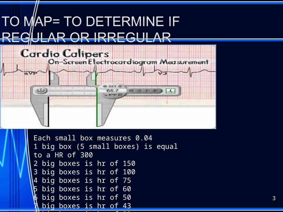
Each small box measures 0.041 big box (5 small boxes) is equal to a HR of 3002 big boxes is hr of 1503 big boxes is hr of 1004 big boxes is hr of 755 big boxes is hr of 606 big boxes is hr of 507 big boxes is hr of 438 big boxes is hr of 38
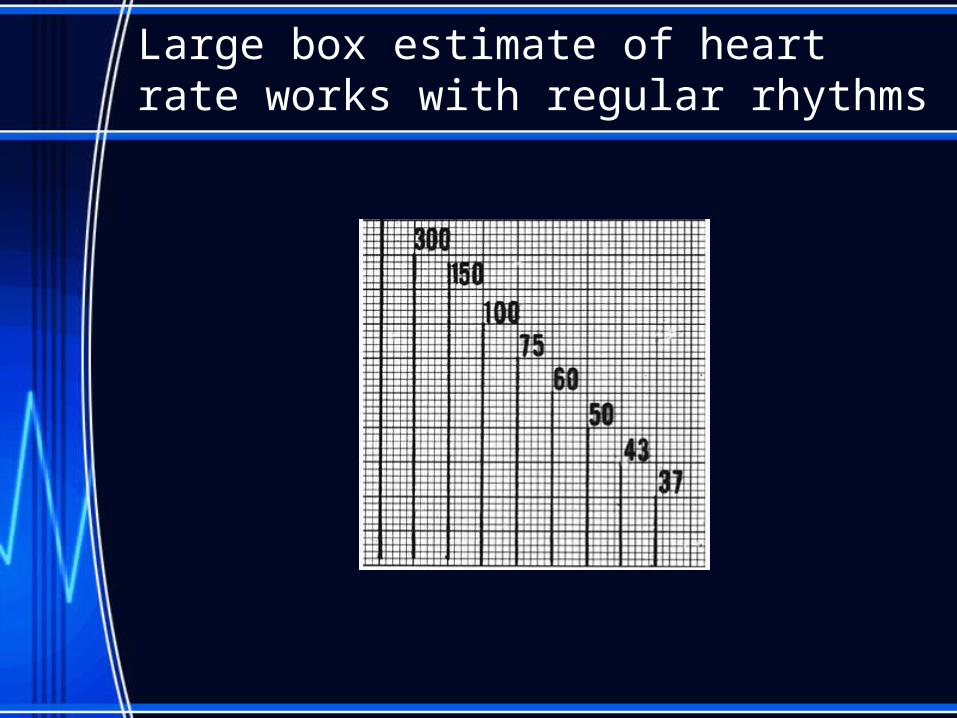
Large box estimate of heart rate works with regular rhythms
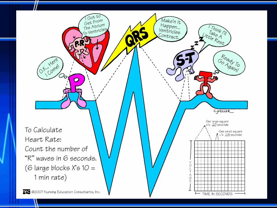
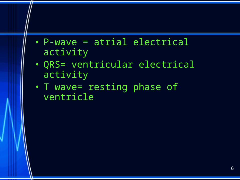
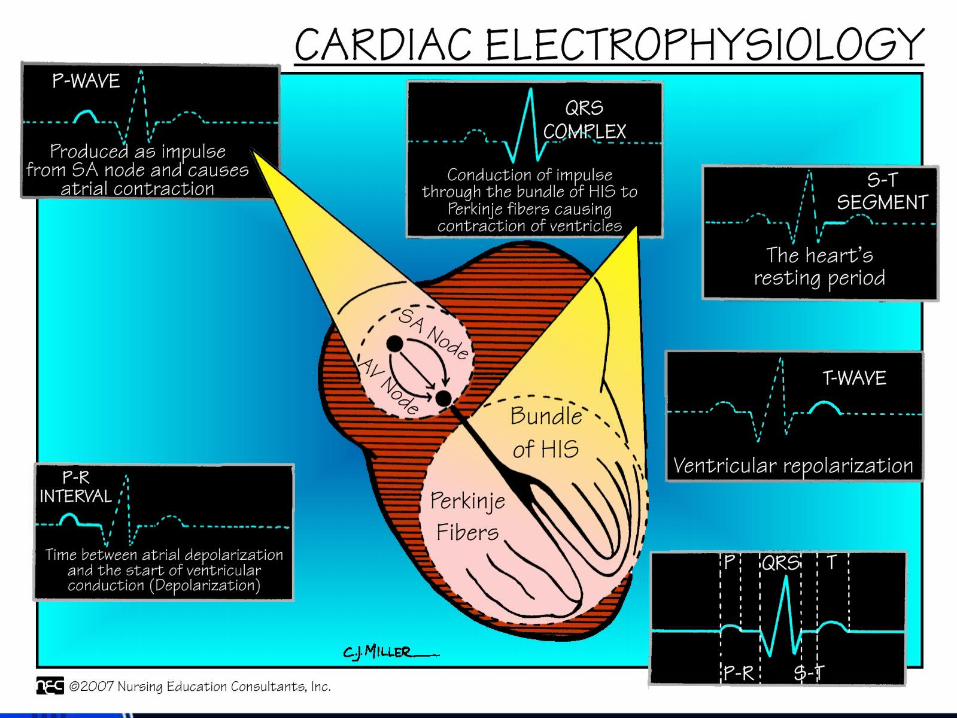
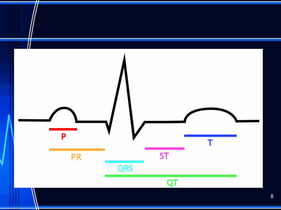
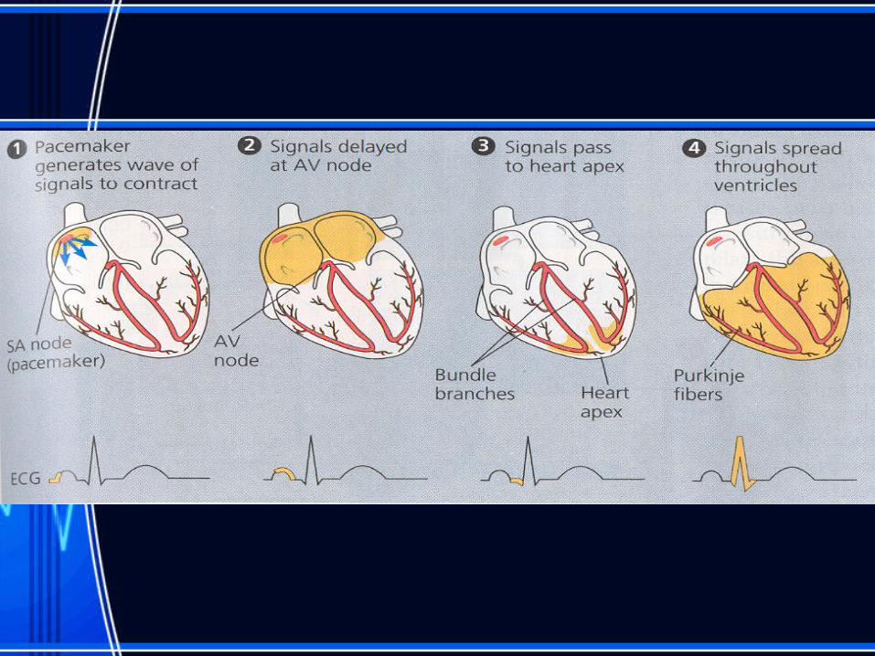
• P-wave = atrial electrical activity• QRS= ventricular electrical activity• T wave= resting phase of ventricle
6
8
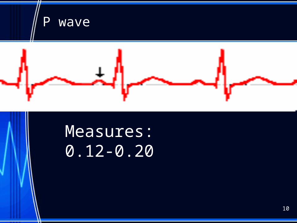
P wave
10
Measures: 0.12-0.20
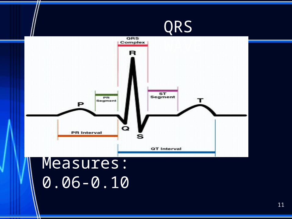
Measures: 0.06-0.10
QRS WAVE
11
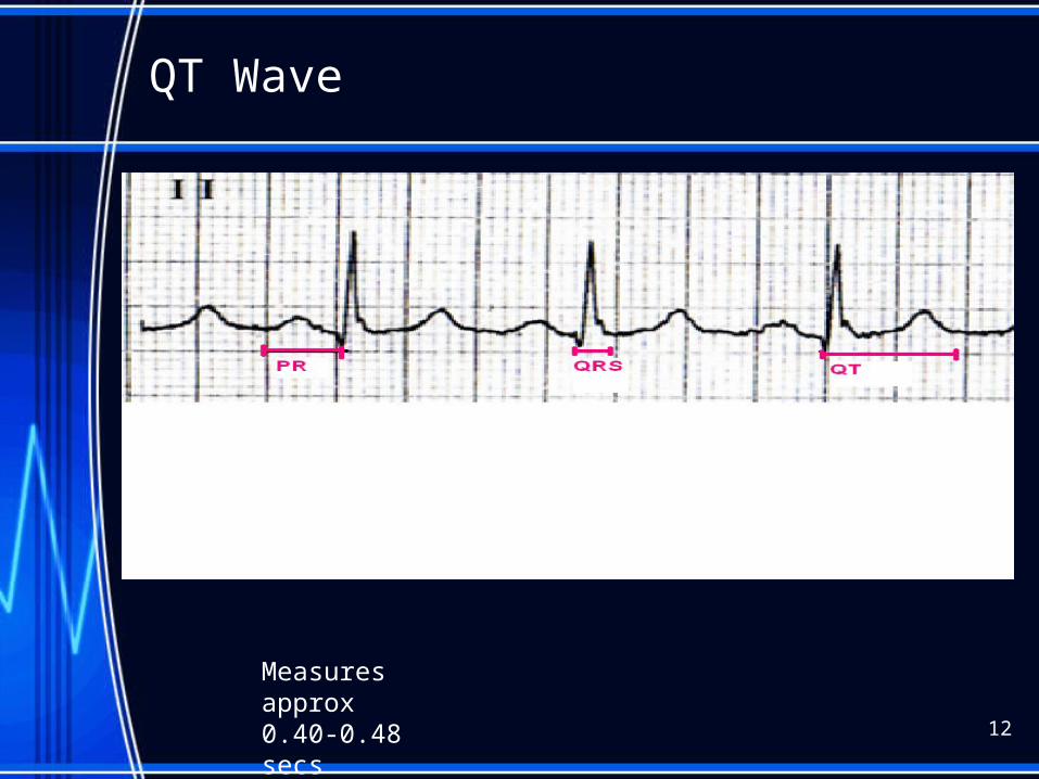
QT Wave
12
Measures approx 0.40-0.48 secs
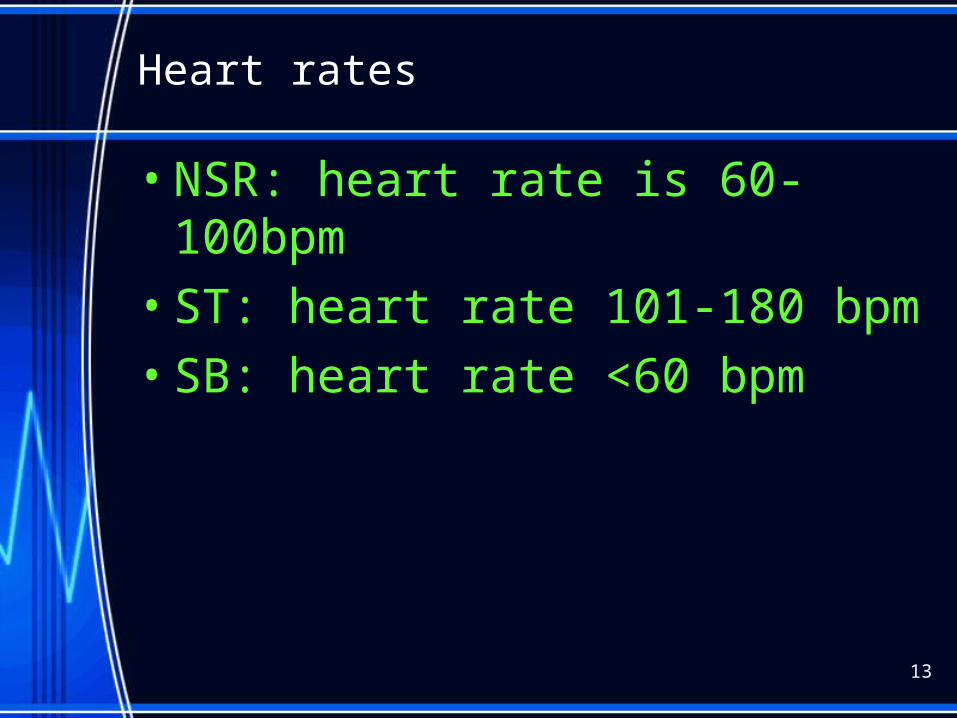
Heart rates
• NSR: heart rate is 60-100bpm
• ST: heart rate 101-180 bpm
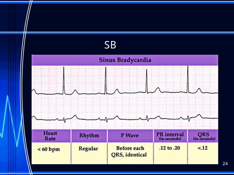
• SB: heart rate <60 bpm
13
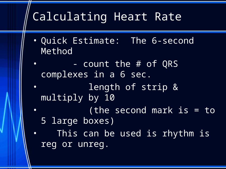
Calculating Heart Rate
• Quick Estimate: The 6-second Method• - count the # of QRS complexes in a 6 sec.• length of strip & multiply by 10• (the second mark is = to 5 large boxes)• This can be used is rhythm is reg or unreg.
• Count small boxes between two R waves. Divide into1500 Gives BPM
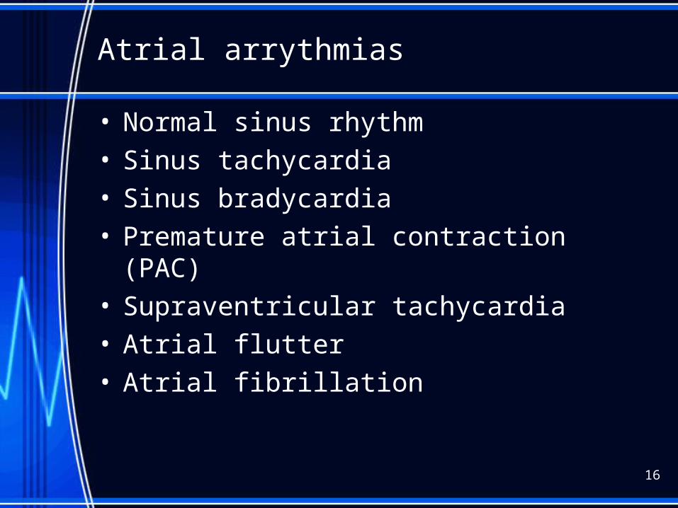
Atrial arrythmias
• Normal sinus rhythm• Sinus tachycardia• Sinus bradycardia• Premature atrial contraction (PAC)• Supraventricular tachycardia• Atrial flutter• Atrial fibrillation
16
Ventricular arrythmias
• Junctional rhythm• AV blocks• Premature junctional rhythm• Premature ventricular contraction (PVC)• Ventricular Tachycardia (V-tach)• Ventricular Fibrillation (V-Fib)• Torsade de Pointes (TdP)• Pulseless electrical activity (PEA)• Asystole
17
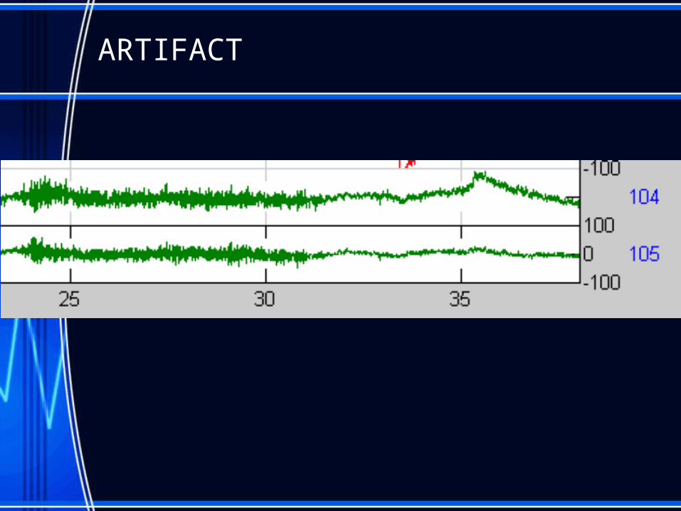
ARTIFACT
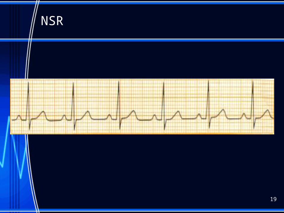
NSR
19
Sinus rhythm
• PR interval- 0.12-0.20sec• QRS-0.06-0.10sec• QT segment 0.36-0.44 sec • Heart rate 60-100
20
21
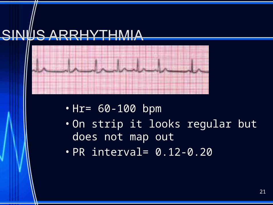
• Hr= 60-100 bpm• On strip it looks regular but does not map
out• PR interval= 0.12-0.20
22
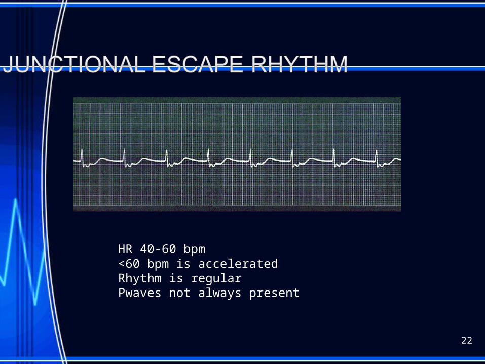
HR 40-60 bpm<60 bpm is acceleratedRhythm is regularPwaves not always present

Junctional Rhythm
23
SB
24
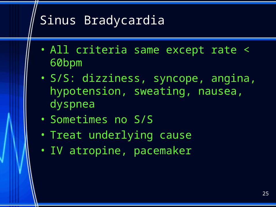
Sinus Bradycardia
• All criteria same except rate < 60bpm• S/S: dizziness, syncope, angina,
hypotension, sweating, nausea, dyspnea• Sometimes no S/S• Treat underlying cause• IV atropine, pacemaker
25
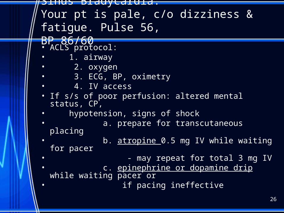
Sinus Bradycardia:Your pt is pale, c/o dizziness & fatigue. Pulse 56,BP 86/60• ACLS protocol:• 1. airway• 2. oxygen • 3. ECG, BP, oximetry• 4. IV access• If s/s of poor perfusion: altered mental status, CP,• hypotension, signs of shock• a. prepare for transcutaneous placing• b. atropine 0.5 mg IV while waiting for pacer• - may repeat for total 3 mg IV• c. epinephrine or dopamine drip while waiting pacer
or• if pacing ineffective
26
ST
27
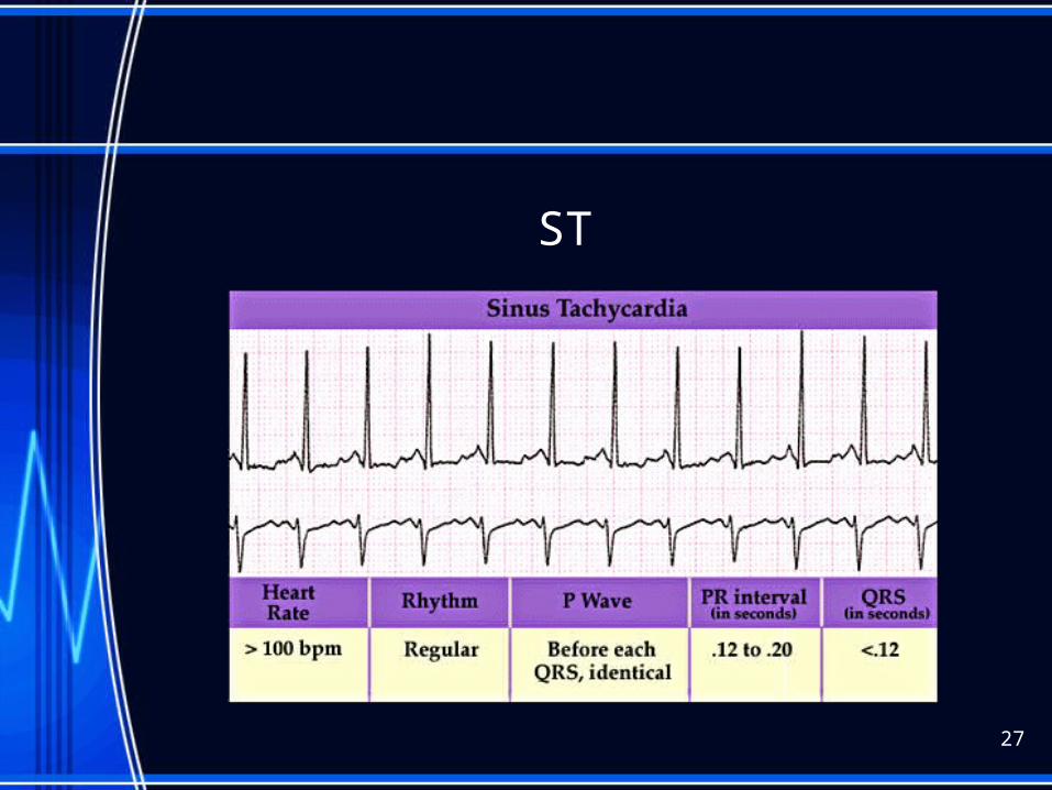
Sinus Tachycardia
• All criteria same as with NSR except rate >100• Causes: fever, dehydration, hypovolemia, increased
sympathetic nervous system stimulation, stress, exercise, AMI
• S/S: Palpations #1, angina and < CO from < V filling time
• Treatment: correct cause, eliminate caffeine, nicotine, alcohol. Beta blockers may be ordered
28

Sinus Tachycardia
• Heart rate greater than 100 but less 180• Caused by external influences (fever, blood• loss, exercise)• Adenosine used• B-blockers may cause condition to worsen ( if MI
limits vent function the heart will compensate by increasing rate then CO will fall)
• Remember to identify and treat cause !!!
29
Supraventricular Tachycardia
30
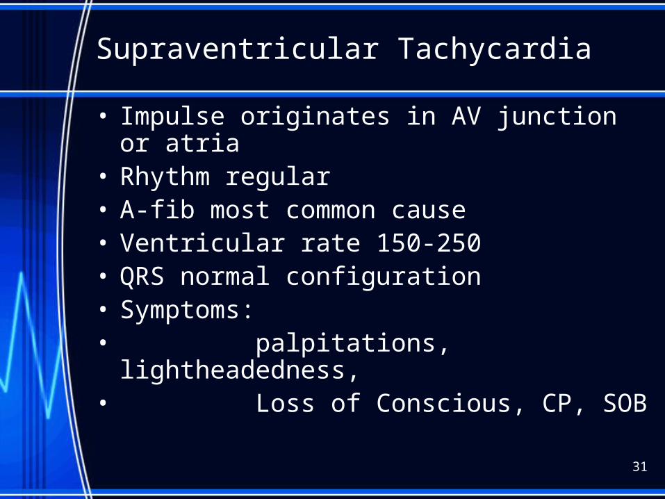
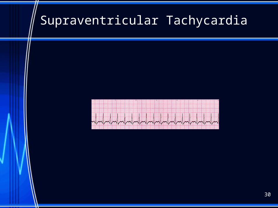
Supraventricular Tachycardia
• Impulse originates in AV junction or atria• Rhythm regular• A-fib most common cause• Ventricular rate 150-250• QRS normal configuration• Symptoms:• palpitations, lightheadedness,• Loss of Conscious, CP, SOB
31
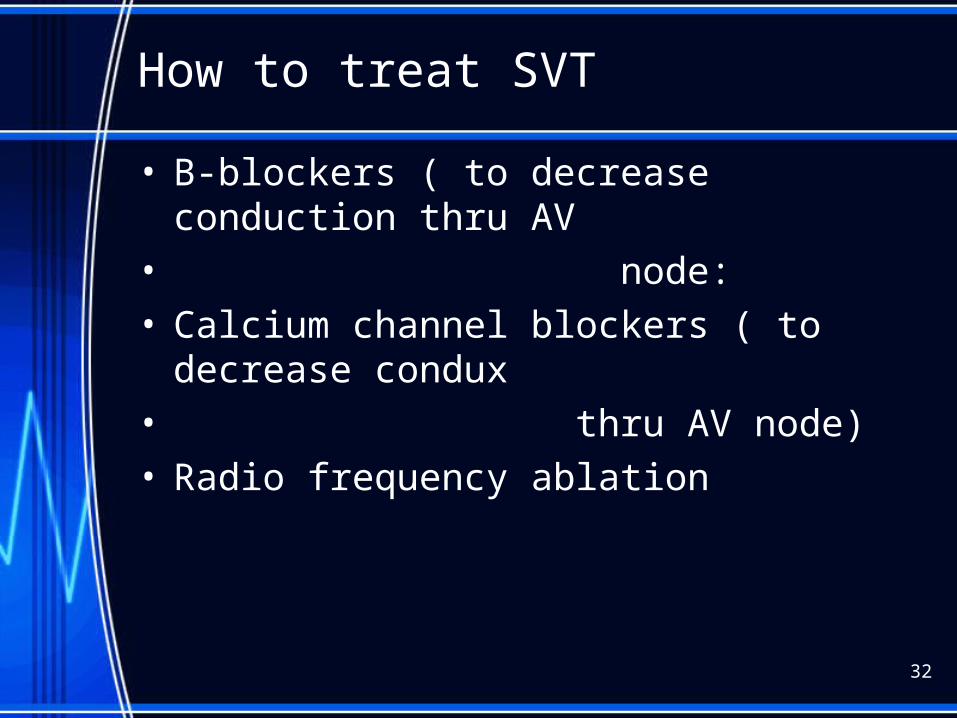
How to treat SVT
• B-blockers ( to decrease conduction thru AV • node:• Calcium channel blockers ( to decrease condux• thru AV node)• Radio frequency ablation
32
33
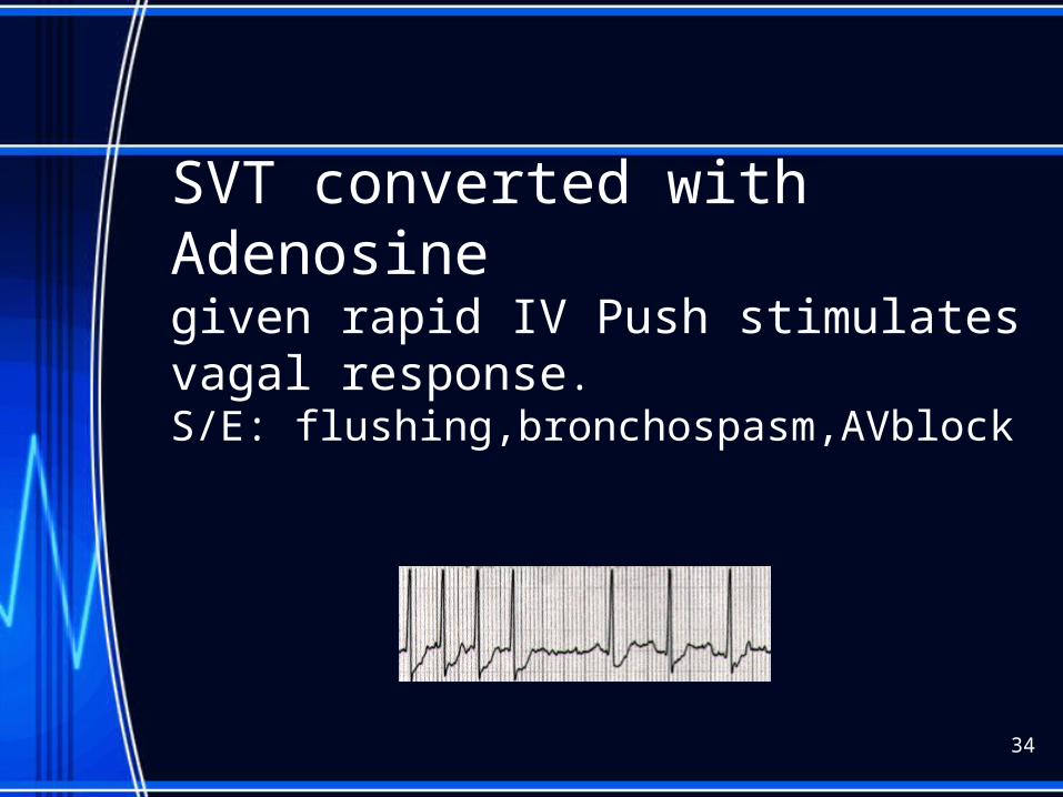
SVT converted with Adenosinegiven rapid IV Push stimulates vagal response. S/E: flushing,bronchospasm,AVblock
34
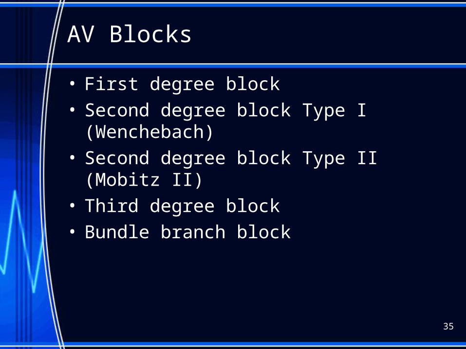
AV Blocks
• First degree block• Second degree block Type I (Wenchebach)• Second degree block Type II (Mobitz II)• Third degree block• Bundle branch block
35
36
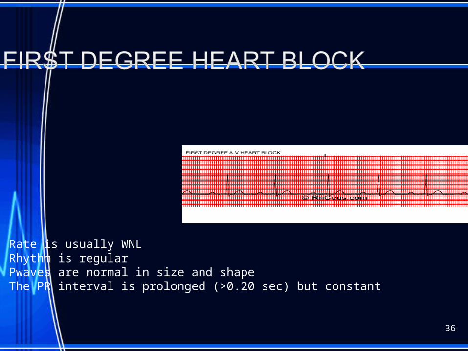
Rate is usually WNLRhythm is regularPwaves are normal in size and shape The PR interval is prolonged (>0.20 sec) but constant
1st degree block
• AV node delays the impulse from the SA node for abnormal length of time
• Causes:• CAD, MI, drugs that act on AV node
(digitalis)• Characteristics:• PR interval >0.20 seconds• Not serious but may progress to 2nd degree
37
1st degree block nursing intervention:
• Document the dysrhythmia• Monitor for progression to slower heart rate or
worsening block• If progression noted, monitor pt, notify
physician
38
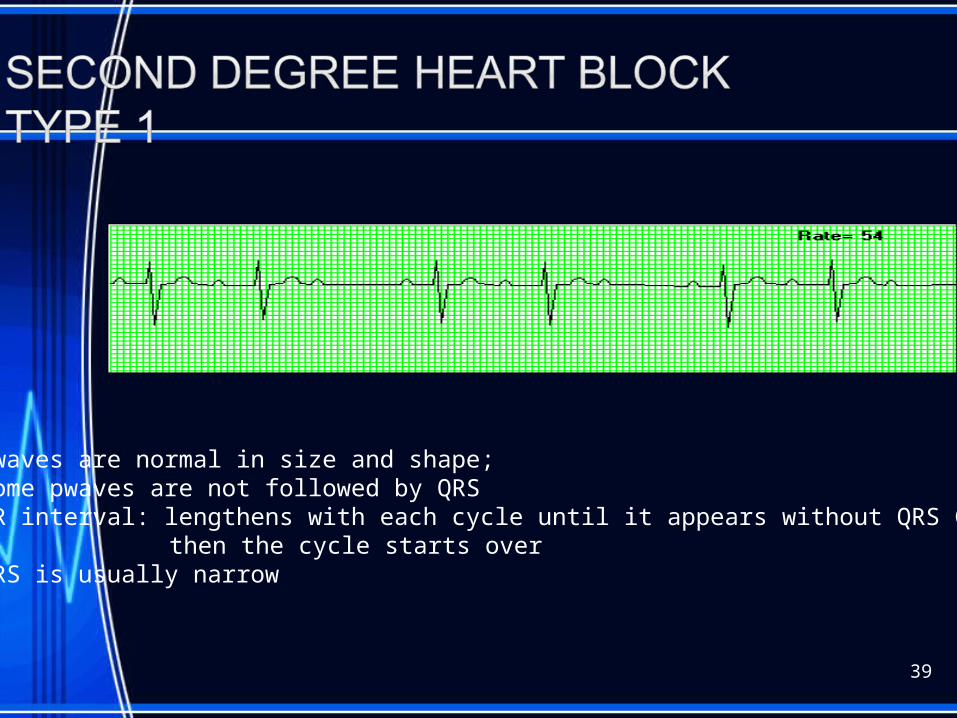
39
Pwaves are normal in size and shape; Some pwaves are not followed by QRSPR interval: lengthens with each cycle until it appears without QRS Complex
then the cycle starts overQRS is usually narrow
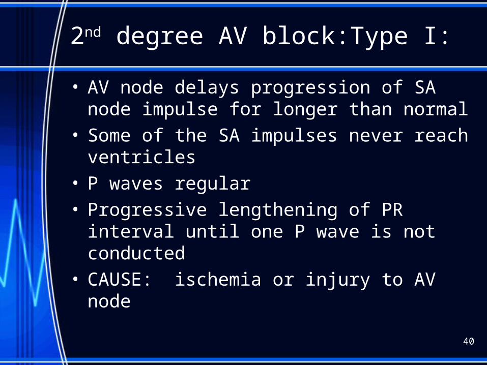
2nd degree AV block:Type I:
• AV node delays progression of SA node impulse for longer than normal
• Some of the SA impulses never reach ventricles• P waves regular• Progressive lengthening of PR interval until one P
wave is not conducted• CAUSE: ischemia or injury to AV node
40
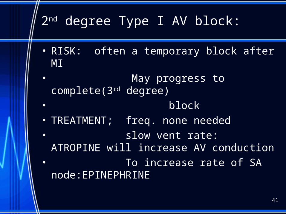
2nd degree Type I AV block:
• RISK: often a temporary block after MI• May progress to complete(3rd degree)• block• TREATMENT; freq. none needed• slow vent rate: ATROPINE will increase
AV conduction• To increase rate of SA
node:EPINEPHRINE
41
2ND degree nursing interventions:Type I• Document• Monitor pt/vitals• If ventricular rate slows enough to produce• symptoms, document , notify physician
42

http://www.youtube.com/watch?v=GVxJJ2DBPiQ&feature=related
44
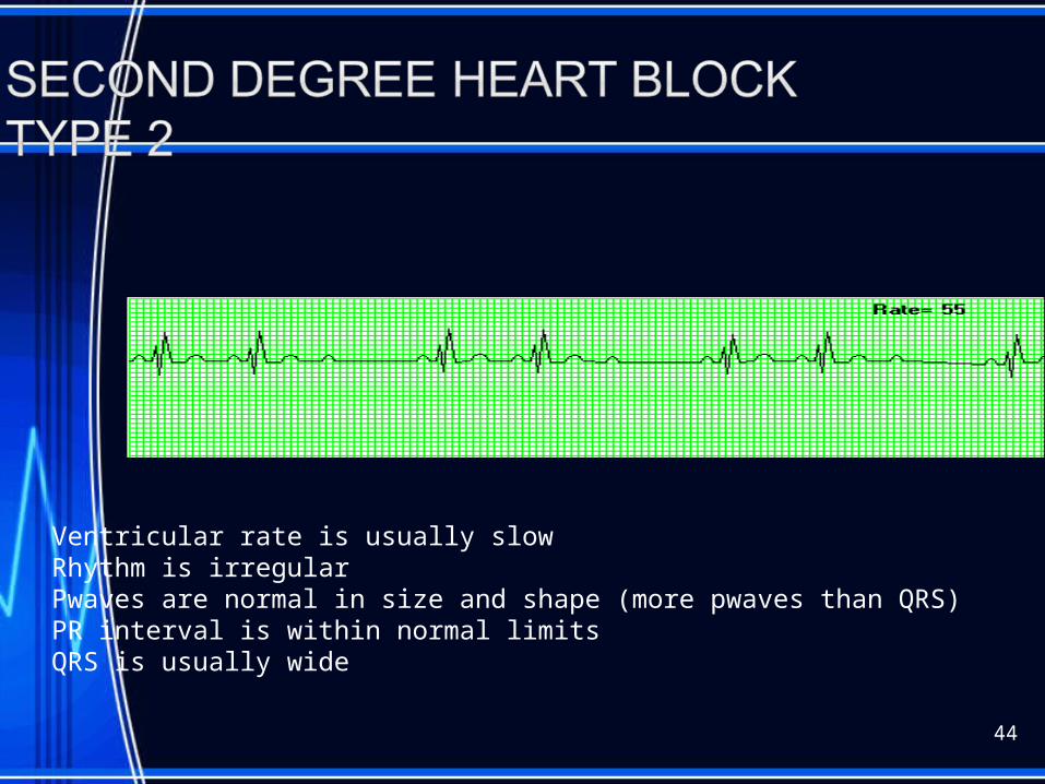
Ventricular rate is usually slowRhythm is irregularPwaves are normal in size and shape (more pwaves than QRS)PR interval is within normal limitsQRS is usually wide

2nd degree Type II(Mobitz Type II)• Atrial rate 60 to 100• More P waves than QRS complexes• Ventricular response 2:1 or 3:1• No change in PR intervals of conducted P waves• CAUSES: disease of AV node, AV junctional
tissue, or His-Purkinje system, inferior MI
45
2nd degree Type II:
• RISK: unpredictable & may suddenly advance to complete hrt block
• Especially common after inferior infarction• A DANGEROUS WARNING DYSRHYTHMIA• TREATMENT: if vent rate slow, atropine or
epinephrine• may need temporary pacer
46
2nd degree Type IINursing Interventions:
• Determine width of QRS• WATCH for widening QRS complex• *width QRS indicates location in the conduction
system of the block• - the wider the complex, the lower in the
bundle branch system the block will be.• IF QRS WIDENS, NOTIFY PHYSICIAN
IMMED.• Prepare for insertion of pacer• Assess vitals
47
48
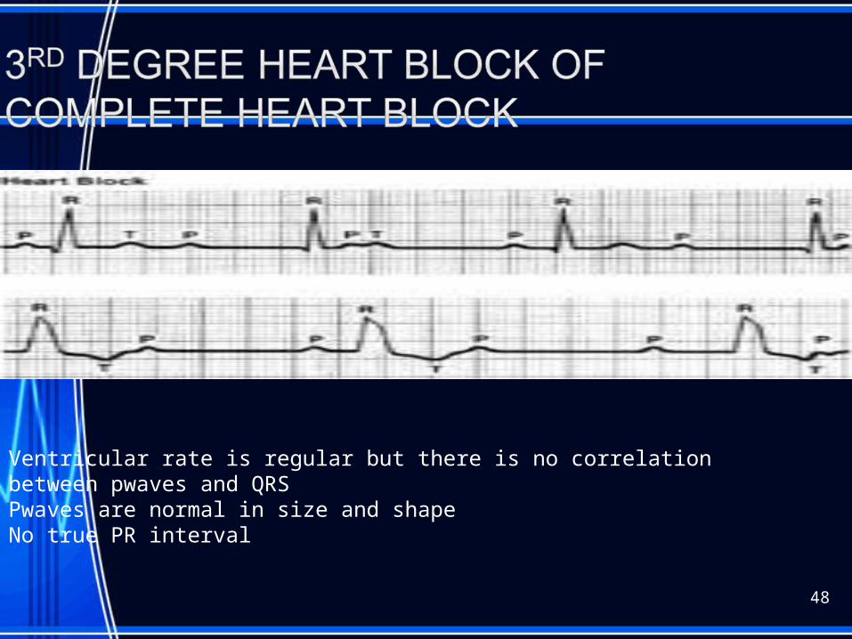
Ventricular rate is regular but there is no correlation between pwaves and QRSPwaves are normal in size and shapeNo true PR interval
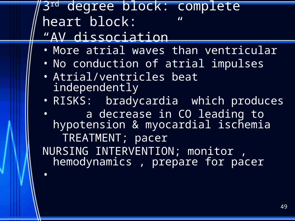
3rd degree block: complete heart block:“AV dissociation”
• More atrial waves than ventricular• No conduction of atrial impulses• Atrial/ventricles beat independently• RISKS: bradycardia which produces• a decrease in CO leading to hypotension
& myocardial ischemia TREATMENT; pacerNURSING INTERVENTION; monitor ,
hemodynamics , prepare for pacer•
49
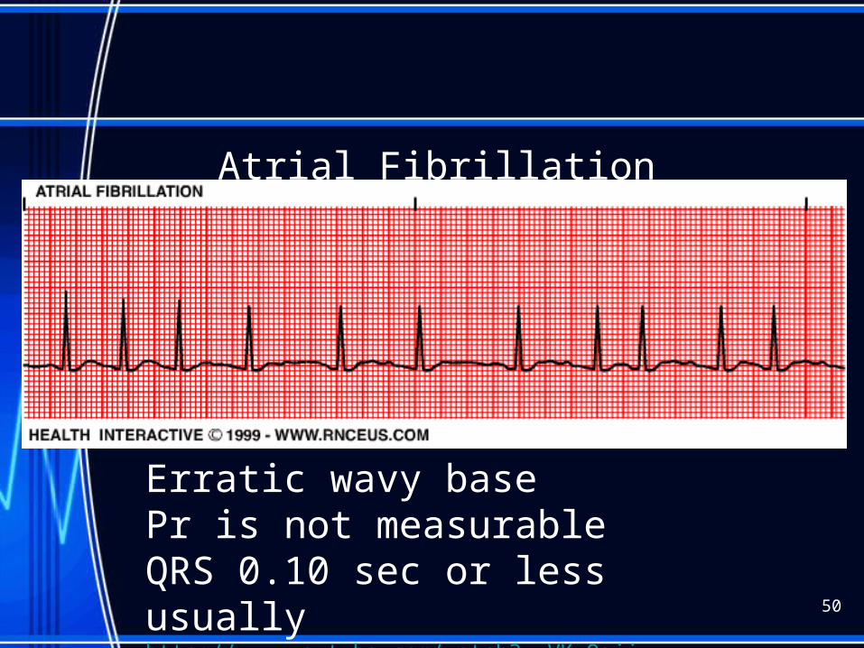
Atrial Fibrillation
50
Erratic wavy basePr is not measurableQRS 0.10 sec or less usuallyhttp://www.youtube.com/watch?v=VKxQgjj2yVU&feature=related
A fib continued
• Atrial rate > 400 bpm with a varying Ventricular rate• Overall rhythm irregular• No P waves, unable to measure PR interval• QRS=normal: Twave undeterminable• Causes: Rheumatic fever, mitral valve stenosis, cad.
HTN, MI, hyperthyroidism, COPD, CHF see pp. 604
51
A fib continued
• Concern with A fib is the development of atrial thrombus and loss of atrial kick from ineffective atrial function.
• Treatment: Ca channel blockers and anti- arrhythmics to convert, beta blockers to < HR, anticoagulants to prevent embolization.
• Synchronized cardioversion
52
Atrial Fibrillation
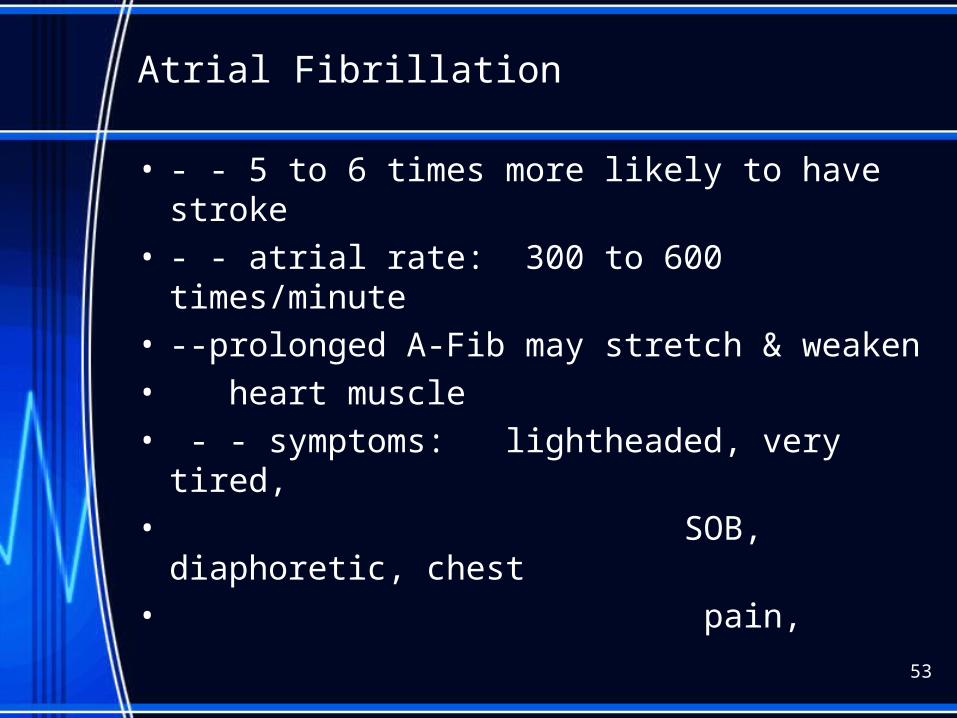
• - - 5 to 6 times more likely to have stroke • - - atrial rate: 300 to 600 times/minute• --prolonged A-Fib may stretch & weaken• heart muscle• - - symptoms: lightheaded, very tired,• SOB, diaphoretic, chest• pain,
53
Afib causes :
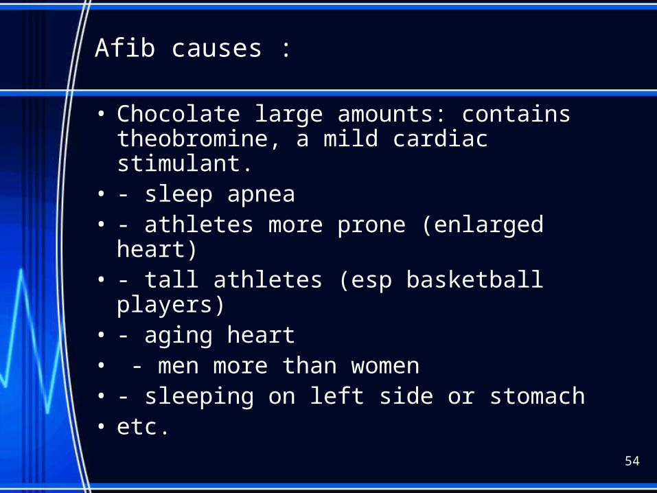
• Chocolate large amounts: contains theobromine, a mild cardiac stimulant.
• - sleep apnea• - athletes more prone (enlarged heart)• - tall athletes (esp basketball players)• - aging heart• - men more than women• - sleeping on left side or stomach• etc.
54

A-fib treatment:
• ASA not as effective as Coumadin in preventing strokes.
• ASA less likely to cause abnorm bleeding• **since hemorrhagic stroke increases with
age & is also increased by taking Coumadin, some Drs. may switch older pts from Coumadin to ASA.
55
A Fib electrical cardioversion:
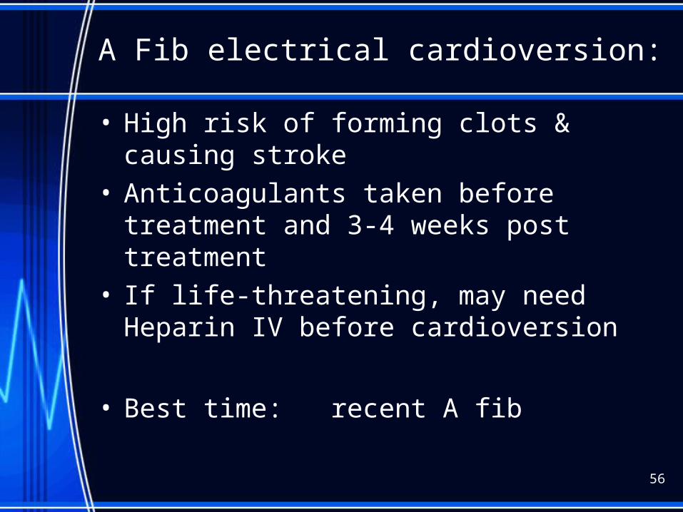
• High risk of forming clots & causing stroke• Anticoagulants taken before treatment and 3-
4 weeks post treatment• If life-threatening, may need Heparin IV
before cardioversion
• Best time: recent A fib
56
57
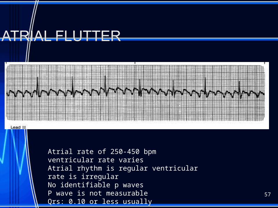
Atrial rate of 250-450 bpm ventricular rate variesAtrial rhythm is regular ventricular rate is irregularNo identifiable p wavesP wave is not measurableQrs: 0.10 or less usually

58
Pacer spike should fall before the P wave unless a dualChamber pacemaker; if it does not there could be a problem
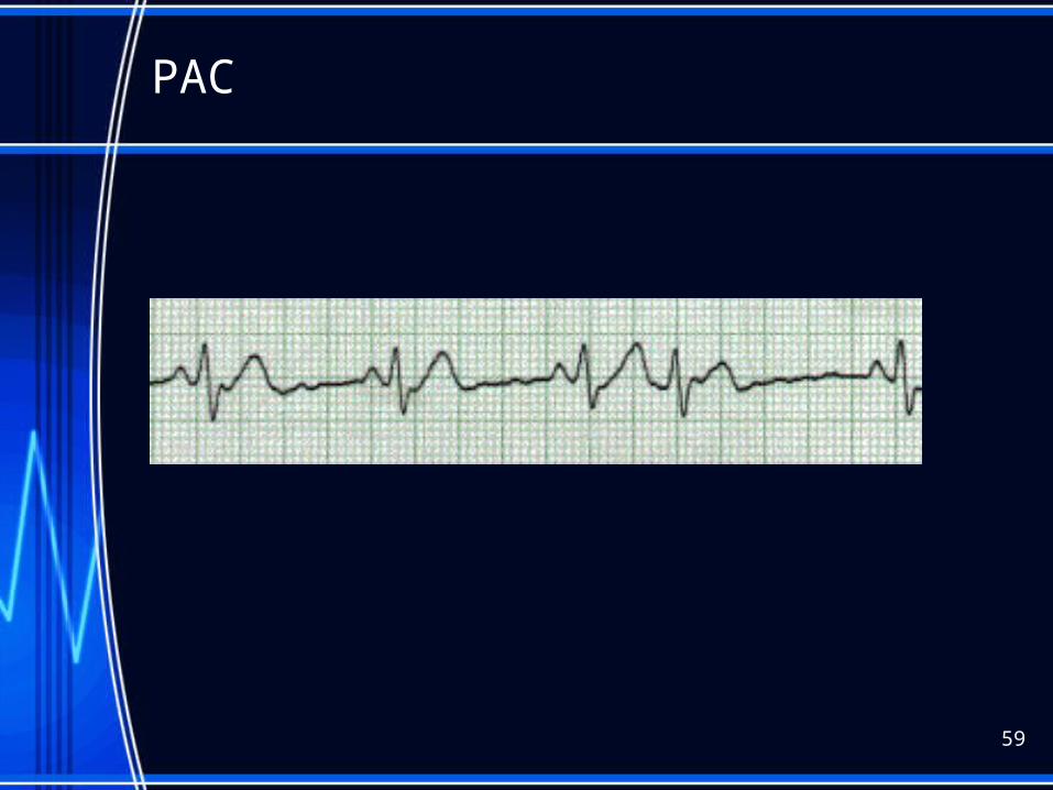
PAC
59
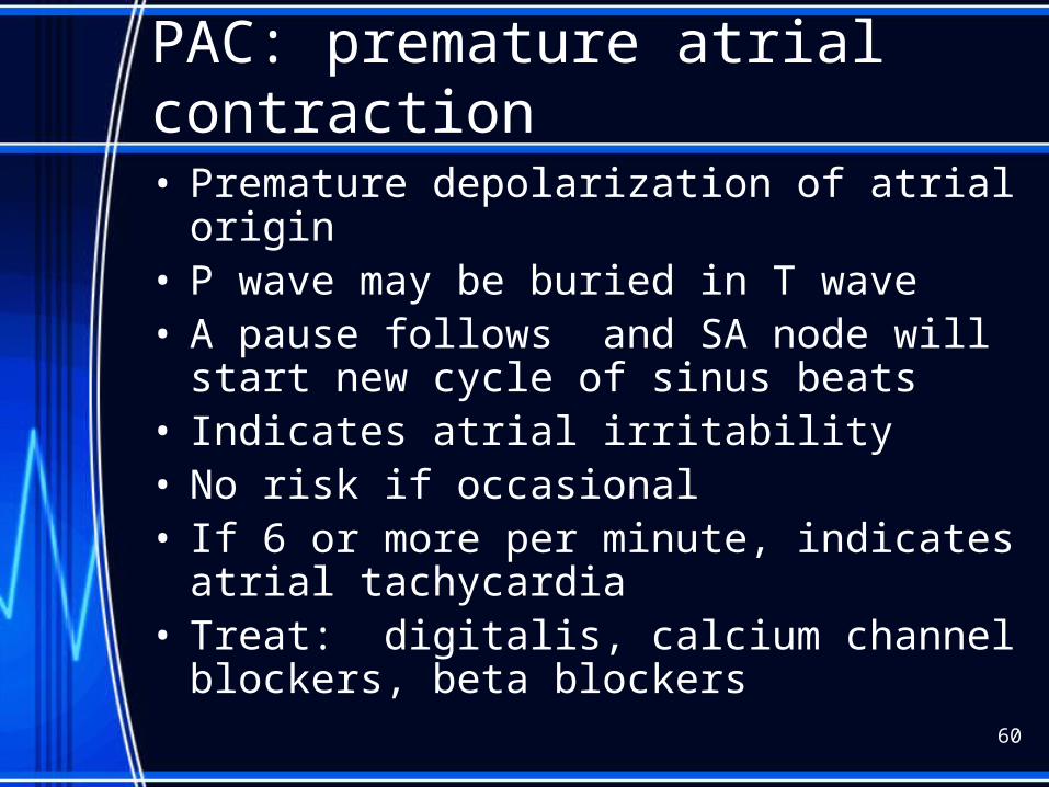
PAC: premature atrial contraction
• Premature depolarization of atrial origin• P wave may be buried in T wave• A pause follows and SA node will start new cycle
of sinus beats• Indicates atrial irritability• No risk if occasional• If 6 or more per minute, indicates atrial
tachycardia• Treat: digitalis, calcium channel blockers, beta
blockers
60
61
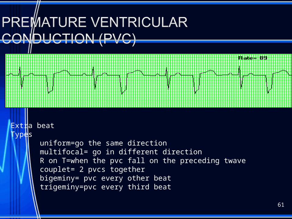
Extra beat Types
uniform=go the same directionmultifocal= go in different directionR on T=when the pvc fall on the preceding twavecouplet= 2 pvcs togetherbigeminy= pvc every other beattrigeminy=pvc every third beat

PVCs (unifocal)
62

PVCs (multifocal)
63
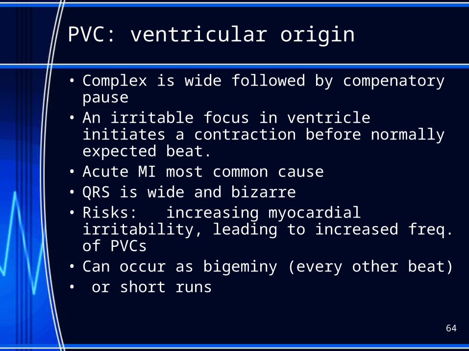
PVC: ventricular origin
• Complex is wide followed by compenatory pause
• An irritable focus in ventricle initiates a contraction before normally expected beat.
• Acute MI most common cause• QRS is wide and bizarre• Risks: increasing myocardial irritability,
leading to increased freq. of PVCs• Can occur as bigeminy (every other beat)• or short runs
64
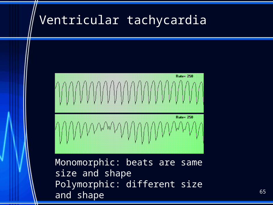
Ventricular tachycardia
65
Monomorphic: beats are same size and shapePolymorphic: different size and shape
V-tach
• Advanced irritability of ventricles due to ASHD, CHF, acute MI electrolye imbal. Hypoxia, acidosis,occas drugs
• RISKS: low to no Cardiac output• Nursing Interventions: monitor, if pt
unconscious,immed. defib
66
67
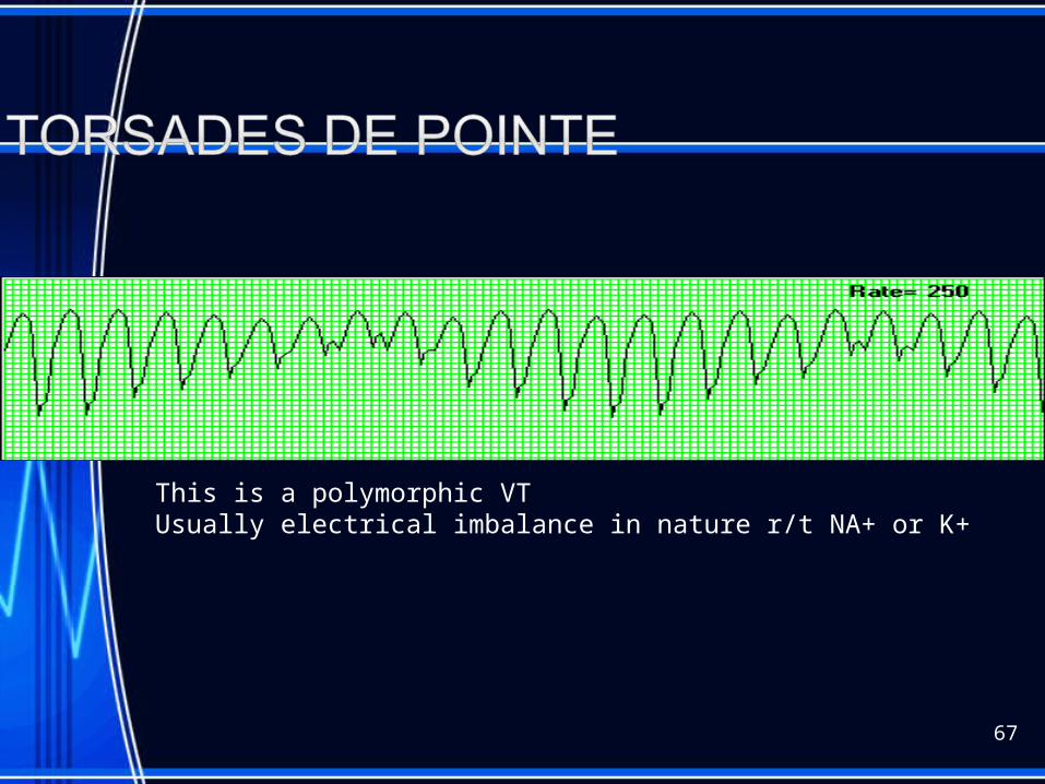
This is a polymorphic VTUsually electrical imbalance in nature r/t NA+ or K+
68
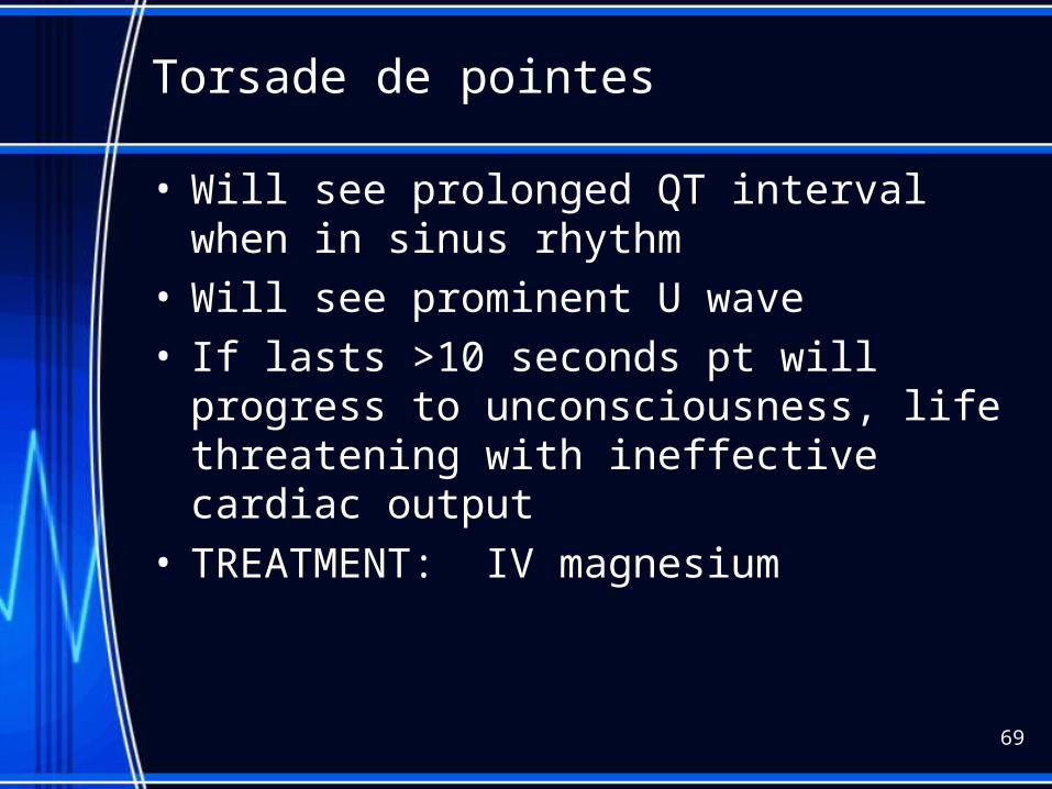
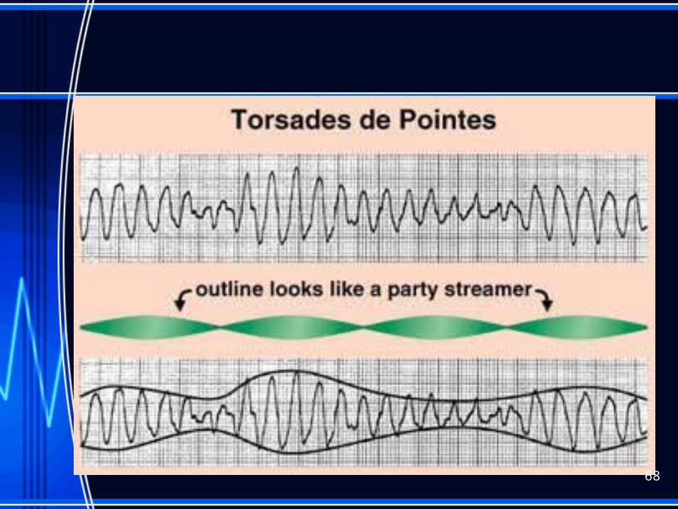
Torsade de pointes
• Will see prolonged QT interval when in sinus rhythm
• Will see prominent U wave• If lasts >10 seconds pt will progress to
unconsciousness, life threatening with ineffective cardiac output
• TREATMENT: IV magnesium
69
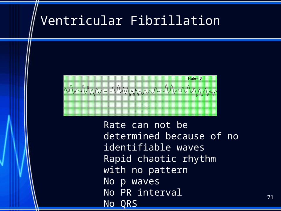
Ventricular Fibrillation
71
Rate can not be determined because of no identifiable wavesRapid chaotic rhythm with no patternNo p wavesNo PR intervalNo QRS
Vtach/Vfib
• Both can be life threatening• VT= V HR 100-250 bpm• Causes: AMI, CAD, hypokalemia, dig toxic• S/S: palpitations, dizzy, angina, <LOC• Treatment: assess for pulse, if none, defib• VF=Rate undeterminable Cause: same• Treatment: CPR
72
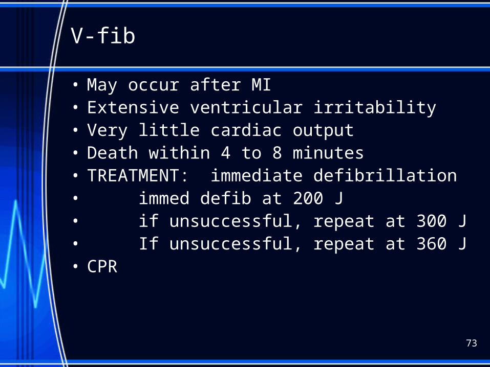
V-fib
• May occur after MI• Extensive ventricular irritability• Very little cardiac output• Death within 4 to 8 minutes• TREATMENT: immediate defibrillation• immed defib at 200 J• if unsuccessful, repeat at 300 J• If unsuccessful, repeat at 360 J• CPR
73
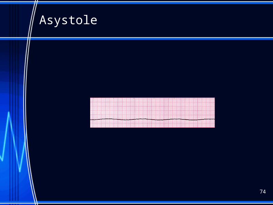
Asystole
74

Asystole and PEA
• CPROxygen
• Epinephrine 1 mg IV/IO (repeat 3-5 minutes)• May give Vasopressin 40U IV/IO to replace• 1st or 2nd dose of epinephrine• Consider Atropine 1 mg IV/IO Repeat every 3 to 5
min (up to 3 doses)
75
What arrthymias are considered PEA?
• See an organized or semi-organized rhythm BUT NO PULSE:
• This includes:• - idioventricular rhythms• - ventricular escape beats• - postdefibrillation
idioventricular•
76

http://www.campaignfornursing.com/events/WINNERS/pennsylvania/
77
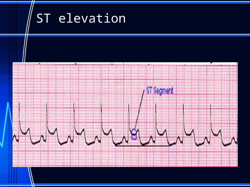
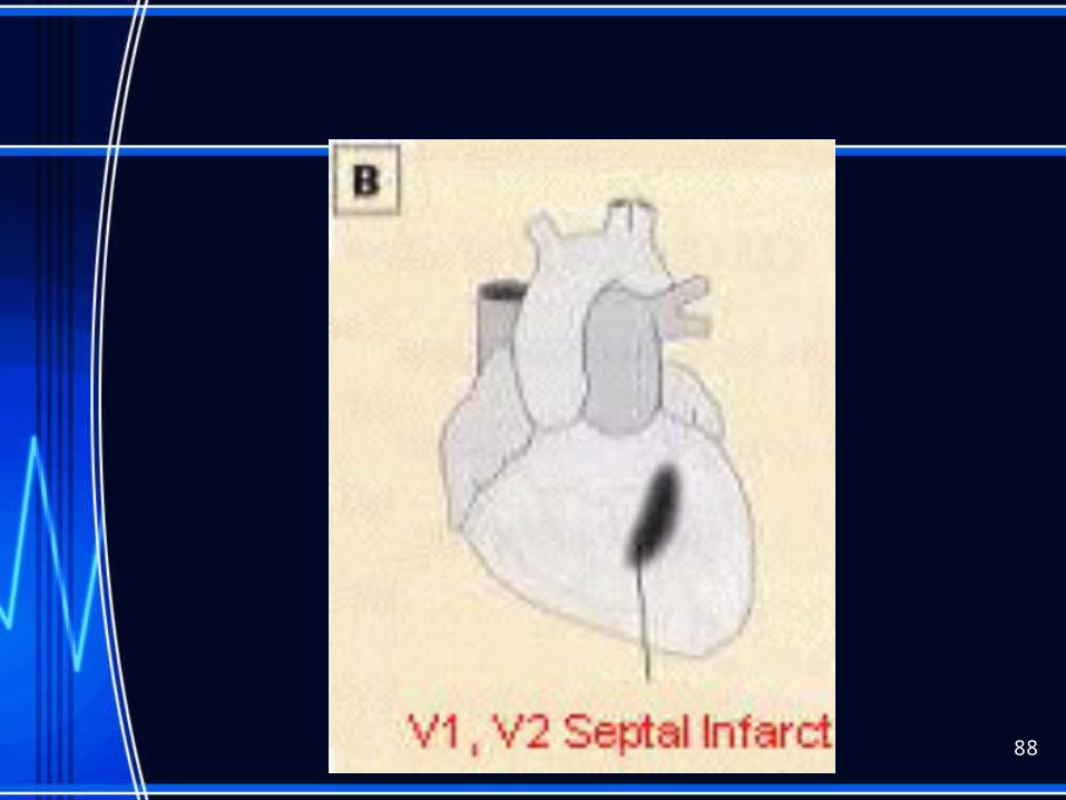
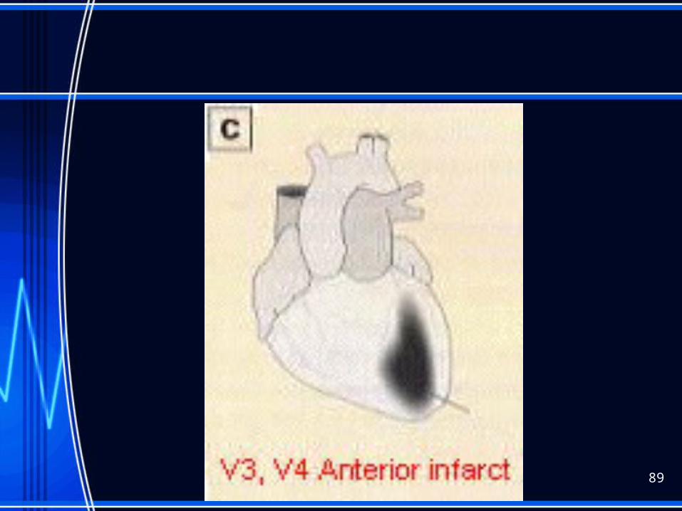
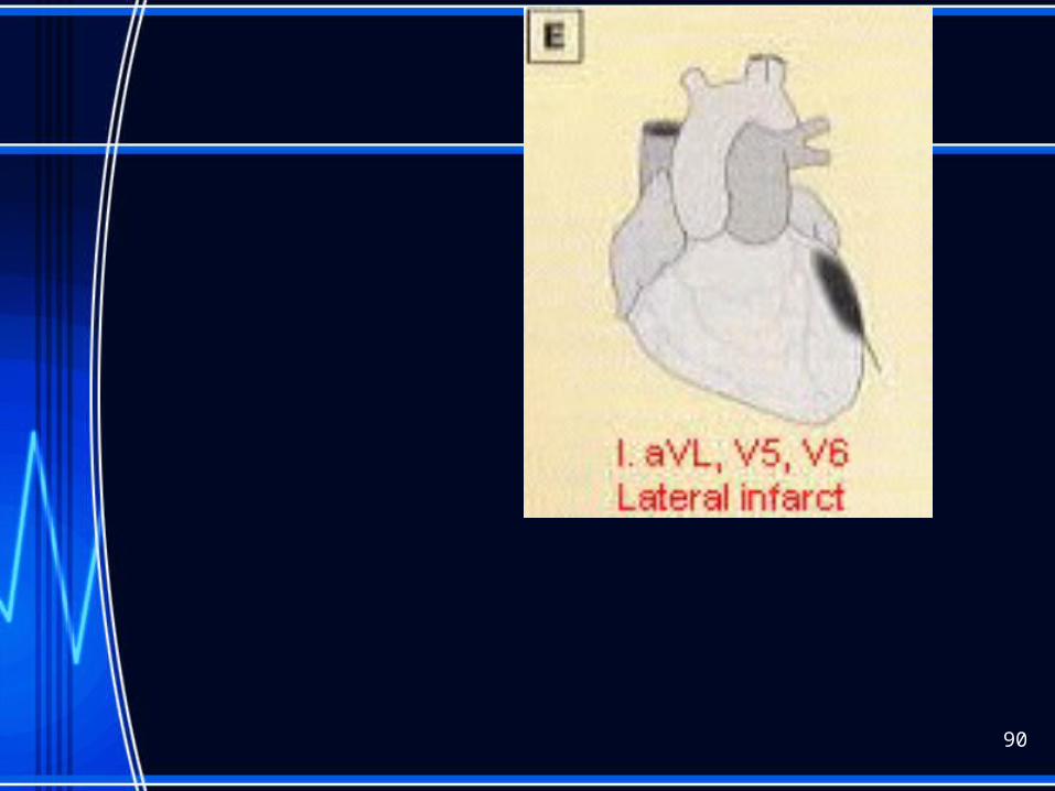
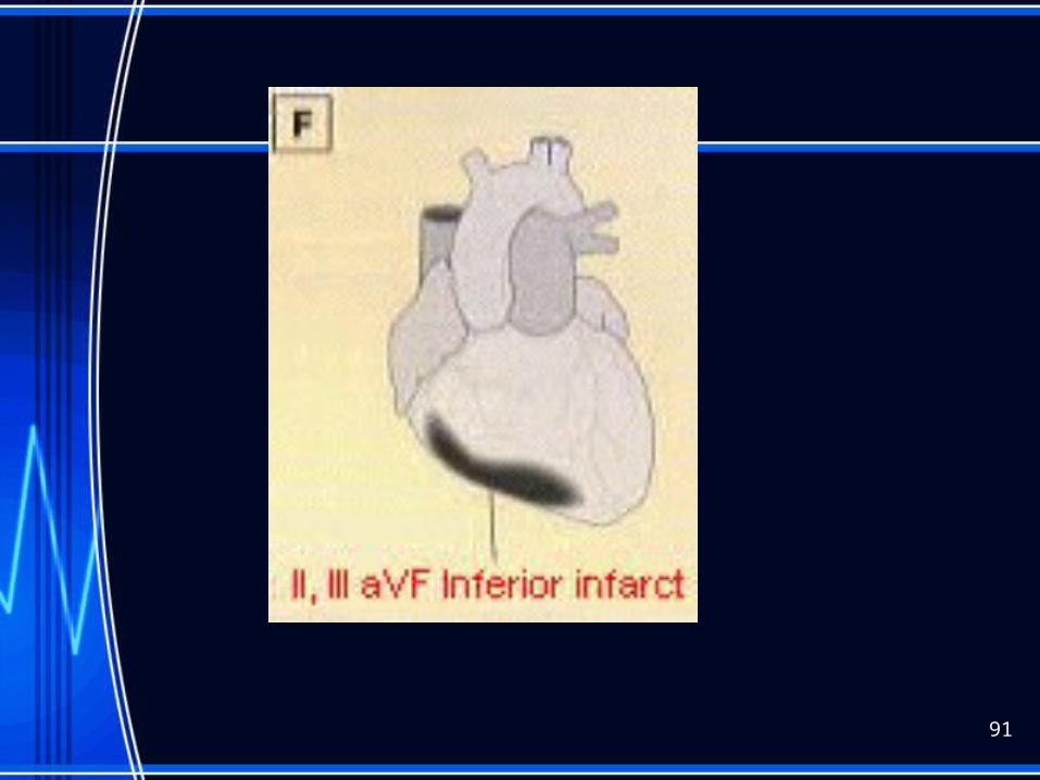
ST elevation
12 lead ekg
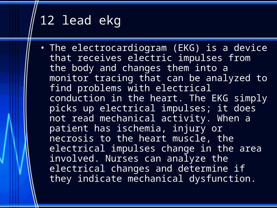
• The electrocardiogram (EKG) is a device that receives electric impulses from the body and changes them into a monitor tracing that can be analyzed to find problems with electrical conduction in the heart. The EKG simply picks up electrical impulses; it does not read mechanical activity. When a patient has ischemia, injury or necrosis to the heart muscle, the electrical impulses change in the area involved. Nurses can analyze the electrical changes and determine if they indicate mechanical dysfunction.
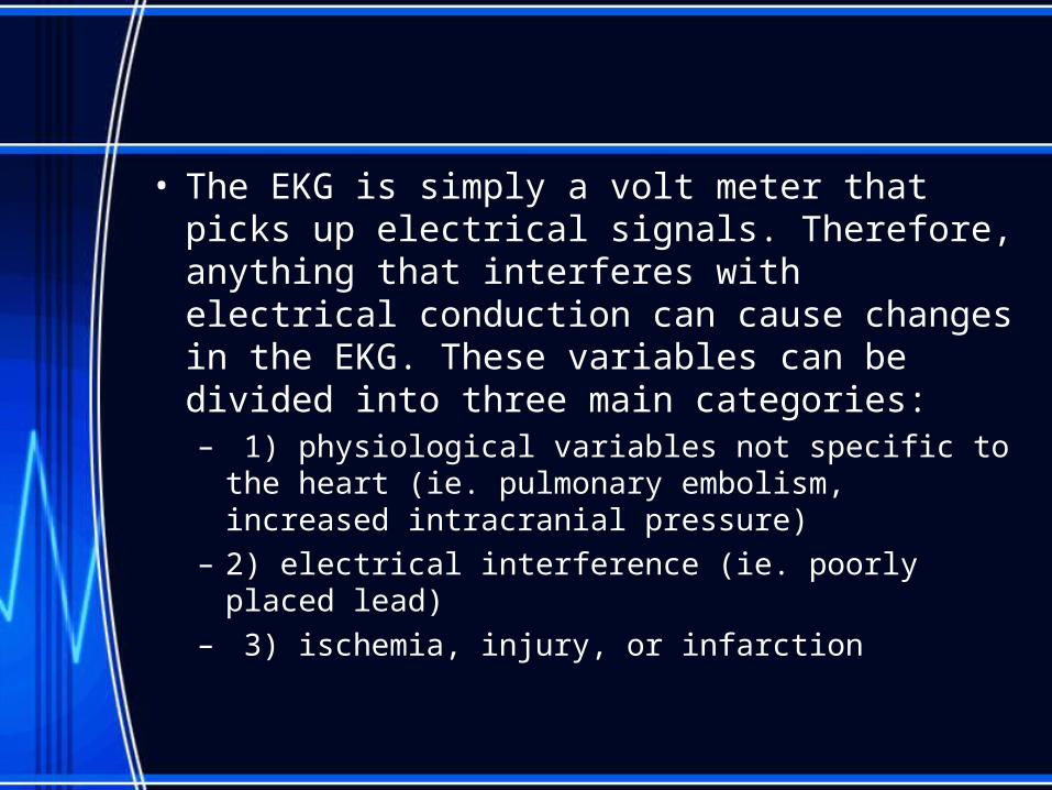
• The EKG is simply a volt meter that picks up electrical signals. Therefore, anything that interferes with electrical conduction can cause changes in the EKG. These variables can be divided into three main categories:– 1) physiological variables not specific to the heart
(ie. pulmonary embolism, increased intracranial pressure)
– 2) electrical interference (ie. poorly placed lead)– 3) ischemia, injury, or infarction
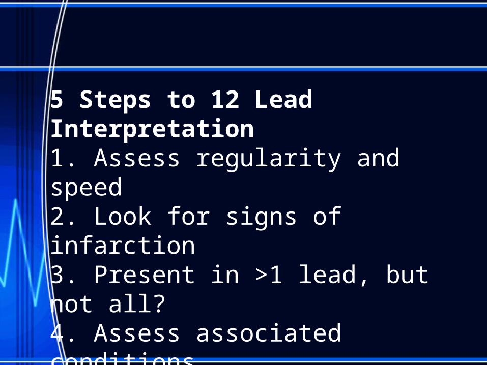
5 Steps to 12 Lead Interpretation1. Assess regularity and speed2. Look for signs of infarction3. Present in >1 lead, but not all?4. Assess associated conditions5. Correlate with clinical condition
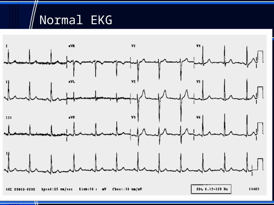
Normal EKG
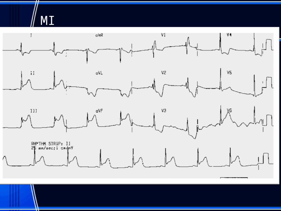
MI
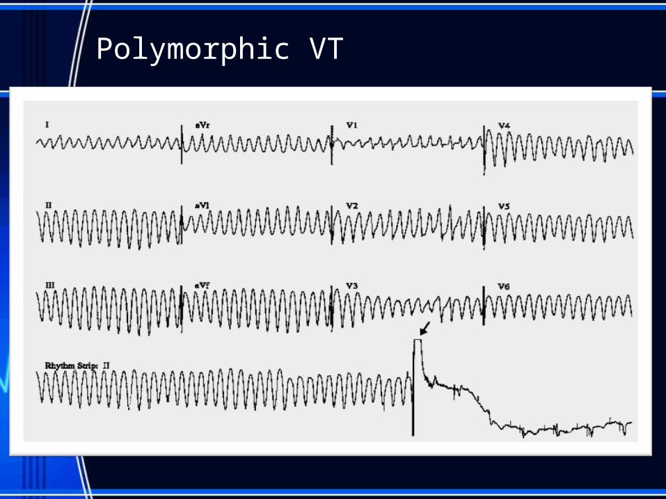
Polymorphic VT
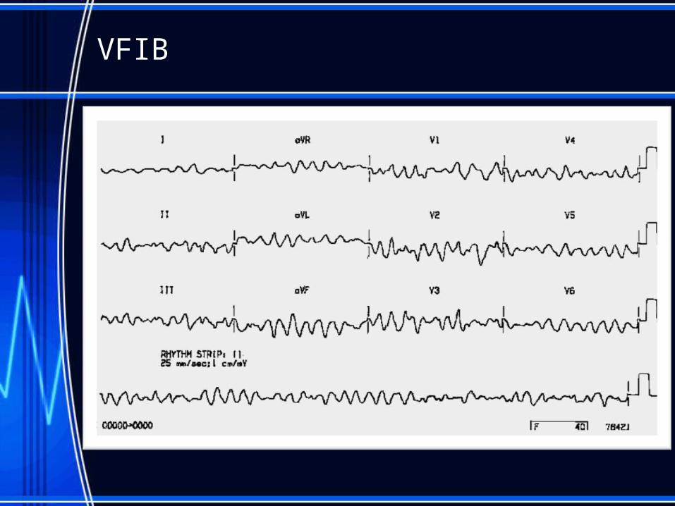
VFIB

88
89
90
91
• http://nursebob.com/
• http://www.usfca.edu/fac_staff/ritter/ekg.htm
• http://ems-safety.com/12-lead-ekg.htm

Rhythms for Cardioversion
• A-fib• A-flutter• Supraventricular tachycardia
93
Electrical cardioversion
• treatment of choice for hemodynamically unstable tachydysrhythmia
• It is used for the treatment of unstable ventricular tachycardia with a pulse
• With cardioversion potentially prevents life-threatening dysrhythmias.
• Cardioversion may either be a planned or emergent procedure.
• Properly done cardioversion will correct the patient’s dysrhythmia with minimal discomfort and maximum safety.
Chemical cardioversion
• IndicationA. Rapid conversion of atrial fibrillation and atrial flutter.B. Ibutilide is moderately effective in patients who have atrial flutter.
• 2. Action.A. Ibutilide prolongs action potential duration.B. Blocks the rapidly activating component of the delayed rectifier potassium
current.C. No significant effect on heart rate, PR interval, or QRS intervalD. Route of elimination: hepatic.
• 3. Administration.A. Ibutilide is available in 10 mL vials containing 0.1 mg/mL (1 mg total).B. For intravenous administration, the recommended dose of Ibutilide is 1mg
over a 10 minute period in patients weighing > 60 kgC. Patients weighing < 60 kg, the recommended dose is 0.01 mg/kg initially, with
a second dose of the same strength 10 minutes later if necessary.D. Ten minutes after the end of the initial infusion, a second 10 minute infusion
of equal strength can be given if the arrhythmia has not terminated.
• Cautions.A. Prolong ventricular repolarizationB. Carries a risk of excessive QT prolongationC. Acquired long-QT syndrome D. Associated polymorphic ventricular tachycardia (torsade de
pointes)E. Careful patient selection and clinical monitoring during drug
administration.• 5. Contraindications.
A. QT interval exceeding 440msB. BradycardiaC. Electrolyte disturbancesD. Other QT-prolonging drugs
• 6. Adverse Effects.A. Ventricular tachycardiaB. Premature ventricular complexesC. HypotensionD. Bundle branch blockE. Atrioventricular
• Post cardioversion care:• 1. generally the care for a patient is the same
as for defibrillation.• 2. If it is a elective procedure, digoxin is
withheld for 48 hours prior to prevent dysrhythmias after the procedure.
• 3. Assess airway and LOC
Indications for pacemaker
• Temporary:• -symptomatic bradycardia (not controlled
by meds)• - ant MI• - drug overdose (dig, beta blocker)• Permanent:• - 2nd degree Mobitz Type II• - 3rd degree Block• - symptomatic bradycardia, arrhythmias• - suppress tachyarrythmias
98
Modes of Pacing
• Synchronous (demand )Mode• - sensitivity is set to patient beats• - pacer will fir when pt rate goes below• that what is set• Asynchronous pacing:• - for asystole, or profound bradycardia• - does not sense any pt beats• - fires at set rate no matter what pt rate is
99
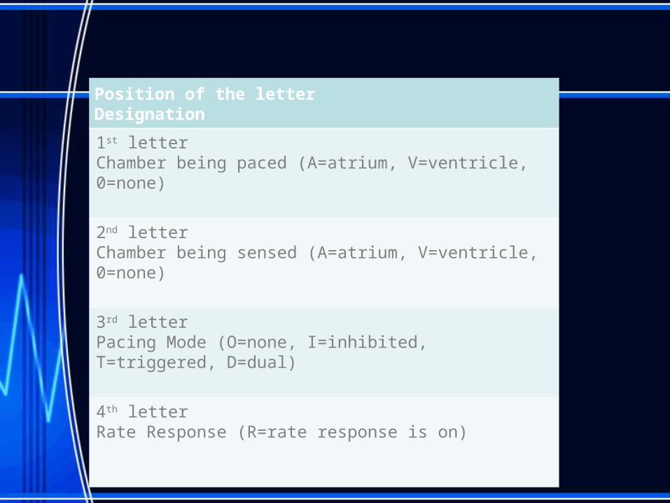
Position of the letterDesignation
1st letterChamber being paced (A=atrium, V=ventricle, 0=none)
2nd letterChamber being sensed (A=atrium, V=ventricle, 0=none)
3rd letterPacing Mode (O=none, I=inhibited, T=triggered, D=dual)
4th letterRate Response (R=rate response is on)
Chambers that can be paced:
AtriumVentricleDual (both atrium and ventricle)ICD (Implantable Cardioverter
Defibrillator)
101
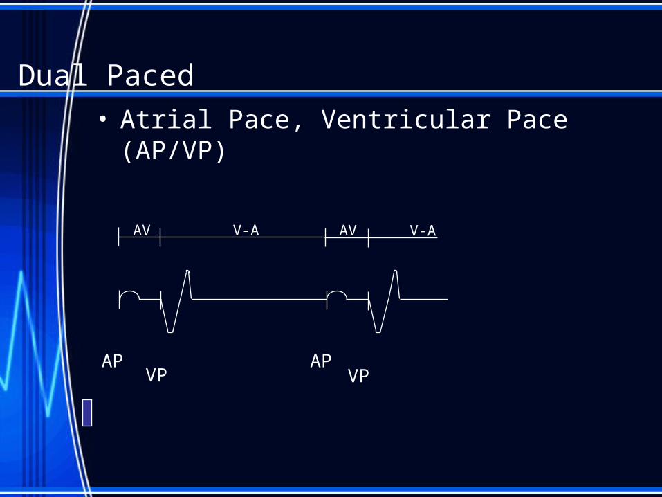
APVP
APVP
V-AAV V-AAV
Dual Paced• Atrial Pace, Ventricular Pace (AP/VP)
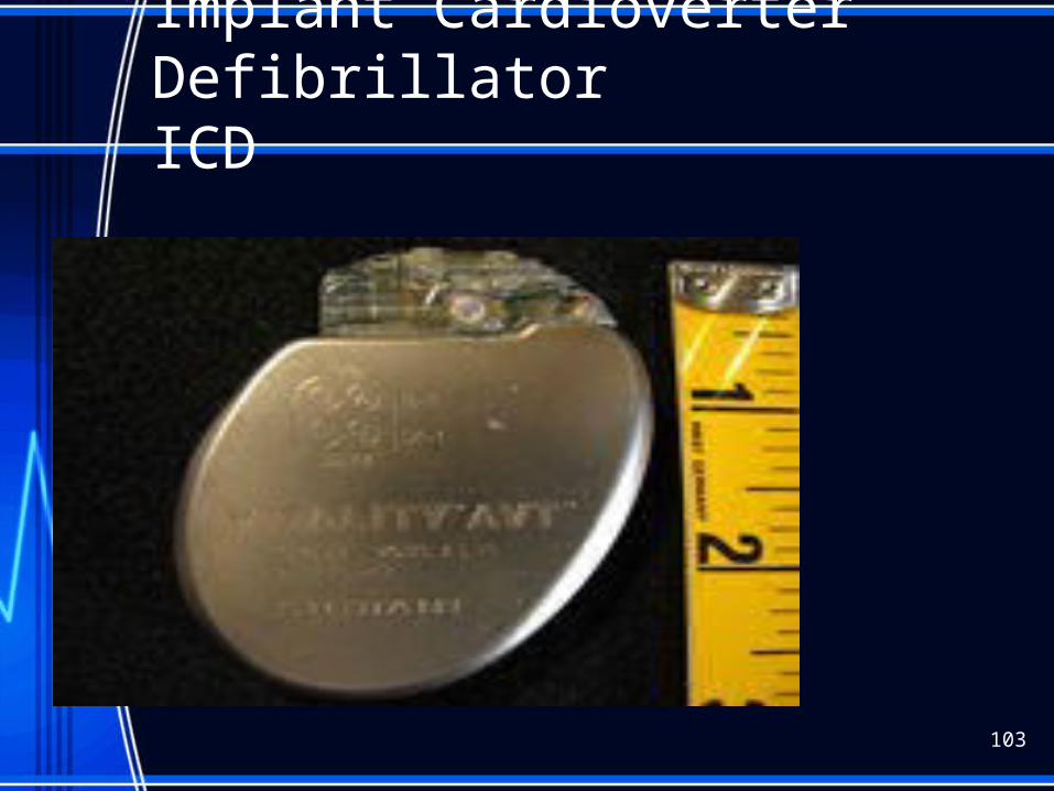
Implant Cardioverter Defibrillator ICD
103
ICD
• - prevents sudden cardiac death due to • V-tach or V-fib.• Pt can feel the shock• -defib felt like “kick in the chest”• that lasts 1 second• - cardiovert feels like “thump in chest• - pt doesn’t feel pacing
104
Problems with pacers
• Failure to fire• Failure to capture• Undersensing (low battery, poor lead • position)• Oversensing (turn down output,
magnetic• interference)
105
Operative failures with pacers:
• Pneumothorax• Pericarditis• Infection• Hematoma• Lead dislodgement (seen on X-ray)• Venous thrombosis (rare but would see• unilateral edema to arm on same
side• as pacer)
106
Pt Education:
• 1. carry ID card (Xray code seen in standard chest Xray)• 2. not allowed to drive for 1 month• 3. no metal detectors or no longer than nec.• 4. MRI interrupts pacing-can’t get one for some time if
new• 5. No power generators (welding)• 6. microwave questionable• 7. radiotherapy (may damage circuits) The• pacer may need to be surgically moved if in• path of radiation field.• 8. TENS (transcutaneous electrical stimulation)
interferes• may need reprogramming• 9. Cell phone use in opposite ear of pacer and store
away• from side of pacer
107
EP with Ablation
An electrophysiology study is simply a study of the electrical function of your heart.
• A (IV) catheter may be placed and be used to continuously administer fluids.
• An EP team doctor will explain why the procedure is necessary and what risks are involved for you.
• Obtain consent
• Prior to the EP study, CHG Bath performed.
• The most common site used is the groin, or the area at the crease of the leg about midway between the center of your body and your hip. Occasionally the forearm, neck or collarbone areas are used
• NPO after midnight the night before the test.
• If the test is not scheduled until later in the day, may have a clear liquid breakfast
• All your medicines will be reviewed, and some may be withdrawn prior to the test.
• . It is important for pt to describe sensations experienced during the test.
• Dentures and Glasses may be worn
• An initial EP study takes an average of two and a half hours; however, they may range from one to six hours.
• After the test, the catheters will be removed. Firm pressure will be applied over the puncture site for approximately 15 minutes.
• Flat bed rest is necessary for two to eight hours after the study. Assessment of site needs performed.
• You will be in the room frequently during the first hour after the study to take blood pressure, heart rate, and check the insertion site for signs of bleeding. The pulses and temperature of the feet will also be checked.
• Pt will be instructed to apply pressure firmly to the insertion site if cough or sneeze and while using the bedpan or urinal.
• Administer pain meds as needed for discomfort
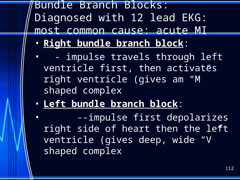
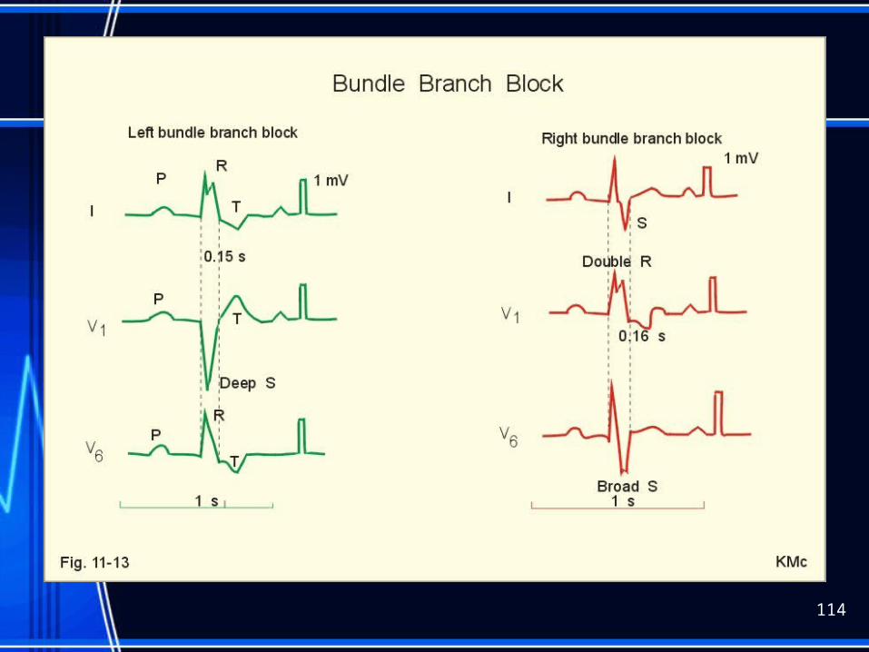
Bundle Branch Blocks: Diagnosed with 12 lead EKG: most common cause: acute MI• Right bundle branch block:• - impulse travels through left ventricle first,
then activates right ventricle (gives am “M” shaped complex
• Left bundle branch block:• --impulse first depolarizes right side of
heart then the left ventricle (gives deep, wide “V” shaped complex
112
Bundle Branch Blocks:
• Risks: can deteriorate to 3rd degree block• then treat with atropine or pacemaker• Pt can be asymptomatic until progresses
113
114
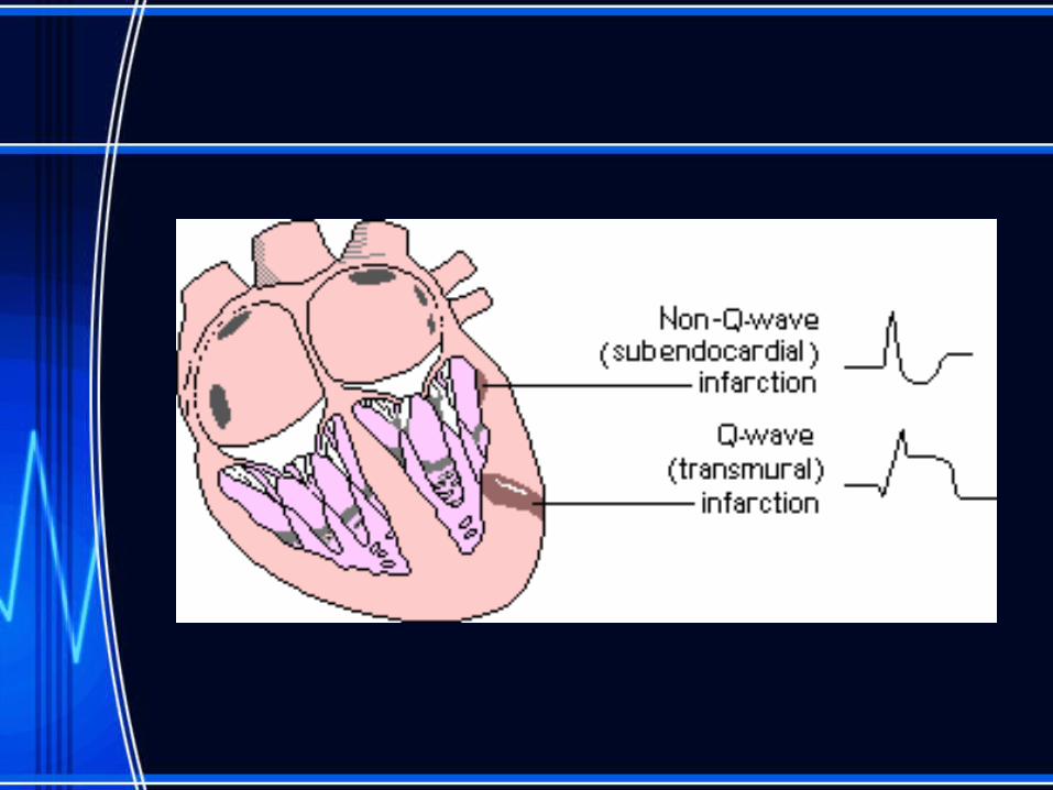
Hyperkalcemia
115
Intro to ACLS
Primary Survey
• Airway: Open airway, look, listen, and feel for breathing
• Breathing: If not breathing slowly give 2 rescue breaths. If breaths go in continue to next step.
• Circulation: Check the carotid artery (Adult) for a pulse. If no pulse begin CPR.
• Defibrillation: Search for and Shock V-Fib/Pulseless V-Tach
Adult ACLS Secondary Survey ABCDs (abbreviated)
• Airway: Intubate if not breathing. Assess bilateral breath sounds for proper tube placement.
• Breathing: Provide positive pressure ventilations with 100% O2.
• Circulation: If no pulse continue CPR, obtain IV access, give proper medications.
• Differential Diagnosis: Attempt to identify treatable causes for the problem.
.
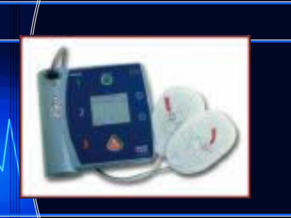
AED
• An AED is a device used in cardiac arrest, or sudden cardiac death, when the heart’s electrical activity is disorganized and there is no effective pumping of blood. The AED is capable of recognizing the heart's electrical activity, and determining if an electric shock is required. If the shock is needed, a voice prompt in the AED is activated, telling the rescuer to push a button to deliver the shock
stress
• Common responses can include: – Feeling a sense of loss, sadness, frustration,
helplessness, or emotional numbness – Experiencing troubling memories from that day – Having nightmares or difficulty falling or staying
asleep – Having no desire for food or a loss of appetite – Having difficulty concentrating – Feeling nervous or on edge
Teaching to cope
• Reach out and talk. • Express yourself. • Watch and listen. • Stay active. • Stay in touch with family. • Take care of yourself.
ANY QUESTIONS???


![Check List Cardioversion I.gallastegi[1]](https://img.pdfslide.net/doc/110x75/55cf8e57550346703b912349/check-list-cardioversion-igallastegi1.jpg)
