Embed Size (px)
Citation preview

1
Revised Manuscript M2:03927
Paclitaxel-resistant human ovarian cancer cells undergo c-Jun NH2-
terminal kinase-mediated apoptosis in response to noscapine
Jun Zhou*†, Kamlesh Gupta‡, Joyce Yao*†, Keqiang Ye§, Dulal Panda‡, Paraskevi
Giannakakou¶, and Harish C. Joshi†||
*Graduate Program in Biochemistry, Cell and Developmental Biology, †Department of
Cell Biology, §Department of Pathology, and ¶Winship Cancer Institute, Emory
University School of Medicine, Altanta, GA 30322; ‡Bhupat and Jyoti Mehta School of
Biosciences and Bioengineering, Indian Institute of Technology, Bombay, Mumbai
400076, India
||To whom correspondence should be addressed: Harish C. Joshi, Department of Cell
Biology, Emory University School of Medicine, 615 Michael Street, Atlanta, GA 30322.
Tel.: 404-727-0445; Fax: 404-727-6256; E-mail: [email protected]
Running title: JNK-mediated apoptosis in paclitaxel-resistant cancer cells
Abbreviations: JNK, c-Jun NH2-terminal kinases; DMSO, dimethyl sulfoxide; PBS,
phosphate-buffered saline; BSA, bovine serum albumin; FITC, fluorescein
isothiocyanate; TUNEL, terminal deoxynuceotidyltransferase-mediated dUTP nick-end
labeling.
Copyright 2002 by The American Society for Biochemistry and Molecular Biology, Inc.
JBC Papers in Press. Published on August 14, 2002 as Manuscript M203927200 by guest on January 4, 2019
http://ww
w.jbc.org/
Dow
nloaded from

2
SUMMARY
We have previously discovered the opium alkaloid noscapine as a microtubule
interacting agent that binds to tubulin, alters the dynamics of microtubule assembly, and
arrests mammalian cells at mitosis (Ye et al., 1998 Proc. Natl. Acad. Sci. USA 95, 1601-
1606; Ye et al., 2001 J. Biol. Chem. 276, 46697-46700; Zhou et al., 2002 J. Biol. Chem.
277, 17200-17208). Here we show that noscapine does not compete with paclitaxel for
tubulin binding and can efficiently inhibit the proliferation of both paclitaxel-sensitive
and paclitaxel-resistant human ovarian carcinoma cells (i.e. the parental cell line 1A9 and
two derivative cell lines, 1A9PTX10 and 1A9PTX22, which harbor β-tubulin mutations
that impair paclitaxel-tubulin interaction, Giannakakou et al., 1997 J. Biol. Chem. 272,
17118-17125). Strikingly, these cells undergo apoptotic death upon noscapine treatment,
accompanied by activation of the c-Jun NH2-terminal kinases (JNK). Furthermore,
inhibition of JNK activity by treatment with antisense oligonucleotide or transfection
with dominant-negative JNK blocks noscapine-induced apoptosis. These findings thus
indicate a great potential for noscapine in the treatment of paclitaxel-resistant human
cancers. In addition, our results suggest that the JNK pathway plays an essential role in
microtubule inhibitor-induced apoptosis.
by guest on January 4, 2019http://w
ww
.jbc.org/D
ownloaded from

3
INTRODUCTION
Microtubules are cytoskeletal components that play a critical role in many cellular
processes, such as maintenance of cell shape and polarity, intracellular transport of
vesicles and organelles, and beating of cilia and flagella. During cell division,
microtubules form a bipolar microtubule array, called the mitotic spindle, that functions
in the distribution of chromosomes into two daughter cells (1). Assembled from αβ-
tubulin heterodimers, microtubules are highly dynamic structures that alternate between
periods of growth and shortening (2), and the dynamic instability property is crucial for
microtubules to carry out many of their cellular functions (3). Disruption of microtubule
dynamics can lead to the formation of abnormally stable or unstable microtubules,
thereby preventing the normal dynamic rearrangements of the microtubule network
required for cell proliferation (4).
One group of agents that target microtubules, such as colchicine, nocodazole, and the
vinca alkaloids, inhibit microtubule polymerization. Another group of agents, such as
taxoids and epothilones, however promote microtubule polymerization and stabilize
microtubules. Nevertheless, they all disrupt microtubule dynamics, block cell cycle
progression at mitosis, and then cause cell death (4). This provides the molecular basis
for the use of microtubule targeting compounds in chemotherapeutic treatment of human
cancers.
Microtubule drugs currently in clinical use for cancer chemotherapy include
paclitaxel (Fig. 1A), docetaxel, and the vinca alkaloids. Specifically, paclitaxel has
proven effective against refractory ovarian cancer, metastatic breast cancer, and non-
small-cell lung cancer (5, 6), and the vinca alkaloids are effective against cancer types
by guest on January 4, 2019http://w
ww
.jbc.org/D
ownloaded from

4
such as lymphoma, leukemia, and Kaposi’s sarcoma (7, 8). Unfortunately, the toxicity
and low aqueous solubility have limited the applicability of taxoids and vinca alkaloids in
cancer chemotherapy. Moreover, their use has been hampered by the development of
drug resistance contributed by multifactorial mechanisms, such as overexpression of P-
glycoprotein (9, 10), altered expression of tubulin isotypes (11), and the presence of
tubulin mutations (12). Therefore, development and/or discovery of microtubule-based
compounds are in demand, especially for the treatment of human cancers resistant to
currently used drugs.
We have previously identified noscapine (Fig. 1A), an opium alkaloid, as a
microtubule targeting agent that binds stoichiometrically to tubulin and alters tubulin
conformation (13). Like many other microtubule drugs, noscapine suppresses
microtubule dynamics, arrests mammalian cells at mitosis, causes apoptosis, and exhibits
potent antitumor activity (13-15). However, distinct from other microtubule drugs,
noscapine does not affect the total polymer mass of tubulin and does not cause gross
deformation of cellular microtubules even at high concentrations (15). In addition, the
low toxicity, water solubility, and feasibility for oral administration are very valuable
advantages of noscapine over many other microtubule drugs for future clinical use in
cancer chemotherapy (16-19).
In this study, we find that noscapine does not compete with paclitaxel for tubulin
binding and can efficiently inhibit the proliferation of both paclitaxel-sensitive and
paclitaxel-resistant human ovarian carcinoma cells. In addition, noscapine causes these
cells to die through the c-Jun NH2-terminal kinase (JNK)-mediated apoptosis pathway.
These findings thus not only offer a rationale for the use of noscapine in the treatment of
by guest on January 4, 2019http://w
ww
.jbc.org/D
ownloaded from

5
paclitaxel-resistant human cancers, but they also provide important insights into the
molecular mechanisms by which microtubule targeting agents inhibit cell proliferation
and cause cell death.
EXPERIMENTAL PROCEDURES
Materials — Goat brain microtubule proteins were isolated in the presence of 1 M
glutamate by two cycles of polymerization and depolymerization (20). Tubulin was
purified from the microtubule proteins by phosphocellulose chromatography as described
previously (21). The tubulin solution was stored at –80oC until future use. Protein
concentration was determined by the method of Bradford using bovine serum albumin
(BSA) as a standard (22). The dominant-negative JNK1 expressing plasmid with a flag-
tag insertion, pCDNA3(flag)-JNK-dn, and the pCDNA3 vector with a flag-tag insertion,
pCDNA3(flag), were kindly provided by Dr. R.J. Davis (University of Massachusetts,
Worcester). Noscapine (97% purity) was purchased from Aldrich (Milwaukee, WI). The
noscapine stock solution was prepared at 100 mM in dimethyl sulfoxide (DMSO) and
stored at –20oC until use. Paclitaxel was from Sigma (St. Louis, MO) and dissolved in
DMSO as a 10 mM stock solution. Fluorescent paclitaxel was from Molecular Probes
(Eugene, OR).
Cell Culture — The 1A9 cell line is a clone of the human ovarian carcinoma cell line,
A2780 (23). The two paclitaxel-resistant cell lines, 1A9PTX10 and 1A9PTX22, were
isolated as individual clones in a single-step selection, by exposing 1A9 cells to 5 ng/ml
paclitaxel in the presence of 5 µg/ml verapamil, a P-glycoprotein antagonist (12). They
were all cultured in RPMI 1640 medium (GIBCO BRL, Grand Island, NY) supplemented
by guest on January 4, 2019http://w
ww
.jbc.org/D
ownloaded from
6
with 10% fetal bovine serum (GIBCO BRL) at 37oC in a 5% CO2/95% air atmosphere as
monolayers in tissue culture plates or on glass coverslips. Paclitaxel-resistant 1A9PTX10
and 1A9PTX22 cell lines were maintained in 15 ng/ml paclitaxel and 5 µg/ml verapamil
continuously but were cultured in drug-free medium for 7 days prior to each experiment.
Competitive Tubulin Binding Assay — The effect of noscapine on paclitaxel binding
to tubulin was measured using fluorescent paclitaxel. In brief, microtubules were
polymerized from purified goat brain tubulin in the presence of 10% DMSO, and
preformed microtubules were incubated with different concentrations of paclitaxel (20
and 40 µM), noscapine (50 and 100 µM), or the vehicle solution DMSO alone as a
control. Fluorescent paclitaxel was then added to each of these solutions and the
fluorescence intensity was measured using a JASCO FP-6500 spectrofluorometer
equipped with a constant temperature water-circulating bath (JASCO International Co.,
Tokyo, Japan). Spectra were taken by multiple scans, and buffer blank values were
subtracted from all measurements. A 0.3-cm pathlength cuvette was used for all
fluorescence measurements in order to minimize the inner filter effects.
In Vitro Cell Proliferation Assay — Cytotoxicity assays were performed in 96-well
plates using the sulforhodamine B assay as previously described (24). In brief, 2 x 10³
cells were seeded in each well and incubated with gradient concentrations of noscapine
for 72 h. The cells were then fixed with 50% trichloroacetic acid and stained with 0.4%
sulforhodamine B dissolved in 1% acetic acid. The cells were then washed with 1%
acetic acid to remove unbound dye. The protein-bound dye was extracted with 10 mM
Tris base to determine the optical density at 564-nm wavelength using a SPECTRAmax
by guest on January 4, 2019http://w
ww
.jbc.org/D
ownloaded from

7
PLUS 384 microplate spectrophotometer (Molecular Devices, Sunnyvale, CA). Each
experiment was repeated three times.
Immunofluorescence Microscopy — Immunofluorescence microscopy was performed
as previously described with minor modifications (15, 25, 26). To visualize microtubules,
human ovarian caricinoma cells grown on glass coverslips were fixed with cold (-20oC)
methanol for 5 min and then washed with phosphate-buffered saline (PBS) for 5 min.
Nonspecific sites were blocked by incubating with 100 µl of 2% BSA in PBS at 37oC for
15 min. A mouse monoclonal antibody against α-tubulin (DM1A, Sigma) was diluted
1:500 in 2% BSA/PBS and incubated (100 µl) with the coverslips at 37οC for 2 h. Cells
were then washed with 2% BSA/PBS for 10 min at room temperature before incubating
with a 1:100 dilution of a fluorescein isothiocyanate (FITC)-labeled donkey anti-mouse
IgG antibody (Jackson ImmunoResearch, Inc., West Grove, PA) at 37oC for 1 h.
Coverslips were then rinsed with 2% BSA/PBS for 10 min and incubated with propidium
iodide (0.5 µg/ml) for 30 sec at room temperature before they were mounted with
AquaMount (Lerner Laboratories, Pittsburgh, PA) containing 0.01% 1,4-
diazobicyclo(2,2,2)octane (DABCO, Sigma). Cells were examined with a Zeiss Axiovert
135 fluorescence microscope using 100 x /1.3 oil lens (Plan-NEOFLUAR, Carl Zeiss,
Inc., Thornwood, NY). Propidium iodide staining of the nuclei was also used to measure
apoptotic percentages in this study. Cells with three or more visible chromatin masses
were considered apoptotic.
Changes of cellular phosphorylated c-Jun level were visualized by
immunofluorescence microscopy as previously described (27). In brief, cells grown on
glass coverslips were fixed with 4% paraformaldehyde in PBS for 20 min at room
by guest on January 4, 2019http://w
ww
.jbc.org/D
ownloaded from

8
temperature, and then permeabilized with 0.5% IGEPAL CA-630 (Sigma) in PBS for 30
min, and blocked in 2% BSA/PBS for 20 min at room temperature. A mouse monoclonal
antibody against phosphorylated c-Jun (Santa Cruz Biotechnology, Santa Cruz, CA) was
diluted 1:500 in 2% BSA/PBS and incubated with the cells for 2 h at 37oC. Cells were
then washed with 2% BSA/PBS for 10 min at room temperature before incubating with a
1:100 dilution of a FITC-labeled donkey anti-mouse IgG antibody (Jackson
ImmunoResearch) at 37oC for 1 h. Coverslips were rinsed with 2% BSA/PBS for 10 min
and then mounted and examined by microscopy as described above.
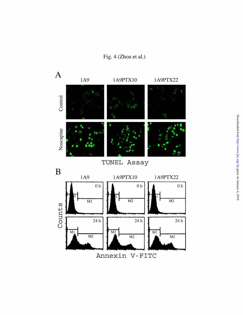
Terminal Deoxynucleotidyltransferase-Mediated dUTP Nick-End Labeling (TUNEL)
Assay — The TUNEL assay was performed by using the in situ cell detection kit (FITC)
following the manufacturer’s instructions (Roche Molecular Biochemicals, Mannheim,
Germany). In brief, cells grown on glass coverslips were fixed by a freshly prepared
paraformaldehyde solution (4% in PBS, pH 7.4) for 1 h at room temperature. Coverslips
were then washed with PBS and incubated in permeabilization solution (0.1% Triton X-
100, 0.1% sodium citrate) for 2 min on ice. Then, 50 µl TUNEL reaction mixture was
added on coverslips and incubated in a humidified chamber for 1 h at 37oC in the dark.
Finally, cells were mounted and examined by microscopy as described above. TUNEL
positive (apoptotic) cells stained bright green (see Fig. 4A). Comparisons in apoptotic cell
ratio between the 1A9 cell line and the two mutant cell lines were analyzed by the
Student’s t test. Statistical difference was considered significant if P values were less than
0.05.
Annexin V Staining Assay — The annexin V staining assay was performed by using
the annexin V apoptosis detection kit (FITC) following the manufacturer’s protocol (BD
by guest on January 4, 2019http://w
ww
.jbc.org/D
ownloaded from

9
PharMingen, San Diego, CA). Briefly, cells were washed with PBS and then resuspended
in 100 µl of binding buffer (10 mM HEPES, pH 7.4, 140 mM NaCl, 2.5 mM CaCl2).
Cells were incubated with 5 µl of FITC-conjugated annexin V for 15 min at room
temperature in the dark. Then 400 µl of binding buffer was added, and cells were
analyzed by flow cytometry using a Coulter Elite flow cytometer (Beckman Coulter, Inc.,
Fullerton, CA). The M1 and M2 gates demarcate annexin V-FITC negative and positive
staining populations, respectively (see Fig. 4B). Comparisons in apoptotic cell ratio
between the 1A9 cell line and the two mutant cell lines were analyzed by the Student’s t
test as described above.
Preparation of Nuclear Extracts — Nuclear extracts were prepared by the method of
Stone and Chambers (28). Briefly, cells grown in plates were collected by gentle scraping
and centrifugation (1500 × g, 10 min). After being washed with cold PBS, cells were
suspended in 3 ml of buffer A (10 mM HEPES, pH 7.9, 1.5 mM MgCl2, 10 mM KCl, 0.5
mM DTT, 20 mM β-glycerophosphate, 1 mM Na3VO4, 20 µg/ml aprotinin, 50 µg/ml
leupeptin, 10 µM pepstatin, 0.1 µM okadaic acid, 1 mM PMSF) and then homogenized
by a Dounce grinder (Fisher Scientific, Pittsburgh, PA) followed by centrifugation at
1000 × g for 10 min to collect the nuclei pellet. The nuclei were then suspended in buffer
A again, rehomogenized and pelleted. Subsequently, the nuclei were lysed in buffer B (20
mM HEPES, pH 7.9, 25% glycerol, 0.6 M NaCl, 1.5 mM MgCl2, 0.2 mM EDTA) for 1 h
followed by centrifugation at 12500 × g for 15 min. The supernatant was collected and
dialyzed against 1 L of buffer C (20 mM HEPES, pH 7.9, 20% glycerol, 0.1 M KCl, 0.2
mM EDTA, 1 mM PMSF, 0.5 mM DTT) and extracts were stored at –70oC.
by guest on January 4, 2019http://w
ww
.jbc.org/D
ownloaded from

10
Immunoprecipitation — Polyclonal antibodies against JNK1 and protein A-agarose
beads were purchased from Santa Cruz Biotechnology. Whole cell lysates were prepared
with the lysis buffer (20 mM Tris, pH 7.4, 200 mM NaCl, 0.1% Nonidet P-40, 1 mM
phenylmethylsulfonyl fluoride, 1 mM Na3VO4, 10 mM NaF), and total protein
concentration was then determined in each fraction by BCA reagents (Pierce, Rockford,
IL). 100 µg of total protein was immunoprecipitated with JNK1 antibody in excess and
protein A-agarose beads at 4oC overnight. The JNK precipitates were washed three times
with the lysis buffer and then used either for JNK activity assay or resuspended in
Laemmli's loading buffer for Western blot analysis of JNK protein level (see below). To
examine the specificity of the immunoprecipitation procedure, protein A-agarose beads
alone and protein A-agarose beads plus polyclonal antibodies against HA (Sigma), an
unrelated antibody, were used as controls.
JNK Activity Assay — The kinase assay of JNK1 was performed according to Wang
et al. (29) with minor modifications. In brief, whole cell lysates were prepared and JNK1
was immunoprecipitated as described above. The precipitates were washed three times
with the cell lysis buffer and then twice with kinase buffer (25 mM HEPES, pH
7.5, 25 mM MgCl2, 25 mM β-glycerophosphate). The kinase reactions were performed
by incubating the immunoprecipitates with 0.2 µg of GST-c-Jun (Santa Cruz
Biotechnology) in the reaction mixture (1 mM dithiothreitol, 0.1 mM Na3VO4, 10 µM
ATP) at room temperature for 30 min. Laemmli's loading buffer was added to stop the
reaction, and the level of c-Jun phosphorylation was revealed by Western blot analysis
using monoclonal antibodies against phosphorylated c-Jun (Santa Cruz Biotechnology).
The level of phosphorylated c-Jun was determined by densitometry as a measure of JNK
by guest on January 4, 2019http://w
ww
.jbc.org/D
ownloaded from
11
activity, using a Lynx video densitometer (Biological Vision Inc., San Mateo, CA). Each
experiment was repeated three times with duplicate densitometric determinations.
To measure the linearity of the JNK activity assay over a range of enzyme
concentration, 30, 53, 93, 163, 286, and 500 µU of pure active JNK (Upstate
Biotechnology, Lake Placid, NY) instead of JNK immunoprecipitate was used. 1 Unit of
JNK is equal to 1 nM phosphate incorporated into GST-c-Jun per min. To measure the
linearity of the JNK activity assay over a range of incubation time, 286 µU pure active
JNK instead of JNK immunoprecipitate was used, and the incubation time varied from 0
to 50 min instead of a constant time of 30 min.
Western Blot Analysis — Proteins were resolved by SDS/polyacrylamide gel
electrophoresis, and the protein bands were electrophoretically transferred onto PVDF
membranes (Millipore, Bedford, MA). The membranes were first incubated with mouse
monoclonal or rabbit polyclonal primary antibodies and then incubated with horseradish
peroxidase-labeled anti-mouse or anti-rabbit secondary antibodies. Specific proteins were
visualized using enhanced chemiluminescence following manufacturer's instructions
(Amersham, Piscataway, NJ).
Antisense Oligonucleotide Treatment — The antisense oligonucleotide experiment for
inhibition of JNK1 expression was performed as previously described (30, 31). The
antisense (5’-GTC ACG CTT GCT TCT GCT CAT GAT-3’) and sense (5’-ATC ATG
AGC AGA AGC AAG CGT GAC-3’) oligonucleotides specific for JNK1 were designed
as reported by Seimiya et al. (30). These sequences represent the amino acid codons –1 to
+7 of JNK1. They were synthesized with phosphorothioate substitutions and purified by
high-performance liquid chromatography (HPLC) (Integrated DNA Technologies,
by guest on January 4, 2019http://w
ww
.jbc.org/D
ownloaded from

12
Coralville, IA). The oligonucleotides were dissolved in sterilized water and added
directly into culture medium 12 h before treatment with 20 µM noscapine or 1 µM
paclitaxel. The protein level and activity of JNK and the percentage of apoptotic cells
were measured 48 h after drug treatment. JNK protein levels were measured by Western
blot analysis performed on the immunoprecipitates and JNK activity was measured by the
chemiluminescent Western blot analysis as described above. Morphological changes in
the nuclear chromatin of cells were detected by staining with propidium iodide as a
measure of apoptosis.
Transient Transfections — 1A9, 1A9PTX10, and 1A9PTX22 cells grown on glass
coverslips in 6-well plates were transfected either with 1 µg pCDNA3(flag)-JNK-dn or
with 1 µg pCDNA3(flag) as a control, using FuGENE 6 reagent (Boehringer Mannheim)
according to the manufacturer’s protocol. Cells were treated with 20 µM noscapine or 1
µM paclitaxel 12 h after transfection. The expression of JNK-dn, the activity of JNK, and
the percentage of apoptotic cells were measured 48 h after drug treatment. To measure
the expression of JNK-dn, cell lysates were immunoprecitated with anti-JNK antibodies,
and Western blot analysis was then performed on the immunoprecipitates, using a mouse
monoclonal antibody against the flag tag to detect the flag-tagged JNK-dn. JNK activity
was measured by the chemiluminescent Western blot analysis as described above.
Transfected cells were identified with the anti-flag antibody, and morphological changes
in the nuclear chromatin of transfected cells were detected by staining with propidium
iodide as a measure of apoptosis.
by guest on January 4, 2019http://w
ww
.jbc.org/D
ownloaded from

13
RESULTS
Noscapine Does Not Compete with Paclitaxel for Tubulin Binding — The binding
conformation of paclitaxel in tubulin has recently been resolved (32-34). The binding
pocket of paclitaxel resides in a deep hydrophobic cleft near the luminal surface of β-
tubulin, where it interacts with the protein by means of three potential hydrogen bonds
and multiple hydrophobic contacts (34). As an initial step to gain structural insights into
the interactions between noscapine and tubulin, we asked whether noscapine binds to
tubulin at sites common with paclitaxel. To test this, we performed a drug competition
experiment using fluorescent paclitaxel (Fig. 1). We found that pre-incubation of
microtubules assembled in vitro from purified αβ-tubulin with unlabeled paclitaxel
significantly inhibited the binding of fluorescent paclitaxel to tubulin, as evident from the
reduction of the fluorescence intensity by unlabeled paclitaxel in a concentration-
dependent manner (Fig. 1B). In contrast, 50 or even 100 µM noscapine did not inhibit the
binding of fluorescent paclitaxel to tubulin, displaying intensity-wavelength curves
overlapping with the control (Fig. 1C). These results show that noscapine is unable to
compete with paclitaxel for binding to tubulin, suggesting that noscapine binds to a site
other than the paclitaxel binding site.
Noscapine Inhibits the Proliferation of Both Pacitaxel-sensitive and
Paclitaxel–resistant Human Ovarian Carcinoma Cells — The non-inhibitory effect of
noscapine on the tubulin binding activity of paclitaxel suggested that human cancer cells
resistant to paclitaxel due to tubulin mutations might be sensitive to noscapine. To test
this, we first examined the effect of noscapine on the cell cycle progression of a set of
human ovarian carcinoma cells, including the parent cell line 1A9, which is sensitive to
by guest on January 4, 2019http://w
ww
.jbc.org/D
ownloaded from
14
paclitaxel treatment, and two derivative, paclitaxel-resistant lines, 1A9PTX10 and
1A9PTX22 (12). The two paclitaxel-resistant cell lines harbor β-tubulin mutations, Phe-
270Val in 1A9PTX10 cells and Ala-364Thr in 1A9PTX22 cells, that might interfere with
paclitaxel binding to tubulin. In addition, 1A9PTX10 and 1A9PTX22 cells exhibit
impaired paclitaxel-driven tubulin polymerization (12). We found that noscapine was
able to arrest these human ovarian carcinoma cells at mitosis. For example, the mitotic
indices for 1A9, 1A9PTX10, and 1A9PTX22 cells were 37.6%, 35.2%, and 35.8%,
respectively, after treatment with 20 µM noscapine for 18 h. The noscapine-arrested cells
have condensed chromosomes with nearly normal bipolar mitotic spindles (Fig. 2, bottom
panels). In contrast, only 5.4%, 5.0%, 5.1% of the control 1A9, 1A9PTX10, and
1A9PTX22 cells were in mitosis, and the rest of them were in interphase showing typical
radial microtubule arrays (Fig. 2, upper panels).
We then used the sulforhodamine B assay to examine the effect of noscapine on the
proliferation of these human ovarian carcinoma cells after a 72-h treatment (24). Our
results revealed that noscapine inhibited the proliferation of 1A9, 1A9PTX10, and
1A9PTX22 cells with similar IC50 values (18.2, 22.7, and 15.4 µM, respectively) (Fig. 3).
The inhibitory effects of noscapine on the proliferation of both paclitaxel-sensitive and
paclitaxel–resistant cells are consistent with the notion that noscapine might bind to
tubulin at a site different from that for paclitaxel, as indicated by the drug competition
experiment (Fig. 1).
Noscapine Induces Apoptosis in Human Ovarian Carcinoma Cells — Since
noscapine can induce apoptosis in a variety of tumor cell lines (13, 14), we asked whether
the human ovarian carcinoma cell line 1A9 and its derivative paclitaxel-resistant cell
by guest on January 4, 2019http://w
ww
.jbc.org/D
ownloaded from

15
lines underwent apoptotic cell death upon noscapine treatment. We first performed
immunofluorescence microscopy to examine the apoptotic percentages, based on the
nuclear morphology, of these cells upon treatment with 20 µM noscapine. As shown in
Table I, the ratio of noscapine-treated cells with apoptotic morphology increased
dramatically over time and reached more than 60% after 48 h of treatment. In addition,
there was no obvious difference in the apoptotic cell ratio among 1A9, 1A9PTX10, and
1A9PTX22 cell lines.
To confirm that noscapine-treated human ovarian carcinoma cells died through the
apoptosis pathway, we performed terminal deoxynucleotidyltransferase-mediated dUTP
nick-end labeling (TUNEL) assay, which detects the fragmentation of DNA, a
characteristic of cells undergoing apoptotic cell death (35). As shown in Fig. 4A, the
apoptotic nuclei stained bright green due to the fluorescence of FITC and were easily
identified in cells treated with 20 µM noscapine. The percentage of TUNEL positive cells
increased with the time of noscapine treatment, indicating the occurrence of apoptosis.
After 48 h of treatment, as many as 56.2%, 50.2%, 60.8% of 1A9, 1A9PTX10 and
1A9PTX22 cells displayed TUNEL positive staining, respectively (Table I).
We then took another approach to quantify apoptotic cell ratio, annexin V-FITC
staining assay, which reports the loss of phosphatidylserine asymmetry of plasma
membrane at the early stage of apoptosis (36). Fig. 4B shows representative annexin V-
FITC staining profiles at two time points after noscapine treatment, 0 h and 24 h,
respectively. Similar to that measured by the TUNEL assay, the percentage of annexin V-
FITC positive cells increased over time and reached 60.8%, 58.6%, and 66.9%,
by guest on January 4, 2019http://w
ww
.jbc.org/D
ownloaded from

16
respectively, for 1A9, 1A9PTX10 and 1A9PTX22 cells after 48 h of noscapine treatment
(Table I).
The results achieved from the above three different apoptosis detection assays were
strikingly comparable to each other. These assays showed similar apoptotic cell ratios for
each individual cell line upon noscapine treatment. In addition, they all revealed a time-
dependent increase in the percentage of apoptotic cells after being treated with noscapine.
JNK Activation upon Noscapine Treatment — The c-Jun NH2-terminal kinases
(JNK), also known as stress-activated MAP kinases (SAPK), are a group of mitogen-
activated protein kinases (MAPK) that bind the NH2-terminal activation domain of the
transcription factor c-Jun and phosphorylate c-Jun on amino acid residues Ser-63 and
Ser-73 (37-39). JNK has been implicated in the regulation of cell proliferation, tumor
transformation, and apoptosis as well as embryonic morphogenesis (40). JNK is activated
when cells are exposed to pro-inflammatory cytokines or environmental stress (e.g. UV-
and γ-radiation, heat shock, osmotic shock, redox stress, etc.), or undergo growth factor
withdrawal (40). Recently, JNK activation has also been found in cells treated with anti-
microtubule agents (28, 29, 41-43). Furthermore, the JNK pathway has been
demonstrated to be required for apoptosis caused by these agents (41, 43). It is thus
conceivable that the JNK pathway might also play a role in noscapine-induced apoptosis
in 1A9, 1A9PTX10, and 1A9PTX22 cells.
To test this, we first examined the activity of JNK in noscapine-treated human
ovarian carcinoma cells by measuring the level of endogenous phosphorylated c-Jun. As
revealed by Fig. 5A, 12 h after noscapine treatment, the level of endogenous
phosphorylated c-Jun was markedly increased in these cells showing bright fluorescence
by guest on January 4, 2019http://w
ww
.jbc.org/D
ownloaded from

17
signal in the nuclei, indicating JNK activation due to noscapine treatment. In contrast, 1
µM paclitaxel could activate JNK in 1A9 cells but not in the two mutant cell lines that are
resistant to paclitaxel (Fig. 5A). We used 1 µM rather than 20 µM paclitaxel as a control
for noscapine experiments in this study (also see figures below), because of the
devastating cytotoxicity of paclitaxel at high concentrations.
We then performed a Western blot analysis of nuclear extracts to further examine the
level of endogenous phosphorylated c-Jun in 1A9, 1A9PTX10, and 1A9PTX22 cells in
response to noscapine treatment (Fig. 5B). Nuclear extracts were prepared using the
method of Stone and Chambers (28) and then subjected to Western blotting with
antibodies against phosphorylated c-Jun. Consistent with the immunofluorescence
staining results (Fig. 5A), the phosphorylation of c-Jun could be clearly visualized as
early as 12 h of noscapine treatment in all the three human ovarian carcinoma cell lines;
however, upon paclitaxel treatment, c-Jun was phosphorylated only in the 1A9 cell line
(Fig. 5B). We also performed immunofluorescence staining and Western blotting with a
phosphorylation-independent c-Jun antibody and found that the level of endogenous c-
Jun remained constant in 1A9, 1A9PTX10, and 1A9PTX22 cells following treatment
with noscapine or paclitaxel (unpublished data).
To further investigate JNK activation in response to noscapine treatment, we
examined JNK activity by immunocomplex JNK activity assay (through the
chemiluminescent Western blot analysis with antibodies against phosphoylated c-Jun) in
1A9, 1A9PTX10, and 1A9PTX22 cells after they were treated with 20 µM noscapine for
0, 12, 24, 36, or 48 h. As shown in Fig. 6A, noscapine induced the activation of JNK in a
time-dependent manner in all the three cell lines. In contrast, paclitaxel induced JNK
by guest on January 4, 2019http://w
ww
.jbc.org/D
ownloaded from

18
activation only in the parental cell line 1A9. We quantified the level of JNK activation by
measuring the level of phosphorylated c-Jun with densitometry and compared the effect
of noscapine with paclitaxel (Table II). After 12, 24, 36, and 48 h of noscapine treatment,
the JNK activity in 1A9 cells was 3.3, 4.5, 4.8, and 6.9-fold, respectively, as high as that
for the untreated 1A9 cells. In contrast, the fold of JNK activation was 2.9, 4.4, 4.9, and
7.1 respectively for 1A9PTX10 cells, and 3.1, 3.9, 4.2, and 6.6 respectively for
1A9PTX22 cells, in response to 12, 24, 36, and 48 h of noscapine treatment (Table II). In
1A9 cells treated with 1 µM paclitaxel for 12, 24, 36, and 48 h, the JNK activity was 3.6,
4.7, 5.3, and 7.9-fold, respectively, as high as that for the untreated 1A9 cells (Table II).
However, in 1A9PTX10 and 1A9PTX22 cells treated with 1 µM paclitaxel, the activity
of JNK is similar to that of the untreated, indicating that JNK is not activated by
paclitaxel in these mutant cell lines.
To confirm that the JNK activity assay is quantitative, we examined the linearity of
this assay (Fig. 6B). Strikingly, phosphorylation of c-Jun occurred linearly (the arbitrary
densitometry units were 0.91, 1.43, 2.29, 3.64, 5.68, and 9.25 respectively) over a 16.67
fold range of the JNK enzyme (ranging from 30 µU to 500 µU), under the same condition
as that for the immunocomplex JNK activity assay except that pure active JNK instead of
JNK immunoprecipitate was used (Fig. 6B, left panel). Furthermore, phosphorylation of
c-Jun also occurred linearly over 40 min of incubation time (Fig. 6B, right panel).
We also examined the specificity of the immunoprecipitation procedure. Protein A-
agarose beads alone and protein A-agarose beads plus antibodies against ΗΑ , an
unrelated antibody, were used as controls (Fig. 6C). We found that JNK protein was
immunoprecipitated and showed activity only when both protein A-agarose beads and
by guest on January 4, 2019http://w
ww
.jbc.org/D
ownloaded from

19
JNK antibody were used (Fig. 6C). We thus conclude that JNK is indeed activated to a
significant extent in response to noscapine treatment
The JNK Pathway is Required for Noscapine-induced Apoptosis — To test whether
the observed JNK activation plays a role in noscapine-induced apoptosis, we selectively
inhibited JNK activity by using JNK-specific antisense oligonucleotides that have proven
highly effective in the inhibition of JNK expression in human cells (30, 31). 1A9,
1A9PTX10, and 1A9PTX22 cells were incubated with the antisense oligonucleotides for
12 h before treatment with noscapine. Consequently, the protein level of JNK in these
human ovarian carcinoma cells was significantly reduced, and noscapine-induced JNK
activation was also remarkably blocked (Fig. 7). In contrast, cells incubated with the
solvent alone or with sense oligonucleotides exhibited much higher JNK protein levels
and activity. We then examined the percentage of apoptotic cells under these conditions.
JNK antisense oligonucleotides clearly prevented 1A9, 1A9PTX10 and 1A9PTX22 cells
from undergoing apoptosis, however the solvent alone or sense oligonucleotides did not
(Fig. 7). 1 µM paclitaxel was also used as a control to examine the effect of
oligonucleotide treatment on JNK protein level and activity and the apoptotic percentage.
Strikingly, 1A9 cells, but not 1A9PTX10 and 1A9PTX22 cells, were sensitive to
paclitaxel-induced JNK activation and apoptosis, which were inhibited by treatment with
the JNK antisense oligonucleotides (Fig. 7). These results thus indicate that the JNK
pathway is required for human ovarian carcinoma cells to undergo apoptosis upon
noscapine treatment.
We tested this idea further by inhibition of JNK activity through dominant negative
transfections. 1A9, 1A9PTX10, and 1A9PTX22 cells were transiently transfected with
by guest on January 4, 2019http://w
ww
.jbc.org/D
ownloaded from

20
pCDNA3(flag)-JNK-dn, which expresses a dominant allele of JNK with a flag tag (44),
and then treated these cells with 20 µM noscapine or 1 µM paclitaxel as a control. As
shown in Fig. 8, JNK activity was largely blocked by expression of the dominant
negative allele. For 1A9, 1A9PTX10, and 1A9PTX22 cells expressing the dominant
negative JNK, only 11.9, 21.2, and 18.9% of them underwent apoptosis, respectively,
after 48 h of noscapine treatment. In contrast, for cells transfected with the control
plasmid, as many as 53.2, 51.0, and 54.9% of them underwent apoptosis, respectively,
upon noscapine treatment (Fig. 8). In addition, 1A9 cells, but not 1A9PTX10 and
1A9PTX22 cells, were sensitive to paclitaxel-induced JNK activation and apoptosis,
which were inhibited by expression of the dominant negative JNK (Fig. 8). Thus, we
conclude that the JNK pathway is required for noscapine-induced apoptosis in human
ovarian carcinoma cells.
DISCUSSION
Microtubules are highly dynamic polymers that are crucial for accurate chromosome
segregation during mitosis through formation of the bipolar mitotic spindle. To ensure
faithful chromosome transmission, eukaryotic cells have evolved a surveillance
mechanism called the spindle assembly checkpoint, which halts mitotic progression when
the spindle has a defect or chromosomes are not properly attached by spindle
microtubules (45). It is not surprising then that many chemical compounds that target
microtubules can arrest cells at mitosis, a property attributed to the use of microtubule
targeting agents in cancer chemotherapy. Notable examples are paclitaxel, docetaxel, and
the vinca alkaloids, which have proven successful in clinical use (5-8). However, the
by guest on January 4, 2019http://w
ww
.jbc.org/D
ownloaded from

21
clinical applicability of these microtubule targeting drugs is hampered by their low
aqueous solubility and toxicity. Moreover, although patients have impressive initial
response to these drugs, many of them relapse after treatment due to the development of
drug resistance. Consequently, considerable effort has been made over the past decade in
order to develop and/or discover novel anti-microtubule agents that have similar modes
of action to the currently used drugs but with improved biological and pharmaceutical
features.
During a systematic screen based on structural similarity of known microtubule
targeting agents, we have identified the opium alkaloid noscapine as a microtubule
targeting agent (13). Noscapine acts mechanistically similar to many other microtubule
agents, in that it binds to the tubulin subunit of microtubules, alters microtubule
dynamics, blocks mitotic progression, causes apoptosis, and has potent antitumor activity
(13-15). In this study, we demonstrate that noscapine can effectively inhibit the
proliferation and induce apoptosis in both paclitaxel-sensitive and paclitaxel-resistant
(due to β-tubulin mutations that affect paclitaxel binding) human ovarian carcinoma cells.
This is in agreement with the assumption that noscapine binds to tubulin at a site different
from the paclitaxel binding site, as indicated by the non-inhibitory effect of noscapine on
paclitaxel binding to tubulin (Fig. 1). It will be of great importance to test in nude mice
model whether noscapine can inhibit the growth of tumors formed from the
transplantation of paclitaxel-resistant human cancer cells.
Unlike many other microtubule targeting drugs, noscapine does not significantly
promote or inhibit microtubule polymerization even at high concentrations, nor does it
affect the total polymer mass of tubulin (15). When applied to tissue culture cells,
by guest on January 4, 2019http://w
ww
.jbc.org/D
ownloaded from

22
noscapine does not cause gross deformations of cellular microtubules. Instead, noscapine
alters the steady-state dynamics of microtubule assembly, which is sufficient to arrest
mitosis (15). This unique feature of noscapine would assume that other microtubule-
dependent cellular events such as organelle distribution and axonal transport are not
affected, which are serious concerns for many currently used microtubule targeting
anticancer drugs. Indeed, noscapine has been found to cause little or no toxicity to the
kidney, heart, liver, bone marrow, spleen, or small intestine, and does not inhibit primary
humoral immune responses in mice (19). Because noscapine is water-soluble and
absorbed after oral administration (16-18), its chemotherapeutic potential in the treatment
of human cancers merits thorough evaluation, particularly for cancers that are resistant to
the currently used drugs.
Several lines of compelling evidence suggest that anti-microtubule agents are able to
induce both morphological and biochemical features of the typical apoptosis, including
chromatin condensation, cell membrane blebbing, and internucleosomal DNA
fragmentation (46-52). It has been thought that this might result from the inhibition of
microtubule polymerization or depolymerization. Since noscapine suppresses
microtubule dynamics without obvious effects on microtubule polymerization or
depolymerization (15), yet can induce characteristic apoptosis in a variety of cell types
(Fig. 4; also see refs. 13, 14), we propose that apoptotic cell death caused by anti-
microtubule agents might reflect the disruption of the normal physiological balance in
microtubule dynamics rather than the action of them on the polymerization and
depolymerization of microtubules.
by guest on January 4, 2019http://w
ww
.jbc.org/D
ownloaded from

23
It has been suggested that anti-microtubule agents might trigger apoptosis through a
variety of phospho-regulatory pathways. For example, signal transduction pathways
mediated by Ras, Raf-1, and ASK1 (apoptosis signal-regulating kinase) have been
implicated in apoptosis induced paclitaxel (29, 41). The JNK pathway, which was
initially thought to participate in apoptosis induced by environmental stress, pro-
inflammatory cytokines, or growth factor withdrawal (40), has been recently shown to
play a role in microtubule dysfunction-induced apoptosis as well (41, 43). It is thus
understandable that noscapine, being a microtubule targeting agent, induces apoptosis
through a similar mechanism.
JNK activation has been observed in cells treated with microtubule targeting agents
such as paclitaxel and vinblastine (28, 29, 41-43). In addition, activation of JNK by
paclitaxel has been suggested to require interactions between microtubules and paclitaxel
(29). We find that noscapine causes nearly equal JNK activation among human ovarian
carcinoma cell lines 1A9 and the two derivative, β-tubulin mutant cell lines, 1A9PTX10
and 1A9PTX22 (Fig. 5 and Fig. 6). This finding is consistent with the observation that
paclitaxel caused JNK activation only in the parental 1A9 line but not in the two
derivative lines harboring β-tubulin mutations that impair paclitaxel binding to tubulin
(Figs. 5-8). Our data suggest that these mutations do not affect noscapine interactions
with tubulin nor the downstream effects leading to apoptosis. In addition, our data also
support the notion that noscapine does not bind at the paclitaxel site on tubulin. At
present, it remains unclear how the microtubule interactions of microtubule targeting
agents mediates the activation of JNK, which in turn leads to the occurrence of apoptosis.
The level of JNK activity in the untreated ovarian carcinoma cells may appear slightly
by guest on January 4, 2019http://w
ww
.jbc.org/D
ownloaded from

24
high relative to some other reports in the field (see for example, ref. 41). Therefore, it is
possible that the JNK pathway may be already slightly activated in these cells before the
experiments start due to their being cultured in paclitaxel-containing medium 7 days prior
to the experiments.
Taken together, we describe here that the anti-microtubule agent noscapine is highly
effective in inhibiting proliferation and inducing apoptosis in human ovarian carcinoma
cells sensitive or resistant to paclitaxel. In addition, we demonstrate that the JNK
pathway plays a role in noscapine-induced apoptosis. In the future, it will be of great
interest to explore the use of noscapine in the treatment of paclitaxel-resistant human
cancers. A better understanding of the molecular mechanisms by which JNK mediates the
apoptotic cell death induced by noscapine may also facilitate its use in cancer
chemotherapy.
Acknowledgments — We thank Dr. Roger J. Davis for reagents and Drs. Erica Werner,
Victor Faundez, Timothy C. Chambers, Haian Fu, John C. Lucchesi, Guy M. Benian,
Dennis C. Liotta, James P. Snyder, and James H. Nettles for helpful discussions and
many good suggestions. We are greatly indebted to the anonymous reviewer for
extremely helpful suggestions about experiments. This work was supported by a grant
from the National Institutes of Health to H.C.J. and an award from the Association for the
Cure of Cancer of the Prostate (CaP CURE) to D.P.
by guest on January 4, 2019http://w
ww
.jbc.org/D
ownloaded from

25
REFERENCES
1. McIntosh, J. R. (1994) in Microtubules, eds. Hyams, J. S. & Lloyd, C. W. (Wiley-
Liss, Inc., New York, NY), pp. 413-434
2. Mitchison, T., and Kirschner, M. (1984) Nature 312, 237-242
3. Desai, A., and Mitchison, T. J. (1997) Annu. Rev. Cell Dev. Biol. 13, 83-117
4. Jordan, M. A., and Wilson, L. (1999) Methods Cell Biol. 61, 267-295
5. Rowinsky, E. K. (1997) Annu. Rev. Med. 48, 353-374
6. Crown, J., and O’Leary, M. (2000) Lancet 355, 1176-1178
7. van Tellingen, O., Sips, J. H., Beijnen, J. H., Bult, A., and Nooijen, W. J. (1992)
Anticancer Res. 12, 1699-1715
8. Haskell, C. M. (1995) in Cancer Treatment, eds. Haskell, C. M. (W. B. Saunders
Co., Philadelphia, PA), pp. 78-165
9. Gottesman, M. M., and Pastan, I. (1993) Annu. Rev. Biochem. 62, 385-427
10. Bradley, G., and Ling, V. (1994) Cancer Metastasis Rev. 13, 223-233
11. Burkhart, C. A., Kavallaris, M., and Horwitz, S. B. (2001) Biochim. Biophys. Acta
1471, O1-O9
12. Giannakakou, P., Sackett, D. L., Kang, Y. K., Zhan, Z., Buters, J. T., Fojo, T., and
Poruchynsky, M. S. (1997) J. Biol. Chem. 272, 17118-17125
13. Ye, K., Ke, Y., Keshava, N., Shanks, J., Kapp, J. A., Tekmal, R. R., Petros, J., and
Joshi, H. C. (1998) Proc. Natl. Acad. Sci. USA 95, 1601-1606
14. Ye, K., Zhou, J., Landen, J. W., Bradbury, E. M., and Joshi, H. C. (2001) J. Biol.
Chem. 276, 46697-46700
by guest on January 4, 2019http://w
ww
.jbc.org/D
ownloaded from

26
15. Zhou, J., Panda, D., Landen, J. W., Wilson, L., and Joshi, H. C. (2002) J. Biol.
Chem. 277, 17200-17208
16. Dahlstrom, B., Mellstrand, T., Lofdahl, C. G., and Johansson, M. (1982) Eur. J.
Clin. Pharmacol. 22, 535-539
17. Haikala, V., Sothmann, A., and Marvola, M. (1986) Eur. J. Clin. Pharmacol. 31,
367-369
18. Karlsson, M. O., Dahlstrom, B., Eckernas, S. A., Johansson, M., and Alm, A. T.
(1990) Eur. J. Clin. Pharmacol. 39, 275-279
19. Ke, Y., Ye, K., Grossniklaus, H. E., Archer, D. R., Joshi, H. C., and Kapp, J. A.
(2000) Cancer Immunol. Immunother. 49, 217-225
20. Hamel, E., and Lin, C. M. (1981) Arch. Biochem. Biophys. 209, 29-40
21. Panda, D., Chakrabarti, G., Hudson, J., Pigg, K., Miller, H. P., Wilson, L., and
Himes, R. H. (2000) Biochemistry 39, 5075-5081
22. Bradford, M. M. (1976) Anal. Biochem. 72, 248-254
23. Eva, A., Robbins, K. C., Andersen, P. R., Srinivasan, A., Tronick, S. R., Reddy,
E. P., Ellmore, N. W., Galen, A. T., Lautenberger, J. A., Papas, T. S., Westin, E.
H., Wong-Staal, F., Gallo, R. C., and Aaronson, S. A. (1982) Nature 295, 116-119
24. Skehan, P., Stroreng, R., Scudiero, D., Monks, A., McMahon, J., Vistica, D.,
Warren, J. T., Bokesch, H., Kenney, S., and Boyd, M. R. (1990) J. Natl. Cancer
Inst. 82, 1107-1112
25. Zhou, J., Shu, H. B., and Joshi, H. C. (2002) J. Cell. Biochem. 84, 472-483
26. Joshi, H. C., and Zhou, J. (2001) Methods Cell Biol. 67, 179-193
by guest on January 4, 2019http://w
ww
.jbc.org/D
ownloaded from

27
27. Figueroa-Masot, X. A., Hetman, M., Higgins, M. J., Kokot, N., and Xia, Z. (2001)
J. Neurosci. 21, 4657-4667
28. Stone, A. A., and Chambers, T. C. (2000) Exp. Cell Res. 254, 110-119
29. Wang, T. H., Wang, H. S., Ichijo, H., Giannakakou, P., Foster, J. S., Fojo, T., and
Wimalasena, J. (1998) J. Biol. Chem. 273, 4928-4936
30. Seimiya, H., Mashima, T., Toho, M., and Tsuruo, T. (1997) J. Biol. Chem. 272,
4631-4636
31. Shiah, S. G., Chuang, S. E., Chau, Y. P., Shen, S. C., and Kuo, M. L. (1999)
Cancer Res. 59, 391-398
32. Nogales, E., Wolf, S. G., and Downing, K. (1998) Nature 391, 121-123
33. Nogales, E., Whittaker, M., Milligan, R. A., and Downing, K.H. (1999) Cell 96,
79-88
34. Snyder, J. P., Nettles, J. H., Cornett, B., Downing, K. H., and Nogales, E. (2001)
Proc. Natl. Acad. Sci. USA 98, 5312-5316
35. Heatwole, V. M. (1999) Methods Mol. Biol. 115, 141-148
3 6 . van Engeland, M., Nieland, L. J., Ramaekers, F. C., Schutte, B., and
Reutelingsperger, C. P. (1998) Cytometry 31, 1-9
37. Pulverer, B.J., Kyriakis, J. M., Avruch, J., Nikolakaki, E., and Woodgett, J. R.
(1991) Nature 353, 670-674
38. Adler, V., Polotskaya, A., Wagner, F., and Kraft, A. S. (1992) J. Biol. Chem. 267,
17001-17005
39. Hibi, M., Lin, A., Smeal, T., Minden, A., and Karin, M. (1993) Genes Dev. 7,
2135-2148
by guest on January 4, 2019http://w
ww
.jbc.org/D
ownloaded from

28
40. Davis, R. J. (2000) Cell 103, 239-252
41. Wang, T. H., Popp, D. M., Wang, H. S., Saitoh, M., Mural, J. G., Henley, D. C.,
Ichijo, H., and Wimalasena, J. (1999) J. Biol. Chem. 274, 8208-8216
42. Shtil, A. A., Mandlekar, S., Yu, R., Walter, R. J., Hagen, K., Tan, T. H.,
Roninson, I. B., and Kong, A. N. (1999) Oncogene 18, 377-384
43. Fan, M., Goodwin, M. E., Birrer, M. J., and Chambers, T. C. (2001) Cancer Res.
61, 4450-4458
44. Johnson, N. L., Gardner, A. M., Diener, K. M., Lange-Carter, C. A., Gleavy, J.,
Jarpe, M. B., Minden, A., Karin, M., Zon, L. I., and Johnson, G. L. (1996) J. Biol.
Chem. 271, 3229-3237
45. Zhou, J., Yao, J., and Joshi, H. C. (2002) J. Cell Sci. in press
46. Bhalla, K., Ibrado, A. M., Tourkina, E., Tang, C., Mahoney, M. E., and Huang, Y.
(1993) Leukemia 7, 563-568
47. Jordan. M. A., Toso, R. J., Thrower, D., and Wilson, L. (1993) Proc. Natl. Acad.
Sci. USA 90, 9552-9556
48. Jordan, M. A., Wendell, K., Gardiner, S., Derry, W. B., Copp, H., and Wilson, L.
(1996) Cancer Res. 56, 816-825
49. Bonfoco, E., Ceccatelli, S., Manzo, L., and Nicotera, P. (1995) Exp. Cell Res.
218, 189-200
50. Woods, C. M., Zhu, J., McQueney, P. A., Bollag, D., and Lazarides, E. (1995)
Mol. Med. 1, 506-526
51. Kawamura, K. I., Grabowski, D., Weizer, K., Bukowski, R., and Ganapathi, R.
(1996) Br. J. Cancer 73, 183-188
by guest on January 4, 2019http://w
ww
.jbc.org/D
ownloaded from

29
52. Yu, D., Jing, T., Liu, B., Yao, J., Tan, M., McDonnell, T. J., and Hung, M. C.
(1998) Mol. Cell 2, 581-591
by guest on January 4, 2019http://w
ww
.jbc.org/D
ownloaded from

30
FIGURE LEGENDS
FIG. 1. Noscapine does not inhibit paclitaxel binding to tubulin. A, structures of
noscapine and paclitaxel. In B and C, microtubules were polymerized from purified goat
brain tubulin in the presence of 10% DMSO, and preformed microtubules were incubated
with different concentrations of paclitaxel (20 and 40 µM), noscapine (50 and 100 µM),
or the vehicle solution DMSO alone as a control. Fluorescent paclitaxel was then added
to each of these solutions and the fluorescence intensity was measured at wavelengths
ranging from 500 nm to 540 nm. The excitation wavelength was 490 nm. Unlabeled
paclitaxel markedly inhibits the binding of fluorescent paclitaxel to tubulin, as evident
from the reduction of the fluorescence intensity by unlabeled paclitaxel in a
concentration-dependent manner (B). In contrast, 50 or even 100 µM noscapine does not
inhibit the binding of fluorescent paclitaxel to tubulin, displaying intensity-wavelength
curves overlapping with the control (C).
FIG. 2. Immunofluorescence micrographs showing microtubules (green) and DNA
(red) in human ovarian carcinoma cells treated with 20 µµµµM noscapine for 18 h or
the vehicle solution DMSO as a control. The formation of nearly normal bipolar
microtubule arrays (mitotic spindles) in noscapine-treated cells (bottom panels) indicates
mitotic arrest (the mitotic indices in 1A9, 1A9PTX10, and 1A9PTX22 cells are 37.6%,
35.2%, and 35.8%, respectively). In contrast, only 5.4%, 5.0%, 5.1% of the control 1A9,
1A9PTX10, and 1A9PTX22 cells were in mitosis, and the rest of them were in interphase
(upper panels), showing typical radial microtubule arrays. Bars, 10 µm.
by guest on January 4, 2019http://w
ww
.jbc.org/D
ownloaded from

31
FIG. 3. Noscapine causes cell death in paclitaxel-resistant, ββββ-tubulin mutant
1A9PTX10 and 1A9PTX22 cell lines as effectively as for their parental cell line 1A9.
Cells were treated with noscapine for 72 h and the survival percentage was then measured
by the sulforhodamine B assay as described under “Experimental Procedures”. Each
value represents the average of three independent experiments with duplicate
determinations.
FIG. 4. Noscapine induces apoptosis in human ovarian carcinoma cells as revealed
by TUNEL assay (A) and annexin V-FITC staining assay (B ). A, a group of
representative TUNEL assay results, showing that TUNEL positive cells stained bright
green. B, representative annexin V-FITC staining profiles of cells treated with noscapine
for 0 h (upper panels) and 24 h (bottom panels). The M1 and M2 gates demarcate
annexin V-FITC negative and positive staining populations, respectively. Please see
Table I for a summary of the quantitation of TUNEL and annexin V-FITC positive 1A9,
1A9PTX10, and 1A9PTX22 cells after 0, 12, 24, 36, or 48 h of noscapine treatment.
FIG. 5. Phosphorylation of c-Jun in human ovarian carcinoma cells treated with
noscapine. A, immunofluorescence micrographs showing the extent of c-Jun
phosphorylation in 1A9, 1A9PTX10, and 1A9PTX22 cells treated with 20 µM noscapine,
1 µM paclitaxel, or the vehicle solution DMSO for 12 h. Phosphorylation of c-Jun was
visualized using antibodies against phosphorylated c-Jun as described under
“Experimental Procedures”. Bar , 10 µm. B, Western blot analysis for the level of
by guest on January 4, 2019http://w
ww
.jbc.org/D
ownloaded from

32
phosphorylated c-Jun (p-c-Jun) in the nuclei of 1A9, 1A9PTX10, and 1A9PTX22 cells
upon treatment with DMSO, 20 µM noscapine, or 1 µM paclitaxel for 12 h. Nuclear
extracts were prepared by the method of Stone and Chambers (28), and Western blotting
was performed using antibodies against phosphorylated c-Jun.
FIG. 6. JNK activation upon noscapine treatment. A, measurement of JNK activity in
1A9, 1A9PTX10, and 1A9PTX22 cells after treatment with 20 µM noscapine or 1 µM
paclitaxel for 0, 12, 24, 36, or 48 h (upper panels). JNK activity was measured by
chemiluminescent Western blot analysis using antibodies against phosphorylated c-Jun
(p-c-Jun) and described in detail under “Experimental Procedures”. The level of p-c-Jun
was determined by densitometry (arbitrary units). Each experiment was repeated three
times with duplicate densitometric determinations (please see Table II for a summary of
the quantitation of JNK activity in noscapine- and paclitaxel-treated 1A9, 1A9PTX10,
and 1A9PTX22 cells). To confirm that equal amounts of JNK were used for kinase
reactions, the level of JNK protein was measured by Western blot analysis (performed on
the immunoprecipitates derived from cell extracts containing100 µg of total protein)
(bottom panels). B, linearity of the JNK activity assay. The level of p-c-Jun was
determined by densitometry (arbitrary units) as a measure of JNK activity, and each value
represents the average of three independent experiments with duplicate determinations.
Left panel, phosphorylation of c-Jun occurs linearly over a 16.67 fold range of the JNK
enzyme under the same condition as that for A except that pure active JNK (ranging from
30 µU to 500 µU) instead of JNK immunoprecipitate was used. 1 Unit of JNK is equal to
1 nM phosphate incorporated into GST-c-Jun per min. Right panel, phosphorylation of c-
by guest on January 4, 2019http://w
ww
.jbc.org/D
ownloaded from

33
Jun occurs linearly over 40 min of incubation time and then attenuates. The kinase
reaction was performed under the same condition as that for A except that pure active
JNK (286 µU) instead of JNK immunoprecipitate was used, and the incubation time
ranged from 0 min to 50 min instead of a constant time of 30 min. C, specificity controls
for immunoprecipitation. Protein A-agarose beads alone and protein A-agarose beads
plus antibodies against ΗΑ, an unrelated antibody, were used as controls. Preparation of
whole cell lysates of untreated 1A9, 1A9PTX10, and 1A9PTX22 cells,
immunoprecipitation, kinase assay, and Western blot analysis were performed as in A.
Note that JNK protein was immunoprecipitated (upper panel) and showed activity
(bottom panel) only when both protein A-agarose beads and JNK antibody were used,
confirming the specificity of the immunoprecipitation procedure.
FIG. 7. Inhibition of JNK activity by specific antisense oligonucleotide blocks
noscapine-induced apoptosis in human ovarian cancer cells. The oligonucleotides
were synthesized in phosphorothioate-modified conditions and purified by HPLC and
added directly into culture medium 12 h before treatment with 20 µM noscapine or 1 µM
paclitaxel. JNK-specific sense oligonucleotide and the solvent, sterilized water, were
used as controls for the JNK-specific antisense oligonucleotide. The protein level and
activity of JNK and the percentage of apoptotic cells were measured 48 h after treatment
with noscapine or paclitaxel. JNK protein levels were measured by Western blot analysis
performed on the immunoprecipitates and JNK activity was measured by the
chemiluminescent Western blot analysis as in Fig. 6A and described in detail under
“Experimental Procedures”. Note that JNK protein levels in 1A9, 1A9PTX10, and
by guest on January 4, 2019http://w
ww
.jbc.org/D
ownloaded from
34
1A9PTX22 cells have significantly decreased upon antisense oligonucleotide treatment.
Morphological changes in the nuclear chromatin of cells were detected by staining with
propidium iodide as a measure of apoptosis. Values and error bars shown in this graph
represent the averages and standard deviations, respectively, of three independent
experiments. p-c-Jun stands for phosphorylated c-Jun.
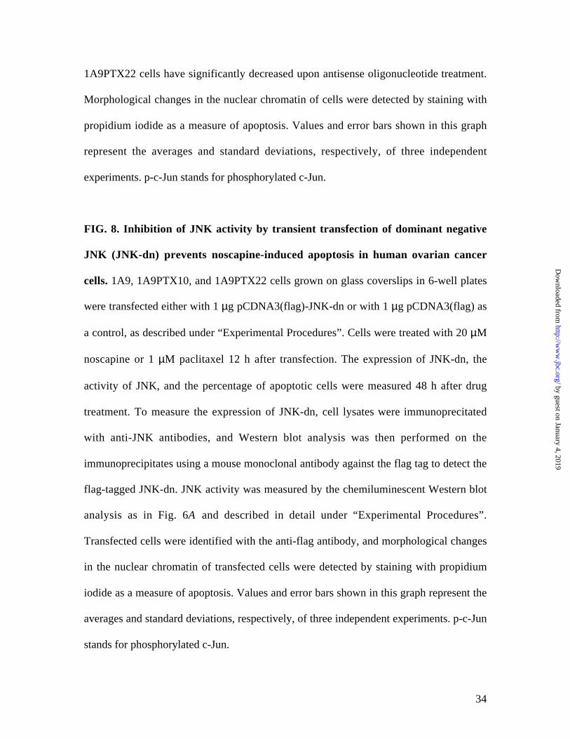
FIG. 8. Inhibition of JNK activity by transient transfection of dominant negative
JNK (JNK-dn) prevents noscapine-induced apoptosis in human ovarian cancer
cells. 1A9, 1A9PTX10, and 1A9PTX22 cells grown on glass coverslips in 6-well plates
were transfected either with 1 µg pCDNA3(flag)-JNK-dn or with 1 µg pCDNA3(flag) as
a control, as described under “Experimental Procedures”. Cells were treated with 20 µM
noscapine or 1 µM paclitaxel 12 h after transfection. The expression of JNK-dn, the
activity of JNK, and the percentage of apoptotic cells were measured 48 h after drug
treatment. To measure the expression of JNK-dn, cell lysates were immunoprecitated
with anti-JNK antibodies, and Western blot analysis was then performed on the
immunoprecipitates using a mouse monoclonal antibody against the flag tag to detect the
flag-tagged JNK-dn. JNK activity was measured by the chemiluminescent Western blot
analysis as in Fig. 6A and described in detail under “Experimental Procedures”.
Transfected cells were identified with the anti-flag antibody, and morphological changes
in the nuclear chromatin of transfected cells were detected by staining with propidium
iodide as a measure of apoptosis. Values and error bars shown in this graph represent the
averages and standard deviations, respectively, of three independent experiments. p-c-Jun
stands for phosphorylated c-Jun.
by guest on January 4, 2019http://w
ww
.jbc.org/D
ownloaded from

35
Table I
Apoptotic percentages of human ovarian carcinoma cells after being treated with 20 µMnoscapine for 0, 12, 24, 36, or 48 h
Apoptotic Cell Ratio (%)Assays of Detection
1A9 1A9PTX10 1A9PTX22
Nuclear Morphology0 h 0.37 ± 0.09 0.41 ± 0.11 0.69 ± 0.1712 h 7.39 ± 1.32 7.14 ± 1.21 7.45 ± 1.4024 h 20.7 ± 4.1 21.3 ± 5.8 22.3 ± 5.136 h 37.4 ± 7.1 37.2 ± 9.7 39.1 ± 8.048 h 66.1 ± 12.8 63.8 ± 11.3 67.5 ± 11.7
TUNEL Assay0 h 2.83 ± 0.32 3.39 ± 0.47 3.14 ± 0.3812 h 7.44 ± 1.10 7.91 ± 1.25 7.09 ± 0.9724 h 17.6 ± 2.5 16.7 ± 2.7 16.2 ± 2.136 h 39.5 ± 5.2 37.2 ± 4.3 39.1 ± 4.848 h 56.2 ± 7.8 50.2 ± 8.1* 60.8 ± 7.6*
Annexin V Staining0 h 0.42 ± 0.12 0.38 ± 0.09 0.43 ± 0.1012 h 11.2 ± 1.65 9.52 ± 1.47 8.18 ± 1.5124 h 37.4 ± 5.4 34.1 ± 4.9 39.4 ± 5.536 h 53.1 ± 5.9 51.1 ± 6.1 57.2 ± 5.8*48 h 60.8 ± 7.5 58.6 ± 8.0 66.9 ± 8.4*
Values shown here are the averages and standard deviations of three independentexperiments. *, Results are significantly different when compared with the 1A9 group (P< 0.05).
by guest on January 4, 2019http://w
ww
.jbc.org/D
ownloaded from

36
Table II
JNK activity in noscapine- and paclitaxel-treated human ovarian carcinoma cells
Relative JNK ActivityTreatment
1A9 1A9PTX10 1A9PTX22
Noscapine (20 µM)0 h 1.0 1.0 1.012 h 3.3 ± 0.7 2.9 ± 0.4 3.1 ± 0.824 h 4.5 ± 0.6 4.4 ± 0.7 3.9 ± 1.036 h 4.8 ± 1.1 4.9 ± 1.3 4.2 ± 0.748 h 6.9 ± 1.2 7.1 ± 1.5 6.6 ± 1.1
Paclitaxel (1 µM)
0 h 1.0 1.0 1.012 h 3.6 ± 0.8 0.9 ± 0.02 0.8 ± 0.0424 h 4.7 ± 0.9 1.1 ± 0.03 1.2 ± 0.0336 h 5.3 ± 1.2 1.2 ± 0.03 0.9 ± 0.0248 h 7.9 ± 1.4 1.0 ± 0.02 1.0 ± 0.03
JNK activity was determined by measuring the level of phosphorylated c-Jun withdensitometry as described in Fig. 6A and under “Experimental Procedures”. Relative JNKactivity refers to the values relative to those in untreated groups (0 h). Values shown inthis table are the averages and standard deviations of three independent experiments withduplicate determinations.
by guest on January 4, 2019http://w
ww
.jbc.org/D
ownloaded from

37
Supple. FIG. 1. The level of endogenous c-Jun remains constant following treatment
with noscapine or paclitaxel. Immunofluorescence staining (A) and Western blotting
(B) were performed as in Fig. 5 except that a phosphorylation-independent c-Jun
antibody was used.
by guest on January 4, 2019http://w
ww
.jbc.org/D
ownloaded from

and Harish C. JoshiJun Zhou, Kamlesh Gupta, Joyce Yao, Keqiang Ye, Dulal Panda, Paraskevi Giannakakou
kinase-mediated apoptosis in response to noscapinePaclitaxel-resistant human ovarian cancer cells undergo c-Jun NH2-terminal
published online August 14, 2002J. Biol. Chem.
10.1074/jbc.M203927200Access the most updated version of this article at doi:
Alerts:
When a correction for this article is posted•
When this article is cited•
to choose from all of JBC's e-mail alertsClick here
by guest on January 4, 2019http://w
ww
.jbc.org/D
ownloaded from


![Cardiologie francophone - franco 2005.ppt [Lecture seule] · 2007-05-14 · AMG Pico Elite Paclitaxel Artax Paclitaxel Aachen Resonance EuroCor Taxcor Paclitaxel Biolimus A9 Biomatrix](https://img.pdfslide.net/doc/110x75/5e42b3f5800daf02232992fa/cardiologie-francophone-franco-2005ppt-lecture-seule-2007-05-14-amg-pico.jpg)

![Original Article Potential biomarkers for paclitaxel ... · Potential biomarkers for paclitaxel sensitivity in ... larynx and oropharynx cancer [5, 15]. ... Biomarkers for paclitaxel](https://img.pdfslide.net/doc/110x75/5af0f1e17f8b9a572b901a03/original-article-potential-biomarkers-for-paclitaxel-biomarkers-for-paclitaxel.jpg)