Embed Size (px)
Citation preview
© Drägerwerk AG & Co. KGaA 1
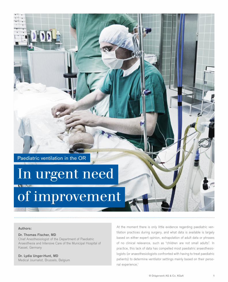
At the moment there is only little evidence regarding paediatric ven-tilation practices during surgery, and what data is available is largely based on either expert opinion, extrapolation of adult data or phrases of no clinical relevance, such as “children are not small adults”. In practice, this lack of data has compelled most paediatric anaesthesio-logists (or anaesthesiologists confronted with having to treat paediatric patients) to determine ventilator settings mainly based on their perso-nal experience.1
In urgent need of improvement
Paediatric ventilation in the OR
Authors:
Dr. Thomas Fischer, MDChief Anesthesiologist of the Department of Paediatric Anaesthesia and Intensive Care of the Municipal Hospital of Kassel, Germany
Dr. Lydia Unger-Hunt, MDMedical Journalist, Brussels, Belgium
D-1
6270
-201
7
PAEDIATRIC VENTILATION: IN URGENT NEED OF IMPROVEMENT
© Drägerwerk AG & Co. KGaA 2
of children for surgical or diagnostic procedures in Europe, and a large variability in the practice of paediatric anaesthesia” and stress that “these findings are substantial enough to warrant attention from national, regional, and specialist societies to target education of anaesthesiologists and their teams and implement strategies for quality improvement in paediatric anaesthesia”.6 In this study, the incidence of perioperative severe critical events was 5.2 percent, with an incidence of respiratory critical events of 3.1 percent (severe critical events included all episodes of laryngospasm, bronchospasm, pulmonary aspiration, drug error, anaphylaxis, cardiovascular instability, neurological damage, perioperative cardiac arrest and emergence stridor).
The aims of the present whitepaper are therefore to provide an overview of the main considerations when ventilating children and newborns in the perioperative setting, to give instructions on how best to proceed in clinical practice and to mention common potential pitfalls that should be avoided in the ventilation of children.
Disclaimer: I have sought to provide sources wherever possible. Wherever evidence was not available, the information and recommendations are based on my clinical expertise and experience (Dr. Thomas Fischer, author).
Quote: “The smaller the baby lung, the greater the potential for unsafe mechanical ventilation.”15 This quote is well suited for this paper, even though the term “baby lung” did not refer to children: Gattinoni named the concept “baby lung” to denote diseased adult lungs that he saw as “as vulnerable as children’s lungs”. However, parameters of mechanical ventilation have the potential to make mechanical ventilation unsafe for the child. It is therefore important to be aware of current practices and the current state of evidence.
The respiratory system: vast differences between children and adultsThe respiratory system differs substantially between children and adults. Early childhood is characterised by a rapid development of the lungs, in particular within the first year of life; functional residual capacity (FRC) increases by a magnitude of 75 times, from 40 ml in infants to 3.000 ml in adults. Postnatal, the distal lungs
Paediatric ventilation: in urgent need of improvementAt the moment there is only little evidence regarding paediatric ventilation practices during surgery, and what data is available is largely based on either expert opinion, extrapolation of adult data or phrases of no clinical relevance, such as “children are not small adults”. In practice, this lack of data has compelled most paediatric anaesthesiologists (or anaesthesiologists confronted with having to treat paediatric patients) to determine ventilator settings mainly based on their personal experience.1
The lack of data together with the particular challenges of anaesthetising newborns, infants and young children is reflected in the higher perioperative mortality of paediatric patients compared to adults2,3 : The anaesthesia-related mortality of children is currently estimated at 1: 30,000 (vs. 1: 250,000 in adults). The incidence of intraoperative cardiac arrests is 1:10,000, and respiratory causes are responsible for about a third of these paediatric mortalities.4 The main factors for the higher mortality in paediatrics include oropharyngeal operative procedures and underlying diseases such as muscular, neurological or pulmonary disorders; the highest mortality is observed in the age group of the 0 to 1-year-olds.
Another factor is the lack of experience of most anaesthesiologists in treating newborns, infants and children.5 The pitfalls and risks for small children and especially newborns were recently confirmed in the APRICOT study, which analysed a total of 31,000 anaesthetic procedures in around 30,000 children treated in 261 centres across 33 European countries. Here, the authors point to “a relatively high rate of severe critical events during the anaesthesia management
D-1
6271
-201
7
PAEDIATRIC VENTILATION: IN URGENT NEED OF IMPROVEMENT
© Drägerwerk AG & Co. KGaA 3
undergo a rapid growth, while airway dimensions show relatively stable changes. The smaller airways of children in relation to lung volume means greater airflow resistance than in adults; in addition, immature airways contain more mucous glands, leading to increased secretions and possible mucous plugging. Also note: In children, smooth muscle and cartilage are concentrated more in the central than in the peripheral airways, leading to a variable response to bronchodilators.7
Lung compliance increases rapidly with increasing height. Compared to adults, the chest wall of an infant is much more compliant; as a consequence, there is less opposition to the tendency of the lung to collapse. This results in low residual lung volume (RV) and distortion of the chest wall, and in turn to a loss of volume during inspiration. Infants have several respiratory system mechanics to partially compensate for the instability of functional residual capacity (FRC) associated with a compliant chest wall, and a tendency for small airway closure during tidal breathing. As a function of total lung capacity (TLC), FRC and RV increase with age throughout childhood, while the closing capacity of the lung decreases significantly with age. As a consequence of the reduced lung volumes, in particular FRC and RV in early childhood, these lungs (until the age of 2-3 years) always work near or on the limit and hence have very restricted capacity for compensation. As of the age of three years, the situation starts to improve, and by the age of six to eight years, the situation is nearly “as good as normal”, i.e. the development of the lungs has almost reached adult status.
An additional aspect is the importance of CO2 and O2 diffusion rate in dependency of age. The smaller the child, the lower the diffusion rate of O2 and CO2. This implies that, especially in newborns, the reduced FRC combined with the lower O2-diffusion rate and the
need for an oxygen uptake twice the amount of adults (per kg bodyweight) will result in a faster desaturation when respiratory problems arise – even without pre-existing underlying diseases of the respiratory, cardiac or cerebral system.
Perioperative Ventilation in Paediatric Patients
GOALS AND STRATEGY
Just like in adults, positive pressure ventilation in the paediatric patient can cause damage to the lungs, i.e. barotrauma and/or volutrauma resulting into ventilator associated lung injury (VILI). Therefore, the aim of mechanical ventilation especially in children is above all to cause no harm to the child. It should be performed with the following targets in mind:
Oxygenation. The aim of mechanical ventilation is adequate oxygenation in the child or newborn.
Note: “Adequate” implies that the oxygenation should be adapted to the situation and the age of the individual patient.
D-1
6272
-201
7
Main points:– Babies and toddlers are vulnerable due to three factors:
functionally reduced lung volume, increased airflow resistance and reduced O2 and CO2 diffusion capacity.
– These lungs work on maximum capacity (unlike adult lungs that work one half throttle), they have no emergency reserve. Note the significantly faster SpO2 drop after cut off of fresh gas / disruption of oxygen supply e. g. during intubation or in cases of FiO2 fluctuation.
– With respect to ventilation, every child under the age of two years is in the highest risk bracket.
– After the age of three years, the situation improves; by the age of six to eight years, the situation is almost “as good as normal” i. e. comparable to adults.
– Due to smaller lung volumes including tidal volume, deadspace is of far greater significance when ventilating paediatric patients. Note that other factors such as leakages may also have a negative impact.
PAEDIATRIC VENTILATION: IN URGENT NEED OF IMPROVEMENT
© Drägerwerk AG & Co. KGaA 4
As mentioned, paediatric patients, and especially newborns, show considerable differences in respiratory physiology compared to adults e.g. with respect to compliance, resistance and CO2/O2 diffusion rate. This different physiology requires specific considerations with respect to ventilator settings, such as tidal volume, respiratory rate and ventilation pressures, in order to achieve adequate oxygenation.
Above all, note that O2 consumption in small children can be as much as 6 - 8ml/kg x min, (vs. 3 - 4ml/kg x min in adults). Coupled with the physiologic specifics mentioned above, one major problem is the reduced apnoea tolerance of children compared to adults, as a study on 50 ASA I patients between the ages of two days and 18 years showed8 : The authors measured the time between removal of the face mask and decrease in oxygen saturation (SpO2 SaO2) from 99-100% to 90%, and found that 1) desaturation started earlier in infants (6 -23 months) than in 2- to 5-year olds (96.5 vs. 160.4 sec), and that 2) children became desaturated faster than adolescents (160.4 vs. 382.4 sec). Children require twice the alveolar ventilation for oxygen demand and CO2 elimination, with a third of the functional residual capacity and twice the O2 consumption.
High settings of fraction of inspired oxygen (FiO2) are frequently avoided in children in order to prevent negative effects of oxygen such as retinopathy. But is it really the FiO2 setting causing this issue? In prematures and newborns up to their 3rd/4th week of life, high SaO2 values over a longer period of time (> 6h) can cause retinopathy. The significant factor is therefore not the level of oxygen supplied ( i.e. FiO2, as previously assumed), but the level of oxygen saturation in the blood. Retinopathy in these newborns is due to saturation-induced neovascularisation.
The possible influence of FiO2 on the development of atelectasis is controversially discussed, mainly because it has only been demonstrated in a single study in adults. The assumption that high FiO2 can cause atelectasis in children is therefore not in any way evidence-based! It seems much more likely that atelectasis develops when a child presses during ventilation and air is unable to escape.Atelectasis due to tube displacement – a common problem – or due to secrete obstructions of the small airways has a much greater impact on diminished oxygenation due to reduced FRC.
Another aspect to keep in mind: The existence of foetal haemoglobin in newborns causes a shift of saturation to lower oxygen partial pressure. In newborns, transfusions of adult haemoglobin – even though they are known to be associated with poorer outcomes – may often be necessary due to intraoperative blood loss and frequent blood sampling (blood sampling amounts to, in relative terms, a lot of blood for preterm babies). However, these adult blood transfusions cause the oxygen-binding curve to shift to the right, with the result that a higher PaO2 is necessary to achieve the same saturation. Adult blood transfusions change the entire situation and should always be borne in mind when measuring saturation. Unfortunately, as yet there is little or no data on the effects of adult blood transfusions regarding ophthalmological or pulmonary changes or changes on the child as a whole. A frequent counter-strategy seems to be to administer iron and erythropoietin, with the aim of reducing the necessary amount of transfused blood.
D-1
6273
-201
7
In healthy children (above 3 weeks of age) with no underlying diseases, saturation should be above 95%. But the specific conditions of the paediatric patient should always be taken into account: In underlying conditions with reduced lung perfusion such as cystic fibrosis or lung stenosis, maximum saturation may be 92- 93%. In cases of single ventricle physiology with a 1:1 Qs/Qp ratio the maximum possible saturation is 80 to 85%. Single ventricle physiology denotes a parallel supply of pulmonary (Qp) and systemic (Qs) blood flow. The equation for a pulmonary blood flow (max saturation 100%) mixed 1:1 with a systemic blood flow (max saturation 60 -70%) is 100+170 = 170, and 170 :2 = 85 %, i.e. the maximum saturation. If more blood flows from the lung, saturation will increase.
PAEDIATRIC VENTILATION: IN URGENT NEED OF IMPROVEMENT
© Drägerwerk AG & Co. KGaA 5
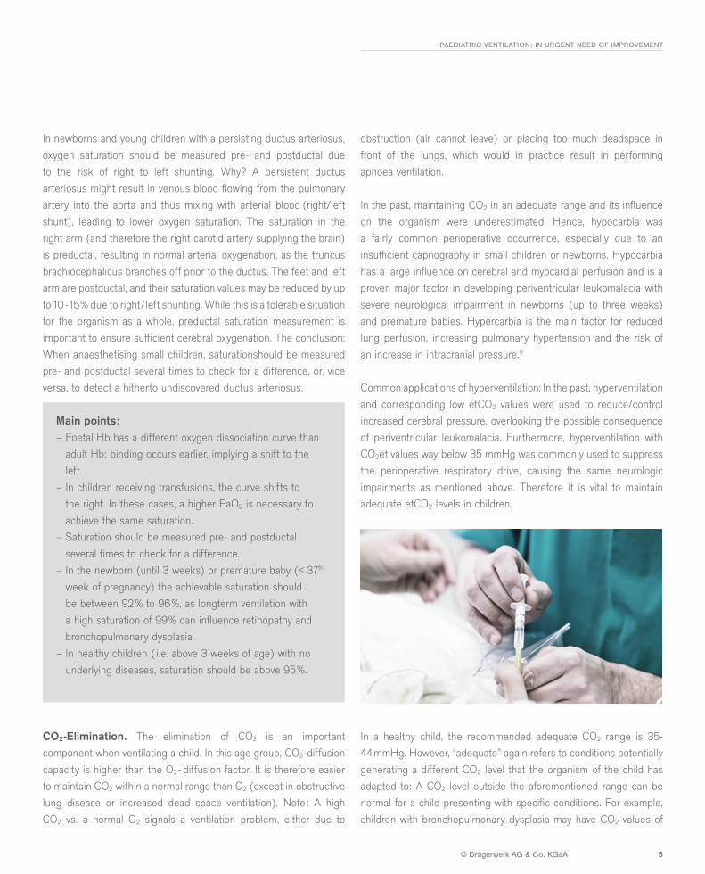
In newborns and young children with a persisting ductus arteriosus, oxygen saturation should be measured pre- and postductal due to the risk of right to left shunting. Why? A persistent ductus arteriosus might result in venous blood flowing from the pulmonary artery into the aorta and thus mixing with arterial blood (right/left shunt), leading to lower oxygen saturation. The saturation in the right arm (and therefore the right carotid artery supplying the brain) is preductal, resulting in normal arterial oxygenation, as the truncus brachiocephalicus branches off prior to the ductus. The feet and left arm are postductal, and their saturation values may be reduced by up to 10 -15% due to right/left shunting. While this is a tolerable situation for the organism as a whole, preductal saturation measurement is important to ensure sufficient cerebral oxygenation. The conclusion: When anaesthetising small children, saturationshould be measured pre- and postductal several times to check for a difference, or, vice versa, to detect a hitherto undiscovered ductus arteriosus.
Main points:– Foetal Hb has a different oxygen dissociation curve than
adult Hb: binding occurs earlier, implying a shift to the left.
– In children receiving transfusions, the curve shifts to the right. In these cases, a higher PaO2 is necessary to achieve the same saturation.
– Saturation should be measured pre- and postductal several times to check for a difference.
– In the newborn (until 3 weeks) or premature baby (< 37th week of pregnancy) the achievable saturation should be between 92% to 96%, as longterm ventilation with a high saturation of 99% can influence retinopathy and bronchopulmonary dysplasia.
– In healthy children ( i.e. above 3 weeks of age) with no underlying diseases, saturation should be above 95%.
CO2-Elimination. The elimination of CO2 is an important component when ventilating a child. In this age group, CO2-diffusion capacity is higher than the O2- diffusion factor. It is therefore easier to maintain CO2 within a normal range than O2 (except in obstructive lung disease or increased dead space ventilation). Note : A high CO2 vs. a normal O2 signals a ventilation problem, either due to
obstruction (air cannot leave) or placing too much deadspace in front of the lungs, which would in practice result in performing apnoea ventilation.
In the past, maintaining CO2 in an adequate range and its influence on the organism were underestimated. Hence, hypocarbia was a fairly common perioperative occurrence, especially due to an insufficient capnography in small children or newborns. Hypocarbia has a large influence on cerebral and myocardial perfusion and is a proven major factor in developing periventricular leukomalacia with severe neurological impairment in newborns (up to three weeks) and premature babies. Hypercarbia is the main factor for reduced lung perfusion, increasing pulmonary hypertension and the risk of an increase in intracranial pressure.9
Common applications of hyperventilation: In the past, hyperventilation and corresponding low etCO2 values were used to reduce/control increased cerebral pressure, overlooking the possible consequence of periventricular leukomalacia. Furthermore, hyperventilation with CO2et values way below 35 mmHg was commonly used to suppress the perioperative respiratory drive, causing the same neurologic impairments as mentioned above. Therefore it is vital to maintain adequate etCO2 levels in children.
In a healthy child, the recommended adequate CO2 range is 35-44mmHg. However, “adequate” again refers to conditions potentially generating a different CO2 level that the organism of the child has adapted to: A CO2 level outside the aforementioned range can be normal for a child presenting with specific conditions. For example, children with bronchopulmonary dysplasia may have CO2 values of
D-1
6269
-201
7
PAEDIATRIC VENTILATION: IN URGENT NEED OF IMPROVEMENT
© Drägerwerk AG & Co. KGaA 6
up to 80 mmHg; their organism has adapted to this situation by increasing the bicarbonate in order to achieve a normal pH of 7.4. Ventilating this patient to achieve the above mentioned “normal” CO2 range would therefore result in a significant shift in pH, as the bicarbonate level takes far longer to adjust. This pH shift will significantly suppress respiratory drive and delay the onset of spontaneous breathing.
TIPP: The Winter’s formula allows the calculation of patients’ normal CO2 level using the bicarbonate value. A delta of 12mmHg of CO2 renders a pH shift of 0.1, and a delta of 6 of bicarbonate also renders a 0.1 shift of pH.
THERMOREGULATION AND HUMIDIFICATION
Due to the very narrow neutral range in the thermoregulation of the infant ( i.e. newborns until around the age of one year), it is necessary to keep thermoregulation well adjusted and under control. Children have a different type of fatty tissue and hence cannot produce heat like adults do; and compared to adults, they have a higher fluid turnover and a higher surface area relative to their body mass. Thermoregulation in children is therefore extremely difficult and requires constant, focused attention, as every external influence factor regarding heat or cold can have a great effect.
Mechanical ventilation has a potential influence due to the cold and dry flow of fresh gas into the system and the length of the circuit
system. This counteracts the heating and the humidification of gas in the system, and dry and cold ventilation may cause obstruction by mucus. The need for an adequate humidification is defined with 30 mg/l H20 content. The used HME (Heat Moisture Exchange) provides the necessary water content and keeps it within the child/newborn. However, according to a study by Hunter et al, the amount of fresh gas flow is less important, as even small fresh gas flows of or even below 1 l/min will be high flow for children, considering their low tidal volumes.10
Therefore, determining the body core temperature and supplying external warming, e.g. by adjusting the operating room temperature and using active warming systems, is crucial in maintaining normothermia in the paediatric patient.
Parameters for mechanical ventilation and technical considerations
Tidal Volume. Ventilator induced lung injury (VILI) is a potential complication after mechanical ventilation, caused by volutrauma and atelectrauma (caused by repetitive opening and closure of alveoli by shear forces). Hence, the use of low tidal volume (Vt) is an integral part of the so-called lung-protective ventilation (LPV) to reduce VILI (experimental research indicates an age-related susceptibility to VILI). However, the optimal tidal volumes in paediatric ventilation are currently still unclear. Although the use of low tidal volumes of 6 - 8 ml/kg bodyweight (BW) is commonly accepted among paediatric anaesthesiologists as being lung protective, a recent metaanalysis including 160 studies showed that in fact high volumes are not associated with higher mortality, and that ultimately the volume used does not impact mortality (i . e. 6, 8, 10, 12 or 14 ml).11
Main points:– In infants, a high CO2 vs. normal O2 signals a ventilation
problem.– Hypocarbia is a proven major factor in developing
periventricular leukomalacia leading to severe neurological impairments.
– Always consider dead space and leakages in the system, as they can significantly influence CO2 measurement.
– In a healthy child, the recommended adequate CO2 level is 35 - 44 mmHg.
– By contrast, children with specific conditions may have much higher CO2 values, and ventilating to achieve 35- 44 mmHg CO2 will adversely affect outcomes.
Main points:– Thermoregulation in children is extremely difficult and
needs constant attention; every external influence factor regarding heat or cold can have a great effect.
– The need for an adequate humidification is defined with 30 mg / l H20 content.
– Supplying external warming is crucial in maintaining normothermia in the paediatric patient.
PAEDIATRIC VENTILATION: IN URGENT NEED OF IMPROVEMENT
© Drägerwerk AG & Co. KGaA 7
Feldman implied as much in his article by stating that “anaesthesia ventilators have traditionally been poorly suited to deliver small tidal volumes accurately”.12 In the light of pressure controlled ventilation potentially delivering varying tidal volumes based on changes in compliance, and the fact that modern anaesthesia devices offer improved delivery of small tidal volumes, Feldman raises the question if volume controlled ventilation could take on a different role in the future. It remains to be seen where this discussion will lead.
With regards to pressure settings during mechanical ventilation of the child, evidence once again is scarce. Available evidence suggests that a mean airway pressure (MAP) should be limited to 10cmH2O, as higher MAPs can demonstrably cause overdistension, pulmonary interstitial emphysema (PIE), pneumothorax and VILI.
In the past, the normal PEEP was not allowed to exceed 5cmH2O, and still today paediatric anaesthesiologists recommend using a PEEP of 3-5 cmH2O in the normal, healthy child. However, a recent study suggested that higher PEEP levels of 8 -10 cmH2O are beneficial in alveolar recruitment and to keep airways open, especially in children with compromised lungs.13 This study found that higher PEEP reduced mortality in small children with acute lung injury. But in general, the ideal PEEP is still unknown and its titration is subject to personal experience.
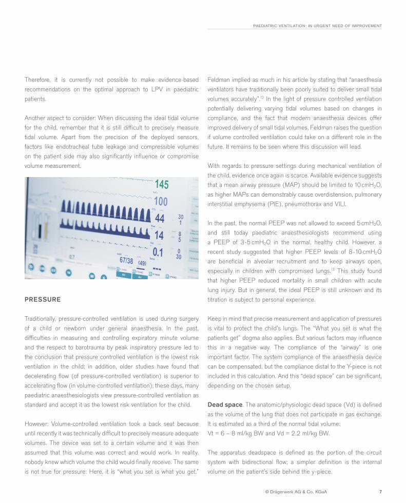
Keep in mind that precise measurement and application of pressures is vital to protect the child’s lungs. The “What you set is what the patients get” dogma also applies. But various factors may influence this in a negative way. The compliance of the “airway” is one important factor. The system compliance of the anaesthesia device can be compensated, but the compliance distal to the Y-piece is not included in this calculation. And this “dead space” can be significant, depending on the chosen setup.
Dead space. The anatomic/physiologic dead space (Vd) is defined as the volume of the lung that does not participate in gas exchange. It is estimated as a third of the normal tidal volume: Vt = 6 – 8 ml/kg BW and Vd = 2.2 ml/kg BW.
The apparatus deadspace is defined as the portion of the circuit system with bidirectional flow; a simpler definition is the internal volume on the patient’s side behind the y-piece.
Therefore, it is currently not possible to make evidence-based recommendations on the optimal approach to LPV in paediatric patients.
Another aspect to consider: When discussing the ideal tidal volume for the child, remember that it is still difficult to precisely measure tidal volume. Apart from the precision of the deployed sensors, factors like endotracheal tube leakage and compressible volumes on the patient side may also significantly influence or compromise volume measurement.
PRESSURE
Traditionally, pressure-controlled ventilation is used during surgery of a child or newborn under general anaesthesia. In the past, difficulties in measuring and controlling expiratory minute volume and the respect to barotrauma by peak inspiratory pressure led to the conclusion that pressure controlled ventilation is the lowest risk ventilation in the child; in addition, older studies have found that decelerating flow (of pressure-controlled ventilation) is superior to accelerating flow (in volume-controlled ventilation); these days, many paediatric anaesthesiologists view pressure-controlled ventilation as standard and accept it as the lowest risk ventilation for the child.
However: Volume-controlled ventilation took a back seat because until recently it was technically difficult to precisely measure adequate volumes. The device was set to a certain volume and it was then assumed that this volume was correct and would work. In reality, nobody knew which volume the child would finally receive. The same is not true for pressure: Here, it is “what you set is what you get.”
D-1
6274
-201
7
PAEDIATRIC VENTILATION: IN URGENT NEED OF IMPROVEMENT
© Drägerwerk AG & Co. KGaA 8
A frequently found setup consists of a y-piece (~ 8 ml), an HME-filter (9 – 20 ml), a closed suction system (~ 10 ml) and extension tubing (up to 25 ml), which results in an apparatus deadspace of potentially 50 ml or even more. Keeping in mind that children require relatively small tidal volumes, the sum of physiologic plus apparatus deadspace can greatly exceed the tidal volume of the patient, potentially resulting in pure dead space ventilation.
Therefore, every child with a body weight of up to 25 kg is at risk through imprudent use of critical device setups with excessive dead space, with the risk of hypercapnia or inadequate Vt/airway pressure to achieve adequate minute ventilation.
Technical. The majority of anaesthetic ventilators use a semi- or closed circuit systems developed for adults and are driven by piston or ascending/descending bellows. The disadvantages of the bellow driven systems can be negative pressure during ventilation, room air aspiration, missing fresh gas decoupling, long response time and the need for driving gas. The ventilator settings should be reliable for children, especially for newborns, with the possibility of a high inspiratory flow, in order to enable short inspiratory times (e.g. Ti = 0.7 sec). The airway resistance of the ventilator should be as low as possible to optimally support spontaneous breathing, as the response on gas modification (time constant of the system) has to be short.
Note that old textbooks focused on I :E ratio (inspiration : expiration), with a target value of 1: 2 or 1 : 1.5; in poorly patients, an I :E of 1 : 1 was considered permissible. These days however, there is less attention on the I:E ratio and more focus on the actual times of inspiration / expiration (Ti, Te) calculated from the respiratory rate. Te is crucial in children: Avoid a drop below 0.4 seconds to prevent air trapping.
Main points:– CO2et, O2 insp/exsp and pressures (peak inspiratory
pressure [PIP], PEEP, MAP) should be measured in real-time, and measured Vt has to be compliance-corrected (“patient get what you set”).
– MAP should be limited to 10 cmH2O, as higher MAPs can demonstrably cause overdistension, pulmonary interstitial emphysema (PIE), pneumothorax and VILI.
– Physiologic plus apparatus deadspace can greatly exceed the tidal volume of the patient, potentially resulting in pure deadspace ventilation.
– Te is crucial in children: Avoid a drop below 0.4 seconds to prevent air trapping.
– PEEP should be, as a rule, 3 -5cm H20; in cases of lung disease, a PEEP of 8 cm H20 is perfectly acceptable.
– The best PEEP is still unknown and its titration is of personal experience.
PAEDIATRIC VENTILATION: IN URGENT NEED OF IMPROVEMENT
© Drägerwerk AG & Co. KGaA 9
Pitfalls to avoid in ventilation:
– Tube displacement is a common error: • Most often, the tube is placed too low in the airways. Airways
of small children are only approximately 2 cm long before bifurcation, hence the high risk of ventilating only the right or only the left lung.
• Do not assume “what is in is in”. • On rare occasions, a tube is placed too high and may easily
slip out. The correct tube placement should be validated (X-ray), especially in longterm intubations.
– Oxygen saturation should be measured pre- and postductal due to the risk of right to left shunting in newborns with persistent Ductus arteriosus.
– Be aware of dead space in infants and toddlers: • Reduce deadspace distal to the Y-piece as much as possible. • In small children, place the HME filter close to the
anaesthesia device, not close to the patient, and critically question the need for additional devices, such as extension tubes etc.
• Weight is crucial: – Above 25 kg BW, deadspace becomes more and more
irrelevant. – Below 25 kg it is deemed highly relevant: – Every person has an anatomic dead space of 2 -3 ml
per kg BW. Therefore, a 30 kg child has a dead space of 60 ml.
– With a system setup distal to the Y-piece, the same amount of dead space may be added, with the risk of hypercapnia or inadequate Vt/airway pressure to achieve adequate minute ventilation.
PD
F-91
0485
9
IMPRINTGERMANYDrägerwerk AG & Co. KGaAMoislinger Allee 53–5523542 Lübeck
www.draeger.com
Discover more on our website www.draeger.com/protective-ventilation
DID YOU FIND THIS ARTICLE USEFUL? Help us to make our articles more relevant and more interesting to you. Please click on one of the icons below!
?
CONCLUSIONOne general rule in paediatric anaesthesiology is: The smaller the patient, the greater the impact an even small error can have. For instance, medications or fluids given to a 10 kg patient will have a 10% dosage error if the weight used is off by 1 kg. The same is true for mechanical ventilation: small changes in delivered tidal volume can lead to unintended hyper- and hypoventilation with hypo-or hypercarbia, with negative impacts on the growing brain and the risk of lung injury. These changes are the main causes of perioperative mortality in children.14
PAEDIATRIC VENTILATION: IN URGENT NEED OF IMPROVEMENT
© Drägerwerk AG & Co. KGaA 10
1 Kneyber MC, Intraoperative mechanical ventilation for the paediatric patient, Best Pract Res Clin Anaesthesiol. 2015 Sep;29(3):371-9
2 Griend BV et al., Postoperative Mortality in Children After 101,885 Anaesthetics at a Tertiary Paediatric Hospital, Anesth Analg 2011;112: 1440-7
3 Ramamoorthy C, et al. Anesthesia-related cardiac arrest in children with heart disease: data from the Paediatric Perioperative Cardiac Arrest (POCA) Registry. Anesth Analg 2010;110:1376–82
4 Bhananker SM et al, Anesthesia-related cardiac arrest in children: update from the Paediatric Perioperative Cardiac Arrest Registry, Anesth Analg. 2007 Aug;105(2):344-50
5 Christensen RE et al. Paediatric Cardiopulmonary Arrest in the Postanesthesia Care Unit, Anesth Analg 2017;124:1231–6
6 Habre W et al., Incidence of severe critical events in paediatric anesthesia (APRICOT): a prospective multicentre observational study in 261 hospitals in Europe. Lancet Respir Med. 2017 May;5(5):412-425
7 MacDonald KD, Volume and pressure modes of mechanical ventilation in pediatric patients; Respir Care Clin. N. Am 1996;2:607-618
8 Patel R et al, Age and the onset of desaturation in apnoeic children; Can J Anaesth. 1994;41:771-774
9 Okumura A et al, Hypocarbia in preterm infants with periventricular leukomalacia: the relation between hypocarbia and mechanical ventilation, Paediatrics 2001;107:469-475
10 Hunter T et al, The temperature and humidity of inspired gases in infants using a pediatric circle system: effects of high and low-flow anesthesia. Paediatric Anaesth. 2005;15:750-754
11 De Jager et al., Tidal volume and mortality in mechanically ventilated children: a systematic review and meta analysis of observational studies. Crit Care Med 2014 (42):2461
12 Feldman JM, Optimal ventilation of the anesthetized paediatric patient; Anesth Analg. 2015 Jan;120(1):165-75
13 Boriosi JP et al, Efficacy and safety of lung recruitment in pediatric patients with acute lung injury. Pediatr Crit Care Med. 2011 Jul;12(4):431-6.
14 Feldman JM, Optimal ventilation of the anesthetized paediatric patient; Anesth Analg. 2015 Jan;120(1):165-75
15 Gattinoni L et al, The concept of “baby lung”, Intensive Care Med. 2005;31:776-784
REFERENCE: