Embed Size (px)
Citation preview
Paper I- August 2010
I. ESSAY
1. Describe the citric acid cycle. How it is regulated? What is its amphibolic
role?[ April 2001, Aug 2006 SN, Aug 2004 Essay]
The citric acid cycle (Krebs cycle, tricarboxylic acid cycle) is a series of reactions in mitochondria that oxidize acetyl residues (as acetyl-CoA) and reduce coenzymes that upon reoxidation are linked to the formation of ATP.
Fig TCA cycle
The citric acid cycle is not only a pathway for oxidation of two-carbon units—it is also a major pathway for interconversion of metabolites arising from transamination and deamination of amino acids. It also provides the substrates for amino acid synthesis by transamination, as well as for gluconeogenesis and fatty acid synthesis. Because it functions in both oxidative and synthetic processes, it is amphibolic
2. Describe the chemistry, absorption, function and deficiency manifestations of
vit A.[ Feb 2005 SN, Aug 2006 Essay]
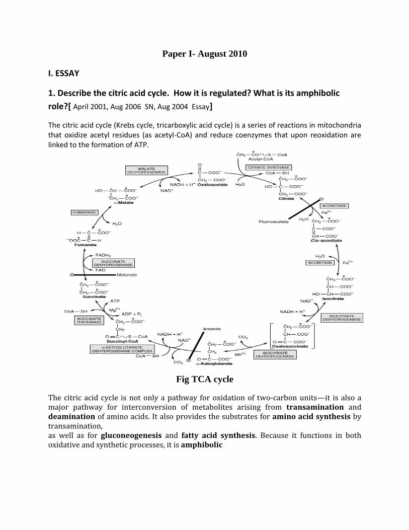
Retinoids comprise retinol, retinaldehyde, and retinoic acid (preformed vitamin A, found only in foods of animal origin); carotenoids, found in plants, comprise carotenes and related compounds, known as provitamin A, as they can be cleaved to yield retinaldehyde and thence retinol and retinoic acid. Although it would appear that one molecule of β-carotene should yield two of retinol, this is not so in practice; 6 μg of β-carotene is equivalent to 1 μg of preformed retinol. The total amount of vitamin A in foods is therefore expressed as micrograms of retinol equivalents. Biochemical functions: Vitamin A Has a Function in Vision In the retina, retinaldehyde functions as the prosthetic group of the light-sensitive opsin proteins, forming rhodopsin (in rods) and iodopsin (in cones). Retinoic Acid Has a Role in the Regulation of Gene Expression & Tissue Differentiation A most important function of vitamin A is in the control of cell differentiation and turnover. All-transretinoic acid and 9-cis-retinoic acid regulate growth, development, and tissue differentiation; they have different actions in different tissues. Deficiency: More prolonged deficiency leads to xerophthalmia: keratinization of the cornea and skin and blindness. Vitamin A also has an important role in differentiation of immune system cells, and mild deficiency leads to increased susceptibility to infectious diseases. Furthermore, the synthesis of retinol-binding protein in response to infection is reduced (it is a negative acute phase protein).
Fig Vit A
II. Write short notes on (10*5=50)
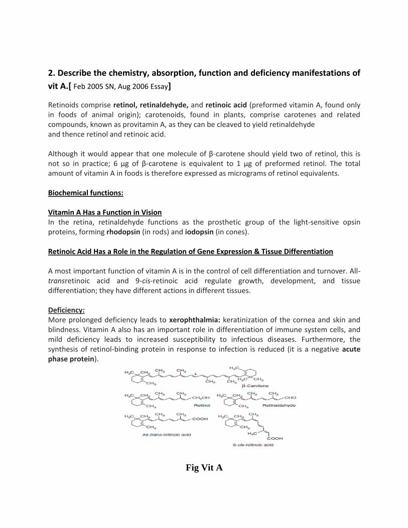
1. Inhibitors of ETC
Fig. Inhibitors of ETC
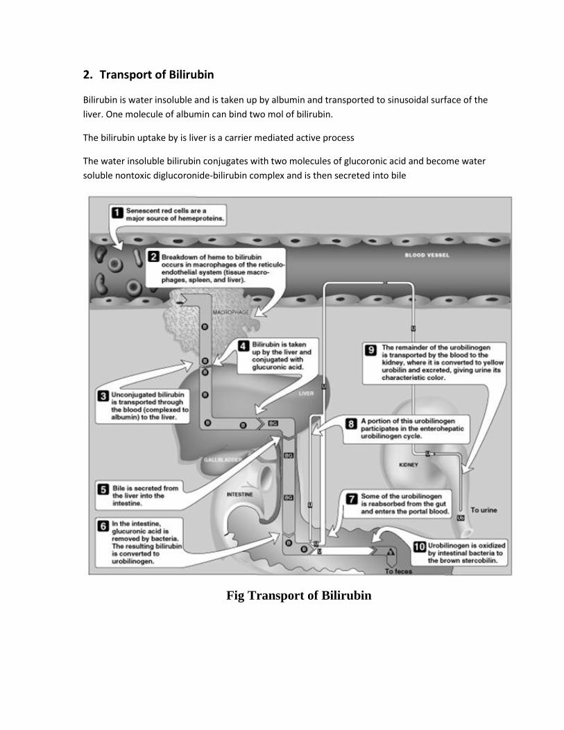
2. Transport of Bilirubin
Bilirubin is water insoluble and is taken up by albumin and transported to sinusoidal surface of the
liver. One molecule of albumin can bind two mol of bilirubin.
The bilirubin uptake by is liver is a carrier mediated active process
The water insoluble bilirubin conjugates with two molecules of glucoronic acid and become water
soluble nontoxic diglucoronide-bilirubin complex and is then secreted into bile
Fig Transport of Bilirubin
3. Vitamin E [Aug 2004 SN]
Other name:
Tocopherol (tokos-child birth; pheros- to bear; ol-alcholol) or anti-infertility vitamin
Normal level: 0.5-1 mg/dl.
RDA:
Males – 10 mg/day
Females – 8 mg/day
Sources:
vegetabTranle oils (wheat germ oil, sunflower oil, safflower oil, cotton seed oil)
Absorption:
Since it is a fat soluble vitamin, it need bile salts for absorption.
It is transported as chylomicron. It is stored in adipose tissue.
Biochemical role:
1. Anti oxidant
2. It protects RBC from hemolysis
3. It boots immune response
4. It reduces pre mature aging
5. It reduces the risk of atherosclerosis by reducing oxidation of LDL.
Inter-relationship with selenium:
Selenium and vit E act synergistically to minimize lipid peroxidation.
Deficiency:
Increased fragility of RBC
Muscular dystrophy
Weakness and creatinuria
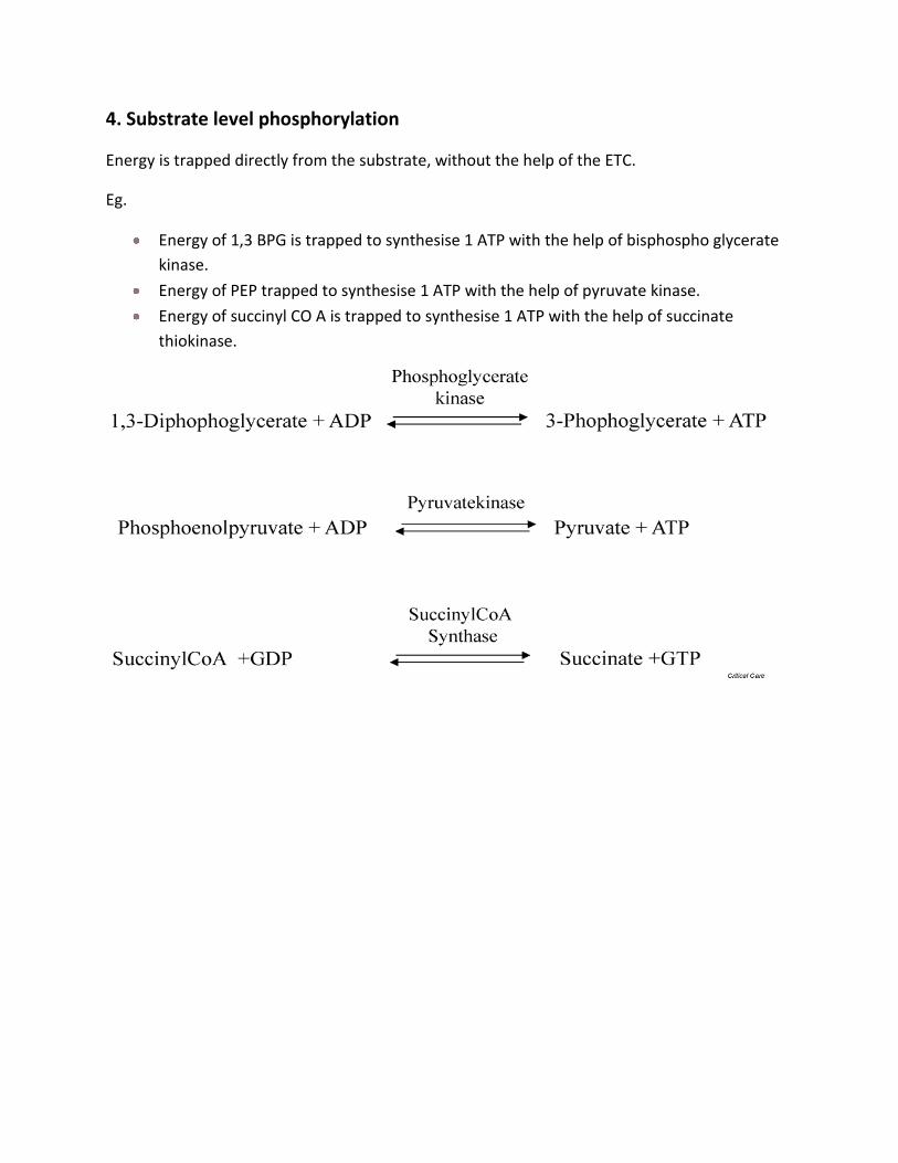
4. Substrate level phosphorylation
Energy is trapped directly from the substrate, without the help of the ETC.
Eg.
Energy of 1,3 BPG is trapped to synthesise 1 ATP with the help of bisphospho glycerate
kinase.
Energy of PEP trapped to synthesise 1 ATP with the help of pyruvate kinase.
Energy of succinyl CO A is trapped to synthesise 1 ATP with the help of succinate
thiokinase.
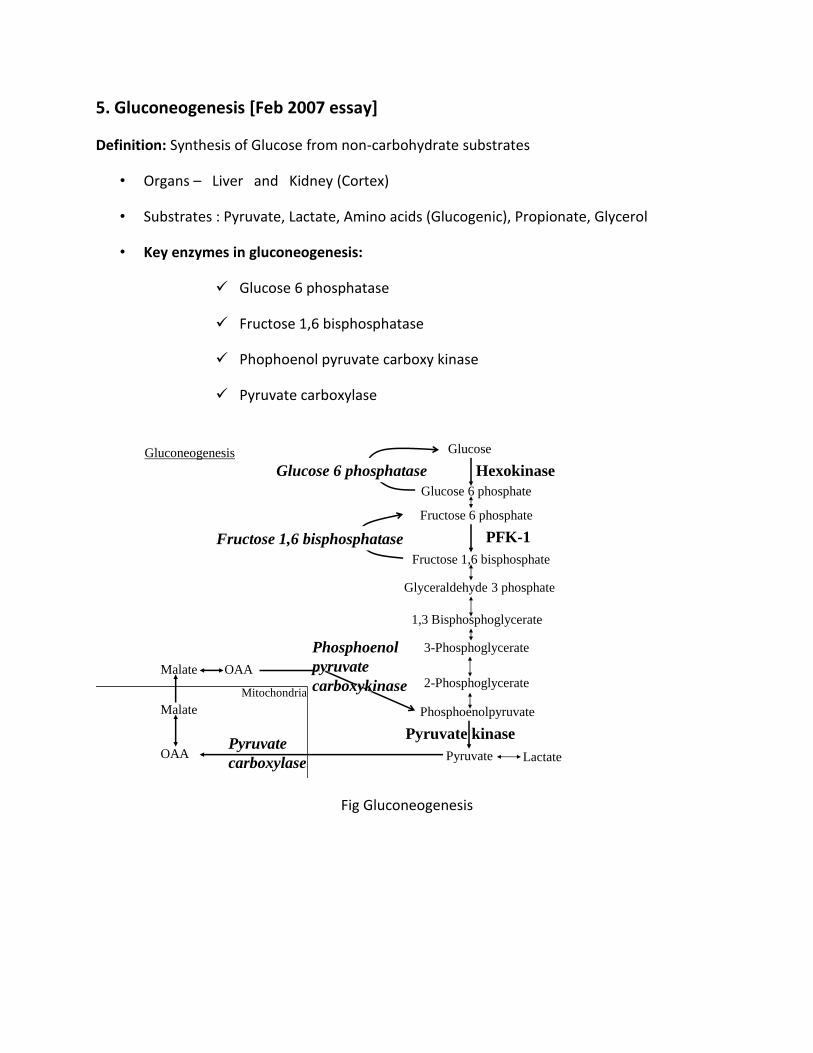
5. Gluconeogenesis [Feb 2007 essay]
Definition: Synthesis of Glucose from non-carbohydrate substrates
• Organs – Liver and Kidney (Cortex)
• Substrates : Pyruvate, Lactate, Amino acids (Glucogenic), Propionate, Glycerol
• Key enzymes in gluconeogenesis:
Glucose 6 phosphatase
Fructose 1,6 bisphosphatase
Phophoenol pyruvate carboxy kinase
Pyruvate carboxylase
Glucose
Glucose 6 phosphate
Fructose 6 phosphate
Fructose 1,6 bisphosphate
Glyceraldehyde 3 phosphate
1,3 Bisphosphoglycerate
Phosphoenolpyruvate
Pyruvate Lactate
Hexokinase
2-Phosphoglycerate
3-Phosphoglycerate
PFK-1
Pyruvate kinase
Glucose 6 phosphatase
Fructose 1,6 bisphosphatase
Pyruvate
carboxylase
Malate
Malate
Phosphoenol
pyruvate
carboxykinaseOAA
Mitochondria
Gluconeogenesis
OAA
Fig Gluconeogenesis
6. Regulation of enzyme activity
In humans, the induction of protein synthesis is a complex multistep process that typically requires hours to produce significant changes in overall enzyme level. By contrast, changes in intrinsic catalytic efficiency effected by binding of dissociable ligands (allosteric regulation) or by covalent modification achieve regulation of enzymic activity within seconds. Changes in protein level serve long-term adaptive requirements, whereas changes in catalytic efficiency are best suited for rapid and transient alterations in metabolite flux. ALLOSTERIC EFFECTORS REGULATE CERTAIN ENZYMES Feedback inhibition refers to inhibition of an enzyme in a biosynthetic pathway by an end product of that pathway. For example, for the biosynthesis of D from A catalyzed by enzymes Enz1 through Enz3, high concentrations of D inhibit conversion of A to B. Inhibition results not from the “backing up” of intermediates but from the ability of D to bind to and inhibit Enz1. Typically, D binds at an allosteric site spatially distinct from the catalytic site of the target enzyme. Feedback inhibitors thus are allosteric effectors and typically bear little or no structural similarity to the substrates of the enzymes they inhibit. In this example, the feedback inhibitor D acts as a negative allosteric effector of Enz1.
Aspartate Transcarbamoylase Is a Model Allosteric Enzyme Aspartate transcarbamoylase (ATCase), the catalyst for he first reaction unique to pyrimidine biosynthesis is feedback-inhibited by cytidine triphosphate. Following treatment with mercurials, ATCase loses its sensitivity to inhibition by CTP but retains its full activity for synthesis of carbamoyl aspartate. This suggests that CTP is bound at a different (allosteric) site from either substrate. ATCase consists of multiple catalytic and regulatory subunits. Each catalytic subunit contains four aspartate (substrate) sites and each regulatory subunit at least two CTP (regulatory) sites
7. Abnormal Hb[ March 2002, Sep 2002 SN]
Haemoglobin is a conjugated protein made up of a prosthetic group called heme and protein
part globin. Globin is a complex tertiary structure composed of two alpha and two beta chains.
The genes for these proteins are located in 16 and 11 respectively. Any mutation in these genes
gives raise to abnormal structure of haemoglobin which shows altered haemoglobin function.
There are plenty abnormal Hb is discovered yet.
The following lists the major types of Hemoglobinopathies –
HbS: Sickle Cell Hb – The glutamic acid in the 6th position of beta chain of Hb is
changed to valine. This change of aminoacid causes sikling of RBC. The sickled RBC
plugs in capillaries and may cause occlusion of major vessels and lead to infarction of
organs.
Hb E: it is the second most variant occurring after HbS. The glutamic acid at 26th
position replace by lysine in beta chain. This variant is most prevalent west Bengal.
HbC: glu at 6th position of beta chain is replaced y lysine. It is mostly seen in blacks.
Homogygous have am mild to moderate haemolytic anaemia
HbD: it does not produce sickling. Is found in Punjab. Beta 21 glu acid is replaced by
glutamine.
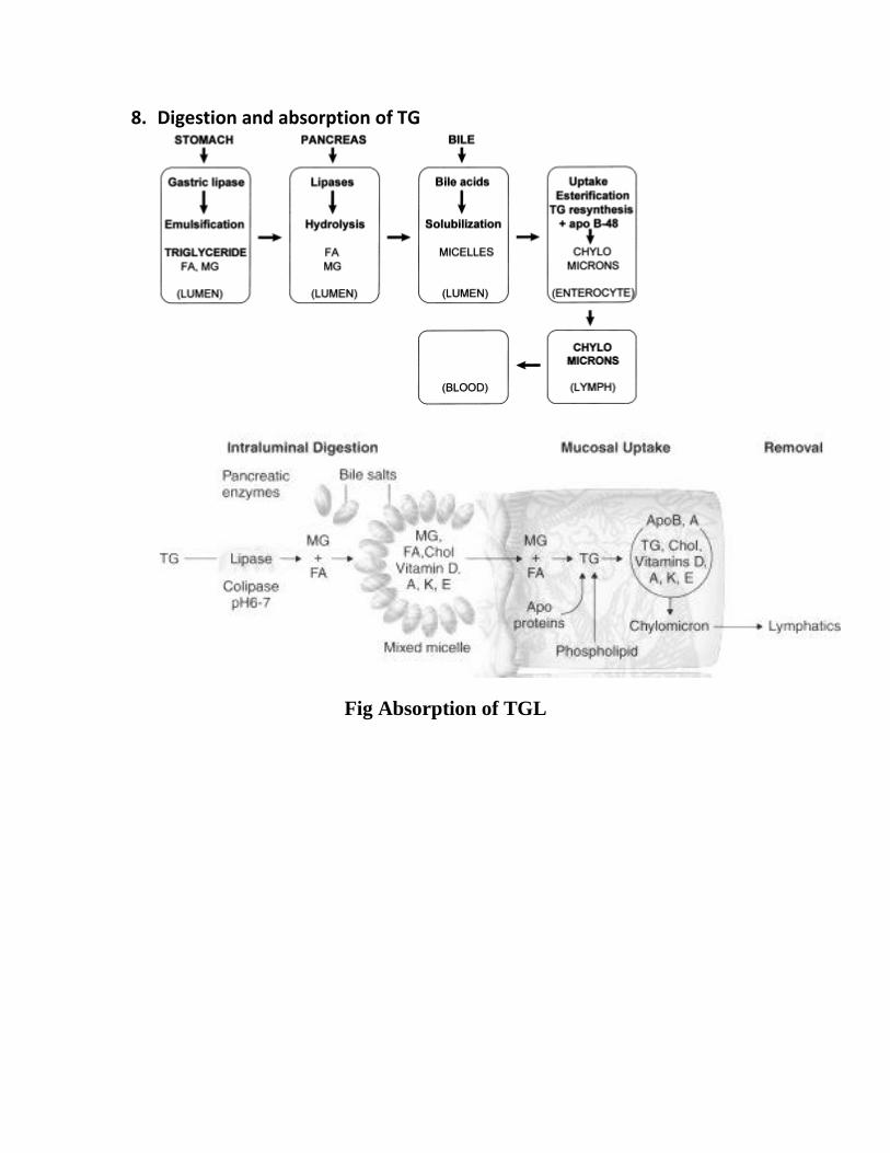
8. Digestion and absorption of TG
Fig Absorption of TGL
III. Short answers 2 marks
1. Mutarotation [Feb 2006 SN]
The α and β anomers are diastereomers of each other and usually have different specific rotations. A solution or liquid sample of a pure α anomer will rotate plane polarised light by a different amount and/or in the opposite direction than the pure β anomer of that compound. The optical rotation of the solution depends on the optical rotation of each anomer and their ratio in the solution.
For example if a solution of β-D-glucopyranose is dissolved in water, its specific optical rotation will be +18.7. Over time, some of the β-D-glucopyranose will undergo mutarotation to become α-D-glucopyranose, which has an optical rotation of +112.2. Thus the rotation of the solution will increase from +18.7 to an equilibrium value of +52.5 as some of the β form is converted to the α form. The equilibrium mixture is actually about 64% of β-D-glucopyranose and about 36% of α-D-glucopyranose, though there are also with traces of the other forms including furanoses and open chained form
2. Subcellular organelles
Mitochondria, nucleus, golgi bodies, lysosomes, nucleus etc.
3. Free radicals
It is a molecule or molecular fragment that contains one or more unpaired electrons in its
outer orbital. It is generally represented by a superscript dot, (R’). They are constantly
produced during the oxidation of foodstuffs, due to leaks in ETC. About 1-4% of oxygen is
converted to free radicals in our body.
Eg. Superoxide anion radical (O2’-)
Lipid peroxide radical (ROO’)
Characteristics:
Extreme reactivity, Short life span, damage to various tissues, generation of new free radical
by chain reaction.
4. BMR
Definition: The energy required by a awaken individual during physical, emotional and
digestive rest.
It is the minimum amount of energy required to perform vital functions such as circulation,
respiration, working of heart etc.
Normal Value: Men - 34-37 k cal/m2/hr
Women - 30-35 k cal/m2/hr
5. Essential amino acids.[ Aug 2007 SN]
Isoleucine, leucine, threonine, lysine, methionine, phenylalanine, tryptophan, and valine.
Histidine and arginine is semi essential. Growing children require in food.
6. Causes of Fatty liver [sep 2002 SN]
Fatty liver refers to the deposition of excess TGL in the liver cells.
Causes of fatty liver:
I. Increased Mobilization of non esterified fatty acids from adipose tissue.
II. Increased lipolysis in adipose tissue in diabetes and starvation.
III. More synthesis of fatty acid from glucose.
IV. Decreased oxidation of fat by hepatic cells.
V. Toxic injury to liver due to poisoning by carbon tetra chloride, arsenic, lead compounds
VI. Hepatitis B infection
VII. Obesity
VIII. Protein energy malnutrition causes reduced apoprotein synthesis and hence fatty liver.
IX. Alcholism
7. Renal glycosuria [ Aug 2007 SN]
Normal renal threshold for glucose 175-180 mg/dl
If it rises above, it starts appear in urine.
Physiological cause:
Pregnancy
Pathological cause:
Fanconi syndrome
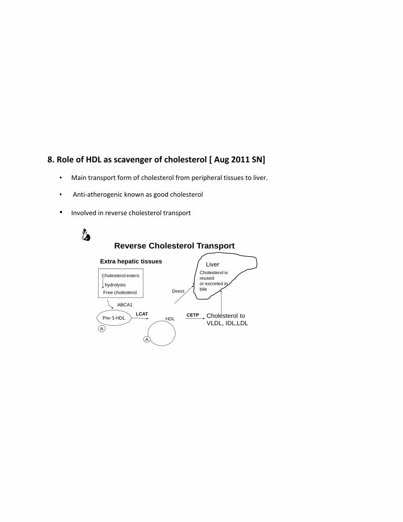
8. Role of HDL as scavenger of cholesterol [ Aug 2011 SN]
• Main transport form of cholesterol from peripheral tissues to liver.
• Anti-atherogenic known as good cholesterol
• Involved in reverse cholesterol transport
CETPLCAT
Free cholesterol
hydrolysis
Reverse Cholesterol Transport
Extra hepatic tissues
Cholesterol esters
Pre- -HDL
A
HDL
A
Cholesterol to
VLDL, IDL,LDL
Liver
Cholesterol is
reused
or excreted in
bile
ABCA1
Direct
9. FIGLU
Histidine is metabolized to formimino glutamic acid from which formiminogroup is removed by
THFA. Therefore in folate deficiency, FIGLU is excreted in urine.
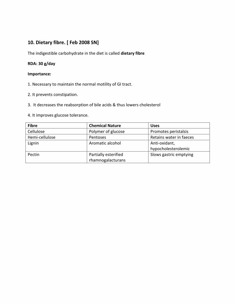
10. Dietary fibre. [ Feb 2008 SN]
The indigestible carbohydrate in the diet is called dietary fibre
RDA: 30 g/day
Importance:
1. Necessary to maintain the normal motility of GI tract.
2. It prevents constipation.
3. It decreases the reabsorption of bile acids & thus lowers cholesterol
4. It improves glucose tolerance.
Fibre Chemical Nature Uses
Cellulose Polymer of glucose Promotes peristalsis
Hemi-cellulose Pentoses Retains water in faeces
Lignin Aromatic alcohol Anti-oxidant, hypocholesterolemic
Pectin Partially esterified rhamnogalacturans
Slows gastric emptying