Embed Size (px)
Citation preview
Paraaortic Sentinel Lymph Nodes: TowardOptimal Detection and IntraoperativeLocalization Using SPECT/CT andIntraoperative Real-Time Imaging
Lenka Vermeeren1, Willem Meinhardt2, Axel Bex2, Henk G. van der Poel2, Wouter V. Vogel1, Cees A. Hoefnagel1,Simon Horenblas2, and Renato A. Valdes Olmos1
1Department of Nuclear Medicine, Netherlands Cancer Institute, Antoni van Leeuwenhoek Hospital, Amsterdam, The Netherlands;and 2Department of Urology, Netherlands Cancer Institute, Antoni van Leeuwenhoek Hospital, Amsterdam, The Netherlands
Paraaortic sentinel node biopsy may be a challenging proce-dure because the sentinel nodes are located retroperitoneallyin close proximity to vital structures. The purpose of this studywas to describe and evaluate the value of preoperative SPECT/CT for lymphatic mapping, and a portable g-camera for intra-operative radioguidance, in patients with paraaortic sentinelnodes. Methods: We evaluated our practice in 18 patients,who were treated at The Netherlands Cancer Institute with sen-tinel lymphadenectomy for different urologic malignancies andshowed paraaortic drainage on preoperative images. Afterintratumoral injection of 99mTc-nanocolloid, the patients under-went sequential planar lymphoscintigraphy, hybrid SPECT/CT,and sentinel lymphadenectomy. Intraoperative node detectionand localization were guided by a laparoscopic g-probe anda portable g-camera. This g-camera was set to display boththe 99mTc signal and the 125I-seed signal. This 125I seed wasplaced on top of the g-probe, functioning as a pointer onscreen, thus enabling real-time sentinel node localization withthe g-camera. Results: In 16 patients with midabdominal drain-age on planar images and in 2 patients with nonvisualization onplanar images, SPECT/CT showed clear localization of para-aortic sentinel nodes in relation to the abdominal vessels.Five patients underwent open surgery, and 13 patients under-went laparoscopy. The paraaortic sentinel nodes were suc-cessfully localized and removed in 15 patients with the help ofthe portable g-camera and g-probe and in 3 patients with theg-probe only. In 1 patient, the paraaortic sentinel node showeda metastasis. Conclusion: If retroperitoneal drainage isexpected, SPECT/CT provides good detection and clear local-ization of sentinel nodes in relation to anatomic structures. De-tection and removal of paraaortic sentinel nodes by means ofa laparoscopic g-probe and real-time imaging with a portableg-camera is a successful method with high intraoperative de-tection rates.
Key Words: sentinel lymph node biopsy; aorta; neoplasm stag-ing; gamma cameras; tomography, emission-computed, single-photon
J Nucl Med 2010; 51:376–382DOI: 10.2967/jnumed.109.071779
In recent decades, the use of sentinel lymph node biopsyhas been introduced as a possible staging procedure inseveral solid malignancies (1–3). Sentinel node biopsy isnow widely used for breast cancer and melanoma staging(4,5), and its possible value in several other malignancies isa reason for worldwide ongoing research. The concept ofsentinel node biopsy is based on the sequential spread ofmetastatic cancer cells through lymphatic flow. The valueof this procedure is based on the idea that if the first drain-ing lymph node from the tumor (sentinel node) is free ofmetastases, the remaining nodes in the same nodal basin willbe as well. Sentinel lymph node mapping is now used asa minimally invasive staging method in several malignan-cies to spare many node-negative patients the significantcomorbidity caused by regional lymph node dissection.
Sentinel node biopsies from axillary and inguinal nodalbasins have been shown to be safe and quick (6–8),probably in consequence of the superficial location of thesenodes and the widespread experience of surgeons withthese cases.
As the use of sentinel lymph node mapping is extending,so is the variety of lymph node basins containing thesentinel nodes. Experience with sentinel lymph nodemapping of retroperitoneal and intraabdominal nodal basinsis less widespread but increasing. The feasibility and valueof sentinel lymph node mapping in prostate cancer, withdrainage mainly to pelvic sentinel nodes, has been welldescribed (9,10). Furthermore, sentinel lymphadenectomyin cervical cancer, endometrial cancer, ovarian cancer, andtesticular cancer has been studied with different results
Received Oct. 21, 2009; revision accepted Dec. 9, 2009.For correspondence contact: Renato A. Valdes Olmos, Netherlands
Cancer Institute, Antoni van Leeuwenhoek Hospital, Plesmanlaan 121,Amsterdam 1066 CX, The Netherlands.
E-mail: [email protected] ª 2010 by the Society of Nuclear Medicine, Inc.
376 THE JOURNAL OF NUCLEAR MEDICINE • Vol. 51 • No. 3 • March 2010
(11,12). Its possible role in gastric cancer and colorectalcancer is reason for debate (13,14).
Exact lymphatic drainage patterns from tumors withexpected retroperitoneal drainage have not been clearlyidentified yet, and considerable interpersonal variation canbe observed. From several abdominal tumors, though,drainage to paraaortic sentinel lymph nodes might beexpected. Precise localization and removal of those para-aortic nodes is a requisite to achieve successful staging butcan be challenging because they are near vital structures.The purpose of the current study was to describe andevaluate our approach to paraaortic sentinel nodes withpreoperative SPECT/CT and the introduction of a portableg-camera for intraoperative radioguidance.
MATERIALS AND METHODS
PatientsWe evaluated our practice of sentinel node detection, localiza-
tion, and excision in 18 patients with paraaortic sentinel nodes.The median age of these patients was 57 y (range, 31–71 y). Fourpatients have also been included in a pilot study of the value ofa portable g-camera for intraoperative imaging of sentinel nodes(15). In all patients, the malignancy was diagnosed at TheNetherlands Cancer Institute between January 2006 and July2009, and all patients were included in different studies regardingthe value and feasibility of sentinel lymphadenectomy. Patientswere included in the current study if preoperative imaging showedparaaortic sentinel nodes. In the case of preoperative nonvisual-ization, patients were excluded from this analysis. The result wasinclusion of all patients with visualized paraaortic sentinel nodessince the introduction of SPECT/CT and intraoperative imaging: 8prostate cancer patients, 6 patients with renal cell carcinoma, and4 patients with testicular cancer. Patient characteristics and resultsper tumor are outlined in Table 1.
Preoperative Sentinel Node DetectionPatients underwent preoperative lymphoscintigraphy after in-
jection of 99mTc-nanocolloid (GE Healthcare).In prostate cancer patients, the tracer was injected, 1 depot of
0.1 mL per quadrant of the prostate, guided by transrectalultrasonography. Each depot of 0.1 mL was followed by flushingwith approximately 0.7 mL of saline. In renal cell carcinoma,intratumoral injection, in 2 depots of 0.2 mL, was guided byultrasonography, and in testicular cancer, 1 depot of 0.2 mL wasinjected intratesticularly. The remaining radioactivity in the in-jection device was subtracted from the injected dose to calculatethe net injected dose. The median injected dose was 205 MBq of99mTc-nanocolloid (range, 59–243 MBq).
All patients underwent planar lymphoscintigraphy and SPECT/CT to evaluate lymphatic drainage. Planar lymphoscintigraphywas performed at least twice: 15 min and 2 h after injection of theradiopharmaceutical. Planar images after 4 h were acquired onlyin the case of renal cell carcinoma, because for these tumorssentinel node mapping protocols have not been validated yet andwe wanted to ensure that we would not miss any drainage.
After the delayed planar images, SPECT and low-dose CT wereperformed, using a hybrid camera (SymbiaT; Siemens). Thissystem consists of a dual-head variable-angle g-camera equippedwith low-energy high-resolution collimators and a multislice
spiral CT component optimized for rapid rotation. The SPECTacquisition (128 · 128 matrix, 60 frames, 25 s/frame) wasperformed using 6� angular steps in a 20-s time frame. For CT(130 kV, 40 mAs, B30s kernel), 5-mm slices were obtained.
After correction for attenuation and scatter, correspondingSPECT and CT axial 5-mm slices were generated using an Esoft2000/Mi Apps application package (Siemens). An iterative re-construction was performed using 3-dimensional fast low-angleshot (8.4-mm gaussian). Images were fused using an Osirix Dicomviewer in a Unix-based operating system (MAC OS X, MacPro;Apple Inc.).
Furthermore, the images were analyzed using 2-dimensionalorthogonal reslicing in axial, sagittal, and coronal directions. Alsoa 3-dimensional presentation, using volume rendering, was gen-erated to localize sentinel nodes in relation to anatomic structures.
All images were available on a separate SPECT/CT screen inthe operation theater.
If a lymphatic channel leading to a paraaortic node was seen,this node was regarded as the sentinel node. If no lymphaticchannel was visualized, the node or nodes that were the first toappear intensely on early planar images were defined as thesentinel node or nodes. Nodes appearing later in the same stationswere considered to be second-echelon nodes. If SPECT/CTshowed additional hot spots in caudal areas or on a side withprevious drainage or no other drainage, those hot spots were alsoconsidered to be sentinel nodes. For prostate cancer patients, thisimplied that if an intense hot spot was seen in the paraaortic areabefore or simultaneously with pelvic nodes, this paraaortic nodewas considered to be a sentinel node. If this paraaortic hot spotappeared after visualization of intense pelvic nodes, it wasregarded as a node further downstream and those patients werenot included in this study.
Intraoperative Sentinel-Node DetectionAll patients with prostate cancer and testicular cancer un-
derwent surgery within 6 h after injection of the tracer. Patientswith renal cell carcinoma underwent surgery the next morning,because for logistic reasons late planar images up to 4 h afterinjection could not be combined with an operation on the sameday.
Sentinel nodes were removed laparoscopically in patients withprostate cancer and testicular cancer. In renal cell carcinoma, thesentinel lymph node biopsy was performed laparoscopically orthrough open surgery, depending on the primary tumor, otherpatient characteristics, and the surgeon’s preference.
For intraoperative sentinel node localization, we introduced theuse of a portable g-camera (Sentinella, S102; Oncovision), incombination with the g-probe (Europrobe; Euro Medical Instru-ments, and Neoprobe; Johnson&Johnson Medical). The portableg-camera was equipped with a 4-mm pinhole collimator and hasa field of view of 4 · 4 cm, which increases to 20 · 20 cm whenthe camera is placed at a distance of 15 cm from the patient’sbody. It uses a CsI(Na) continuously scintillating crystal and hasa 1.3-mm intrinsic resolution (15).
Before the start of sentinel node seeking, a 125I seed (.10MBq) was placed on the top of the g-probe. In the case oflaparoscopic sentinel lymphadenectomy, the 125I seed was placedon the laparoscopic probe. During the operation, this 125I seed wasused as a pointer, being displayed separately (as a green circle) onthe screen of the portable g-camera. To provide better orientationfor the surgeon and to avoid attenuation of the signal, the location
PARAAORTIC SENTINEL NODE LOCALIZATION • Vermeeren et al. 377

TA
BL
E1
Patient
Chara
cte
ristics
and
Results
Fin
din
gs
Typ
eo
f
malig
nancy
nA
ge
(y)
Inje
cte
dd
ose
(MB
q)
Pla
nar
imag
es
SP
EC
T/C
TS
urg
ery
Intr
ao
pera
tive
localiz
atio
n
Po
sto
pera
tive
co
urs
eP
ath
olo
gy
Pro
sta
te
cancer
8M
ed
ian:
68;
rang
e:
56–71
Med
ian:
205;
rang
e:
147–2
39
8m
idab
do
min
al
SN
s;16
pelv
ico
rp
resa
cra
l
SN
s
8S
Ns
aro
und
ao
rtic
bifurc
atio
n(e
xact
localiz
atio
n);
localiz
atio
no
f20
oth
er
SN
s
Lap
aro
sco
pic
sentinel
lym
phad
e-
necto
my
Dete
ctio
nand
localiz
atio
no
fall
SN
sw
ith
po
rtab
leg-c
am
era
and
pro
be
Unco
mp
licate
d:
7p
ts;
lym
pho
cele
:
1p
t
Para
ao
rtic
SN
po
sitiv
e:
1p
t;p
ara
iliac
SN
po
sitiv
e:
2p
ts
Renalcell
carc
ino
ma
6M
ed
ian:
54;
rang
e:
45–59
Med
ian:
224;
rang
e:
95–243
4m
idab
do
min
al
SN
s;1
para
ste
rnal
SN
;no
nvis
:
2p
ts
8p
ara
-and
inte
rao
rto
caval
SN
s(e
xact
localiz
atio
n);
1p
ara
ste
rnalS
N
Lap
aro
sco
pic
nep
hre
cto
my
and
sentinel
lym
phad
e-
necto
my:
1p
t;o
pen
pro
ced
ure
:
5p
ts
Vis
ualiz
atio
no
fS
Nw
ith
po
rtab
le
g-c
am
era
:3
pts
;
dete
ctio
np
ossib
le
only
with
g-p
rob
e:
3p
ts;
dete
ctio
no
f
ad
ditio
nalho
t
no
de
in1
pt*
Unco
mp
licate
d:
4p
ts;
infe
cte
d
wo
und
:1
pt;
cic
atr
icia
lhern
ia
of
nep
hre
cto
my
wo
und
:1p
t
All
SN
sneg
ative
Testicula
r
cancer
4M
ed
ian:
32;
rang
e:
31–40
Med
ian:
77;
rang
e:
59–97
7m
idab
do
min
al
SN
s
5in
tera
ort
ocavalS
Ns;
2le
ftp
ara
ao
rtic
SN
s;
1S
Nalo
ng
testicula
r
vein
(exact
localiz
atio
n);
localiz
atio
no
f1
para
iliac
SN
Lap
aro
sco
pic
sentinel
lym
phad
e-
necto
my
Dete
ctio
nand
localiz
atio
no
fall
SN
sw
ith
po
rtab
le
g-c
am
era
and
pro
be
Unco
mp
licate
d:
3p
ts;
hyd
ronep
hro
sis
:
1p
t
All
SN
s
neg
ative
*Tw
op
ara
ao
rtic
sentinelno
des
were
found
and
rem
oved
during
surg
ery
,w
here
as
pre
op
era
tive
SP
EC
T/C
Thad
vis
ualiz
ed
only
1p
ara
ao
rtic
sentinelno
de.
No
nvis
5no
nvis
ualiz
atio
n;
Pt
5p
atient;
SN
5sentinelno
de.
378 THE JOURNAL OF NUCLEAR MEDICINE • Vol. 51 • No. 3 • March 2010
of the 125I seed on the top of the g-probe was marked with a thinblack line. In this way, the location of the seed on the probe wasvisible. In the case of laparoscopy, the thin black line was applied
to the top of the laparoscopic g-probe and to the grip of thelaparoscopic probe. In this way, the surgeon could see where onthe probe the seed was located.
The collimator of the g-camera was sterile-wrapped to allowmanipulation by the surgeon and was placed above the previouslymarked sentinel node levels, using a laser pointer. Then, thecamera was set to display the 2 different signals: the 99mTc signalfor sentinel node localization and the 125I signal displayed asa green circle, functioning as a pointer. Matching of the 2 signalson the screen of the portable g-camera indicated correct localiza-tion of the sentinel node, which was subsequently removed. Thisintraoperative procedure is further explained in Figure 1.
All detected hot spots near the marked areas (sentinel nodelevels) were considered to be sentinel nodes and therefore werelocalized with the help of the g-camera and removed. Second-echelon nodes, as identified preoperatively, were left in place. Allremoved nodes were examined by experienced pathologists.
RESULTS
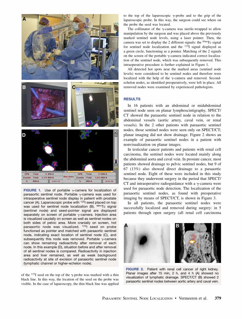
In 16 patients with an abdominal or midabdominalsentinel node seen on planar lymphoscintigraphy, SPECT/CT showed the paraaortic sentinel node in relation to theabdominal vessels (aortic artery, caval vein, or renalvessels). In the 2 other patients with paraaortic sentinelnodes, those sentinel nodes were seen only on SPECT/CT;planar imaging did not show drainage. Figure 2 shows anexample of paraaortic sentinel nodes in a patient withnonvisualization on planar images.
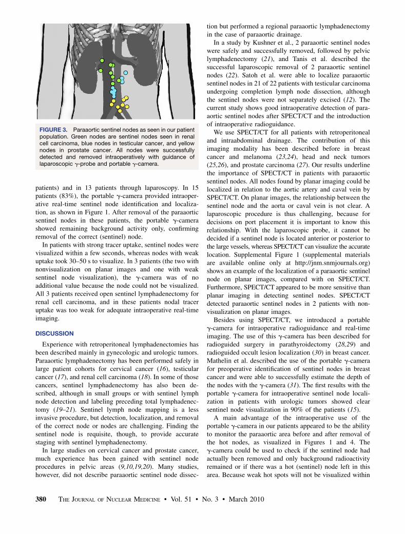
In testicular cancer patients and patients with renal cellcarcinoma, the sentinel nodes were located mainly alongthe abdominal aorta and caval vein. In prostate cancer, mostpatients showed drainage to pelvic sentinel nodes, but 9 of67 (13%) also showed direct drainage to a paraaorticsentinel node. Eight of these were included in this studybecause they underwent surgery in the period that SPECT/CT and intraoperative radioguidance with a g-camera wereused for paraaortic node detection. The localization of theparaaortic sentinel nodes, as found with preoperativeimaging by means of SPECT/CT, is shown in Figure 3.
In all patients, the paraaortic sentinel nodes weresuccessfully localized and removed during surgery: in 5patients through open surgery (all renal cell carcinoma
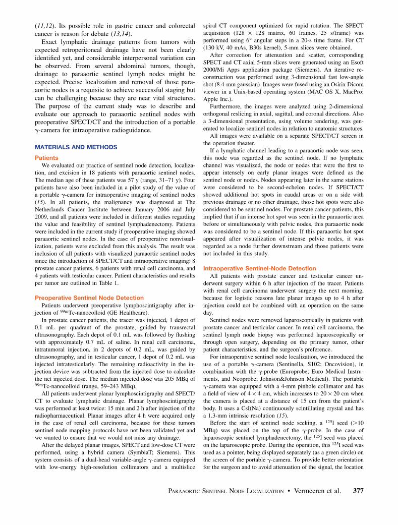
FIGURE 1. Use of portable g-camera for localization ofparaaortic sentinel node. Portable g-camera was used forintraoperative sentinel node display in patient with prostatecancer (A). Laparoscopic probe with 125I seed placed on topwas used for sentinel node localization (B). 99mTc signal(sentinel node) and seed-pointer signal are displayedseparately on screen of portable g-camera. Injection areais visualized caudally on screen as well as sentinel nodes onboth sides of pelvic area. More cranially on screen, hotparaaortic node was visualized. 125I seed on probefunctioned as pointer and matched with paraaortic sentinelnode, indicating exact location of sentinel node (C), andsubsequently this node was removed. Portable g-cameracan show remaining radioactivity after removal of eachnode. In this example (D), situation before and after removalof all sentinel nodes is compared. Radioactivity in injectionarea and liver remained, as well as weak backgroundradioactivity at site of excision of paraaortic sentinel node(lymphatic channel or higher-echelon node).
FIGURE 2. Patient with renal cell cancer of right kidney.Planar images after 15 min, 2 h, and 4 h (A) showed novisualization of lymphatic drainage. SPECT/CT (B) showed 2paraaortic sentinel nodes between aortic artery and caval vein.
PARAAORTIC SENTINEL NODE LOCALIZATION • Vermeeren et al. 379
patients) and in 13 patients through laparoscopy. In 15patients (83%), the portable g-camera provided intraoper-ative real-time sentinel node identification and localiza-tion, as shown in Figure 1. After removal of the paraaorticsentinel nodes in these patients, the portable g-camerashowed remaining background activity only, confirmingremoval of the correct (sentinel) node.
In patients with strong tracer uptake, sentinel nodes werevisualized within a few seconds, whereas nodes with weakuptake took 30–50 s to visualize. In 3 patients (the two withnonvisualization on planar images and one with weaksentinel node visualization), the g-camera was of noadditional value because the node could not be visualized.All 3 patients received open sentinel lymphadenectomy forrenal cell carcinoma, and in these patients nodal traceruptake was too weak for adequate intraoperative real-timeimaging.
DISCUSSION
Experience with retroperitoneal lymphadenectomies hasbeen described mainly in gynecologic and urologic tumors.Paraaortic lymphadenectomy has been performed safely inlarge patient cohorts for cervical cancer (16), testicularcancer (17), and renal cell carcinoma (18). In some of thosecancers, sentinel lymphadenectomy has also been de-scribed, although in small groups or with sentinel lymphnode detection and labeling preceding total lymphadenec-tomy (19–21). Sentinel lymph node mapping is a lessinvasive procedure, but detection, localization, and removalof the correct node or nodes are challenging. Finding thesentinel node is requisite, though, to provide accuratestaging with sentinel lymphadenectomy.
In large studies on cervical cancer and prostate cancer,much experience has been gained with sentinel nodeprocedures in pelvic areas (9,10,19,20). Many studies,however, did not describe paraaortic sentinel node dissec-
tion but performed a regional paraaortic lymphadenectomyin the case of paraaortic drainage.
In a study by Kushner et al., 2 paraaortic sentinel nodeswere safely and successfully removed, followed by pelviclymphadenectomy (21), and Tanis et al. described thesuccessful laparoscopic removal of 2 paraaortic sentinelnodes (22). Satoh et al. were able to localize paraaorticsentinel nodes in 21 of 22 patients with testicular carcinomaundergoing completion lymph node dissection, althoughthe sentinel nodes were not separately excised (12). Thecurrent study shows good intraoperative detection of para-aortic sentinel nodes after SPECT/CT and the introductionof intraoperative radioguidance.
We use SPECT/CT for all patients with retroperitonealand intraabdominal drainage. The contribution of thisimaging modality has been described before in breastcancer and melanoma (23,24), head and neck tumors(25,26), and prostate carcinoma (27). Our results underlinethe importance of SPECT/CT in patients with paraaorticsentinel nodes. All nodes found by planar imaging could belocalized in relation to the aortic artery and caval vein bySPECT/CT. On planar images, the relationship between thesentinel node and the aorta or caval vein is not clear. Alaparoscopic procedure is thus challenging, because fordecisions on port placement it is important to know thisrelationship. With the laparoscopic probe, it cannot bedecided if a sentinel node is located anterior or posterior tothe large vessels, whereas SPECT/CT can visualize the accuratelocation. Supplemental Figure 1 (supplemental materialsare available online only at http://jnm.snmjournals.org)shows an example of the localization of a paraaortic sentinelnode on planar images, compared with on SPECT/CT.Furthermore, SPECT/CT appeared to be more sensitive thanplanar imaging in detecting sentinel nodes. SPECT/CTdetected paraaortic sentinel nodes in 2 patients with non-visualization on planar images.
Besides using SPECT/CT, we introduced a portableg-camera for intraoperative radioguidance and real-timeimaging. The use of this g-camera has been described forradioguided surgery in parathyroidectomy (28,29) andradioguided occult lesion localization (30) in breast cancer.Mathelin et al. described the use of the portable g-camerafor preoperative identification of sentinel nodes in breastcancer and were able to successfully estimate the depth ofthe nodes with the g-camera (31). The first results with theportable g-camera for intraoperative sentinel node locali-zation in patients with urologic tumors showed clearsentinel node visualization in 90% of the patients (15).
A main advantage of the intraoperative use of theportable g-camera in our patients appeared to be the abilityto monitor the paraaortic area before and after removal ofthe hot nodes, as visualized in Figures 1 and 4. Theg-camera could be used to check if the sentinel node hadactually been removed and only background radioactivityremained or if there was a hot (sentinel) node left in thisarea. Because weak hot spots will not be visualized within
FIGURE 3. Paraaortic sentinel nodes as seen in our patientpopulation. Green nodes are sentinel nodes seen in renalcell carcinoma, blue nodes in testicular cancer, and yellownodes in prostate cancer. All nodes were successfullydetected and removed intraoperatively with guidance oflaparoscopic g-probe and portable g-camera.
380 THE JOURNAL OF NUCLEAR MEDICINE • Vol. 51 • No. 3 • March 2010
a few seconds, we decided that imaging of at least 1 min isrequired for accurate postexcision monitoring. However,whether a very hot node has been correctly removed will beclear within 20–30 s.
In laparoscopic procedures, where spatial mobility islimited, SPECT/CT images and the visualization of the 125Iseed on the laparoscopic probe can lead the surgeon to thesentinel node. In this way, intraoperative identification andlocalization of the sentinel nodes appears to improve. Theportable g-camera can visualize and identify sentinel nodesclose to the injection area, which is nearly impossible withthe g-probe. The 125I-seed signal is not hampered by the
99mTc signal from the injection area, and therefore, espe-cially nodes near the injection area are more easilylocalized with this method. If underneath the injectionarea, however, the node is not visible on the screen of theportable g-camera. That situation was never the case in ourgroup of patients because the primary tumors were not inthe preaortic region.
In 3 patients with weak nodal tracer uptake, the para-aortic sentinel nodes could not be depicted on the screenof the portable g-camera. Sufficient tracer uptake is re-quired for adequate intraoperative real-time imaging withg-cameras.
In our practice, there are no additional costs for the useof the 125I seed as a pointer, because remaining seeds thathave become too weak for brachytherapy can be used aslong as radioactivity is above 10 MBq. We choose to use aniodine source because it is freely available at our institute.Another reason is that the energy peak of 125I (35 keV)differs greatly from the energy peak of 99mTc (140 keV),making optimal separation of the 2 signals on the screenof the portable g-camera possible. Other g-ray–emittingsources (e.g., barium, armarium, or gadolinium) could alsobe used as a pointer on the screen of the portable g-camera.However, to be able to distinguish both signals, the energypeak from the isotope used as the point source should differsubstantially from the energy peak of the tracer (99mTc).
CONCLUSION
If preoperative planar lymphoscintigraphy shows possi-ble paraaortic lymph nodes, SPECT/CT will show the exactlocation of those nodes in relation to abdominal anatomicstructures (mainly the large vessels) and will thereforemoderate intraoperative tracing of those nodes. Further-more, SPECT/CT can detect sentinel nodes if they are notvisualized on planar images. If drainage to retroperitonealnodes is expected, routine use of SPECT/CT is advisable.Intraoperative localization of paraaortic sentinel nodes bymeans of a laparoscopic g-probe and intraoperative real-time imaging with a portable g-camera has shown gooddetection and removal rates in our population. The portableg-camera can also provide certainty about the completenessof the sentinel lymphadenectomy, showing remainingradioactivity after removal of the node.
REFERENCES
1. Cabanas RM. An approach for the treatment of penile carcinoma. Cancer.
1977;39:456–466.
2. Gould EA, Winship T, Philbin PH, Kerr HH. Observations on a ‘‘sentinel node’’
in cancer of the parotid. Cancer. 1960;13:77–78.
3. Morton DL, Wen DR, Wong JH, et al. Technical details of intraoperative
lymphatic mapping for early stage melanoma. Arch Surg. 1992;127:392–399.
4. Morton DL, Thompson J, Cochran A, et al. Sentinel-node biopsy or nodal
observation in melanoma. N Engl J Med. 2006;355:1307–1317.
5. Veronesi U, Paganelli G, Galimberti V, et al. Sentinel-node biopsy to avoid
axillary dissection in breast cancer with clinically negative lymph-nodes. Lancet.
1997;349:1864–1867.
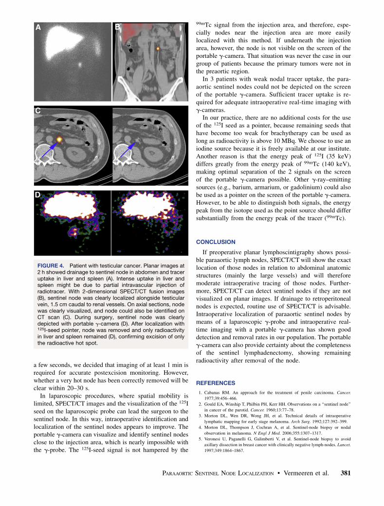
FIGURE 4. Patient with testicular cancer. Planar images at2 h showed drainage to sentinel node in abdomen and traceruptake in liver and spleen (A). Intense uptake in liver andspleen might be due to partial intravascular injection ofradiotracer. With 2-dimensional SPECT/CT fusion images(B), sentinel node was clearly localized alongside testicularvein, 1.5 cm caudal to renal vessels. On axial sections, nodewas clearly visualized, and node could also be identified onCT scan (C). During surgery, sentinel node was clearlydepicted with portable g-camera (D). After localization with125I-seed pointer, node was removed and only radioactivityin liver and spleen remained (D), confirming excision of onlythe radioactive hot spot.
PARAAORTIC SENTINEL NODE LOCALIZATION • Vermeeren et al. 381

6. Morton DL, Cochran AJ, Thompson JF, et al. Sentinel node biopsy for early-
stage melanoma: accuracy and morbidity in MSLT-I, an international multicenter
trial. Ann Surg. 2005;242:302–311.
7. Veronesi U, Paganelli G, Viale G, et al. A randomized comparison of sentinel-
node biopsy with routine axillary dissection in breast cancer. N Engl J Med.
2003;349:546–553.
8. Van der Ploeg IM, Nieweg OE, van Rijk MC, Valdes Olmos RA, Kroon BBR.
Axillary recurrence after a tumour-negative sentinel node biopsy in breast cancer
patients: a systematic review and meta-analysis of the literature. Eur J Surg
Oncol. 2008;34:1277–1284.
9. Jeschke S, Nambirajan T, Leeb K, Ziegerhofer J, Sega W, Janetschek G.
Detection of early lymph node metastases in prostate cancer by laparoscopic
radioisotope guided sentinel lymph node dissection. J Urol. 2005;173:1943–1946.
10. Weckermann D, Dorn R, Trefz M, Wagner T, Wawroschek F, Harzmann R.
Sentinel lymph node dissection for prostate cancer: experience with more than
1,000 patients. J Urol. 2007;177:916–920.
11. El-Ghobashy AE, Saidi SA. Sentinel lymph node sampling in gynaecological
cancers: techniques and clinical applications. Eur J Surg Oncol. 2009;35:
675–685.
12. Satoh M, Ito A, Kaiho Y, et al. Intraoperative, radio-guided sentinel lymph node
mapping in laparoscopic lymph node dissection for stage I testicular carcinoma.
Cancer. 2005;103:2067–2072.
13. Govindarajan A, Baxter NN. Lymph node evaluation in early-stage colon cancer.
Clin Colorectal Cancer. 2008;7:240–246.
14. Ozmen MM, Ozmen F, Zulfikaroglu B. Lymph nodes in gastric cancer. J Surg
Oncol. 2008;98:476–481.
15. Vermeeren L, Valdes Olmos RA, Meinhardt W, et al. Intraoperative radio-
guidance with a portable gamma camera: a novel technique for laparoscopic
sentinel node localization in urological malignancies. Eur J Nucl Med Mol
Imaging. 2009;36:1029–1036.
16. Querleu D, Leblanc E, Cartron G, Narducci F, Ferron G, Martel P. Audit of
preoperative and early complications of laparoscopic lymph node dissection in
1000 gynecologic cancer patients. Am J Obstet Gynecol. 2006;195:1287–1292.
17. LeBlanc E, Caty A, Dargent D, Querleu D, Mazeman E. Extraperitoneal
laparoscopic para-aortic lymph node dissection for early stage nonseminomatous
germ cell tumors of the testis with introduction of a nerve sparing technique:
description and results. J Urol. 2001;165:89–92.
18. Blom JH, van Poppel H, Marechal J, et al. Radical nephrectomy with and
without lymph-node dissection: final results of European Organization for
Research and Treatment of Cancer (EORTC) randomized phase 3 trial 30881.
Eur Urol. 2009;55:28–34.
19. Coutant C, Morel O, Delpech Y, Uzan S, Daraı E, Barranger E. Laparoscopic
sentinel node biopsy in cervical cancer using a combined detection: 5-years
experience. Ann Surg Oncol. 2007;14:2392–2399.
20. Lavoue V, Bats A, Rouzier R, Coutant C, Barranger E, Daraı E. Sentinel lymph
node procedure followed by laparoscopic pelvic and paraaortic lymphadenec-
tomy in women with IB2-II cervical cancer. Ann Surg Oncol. 2007;14:2654–
2661.
21. Kushner DM, Connor J, Wilson M, et al. Laparoscopic sentinel lymph node
mapping for cervix cancer: a detailed evaluation and time analysis. Gynecol
Oncol. 2007;106:507–512.
22. Tanis PJ, Horenblas S, Valdes Olmos RA, Hoefnagel CA, Nieweg OE.
Feasibility of sentinel node lymphoscintigraphy in stage I testicular cancer.
Eur J Nucl Med Mol Imaging. 2002;29:670–673.
23. Van der Ploeg IM, Valdes Olmos RA, Kroon BBR, et al. The yield of SPECT/CT
for anatomical lymphatic mapping in patients with melanoma. Ann Surg Oncol.
2009;16:1537–1542.
24. Van der Ploeg IM, Nieweg OE, Kroon BBR, et al. The yield of SPECT/CT for
anatomical lymphatic mapping in patients with breast cancer. Eur J Nucl Med
Mol Imaging. 2009;36:903–909.
25. Bilde A, Von Buchwald C, Mortensen J, et al. The role of SPECT-CT in the
lymphoscintigraphic identification of sentinel nodes in patients with oral cancer.
Acta Otolaryngol. 2006;126:1096–1103.
26. Khafif A, Schneebaum S, Fliss D, et al. Lymphoscintigraphy for sentinel node
mapping using a hybrid single photon emission CT (SPECT)/CT system in oral
cavity squamous cell carcinoma. Head Neck. 2006;28:874–879.
27. Vermeeren L, Valdes Olmos RA, Meinhardt W, et al. Value of SPECT/CT for
detection and anatomical localization of sentinel lymph nodes before
laparoscopic sentinel node lymphadenectomy in prostate carcinoma. J Nucl
Med. 2009;50:865–870.
28. Kitagawa W, Shimizu K, Akasu H. Radioguided parathyroidectomy for primary
hyperparathyroidism using the solid-state, multi-crystal gamma camera. Med Sci
Monit. 2003;9:CS53–CS56.
29. Ortega J, Ferrer-Rebolleda J, Cassinello N, Lledo S. Potential role of a new
hand-held miniature gamma camera in performing minimally invasive para-
thyroidectomy. Eur J Nucl Med Mol Imaging. 2007;34:165–169.
30. Paredes P, Vidal-Sicart S, Zanon G, et al. Radioguided occult lesion localization
in breast cancer using an intraoperative portable gamma camera: first results. Eur
J Nucl Med Mol Imaging. 2008;35:230–235.
31. Mathelin C, Salvador S, Huss D, Guyonnet JL. Precise localization of sentinel
lymph nodes and estimation of their depth using a prototype intraoperative mini
gamma-camera in patients with breast cancer. J Nucl Med. 2007;48:623–629.
382 THE JOURNAL OF NUCLEAR MEDICINE • Vol. 51 • No. 3 • March 2010