Embed Size (px)
Citation preview
Department of Medical Biology and Parasitology
Wrocław Medical University
Parasitology
Exercise booklet
Prepared by (in alphabetic order):
Agnieszka Cisowska, Andrzej Hendrich, Marta Kicia, Dorota
Tichaczek-Goska, Maria Wesołowska, Dorota Wojnicz
Wrocław 2014
Parasitology exercise booklet 2014/2015
Department of Medical Biology and Parasitology
2
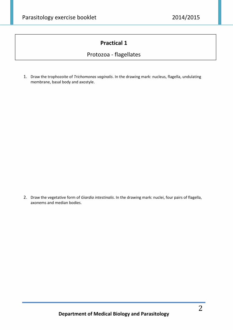
Practical 1
Protozoa - flagellates
1. Draw the trophozoite of Trichomonas vaginalis. In the drawing mark: nucleus, flagella, undulating membrane, basal body and axostyle.
2. Draw the vegetative form of Giardia intestinalis. In the drawing mark: nuclei, four pairs of flagella, axonems and median bodies.
Parasitology exercise booklet 2014/2015
Department of Medical Biology and Parasitology
3
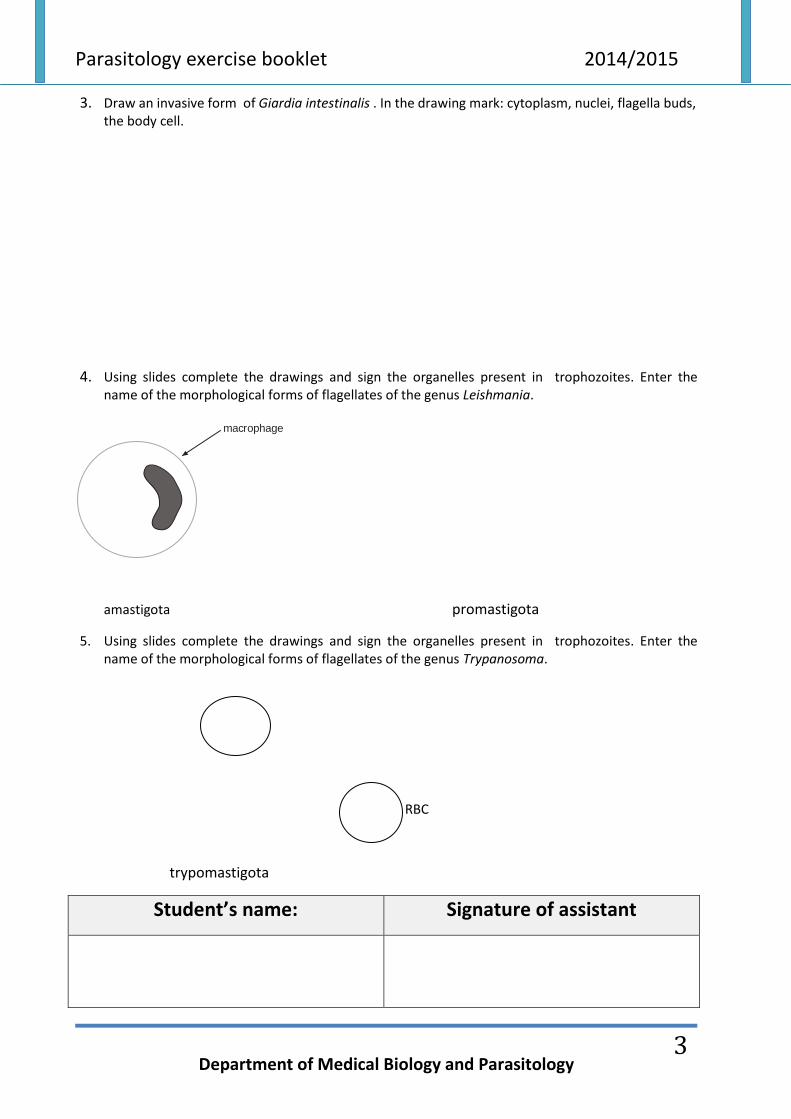
3. Draw an invasive form of Giardia intestinalis . In the drawing mark: cytoplasm, nuclei, flagella buds, the body cell.
4. Using slides complete the drawings and sign the organelles present in trophozoites. Enter the name of the morphological forms of flagellates of the genus Leishmania.
macrophage
amastigota promastigota
5. Using slides complete the drawings and sign the organelles present in trophozoites. Enter the name of the morphological forms of flagellates of the genus Trypanosoma.
RBC
trypomastigota
Student’s name: Signature of assistant
Parasitology exercise booklet 2014/2015
Department of Medical Biology and Parasitology
4
Practical 2
Protozoa - Amoebae and Apicomplexa
1. Draw the trophozoite of Entamoeba histolytica and mark the following elements: ectoplasm
(pseudopodium), endoplasm, nucleus, kariosome, erythrocytes in vacuoles.
2. Draw the cyst of Entamoeba histolytica and mark the following: cytoplasm, nuclei, chromatoidal
bars.
Parasitology exercise booklet 2014/2015
Department of Medical Biology and Parasitology
5
3. Draw and describe the trophozoite of Acanthamoeba castellanii.
4. Draw and describe two stages of Plasmodium falciparum, a signet ring form in the erythrocyte,
gametocyte. In the signet ring stage and in the gametocyte point the nucleus.
5. Draw a schizont of the Plasmodium vivax in erythrocyte, mark the nuclei of the schizont after the division.
Parasitology exercise booklet 2014/2015
Department of Medical Biology and Parasitology
6
6. Draw the trophozoite of Toxoplasma gondii, mark the nucleus.
7. Draw an oocyst of Cryptosporidium with four sporozoites.
Student’s name: Signature of assistant
Parasitology exercise booklet 2014/2015
Department of Medical Biology and Parasitology
7
Practical 3
Platyhelminthes (flatworms) - Trematodes
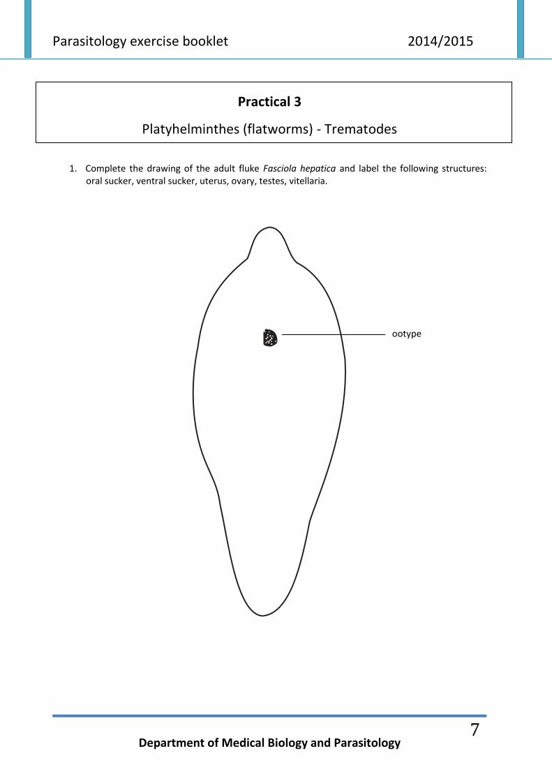
1. Complete the drawing of the adult fluke Fasciola hepatica and label the following structures:
oral sucker, ventral sucker, uterus, ovary, testes, vitellaria.
ootype
Parasitology exercise booklet 2014/2015
Department of Medical Biology and Parasitology
8
2. Taking into account the differences in size draw (from the largest to the smallest) and label the
eggs of the following flukes: Clonorchis sinensis, Fasciola hepatica, Schistosoma mansoni.
………………………………… …………………………………. ……………………………………
Student’s name: Signature of assistant
Parasitology exercise booklet 2014/2015
Department of Medical Biology and Parasitology
9
Practical 4
Platyhelminthes (flatworms) - Cestodes
1. Draw the mature proglottid of Diphyllobothrium latum and label the following structures: uterus,
genital pore, follicles of testes and vitellaria, excretory ducts.
2. Draw an egg of Diphyllobothrium latum taking into account localization of the embryo and
operculum (in proper proportions).
Parasitology exercise booklet 2014/2015
Department of Medical Biology and Parasitology
10
3. Draw and label the gravid proglottid of Taenia saginata with consideration to the correct number of primary branches of the uterus and the localization of the genital pore.
4. Draw an invasive egg of Taenia spp. and label larvae (oncosphere) and the sheath surrounding it (embryophore).
Parasitology exercise booklet 2014/2015
Department of Medical Biology and Parasitology
11
5. Draw a fragment of the strobila of Hymenolepis nana (3-4 gravid proglottids) and in one of them draw and label uterus filled with eggs.
6. Observe the protoscolex of Echinococcus granulosus under microscope.
Student’s name: Signature of assistant
Parasitology exercise booklet 2014/2015
Department of Medical Biology and Parasitology
12
Practical 5
Nemathelminthes (roundworms) - Nematodes
1. Draw female and male Ascaris lumbricoides hominis taking into account sexual dimorphism. Label
the posterior end of male’s body. 2. Draw an egg of Ascaris lumbricoides hominis and label the protein coat. 3. Draw female and male Trichuris trichiura taking into account sexual dimorphism. Label the anterior
and posterior end of the body.
Parasitology exercise booklet 2014/2015
Department of Medical Biology and Parasitology
13
4. Draw Trichuris trichiura egg and label the egg shell and two polar plugs. 5. Draw an egg of Enterobius vermicularis. 6. Draw the encysted larva of Trichinella spiralis located in the striated muscles. Label: muscle fibers,
nurse cell (capsule), L2 larva.
Parasitology exercise booklet 2014/2015
Department of Medical Biology and Parasitology
14
7. Draw Loa loa microfilaria and label the following structures: elastic sheath, nerve ring, excretory pore and anal pore.
8. Observe male and female of Nippostrongylus brasiliensis under microscope.
Student’s name: Signature of assistant