Embed Size (px)
Citation preview

Part 1: Assessment and Management of Pressure Ulcers
Part A: Educational Workshop for RNs and RPNsAssessment and Management of Pressure Ulcers
Based on the Registered Nurses’ Association of Ontario
Best Practice Guideline:
Assessment and Management of Stage I to IV Pressure Ulcers

Educational Workshop for RNs and RPNs: Assessment and Management of Pressure Ulcers
Nursing Best Practice Guidelines Program Registered Nurses’ Association of Ontario
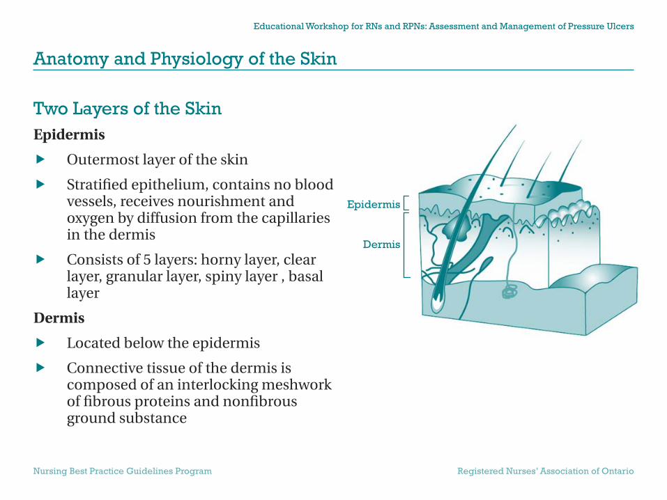
Anatomy and Physiology of the Skin
The Skin Defi nes us
Largest organ in the body
Weighs 3kg
(Molony et al., 1999)
Educational Workshop for RNs and RPNs: Assessment and Management of Pressure Ulcers
Anatomy and Physiology of the Skin
Two Layers of the SkinEpidermis
Outermost layer of the skin
Stratifi ed epithelium, contains no blood vessels, receives nourishment and oxygen by diffusion from the capillaries in the dermis
Consists of 5 layers: horny layer, clear layer, granular layer, spiny layer , basal layer
Dermis
Located below the epidermis
Connective tissue of the dermis is composed of an interlocking meshwork of fi brous proteins and nonfi brous ground substance
Nursing Best Practice Guidelines Program Registered Nurses’ Association of Ontario
Epidermis
Dermis

Educational Workshop for RNs and RPNs: Assessment and Management of Pressure Ulcers
Anatomy and Physiology of the Skin
Nursing Best Practice Guidelines Program Registered Nurses’ Association of Ontario
Subcutaneous Tissue
Subcutaneous TissueComposed of:
Loose connective tissue
Adipose tissue
Elements of peripheral vasculature

Educational Workshop for RNs and RPNs: Assessment and Management of Pressure Ulcers
Anatomy and Physiology of the Skin
Nursing Best Practice Guidelines Program Registered Nurses’ Association of Ontario
Six Functions of the Skin Supports underlying body structures
Maintains thermoregulation
(body temperature)
Source of sensation
Eliminates waste
Protects
Synthesizes vitamin D
(Molony et al., 1999)

Educational Workshop for RNs and RPNs: Assessment and Management of Pressure Ulcers
Anatomy and Physiology of the Skin
Nursing Best Practice Guidelines Program Registered Nurses’ Association of Ontario
Factors that Affect Skin Condition Dryness
Age
Nutrition
Hydration
Environment

Educational Workshop for RNs and RPNs: Assessment and Management of Pressure Ulcers
Nursing Best Practice Guidelines Program Registered Nurses’ Association of Ontario
Assessing Risk Factors for Developing Pressure Ulcers
Pressure UlcerDefi nition
Any lesion caused by unrelieved pressure that results in damage to underlying tissue
Usually occurs over a bony prominence
Staged to classify the degree of tissue damage observed
(National Pressure Ulcer Advisory Panel, 1989)
Educational Workshop for RNs and RPNs: Assessment and Management of Pressure Ulcers
Nursing Best Practice Guidelines Program Registered Nurses’ Association of Ontario
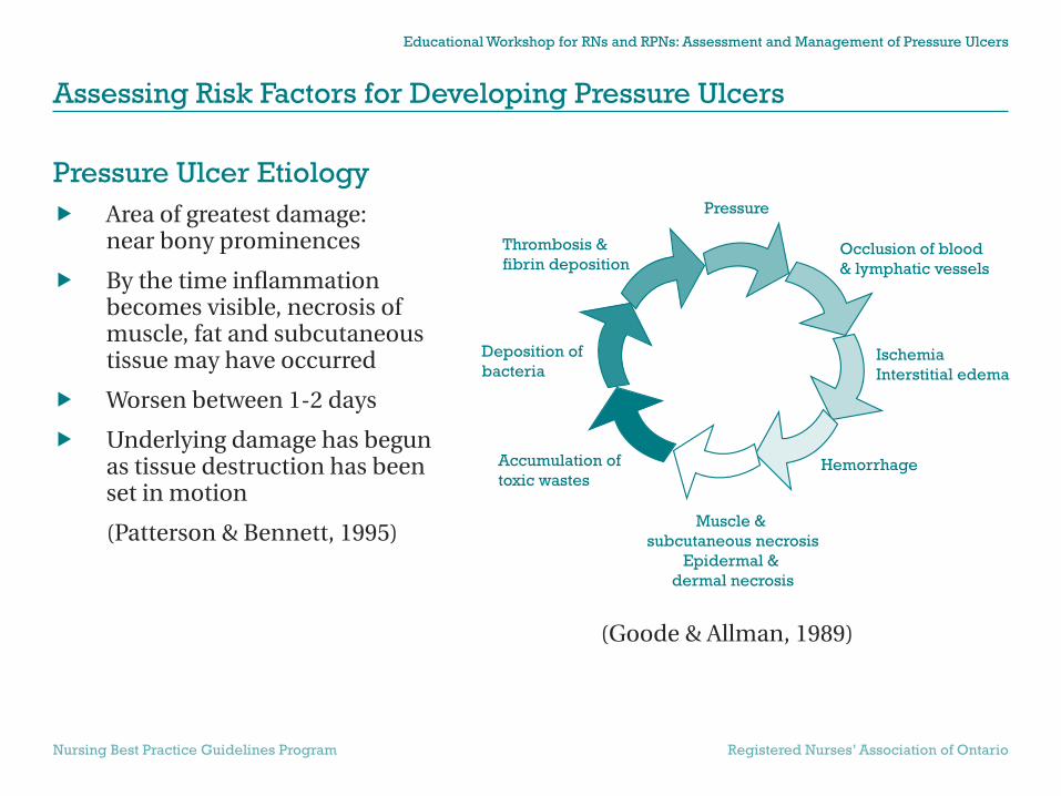
Assessing Risk Factors for Developing Pressure Ulcers
Pressure Ulcer Etiology Area of greatest damage:
near bony prominences
By the time infl ammation becomes visible, necrosis of muscle, fat and subcutaneous tissue may have occurred
Worsen between 1-2 days
Underlying damage has begun as tissue destruction has been set in motion
(Patterson & Bennett, 1995)
(Goode & Allman, 1989)
Pressure
Accumulation oftoxic wastes
Deposition ofbacteria
Thrombosis &fibrin deposition
Occlusion of blood& lymphatic vessels
IschemiaInterstitial edema
Hemorrhage
Muscle &subcutaneous necrosis
Epidermal &dermal necrosis

Educational Workshop for RNs and RPNs: Assessment and Management of Pressure Ulcers
Nursing Best Practice Guidelines Program Registered Nurses’ Association of Ontario
Assessing Risk Factors for Developing Pressure Ulcers
Risk Factors Mobility
Activity
Sensory Perception
Nutrition
Arteriolar Pressure
Pressure
Moisture
Friction
Shear
Age
(Braden, 1987)

Educational Workshop for RNs and RPNs: Assessment and Management of Pressure Ulcers
Nursing Best Practice Guidelines Program Registered Nurses’ Association of Ontario
Assessing Risk Factors for Developing Pressure Ulcers
Common Pressure Ulcer SitesSupine Position
heels, sacrum, elbows, scapulae, back of head
Lateral Position
malleous, medial and lateral condyles, greater trochanter, ribs, acromion process, ear
Prone Position
toes, knees, genitalia (men), breasts (women), acromion process, cheek and ear
Sitting Position
elbow, sacrum, ischium
Illustrated by Nancy A. Bauer, BA, Bus Admin, RN, ET

Educational Workshop for RNs and RPNs: Assessment and Management of Pressure Ulcers
Nursing Best Practice Guidelines Program Registered Nurses’ Association of Ontario
Assessing Risk Factors for Developing Pressure Ulcers
When to Assess Risk On admission
Weekly basis
Change in patient/residents’ health status
Before transfer to another facility

Educational Workshop for RNs and RPNs: Assessment and Management of Pressure Ulcers
Nursing Best Practice Guidelines Program Registered Nurses’ Association of Ontario
Assessing Risk Factors for Developing Pressure Ulcers
The Braden Scale validated instrument
used to aid professional and clinical judgment
full patient profi le context
composed of six sub-scales that refl ect sensory perception, skin moisture, activity, mobility, friction and shear, and nutritional status
fi ve of the six sub-scales are rated from one (1) (least favourable) to four (4) (most favourable); friction and shear sub-scale is rated from one (1) to three (3). A total of 23 points is possible. The lower the score, the higher the risk for pressure ulcer development
total score is only a number to guide interventions
(Braden, 2001; Ayello, 1999)

Educational Workshop for RNs and RPNs: Assessment and Management of Pressure Ulcers
Nursing Best Practice Guidelines Program Registered Nurses’ Association of Ontario
Assessing Risk Factors for Developing Pressure Ulcers
The Braden Scale for Predicting Pressure Sore Risk
1. Completely limitedUnresponsive (does not moan, flinch or grasp) to painful stimuli due to diminished level ofconsciousness or sedation, or limited ability tofeel pain over most of body surface.
2. Very limitedResponds only to painful stimuli. Cannot communicate discomfort except by moaning orrestlessness, or has a sensory impairment thatlimits the ability to feel pain or discomfort overhalf of body.
Sensory PerceptionAbility to respond meaningfully to pressure-related discomfort.
MoistureDegree to which skin is exposed to moisture.
ActivityDegree of physical activity.
MobilityAbility to change and control body position.
NutritionUsual food intake
pattern.
Friction and Shear
1. Constantly moistSkin is kept moist almost constantly by perspiration, urine, etc. Dampness is detectedevery time patient is moved or turned.
2. Very moistSkin is often, but not always, moist. Linen must be changed at least once a shift.
1. BedfastConfined to bed.
2. ChairfastAbility to walk severely limited or non-existent. Cannot bear own weight and/ormust be assisted into chair or wheelchair.
1. Completely immobileDoes not make even slight changes in body or extremity position without assistance.
2. Very limitedMakes occasional, slight changes in body or extremity position but unable to make frequentor significant changes independently.
1. Very poorNever eats a complete meal. Rarely eats morethan 1/3 of any food offered. Eats 2 servings orless of protein (meat or dairy products) perday. Takes fluids poorly. Does not take a liquiddietary supplement, or is NPO and/or maintainedon clear liquids or IVs for more than 5 days.
2. Probably inadequateRarely eats a complete meal and generallyeats only about half of any food offered.Protein intake includes only 3 servings of meator dairy products per day. Occasionally will takea dietary supplement, or receives less than opti-mum amount of liquid diet or tube feeding.
1. ProblemRequires moderate to maximum assistance inmoving. Complete lifting without slidingagainst sheets is impossible. Frequently slidesdown in bed or chair, requiring frequentrepositioning with maximum assistance.Spasticity, contractures or agitation lead toalmost constant friction.
2. Potential ProblemsMoves feebly or requires minimum assis-tance. During a move skin probably slides tosome extent against sheets, chair restraints,or other devices. Maintains relatively goodposition in chair or bed most of the time butoccasionally slides down.
(Copyright, 1988. Reprinted with permission)
3. Slightly limitedResponds to verbal commands but cannot always communicate discomfort or need to beturned, or has some sensory impairment thatlimits ability to feel pain or discomfort in 1 or 2extremities.
4. No impairmentResponds to verbal commands, has no sensory deficit that would limit ability to feel or voicepain or discomfort.
3. Occasionally moistSkin is occasionally moist, requiring an extralinen change approximately once a day.
4. Rarely moistSkin is usually dry, linen only requires changing at routine intervals.
3. Walks occasionallyWalks occasionally during day, but for very short distances with or without assistance.Spends majority of each shift in bed or chair.
4. Walks frequentlyWalks outside the room at least twice a day and inside room at least every 2 hours duringwaking hours.
3. Slightly limitedMakes frequent though slight changes in body or extremity position independently.
4. Walks frequentlyMakes major and frequent changes in position without assistance.
3. AdequateEats over half of most meals. Eats a total of 4 servings of protein (meat, dairy products)each day. Occasionally will refuse a meal, butwill usually take a supplement if offered, or ison a tube feeding or TPN regimen, whichmeets most of nutritional needs.
4. ExcellentEats most of every meal. Never refuses a meal. Usually eats a total of 4 or moreservings of meat and dairy products.Occasionally eats between meals. Does not require supplementation.
3. No apparent problemMoves in bed and in chair independently andhas sufficient muscle strength to lift up com-pletely during move. Maintains good positionin bed or chair at all times.
NOTE: Patients with a total score of 16 or lessare considered to be at risk of developing pressure ulcers. (15 or 16=low risk; 13 or 14=moderate risk; 12 or less=high risk)
TOTAL SCORE
SCORE
© Copyright Barbara Braden and Nancy Bergstrom, 1988. Reproduced with permission.

Educational Workshop for RNs and RPNs: Assessment and Management of Pressure Ulcers
Nursing Best Practice Guidelines Program Registered Nurses’ Association of Ontario
Preventative Skin Care
Identify patient/residents at risk for skin breakdown
Utilize the Braden scale to identify the level of patient/resident risk
Implement a plan to reduce risk factors and prevent skin breakdown
Monitor skin integrity
Prevent dehydration and malnutrition

Educational Workshop for RNs and RPNs: Assessment and Management of Pressure Ulcers
Nursing Best Practice Guidelines Program Registered Nurses’ Association of Ontario
Preventative Skin Care
Prevent pressure and trauma in order to maintain skin integrityDo’s
Prevent local areas of pressure
Provide pressure reduction via use of mattress overlays, cushions, or foams
Don’ts
Sheepskin provides comfort only, not pressure reduction
Eggcrate mattresses and donut devices do not provide pressure reduction

Educational Workshop for RNs and RPNs: Assessment and Management of Pressure Ulcers
Nursing Best Practice Guidelines Program Registered Nurses’ Association of Ontario
Preventative Skin Care
Prevent maceration, irritation and bacterial growth
Keep skin hydrated and supple
Keep skin clean and dry
Wash skin gently with water; pH balanced soaps or skin cleansers
Investigate and manage incontinence
Educational Workshop for RNs and RPNs: Assessment and Management of Pressure Ulcers
Nursing Best Practice Guidelines Program Registered Nurses’ Association of Ontario
Preventative Skin Care
Prevent moisture retention and excessive warmth
Avoid use of plastics (underpads and diapers) choose liner or fabric instead
Increase vigilance when patient is diaphoretic
Protect skin by applying barrier creams, gels or pastes
Avoid applying lotion between toes
(Barton & Parslow, 1996)

Educational Workshop for RNs and RPNs: Assessment and Management of Pressure Ulcers
Nursing Best Practice Guidelines Program Registered Nurses’ Association of Ontario
A Multidisciplinary Approach to Ulcer Care
The Role of the Wound and Skin Care SpecialistConsultation
Assess and recommend appropriate dressing and/or support surfaces
Education
Provide education on prevention and management of pressure ulcers to the patient, family and team
Program Development
Develop policies and procedures that are research and evidence based to advance the clinical practice of nursing staff

Educational Workshop for RNs and RPNs: Assessment and Management of Pressure Ulcers
Nursing Best Practice Guidelines Program Registered Nurses’ Association of Ontario
A Multidisciplinary Approach to Ulcer Care
Involvement of Physicians and Surgeons General Practitioners
Geriatricians
Dermatologists
Plastic Surgeons
Vascular Surgeons

Educational Workshop for RNs and RPNs: Assessment and Management of Pressure Ulcers
Nursing Best Practice Guidelines Program Registered Nurses’ Association of Ontario
How to Approach Pressure Ulcer Management
History and Physical ExaminationsPressure ulcers should be assessed in the context of the patient/resident’s overall physical and psychological health
Intrinsic Factors – Relate to the aspects of the client’s physical, medical or psychosocial condition.
Physical
Nutritional status
Reduced mobility/immobility
Posture/contractures
Repetitive stress syndrome
Neurological/sensory impairment
Incontinence
Age
Level of consciousness
Medical
Acute illness
History of previous pressure damage
Vascular disease
Chronic/terminal illness
Psychosocial
Stress and anxiety
Sleep disturbances

Educational Workshop for RNs and RPNs: Assessment and Management of Pressure Ulcers
Nursing Best Practice Guidelines Program Registered Nurses’ Association of Ontario
How to Approach Pressure Ulcer Management
History and Physical Examinations, cont’dExtrinsic Factors – Are derived from the environment
Friction
Pressure
Shearing
Hygiene
Living Conditions
Medication
Garments
Transfer slings
Restraint use
Support systems for pressure relief

Educational Workshop for RNs and RPNs: Assessment and Management of Pressure Ulcers
Nursing Best Practice Guidelines Program Registered Nurses’ Association of Ontario
How to Approach Pressure Ulcer Management
Psychosocial Assessment and ManagementAbility and motivation to comprehend treatment program
1. Mental Status
2. Learning Ability
3. Depression
4. Social Support
5. Polypharmacy/Overmedication
6. Alcohol/Drug Use
7. Goals/Values and Lifestyle
8. Sexuality
9. Culture and Ethnicity
10. Pain as a symptom
11. Stressors
12. Resources (availability and skill of caregivers, fi nances, equipment) of individuals being treated for pressure ulcers in the home

Educational Workshop for RNs and RPNs: Assessment and Management of Pressure Ulcers
Nursing Best Practice Guidelines Program Registered Nurses’ Association of Ontario
How to Approach Pressure Ulcer Management
Quality of Life How do pressure ulcers affect the patient’s
quality of life?
Ask patient to describe his/her current health status
Ask patient how the pressure ulcer impacts on his/her day to day living

Educational Workshop for RNs and RPNs: Assessment and Management of Pressure Ulcers
Nursing Best Practice Guidelines Program Registered Nurses’ Association of Ontario
How to Approach Pressure Ulcer Management
Pressure Ulcer Assessment Location
Depth/Stage
Size (cm)
Odour
Sinus Tracts
Undermining
Tunnelling
Exudate
Wound Bed
Appearance/Condition of Surrounding Skin (Periwound) and Wound Edges
Educational Workshop for RNs and RPNs: Assessment and Management of Pressure Ulcers
Nursing Best Practice Guidelines Program Registered Nurses’ Association of Ontario
How to Approach Pressure Ulcer Management
LocationMost common sites
Sacrum (60% of all sores)
Ischial Tuberosities in the sitting patient, greater trochanter, heel (15%)
Uncommon Sites
Elbow
Knee
Ankle
Occiput
(Barton & Parslow, 1996)

Educational Workshop for RNs and RPNs: Assessment and Management of Pressure Ulcers
Nursing Best Practice Guidelines Program Registered Nurses’ Association of Ontario
How to Approach Pressure Ulcer Management
Measurement Size (how to measure)
Use a ruler, transparency tracings or photography
Measure the width/depth/breadth
Weekly measurement are usually suffi cient
Depth
Sinus Tracts
Undermining (clock measurement)
Tunnelling
Illustrated by Nancy A. Bauer, Hon BA, B. Comm, RN, CETN

Educational Workshop for RNs and RPNs: Assessment and Management of Pressure Ulcers
Nursing Best Practice Guidelines Program Registered Nurses’ Association of Ontario
How to Approach Pressure Ulcer Management
ExudateType
Serous: Clear fl uid with visual absence of blood, pus or other debris
Sanguinous: Bloody, appearing to be composed entirely of blood
Serosanguinous: Blood mixed with obvious quantities of clear fl uid
Purulent: Pus like in appearance, cloudy and viscous

Educational Workshop for RNs and RPNs: Assessment and Management of Pressure Ulcers
Nursing Best Practice Guidelines Program Registered Nurses’ Association of Ontario
How to Approach Pressure Ulcer Management
Exudate, cont’dAmount
Dry: Wound does not produce exudates
Low: Wound is moist
Moderate: Surrounding skin is wet
High:
Surrounding skin is saturated (sometimes macerated)
Wound is bathed in fl uid

Educational Workshop for RNs and RPNs: Assessment and Management of Pressure Ulcers
Nursing Best Practice Guidelines Program Registered Nurses’ Association of Ontario
How to Approach Pressure Ulcer Management
Wound Base black eschar, necrotic cleanse/debride
yellow fi brin or slough cleanse/debride
pink/red granulation protect
Educational Workshop for RNs and RPNs: Assessment and Management of Pressure Ulcers
Nursing Best Practice Guidelines Program Registered Nurses’ Association of Ontario
How to Approach Pressure Ulcer Management
Wound HealingDefi nition
A cascade of events of the biologic and immunologic system (CREST, 1998)
The recognized end point in healing is total wound closure (Robson et al., 1998)
Acute Wounds
Proceed normally through the repair process from injury to healing
Chronic Wounds
Indolent and fail to heal in a timely and orderly process (Eaglstein and Falanga, 2000)
Viability of tissue will determine the course and quality of healing (West and Grimbel, 2000)

Educational Workshop for RNs and RPNs: Assessment and Management of Pressure Ulcers
Nursing Best Practice Guidelines Program Registered Nurses’ Association of Ontario
How to Approach Pressure Ulcer Management
Phases of Wound HealingHemostasis
Protects the body from excessive blood loss and increased exposure to bacterial contamination
Vasoconstriction controls blood loss
Vasodilation and increase capillary permeability to leukocytes and platelets
Formation of clot
Infl ammation
Prepares wound bed for healing by natural autolysis
Disintegration of liquefaction of tissue or cells by leukocytes and enzyme

Educational Workshop for RNs and RPNs: Assessment and Management of Pressure Ulcers
How to Approach Pressure Ulcer Management
Nursing Best Practice Guidelines Program Registered Nurses’ Association of Ontario
Phases of Wound Healing, cont’dProliferation
Filling in and coverage of the wound bed
Neoangiogenesis is the production of a capillary and arteriole network
Granulation is the development of connective tissue
Contraction is the mobilizing force of pulling the wound edges together
Epithelialization is the resurfacing and closure of the wound
Remodelling
Maturation of the wound
Tensile strength of the scar tissue increases to not more than 80% of the tensile strength of non-wounded tissue
Educational Workshop for RNs and RPNs: Assessment and Management of Pressure Ulcers
How to Approach Pressure Ulcer Management
Nursing Best Practice Guidelines Program Registered Nurses’ Association of Ontario
Impediments to Wound HealingBacterial Impediment
Bacterial contamination may prevent the wound from healing
Primary goal of wound care is to prevent microbial contamination

Educational Workshop for RNs and RPNs: Assessment and Management of Pressure Ulcers
How to Approach Pressure Ulcer Management
Nursing Best Practice Guidelines Program Registered Nurses’ Association of Ontario
Impediments to Wound Healing, cont’dMechanical Impediment
Dead tissue in a wound slows wound healing by impeding the migration of epithelial cells from wound edges
Eschar prevents the wound edges from drawing together and is a breeding ground for bacteria
Foreign material (lint and pieces of dressing) can impede epithelial migration and increase likelihood of infection
A dry wound site can impede the viability of the cells and tissues involved in wound healing

Educational Workshop for RNs and RPNs: Assessment and Management of Pressure Ulcers
How to Approach Pressure Ulcer Management
Nursing Best Practice Guidelines Program Registered Nurses’ Association of Ontario
Impediments to Wound Healing, cont’dChemical Impediment
When applied to healthy tissue, some antimicrobial preparations and cleansing agents may delay wound healing and may even be toxic to viable tissue
Example: Full-strength hydrogen peroxide can damage newly forming cells that remain in the wound

Educational Workshop for RNs and RPNs: Assessment and Management of Pressure Ulcers
How to Approach Pressure Ulcer Management
Nursing Best Practice Guidelines Program Registered Nurses’ Association of Ontario
Wound Cleansing Wound cleansing refers to the process of using
fl uids to remove infl ammatory contaminants from the wound surface
Healing cannot take place until the foreign bodies that are responsible for infl ammation have been removed
Wound cleansing must be done in such a way as to minimize wound trauma

Educational Workshop for RNs and RPNs: Assessment and Management of Pressure Ulcers
How to Approach Pressure Ulcer Management
Nursing Best Practice Guidelines Program Registered Nurses’ Association of Ontario
How to Cleanse the Wound Cleanse wounds with normal saline at each
dressing change
To reduce surface bacteria and tissue trauma, irrigate the wound gently with 100 to 150 ml of normal saline. Use enough irrigation pressure to enhance wound cleansing without causing trauma to the wound bed
4-15 psi is the safe and effective irrigation pressure range
To achieve 4-15 psi, use a 20-35 ml syringe with a 19 gauge angiocath
Normal saline used in cleansing wounds should be at room temperature

Educational Workshop for RNs and RPNs: Assessment and Management of Pressure Ulcers
How to Approach Pressure Ulcer Management
Nursing Best Practice Guidelines Program Registered Nurses’ Association of Ontario
How to Cleanse the Wound, cont’dReminder: Do not use skin cleansers or antiseptics to clean the wound. For example:
Povidine Iodine (Betadine)
Iodophor
Sodium Hypochlorite Solution
Hydrogen Peroxide
Acetic Acid

Educational Workshop for RNs and RPNs: Assessment and Management of Pressure Ulcers
How to Approach Pressure Ulcer Management
Nursing Best Practice Guidelines Program Registered Nurses’ Association of Ontario
Debridement Debridement is often necessary to remove
devitalized tissue and exudates, reduce the risk of infection, prepare the wound bed and promote healing. Debridement can be:
Autolytic
Mechanical
Sharp
Enzymatic
Note: Subcutaneous debridement with scalpel is a controlled act and must be carried out by a physician or delegate.

Educational Workshop for RNs and RPNs: Assessment and Management of Pressure Ulcers
How to Approach Pressure Ulcer Management
Nursing Best Practice Guidelines Program Registered Nurses’ Association of Ontario
Assessment and Management of Infected Wounds
Infection is diagnosed when >105 bacterial/gram tissue is present
Notable exception to this is B-hemolytic streptococcus, where a wound infection may be diagnosed at a lower bacterial count
Educational Workshop for RNs and RPNs: Assessment and Management of Pressure Ulcers
How to Approach Pressure Ulcer Management
Nursing Best Practice Guidelines Program Registered Nurses’ Association of Ontario
Clinical Signs of Infection Delayed healing/dehiscence
Increased wound pain
Malodour
Abscess/sinus formation
Localized swelling/redness/heat
Increased level of exudates/purulent discharge
Pyrexia, rigours
Tachycardia

Educational Workshop for RNs and RPNs: Assessment and Management of Pressure Ulcers
How to Approach Pressure Ulcer Management
Nursing Best Practice Guidelines Program Registered Nurses’ Association of Ontario
Management Strategies for Wound InfectionInfection Control
Follow Body Substance Precautions (BSP) when treating pressure ulcers
Use clean gloves for each patient
Contamination
For multiple ulcers, attend to most contaminated ulcer last
Protect pressure ulcers from sources of contamination (eg. fecal matter)
Educational Workshop for RNs and RPNs: Assessment and Management of Pressure Ulcers
How to Approach Pressure Ulcer Management
Nursing Best Practice Guidelines Program Registered Nurses’ Association of Ontario
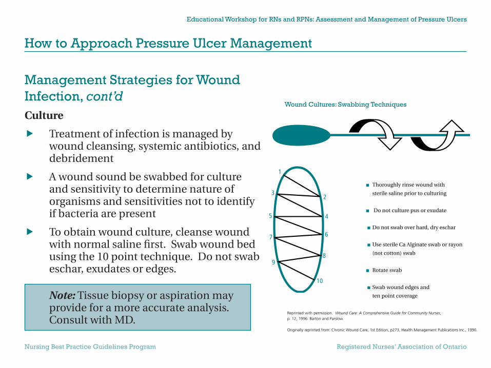
Management Strategies for Wound Infection, cont’dCulture
Treatment of infection is managed by wound cleansing, systemic antibiotics, and debridement
A wound sound be swabbed for culture and sensitivity to determine nature of organisms and sensitivities not to identify if bacteria are present
To obtain wound culture, cleanse wound with normal saline fi rst. Swab wound bed using the 10 point technique. Do not swab eschar, exudates or edges.
Note: Tissue biopsy or aspiration may provide for a more accurate analysis. Consult with MD.
1
23
10
98
7 6
5 4
Thoroughly rinse wound with
sterile saline prior to culturing
Do not culture pus or exudate
Do not swab over hard, dry eschar
Use sterile Ca Alginate swab or rayon
(not cotton) swab
Rotate swab
Swab wound edges and
ten point coverage
Wound Cultures: Swabbing Techniques
Reprinted with permission. Wound Care: A Comprehensive Guide for Community Nurses,p. 12, 1996. Barton and Parslow.
Originally reprinted from: Chronic Wound Care, 1st Edition, p273, Health Management Publications Inc., 1990.
Educational Workshop for RNs and RPNs: Assessment and Management of Pressure Ulcers
How to Approach Pressure Ulcer Management
Nursing Best Practice Guidelines Program Registered Nurses’ Association of Ontario
Management Strategies for Wound Infection, cont’dAntibiotics
Systemic antibiotics are not required for pressure ulcers with only clinical signs of local infection
Exceptions with locally infected wounds requiring systemic antibiotics
Systemic antibiotics are used when the virulence of the organism is high and the host’s defenses are compromised

Educational Workshop for RNs and RPNs: Assessment and Management of Pressure Ulcers
How to Approach Pressure Ulcer Management
Nursing Best Practice Guidelines Program Registered Nurses’ Association of Ontario
Management Strategies for Wound Infection, cont’dAntiseptics
Use of cytotoxic antiseptics to reduce bacteria in wound tissue is not recommended
Typical management of infected wounds includes the use of topical antimicrobials rather than antibiotics or antiseptics
Dressings
Sterile dressings should be used in all care settings
Avoid all occlusive dressings if anaerobic infection is suspected or cultured
Protect non-infected ulcers with occlusive dressings
Note: Consult Wound and Skin Care Specialist, MD or Plastic Surgeon if debriding is needed
Educational Workshop for RNs and RPNs: Assessment and Management of Pressure Ulcers
How to Approach Pressure Ulcer Management
Nursing Best Practice Guidelines Program Registered Nurses’ Association of Ontario
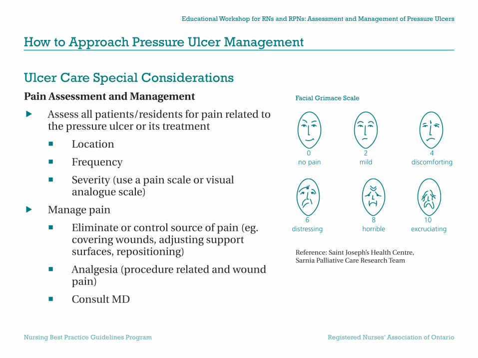
Ulcer Care Special ConsiderationsPain Assessment and Management
Assess all patients/residents for pain related to the pressure ulcer or its treatment
Location
Frequency
Severity (use a pain scale or visual analogue scale)
Manage pain
Eliminate or control source of pain (eg. covering wounds, adjusting support surfaces, repositioning)
Analgesia (procedure related and wound pain)
Consult MD
6 8 10distressing horrible excruciating
0 2 4no pain mild discomforting
Facial Grimace Scale
Reference: Saint Joseph’s Health Centre, Sarnia Palliative Care Research Team
Educational Workshop for RNs and RPNs: Assessment and Management of Pressure Ulcers
How to Approach Pressure Ulcer Management
Nursing Best Practice Guidelines Program Registered Nurses’ Association of Ontario
Ulcer Care Special Considerations, cont’dVascular Assessment
Recommended for ulcers in lower extremities to rule out vascular compromise
Ankle/brachial index (ABI)
Toe pressure
Note: Healability—Some wounds will not heal and treatment goal should focus on preventing infection or further deterioration so that quality of life is maintained.

Educational Workshop for RNs and RPNs: Assessment and Management of Pressure Ulcers
How to Approach Pressure Ulcer Management
Nursing Best Practice Guidelines Program Registered Nurses’ Association of Ontario
Ulcer Care Special Considerations, cont’dOperative Repair of Pressure Ulcers
Wounds can be closed by direct closure, skin grafting, skin fl aps, musculocutaneous fl aps and free fl aps
Procedure is performed by MD and plastic surgeon
Candidates for operative repair are medically stable, adequately nourished, and can tolerate operative blood loss and postoperative immobility
Other considerations are quality of life, patient preferences, treatment goals, risk of recurrence, and expected rehabilitative outcome
Postoperative Care:
Have patient slowly increase periods of time sitting or lying on the fl ap to increase its tolerance to pressure
Monitoring the fl ap for pallor, redness, or both that do not resolve in 10 minutes or pressure relief

Educational Workshop for RNs and RPNs: Assessment and Management of Pressure Ulcers
How to Approach Pressure Ulcer Management
Nursing Best Practice Guidelines Program Registered Nurses’ Association of Ontario
Ulcer Care Special Considerations, cont’dDischarge/Transfer of Care Arrangements
Patients/residents moving between care setting should have the following information provided:
Risk factors identifi ed
Details of pressure points and skin condition prior to transfer
Type of bed/mattress required
Details of healed ulcers
Stage, site and size of existing ulcers
History of ulcers, previous treatments and dressings used
Type of dressing currently used and frequency of change
Allergies to dressing products
Need for on-going nutritional support

Educational Workshop for RNs and RPNs: Assessment and Management of Pressure Ulcers
Nursing Best Practice Guidelines Program Registered Nurses’ Association of Ontario
Stage I Non-blanchable erythema of intact skin, the
heralding lesion of skin ulceration
In individuals with darker skin, discolouration of the skin may be purplish/bluish or violaceous (egg plant-like colour) accompanied by heat, edema, induration or hardness
(Bennett, 1995; NPUAP, 1998)
Staging of Pressure Ulcers

Educational Workshop for RNs and RPNs: Assessment and Management of Pressure Ulcers
Nursing Best Practice Guidelines Program Registered Nurses’ Association of Ontario
Stage II Partial thickness skin loss involving epidermis,
dermis or both
Ulcer is usually superfi cial and presents clinically as an abrasion, blister or shallow crater
(NPUAP, 1998)
Staging of Pressure Ulcers

Educational Workshop for RNs and RPNs: Assessment and Management of Pressure Ulcers
Nursing Best Practice Guidelines Program Registered Nurses’ Association of Ontario
Stage III Full thickness skin loss involving damage to
or necrosis of subcutaneous tissue that may extend down to, but not through underlying fascia
Ulcer presents clinically as a deep crater with or without undermining of adjacent tissue
(NPUAP, 1998)
Staging of Pressure Ulcers

Educational Workshop for RNs and RPNs: Assessment and Management of Pressure Ulcers
Nursing Best Practice Guidelines Program Registered Nurses’ Association of Ontario
Stage IV Full thickness skin loss with extensive
destruction, tissue necrosis, or damage to muscle, bone or supporting structures (ie. tendon joint capsule)
Undermining and sinus tracts also may be associated with Stage IV ulcers
(NPUAP, 1998)
Staging of Pressure Ulcers
Educational Workshop for RNs and RPNs: Assessment and Management of Pressure Ulcers
Nursing Best Practice Guidelines Program Registered Nurses’ Association of Ontario
Stage X Slough or necrotic tissue and/or black
discoloured tissue is present
Diffi cult to stage
Staging requires the removal of eschar
(NPUAP, 1998)
Staging of Pressure Ulcers
Educational Workshop for RNs and RPNs: Assessment and Management of Pressure Ulcers
Nursing Best Practice Guidelines Program Registered Nurses’ Association of Ontario
Reverse Staging of Pressure Ulcers It is incorrect to describe a healing pressure
ulcer by using the staging of I to IV in reverse order
Reverse staging should not be used to describe the healing process of a wound for the following reasons:
When pressure ulcers heal, they do not regenerate the same lost tissue
Wound heals with granulation tissue composed of endothelial cells, fi broblasts, collagen and an extracellular matrix
(NPUAP, 1995)
Staging of Pressure Ulcers

Educational Workshop for RNs and RPNs: Assessment and Management of Pressure Ulcers
Nursing Best Practice Guidelines Program Registered Nurses’ Association of Ontario
Stage 1Goals:
To reduce further skin breakdown and prevent skin loss
Protect against moisture and friction
Interventions:
Protect area from friction, shear, and maceration using a transparent fi lm dressing or thin hydrocolloids
Provide pressure relieving devices to reduce friction and shearing forces
Setting the Treatment Goals

Educational Workshop for RNs and RPNs: Assessment and Management of Pressure Ulcers
Nursing Best Practice Guidelines Program Registered Nurses’ Association of Ontario
Stage IIGoals:
To reduce further skin breakdown and prevent skin loss
To protect the surrounding skin from moisture by managing exudates and providing a moist wound environment to promote healing
Interventions:
Clean the ulcer with normal saline
Protect the wound by covering it with a transparent dressing or hydrocolloid
For moderate amount of exudates, use an absorbent foam dressing
Use liquid or solid barriers to protect periwound skin from maceration damage
Setting the Treatment Goals
Educational Workshop for RNs and RPNs: Assessment and Management of Pressure Ulcers
Nursing Best Practice Guidelines Program Registered Nurses’ Association of Ontario
Stage III & IVGoals:
To remove cell debris and promote autolysis
To provide clean, moist environment for the healing process to begin
To absorb exudates
To protect from contamination and trauma
To decrease dressing changes
To protect surrounding skin
Setting the Treatment Goals

Educational Workshop for RNs and RPNs: Assessment and Management of Pressure Ulcers
Nursing Best Practice Guidelines Program Registered Nurses’ Association of Ontario
Stage III & IVInterventions: Dry Cavity
Irrigate with normal saline using a 20-35 ml syringe and 19 gauge needle or angiocath
Protect periwound skin with a protective barrier
Fill dead space with appropriate fi ller (including sinus tracts)
Line cavity with gel and place 4 x 4 gauze packed loosely
Protect from contamination by use of an absorbent outer semi-occlusive dressing
Setting the Treatment Goals

Educational Workshop for RNs and RPNs: Assessment and Management of Pressure Ulcers
Nursing Best Practice Guidelines Program Registered Nurses’ Association of Ontario
Stage III & IVInterventions: Exudating Cavity
Irrigate with normal saline using a 20-35 ml syringe and 19 gauge needle or angiocath
Protect periwound skin using a protective barrier
Fill dead space with appropriate fi ller (including sinus tracts)
Use absorbent foam dressings
Protect from contamination by use of an outer semi-occlusive dressing
Setting the Treatment Goals
Educational Workshop for RNs and RPNs: Assessment and Management of Pressure Ulcers
Nursing Best Practice Guidelines Program Registered Nurses’ Association of Ontario
Stage XGoals:
To debride and remove dead tissue
To rehydrate the eschar by providing a clean, moist environment for the healing process to begin
To promote closure/healing
Interventions:
Clean with normal saline
Surgical debridement by MD or trained person
Autolytic debridement using gels
Protect periwound skin using a protective barrier
Cover with transparent dressing
Setting the Treatment Goals
Educational Workshop for RNs and RPNs: Assessment and Management of Pressure Ulcers
Nursing Best Practice Guidelines Program Registered Nurses’ Association of Ontario
ReferencesAyello, E. A. (1999). Predicting pressure ulcer sore risk. Journal of Gerontological Nursing, 25, 7-9.
Barton, P. & Parslow, N. (1996). Wound care: A comprehensive guide for community nurses. Toronto, Ontario, St. Elizabeth Health Care.
Bergstrom, N., Braden, B. J., Laguzza, A., & Homan, V. (1987). The Braden Scale for predicting pressure sore risk. Nursing Research, 36, 205-210.
Braden, B.J. (2001). Risk Assessment in Pressure Ulcer Prevention. In D. Krasner, G. Rodenheaver and R.G. Sibbald (Eds.), Chronic Wound Care: A Clinical Source book for Healthcare Professionals (3rd ed. pp. 641-651). Wayne, PA: HMP Communications.
Brignell, A. (ed) 2000. Guideline for developing a pain management program. A resource guide for long-term care facilities, 3rd edition. Sarnia, Ontario: Saint Joseph’s Health Centre.
Clinical Resource Effi ciency Support Team (1998). Guidelines for the prevention and management of pressure sores. Belfast, Northern Ireland: CREST Secretariat.
Eaglstein & Falanga. (1997). Chronic Wounds. Surg Clin North American Wound Healing, 77, 689-700.
Goode, P. S. & Allman, R. M. (1989). The prevention and management of pressure ulcers. Med Clin North Am., 73, 1511-1524.
Molony, S., Waszynski, C. & Lyder, C. (Eds.) (1999). Gerontological Nursing: An Advanced Practice Approach. New York: Appleton and Lange.
Assessment and Management of Pressure Ulcers

Educational Workshop for RNs and RPNs: Assessment and Management of Pressure Ulcers
Nursing Best Practice Guidelines Program Registered Nurses’ Association of Ontario
Assessment and Management of Pressure Ulcers
References, cont’dNational Pressure Ulcer Advisory Panel (1989). Pressure ulcers prevalence, cost and risk assessment: Consensus development conference statement. Decubitis, 2, 24-28.
National Pressure Ulcer Advisory Panel (1995). NPUAP statement on reverse staging of pressure ulcers (Rep. No. 4(2)).
National Pressure Ulcer Advisory Panel. (1998). Stage 1 assessment in darkly pigmented skin.
Patterson, J. A. & Bennett, R. G. (1995). Prevention and treatment of pressure sores. J Am Geriatr Soc., 43, 919-927.
West, J. & Gimbel, M. (2000). Acute surgical and traumatic wound healing. In R.Bryant (Ed.), Acute & Chronic Wounds: Nursing Management (2nd Edition ed., pp. 189-196). St. Louis: Mosby.

Part 2: Positioning Techniques and Devices in Wound Management
Part A: Educational Workshop for RNs and RPNsAssessment and Management of Pressure Ulcers
Based on the Registered Nurses’ Association of Ontario
Best Practice Guideline:
Assessment and Management of Stage I to IV Pressure Ulcers
Educational Workshop for RNs and RPNs: Assessment and Management of Pressure Ulcers
Positioning Techniques and Devices in Wound Management
Nursing Best Practice Guidelines Program Registered Nurses’ Association of Ontario
The Role of PT and OT in the Management of Pressure Ulcers
Assess mobility and function
Improve/maximize mobility and function
Assess for and recommend positioning/seating needs and devices
Prescribe indicated devices and monitor use
Provide education to the patient, family, and team

Educational Workshop for RNs and RPNs: Assessment and Management of Pressure Ulcers
Positioning Techniques and Devices in Wound Management
Nursing Best Practice Guidelines Program Registered Nurses’ Association of Ontario
The Role of PT and OT... cont’dAssess Mobility and Function
AROM, PROM
Contractures, deformities, posture
Bed mobility, transitional movements, transfers
Cognitive level and function
ADL function and independence
Previous physical functional status
Precautions secondary to medical conditions and/or surgeries

Educational Workshop for RNs and RPNs: Assessment and Management of Pressure Ulcers
Positioning Techniques and Devices in Wound Management
Nursing Best Practice Guidelines Program Registered Nurses’ Association of Ontario
The Role of PT and OT... cont’dImprove/Maximize Mobility and Function
Develop appropriate goals
Increase patient independence and therefore aid in prevention/healing process
Improve circulation and healing
Assess for and Recommend Positioning/Seating Needs
Positioning in bed
Appropriate seating systems

Educational Workshop for RNs and RPNs: Assessment and Management of Pressure Ulcers
Positioning Techniques and Devices in Wound Management
Nursing Best Practice Guidelines Program Registered Nurses’ Association of Ontario
The Role of PT and OT... cont’dPrescribe Indicated Devices and Monitor Use
Seating systems and overlays for bed
Protection and prevention devices
Education to patient, family, and team re: use and monitoring of devices
Education to Patient, Family and Team
Fosters empowerment and independence
Coordinated effort
Promotes adherence

Educational Workshop for RNs and RPNs: Assessment and Management of Pressure Ulcers
Positioning Techniques and Devices in Wound Management
Nursing Best Practice Guidelines Program Registered Nurses’ Association of Ontario
Extrinsic Forces to Consider in Seating and PositioningDirect pressure/compression
Force/area; the greater the area of contact with a surface, the less compression force over a specifi c area
Friction
Destructive force created by the rubbing contact between the skin and a surface material
Educational Workshop for RNs and RPNs: Assessment and Management of Pressure Ulcers
Positioning Techniques and Devices in Wound Management
Nursing Best Practice Guidelines Program Registered Nurses’ Association of Ontario
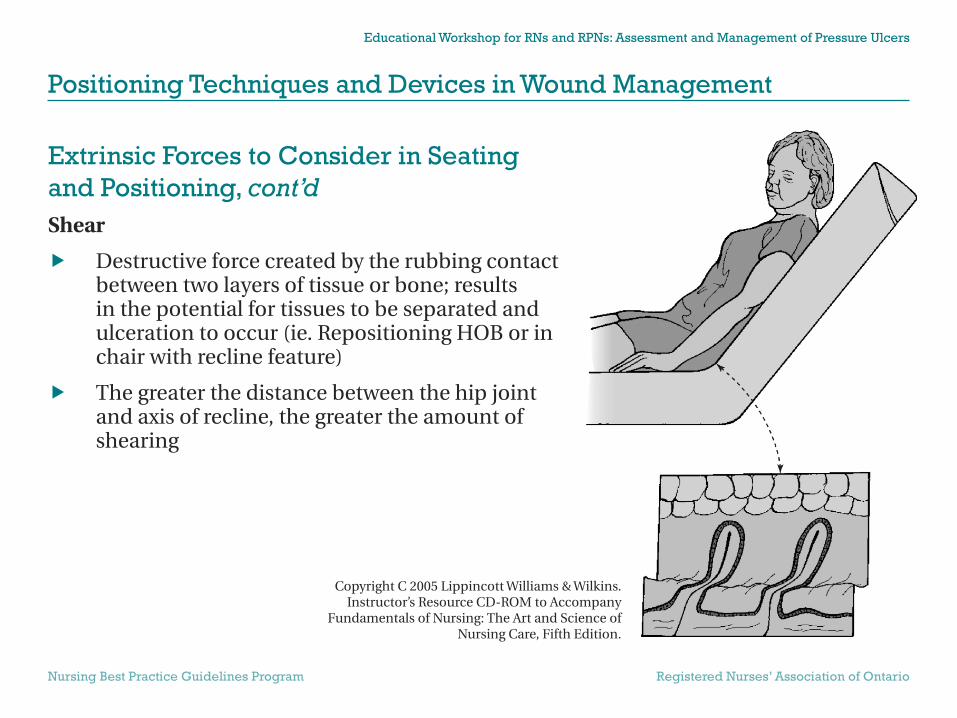
Extrinsic Forces to Consider in Seating and Positioning, cont’dShear
Destructive force created by the rubbing contact between two layers of tissue or bone; results in the potential for tissues to be separated and ulceration to occur (ie. Repositioning HOB or in chair with recline feature)
The greater the distance between the hip joint and axis of recline, the greater the amount of shearing
Copyright C 2005 Lippincott Williams & Wilkins. Instructor’s Resource CD-ROM to Accompany
Fundamentals of Nursing: The Art and Science of Nursing Care, Fifth Edition.

Educational Workshop for RNs and RPNs: Assessment and Management of Pressure Ulcers
Positioning Techniques and Devices in Wound Management
Nursing Best Practice Guidelines Program Registered Nurses’ Association of Ontario
Goals of Seating and PositioningPromote:
Postural alignment
Even weight distribution
Balance and stability
Support for independent function (ie. ADL, mobility)
Pressure relief
Reduce compression, friction, shear forces
Reduce agitated/aggressive behaviour
Increase tolerance
Facilitate ease of repositioning by self or caregivers

Educational Workshop for RNs and RPNs: Assessment and Management of Pressure Ulcers
Positioning Techniques and Devices in Wound Management
Nursing Best Practice Guidelines Program Registered Nurses’ Association of Ontario
Goals of Seating and Positioning, cont’dReduce Pressure, Friction, Shear
Eliminate if possible; contact of affected or at risk areas on surfaces
Distribute pressure across larger surface area
Eg. Decrease HOB angle, use of various support surfaces, use of devices
Utilize proper transferring and repositioning techniques

Educational Workshop for RNs and RPNs: Assessment and Management of Pressure Ulcers
Positioning Techniques and Devices in Wound Management
Nursing Best Practice Guidelines Program Registered Nurses’ Association of Ontario
Goals of Seating and Positioning, cont’dReduce Agitated/Aggressive Behaviour
Poor positioning may result in increased abrasions, shear, and friction forces
Frequent position changes, comfortable seating and positioning may increase tolerance
Independent mobility and ADL function promoted by proper positioning may reduce agitation/agression

Educational Workshop for RNs and RPNs: Assessment and Management of Pressure Ulcers
Positioning Techniques and Devices in Wound Management
Nursing Best Practice Guidelines Program Registered Nurses’ Association of Ontario
Goals of Seating and Positioning, cont’dFacilitate Ease of Repositioning by Self or Caregivers
Introduce proper body mechanics, techniques
Education
Reduce friction/shear during repositioning with proper seating positioning devices
Educational Workshop for RNs and RPNs: Assessment and Management of Pressure Ulcers
Positioning Techniques and Devices in Wound Management
Nursing Best Practice Guidelines Program Registered Nurses’ Association of Ontario
Positioning and Seating Surfaces Pressure Reducing Surface
A surface that lowers pressure compared to a standard product such as a mattress or chair surface (IAET; Standards of Care, 1987)
Does not maintain pressure below capillary closing pressure
Pressure Relieving Surface
A surface that consistently reduces pressure below capillary closing pressure (ie. 32mmHg) (IAET; Standards of Care, 1987)
Foam
Has “memory”, compresses and breaks down (needs to be replaced in 6-9 months), diffi cult to clean, forces the body to conform to its shape, inexpensive

Educational Workshop for RNs and RPNs: Assessment and Management of Pressure Ulcers
Positioning Techniques and Devices in Wound Management
Nursing Best Practice Guidelines Program Registered Nurses’ Association of Ontario
Positioning and Seating Surfaces, cont’d Air
Lightweight, does not “bottom out”, able to customize, conforms to body position, minimal shearing, redistributes pressure, expensive
Seating cushions are grouped into categories: maximum, excellent, medium, low pressure reduction
Avoid “donut” devices as they create a ring of direct pressure
Educational Workshop for RNs and RPNs: Assessment and Management of Pressure Ulcers
Positioning Techniques and Devices in Wound Management
Nursing Best Practice Guidelines Program Registered Nurses’ Association of Ontario
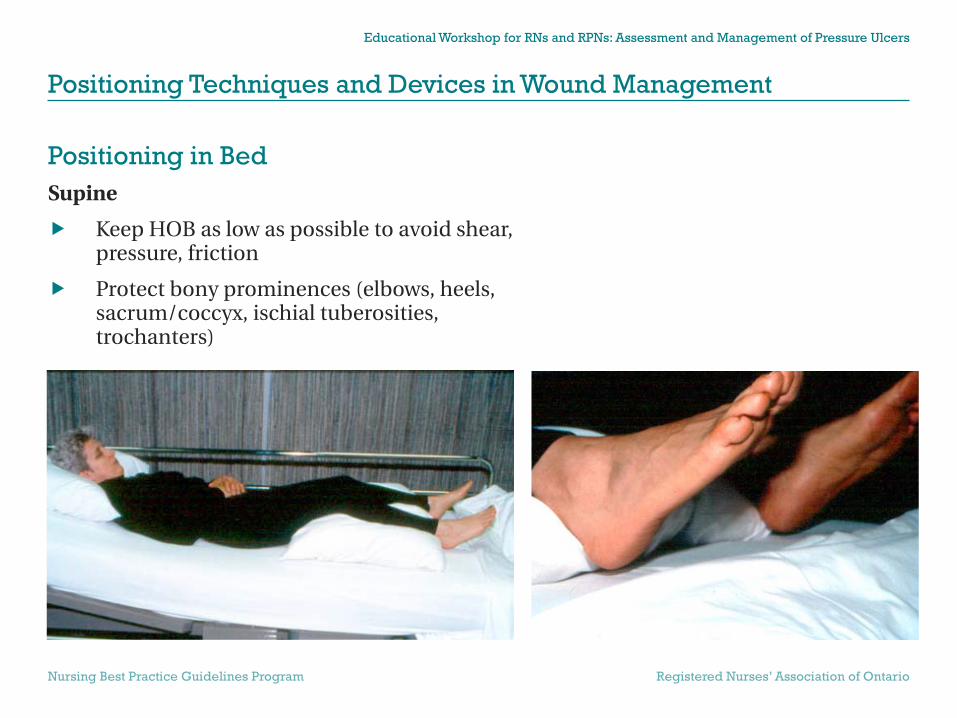
Positioning in BedSupine
Keep HOB as low as possible to avoid shear, pressure, friction
Protect bony prominences (elbows, heels, sacrum/coccyx, ischial tuberosities, trochanters)

Educational Workshop for RNs and RPNs: Assessment and Management of Pressure Ulcers
Positioning Techniques and Devices in Wound Management
Nursing Best Practice Guidelines Program Registered Nurses’ Association of Ontario
Positioning in Bed, cont’dSidelying
30 degree laterally inclined
avoids pressure on greater trochanter and sacrum
Frequent position changes between the 3 positions
Every 1-2 hours

Educational Workshop for RNs and RPNs: Assessment and Management of Pressure Ulcers
Positioning Techniques and Devices in Wound Management
Nursing Best Practice Guidelines Program Registered Nurses’ Association of Ontario
Positioning in Bed, cont’dDevices
Reduce pressure
Pillows, foam wedges, egg crate, heel relief splints
Reduce shear/friction
Gel
Things to Avoid
Any donut type devices
Any direct pressure on bony prominences
Restraint devices if possible
Educational Workshop for RNs and RPNs: Assessment and Management of Pressure Ulcers
Positioning Techniques and Devices in Wound Management
Nursing Best Practice Guidelines Program Registered Nurses’ Association of Ontario
Seating and Positioning Obtain OT/PT assessment of functional level,
mobility, pressure reduction needs, transferring methods, assistive device needs
Ischial tuberosities are the main points of weightbearing in a seated person with an upright posture
Sacral sitting
Clients who “slide” out of their chairs, who have kyphosis have an increased risk for skin breakdown secondary to friction/shear forces, agitated behaviour and uneven pressure distribution
Goals of seating:
To facilitate function and mobility, and to promote comfort, postural alignment, and pressure relief/reduction

Educational Workshop for RNs and RPNs: Assessment and Management of Pressure Ulcers
Positioning Techniques and Devices in Wound Management
Nursing Best Practice Guidelines Program Registered Nurses’ Association of Ontario
Seating and Positioning, cont’d Cushions
Prescribed by therapists after an assessment of posture, functional level and needs, and pressure reduction/relief needs (ie. Foam, gel, air)
Sling seats and backs
Promote internally rotated hips and kyphotic postures – results in increased shear/friction forces and increased direct pressure on bony prominences on back and sacrum/coccyx regions

Educational Workshop for RNs and RPNs: Assessment and Management of Pressure Ulcers
Positioning Techniques and Devices in Wound Management
Nursing Best Practice Guidelines Program Registered Nurses’ Association of Ontario
Seating and Positioning, cont’dComponents that Affect Positioning
Seat width
Too wide, too narrow
Seat depth
Too deep, too short
Armrests
Properly adjusted armrests will reduce pressure under the ischial tuberosities’s by about 25% to 35% (C.A. Fleck, Crown Therapeutics Inc., 2000)
Foot Rest Height
Too low, too high
Seat Height
Too low, too high

Educational Workshop for RNs and RPNs: Assessment and Management of Pressure Ulcers
Positioning Techniques and Devices in Wound Management
Nursing Best Practice Guidelines Program Registered Nurses’ Association of Ontario
Seating and Positioning, cont’dTilt and Recline
Tilt
Back to seat/hip/knee angles remains constant
Rotation of body around a pivot point on the horizontal axis
Position of body relative to the pull of gravity is altered thus redistributing pressure
Pressure reduction at 45 degrees of tilt

Educational Workshop for RNs and RPNs: Assessment and Management of Pressure Ulcers
Positioning Techniques and Devices in Wound Management
Nursing Best Practice Guidelines Program Registered Nurses’ Association of Ontario
Seating and Positioning, cont’dTilt and Recline
Recline
Back to seat/hip angle opens up (obtuse), the seat remains parallel to the ground
Increase in the angle at the pivot point on the horizontal axis
Pull of gravity is spread over a larger body surface, thus decreasing the average PSI (pounds per square inch)
May promote shear/friction as a result of sliding in chair or during repositioning

Educational Workshop for RNs and RPNs: Assessment and Management of Pressure Ulcers
Positioning Techniques and Devices in Wound Management
Nursing Best Practice Guidelines Program Registered Nurses’ Association of Ontario
Seating and Positioning, cont’dPoor positioning in sitting can result in:
Increased pressure forces (ie. Tissue/skin breakdown)
Decreased communication and altertness
Decreased function and mobility
Increased agitated behaviours (ie. Increased need for restraints – more pressure forces)
Altered moods
Educational Workshop for RNs and RPNs: Assessment and Management of Pressure Ulcers
Positioning Techniques and Devices in Wound Management
Nursing Best Practice Guidelines Program Registered Nurses’ Association of Ontario
Transfers and Repositioning, cont’dTransfer Methods
Avoid increased pressure, more importantly shear and friction
Common problems
Transfer boards
Leaving lift sheets under patients
Tugging pants and failure to readjust
Solutions
Transfer belts
Remove lift sheets after use
Loosen tight clothing after transfers
Adequate assistance

Educational Workshop for RNs and RPNs: Assessment and Management of Pressure Ulcers
Positioning Techniques and Devices in Wound Management
Nursing Best Practice Guidelines Program Registered Nurses’ Association of Ontario
Transfers and Repositioning, cont’dRepositioning
Bed
Avoid sliding
Have patient shift hips
Crooklying position
Effective use of lift sheets
Adequate assistance
Chair
Avoid tugging pants
Teach patients to shift hips, pushups, side-leans, front-back rocking
Up and back method
Use patient tilt to prevent sliding

Educational Workshop for RNs and RPNs: Assessment and Management of Pressure Ulcers
Positioning Techniques and Devices in Wound Management
Nursing Best Practice Guidelines Program Registered Nurses’ Association of Ontario
Summary Proper seating and positioning can facilitate:
Reduction of compression, shear and friction forces
Independent mobility and ADL function
Ease in transfers/repositioning/caregiving
Reduction of agitated behaviour and need for restraints
Minor adjustments in seating systems and bed positioning can have a signifi cant impact on pressure distribution
Refer to PT and OT for assessment and assist with follow through of recommendations

Educational Workshop for RNs and RPNs: Assessment and Management of Pressure Ulcers
Positioning Techniques and Devices in Wound Management
Nursing Best Practice Guidelines Program Registered Nurses’ Association of Ontario
ReferencesBabinec, M. (1999). Tilt comparison: Weight shifting vs. non-weight shifting power and manual bases. Canadian Seating and Mobility Conference Proceedings Manual: The Evolution of Revolution. Toronto (pp. 130).
Buck, S. (1999). Wheelchair foot propulsion: The complexities and mysteries of wheelchair and seating system set-up. Canadian Seating and Mobility Conference Proceedings Manual: The Evolution of Revolution. Toronto (pp. 83-89).
Fleck, C. (1999). Pressure ulcer prevention and management. Canadian Seating and Mobility Conference Proceedings Manual: The Evolution of Revolution. Toronto. (pp. 88-89)
IAET (1987) Standards of Care for Dermal Wounds: Pressure Sores. Irvine: International Association of Enterostomal Therapy.
Lachaine, C. (2000). What, when and why of specialty mattresses. Canadian Seating and Mobility Conference Proceedings Manual: Flexible Solutions Shaping Tomorrow. Toronto (pp. 23-26).
Pratt, S. (1999). Assessment techniques, the supine and sitting evaluation. Canadian Seating and Mobility Conference proceedings Manual: The Evolution of Revolution. Toronto (pp. 58-61).
Taylor, V.C. (1999). Pressure mapping clinical protocol. Canadian Seating and Mobility Conference Proceedings Manual: The Evolution of Revolution. Toronto (pp. 66-67).
Part 3: Nutritional Intervention
Part A: Educational Workshop for RNs and RPNsAssessment and Management of Pressure Ulcers
Based on the Registered Nurses’ Association of Ontario
Best Practice Guideline:
Assessment and Management of Stage I to IV Pressure Ulcers

Educational Workshop for RNs and RPNs: Assessment and Management of Pressure Ulcers
Nutrition Intervention
Nursing Best Practice Guidelines Program Registered Nurses’ Association of Ontario
The role of nutrition in a hospital setting Prevalence of malnutrition in institutional
settings is well documented
Malnutrition is associated with increased:
Morbidity and mortality
Length of hospital stay
Costs
Nutrition support will allow for:
Faster recovery and increased strength
Improved wound healing
Decreased risk of infection

Educational Workshop for RNs and RPNs: Assessment and Management of Pressure Ulcers
Nutrition Intervention
Nursing Best Practice Guidelines Program Registered Nurses’ Association of Ontario
Patients at Nutritional Risk Inadequate intake
CVA, elderly, access to food, poor dentition or mouth sores, dysphagia, esophagitis or recent surgery
Inadequate absorptionIrritable Bowel Disease (IBD), Crohn’s, Colitis, diarrhea or vomiting
Increased lossesColostomy, ileostomy, wounds or fi stula
Increased requirementsCHF, COPD, pneumonia, asthma, wound healing

Educational Workshop for RNs and RPNs: Assessment and Management of Pressure Ulcers
Nutrition Intervention
Nursing Best Practice Guidelines Program Registered Nurses’ Association of Ontario
Indicators of Malnutrition Weight loss greater than 10% of usual body
weight over 3 months
BMI (body mass index) less than 18
Albumin < 35 g/L
Total protein < 65 g/L

Educational Workshop for RNs and RPNs: Assessment and Management of Pressure Ulcers
Nutrition Intervention
Nursing Best Practice Guidelines Program Registered Nurses’ Association of Ontario
Oral Nutrition SupplementsDefi nition of eating well – Consuming at least 2/3 of the meal tray at least 75% of the time
For patients who are not consuming an adequate quantity of food to meet their nutritional requirements

Educational Workshop for RNs and RPNs: Assessment and Management of Pressure Ulcers
Nutrition Intervention
Nursing Best Practice Guidelines Program Registered Nurses’ Association of Ontario
Nutrition Support Nutrition support is an alternative to oral
nutrition when a patient is unable to meet greater than 50% of their requirements for greater than 3 days
Enteral and parental nutrition meet 100% of patient’s nutritional requirements
Educational Workshop for RNs and RPNs: Assessment and Management of Pressure Ulcers
Nutrition Intervention
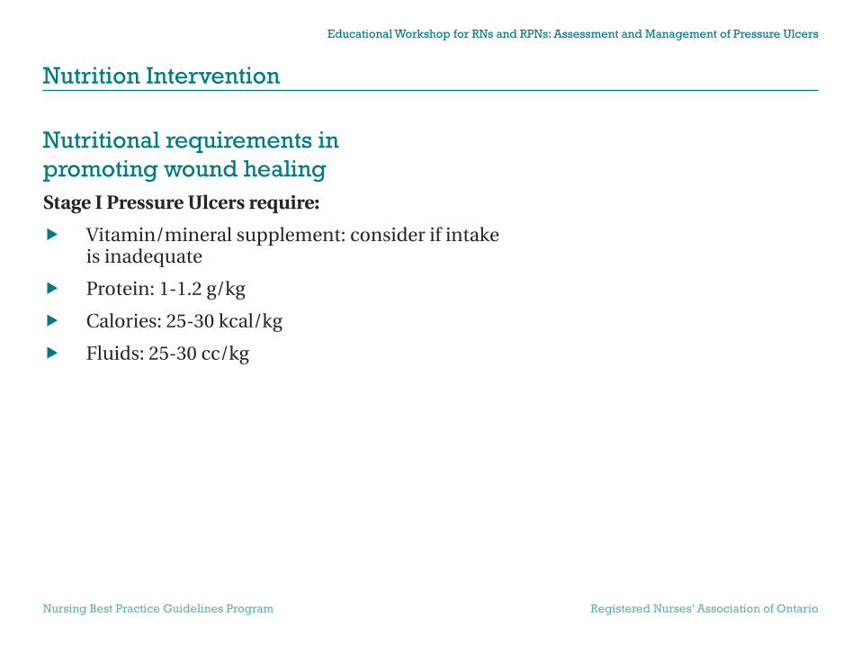
Nursing Best Practice Guidelines Program Registered Nurses’ Association of Ontario
Nutritional requirements in promoting wound healingStage I Pressure Ulcers require:
Vitamin/mineral supplement: consider if intake is inadequate
Protein: 1-1.2 g/kg
Calories: 25-30 kcal/kg
Fluids: 25-30 cc/kg

Educational Workshop for RNs and RPNs: Assessment and Management of Pressure Ulcers
Nutrition Intervention
Nursing Best Practice Guidelines Program Registered Nurses’ Association of Ontario
Nutritional requirements in promoting wound healing, cont’dStage II Pressure Ulcers require:
Vitamin/mineral supplement
Protein: 1.25-1.4 g/kg
Calories: 25-30 kcal/kg
Fluid: 25-35 cc/kg (increased fl uid loss from exudate)

Educational Workshop for RNs and RPNs: Assessment and Management of Pressure Ulcers
Nutrition Intervention
Nursing Best Practice Guidelines Program Registered Nurses’ Association of Ontario
Nutritional requirements in promoting wound healing, cont’dStage III Pressure Ulcers require:
Vitamin/mineral supplement: Vitamin C 500 mg OD; Zinc 25 mg elemental BID
Protein: 1.5 g/kg
Calories: 30-35 kcal/kg
Fluid: 30-40 cc/kg

Educational Workshop for RNs and RPNs: Assessment and Management of Pressure Ulcers
Nutrition Intervention
Nursing Best Practice Guidelines Program Registered Nurses’ Association of Ontario
Nutritional requirements in promoting wound healing, cont’dStage IV Pressure Ulcers require:
Vitamin/mineral supplement: Vitamin C 500 mg OD; Zinc 25 mg elemental BID
Protein: 1.5-2.0 g/kg
Calories: 30-40 kcal/kg
Fluid: 35-45 cc/kg
Note: Zinc should be reassessed at 10 days and discontinued if within the normal range.

Educational Workshop for RNs and RPNs: Assessment and Management of Pressure Ulcers
Nutrition Intervention
Nursing Best Practice Guidelines Program Registered Nurses’ Association of Ontario
ReferencesAllman, R.M., Goode, P.S., Burst, N., Bartolucci, A. A., & Thomas, D. R. (1999). Pressure ulcers, hospital complications, disease severity: Impact on hospital costs and length of stay. Advances in Wound Care, 12(1), 22-30.
Andrews, M., & Gallagher-Allred, C. (1999). The role of zinc in wound healing. Advances in Wound Care, 12(3), 137-138.
Bergstrom, N., Bennett, M. A., Carlson, C. E. et al. (1994). Clinical Practice Guideline, No. 15, Rockville, Md: US Department of Health and Human Services. Agency for Health Care Policy and Research. AHCPR Publication No. 95-0652.
Gilmore, S. A., Robinson, G., Posthauer, M. E., & Raymond, J. (1995). Clinical indicators associated with unintentional weight loss and pressure ulcers in elderly residents of nursing facilities. Journal of the American Dietetic Association, 95(9), 948-992.
Granick, M.S., McGowan, E., & Long, C. D. (1998). Outcome assessment of an in-hospital cross-functional wound care team. Plastic and Reconstructive Surgery, 101(5), 1243-1247.
Himes, D. (1997). Nutritional supplements in the treatment of pressure ulcers: Practical perspectives. Advances in Wound Care, 10(1), 30-31.
Hoffman, D. R. (1996). The false claims act as a remedy to the inadequate provision of nutrition and wound care to nursing home residents. Advances in Wound Care, 9(5), 25-29.

Educational Workshop for RNs and RPNs: Assessment and Management of Pressure Ulcers
Nutrition Intervention
Nursing Best Practice Guidelines Program Registered Nurses’ Association of Ontario
References, cont’dJackobs, M. K. (1999). The cost of medical nutrition therapy in healing pressure ulcers. Topics in Clinical Nutrition, 14(2), 41-47.
Lipschitz, D. A. (1995). Approaches to the nutritional support of the older patient. Clinics in Geriatric Medicine, 11(4), 715-724.
Mazzotta, M. (1994). Nutrition and wound healing. Journal of the American Podiatric Medical Association, 84(9), 456-461.
Molony, S., Waszynski, C. & Lyder, C. (Eds.) (1999). Gerontological Nursing: An Advanced Practice Approach. New York: Appleton and Lange.
Poduch, C. (1996). The dietitian as a cost effi cient agent. Canadian Nursing Homes, 7(1), 5-7.
Smith, P. W., Black, J. M., Black, S. B. (1999). Infected pressure ulcers in the long-term care facility. Infection Control and Hospital Epidemiology, 20(5), 358-361.
Spoelhof, G. D., & Ide, K. (1993). Pressure ulcers in nursing home patients. American Family Physician, 47(5), 1207-1215.
Strauss, E. A., & Margolis, D. J. (1996). Malnutrition in patients with pressure ulcers: Morbidity, morality, and clinically practical assessments. Advances in Wound Care, 9(5), 37-40.
Williams, C. M., Lines, C. M., & McKay, E. C. (1988). Iron and zinc status in multiple sclerosis patients with pressure sores. European Journal of Clinical Nutrition, 42(4), 321-328.
Xakellis, G. C. & Frantz, R. (1996). The cost of healing pressure ulcers across multiple health care settings. Advances in Wound Care, 9(6), 18-22.