Embed Size (px)
Citation preview
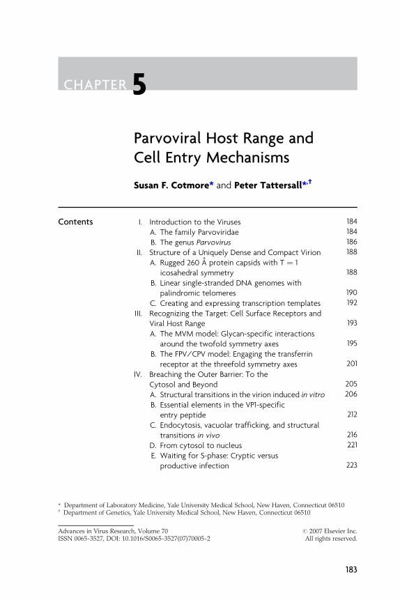
CHAPTER 5
Advances in Virus ResearchISSN 0065-3527, DOI: 10.1
* Department of Laborator{ Department of Genetics,
Parvoviral Host Range andCell Entry Mechanisms
Susan F. Cotmore* and Peter Tattersall*, †
Contents I. Introduction to the Viruses 184
, Vol016/S
y MeYale
ume 70 # 20070065-3527(07)70005-2 All righ
dicine, Yale University Medical School, New Haven, ConnecticuUniversity Medical School, New Haven, Connecticut 06510
Elsts
t 06
A
. T he family Parvoviridae 184B
. T he genus Parvovirus 186II. S
tructure of a Uniquely Dense and Compact Virion 188A
. R ugged 260 A protein capsids with T ¼ 1icosahedral symmetry
188B
. L inear single-stranded DNA genomes withpalindromic telomeres
190C
. C reating and expressing transcription templates 192III. R
ecognizing the Target: Cell Surface Receptors andViral Host Range
193A
. T he MVM model: Glycan-specific interactionsaround the twofold symmetry axes
195B
. T he FPV/CPV model: Engaging the transferrinreceptor at the threefold symmetry axes
201IV. B
reaching the Outer Barrier: To theCytosol and Beyond
205A
. S tructural transitions in the virion induced in vitro 206B
. E ssential elements in the VP1-specificentry peptide
212C
. E ndocytosis, vacuolar trafficking, and structuraltransitions in vivo
216D
. F rom cytosol to nucleus 221E
. W aiting for S-phase: Cryptic versusproductive infection
223evier Inc.reserved.
510
183
184 Susan F. Cotmore and Peter Tattersall
Ackn
owledgments 225Refer
ences 225Abstract Parvoviruses elaborate rugged nonenveloped icosahedral capsids of
�260 A in diameter that comprise just 60 copies of a common core
structural polypeptide. While serving as exceptionally durable shells,
capable of protecting the single-stranded DNA genome from envi-
ronmental extremes, the capsid also undergoes sequential conforma-
tional changes that allow it to translocate the genome from its initial
host cell nucleus all the way into the nucleus of its subsequent host.
Lacking a duplex transcription template, the virus must then wait for
its host to enter S-phase before it can initiate transcription and usurp
the cell’s synthetic pathways. Here we review cell entry mechanisms
used by parvoviruses. We explore two apparently distinct modes of
host cell specificity, first that used by Minute virus of mice, where
subtle glycan-specific interactions between host receptors and resi-
dues surrounding twofold symmetry axes on the virion surface medi-
ate differentiated cell type target specificity, while the second
involves novel protein interactions with the canine transferrin recep-
tor that allow a mutant of the feline leukopenia serotype, Canine
parvovirus, to bind to and infect dog cells. We then discuss confor-
mational shifts in the virion that accompany cell entry, causing
exposure of a capsid-tethered phospholipase A2 enzymatic core
that acts as an endosomolytic agent to mediate virion translocation
across the lipid bilayer into the cell cytoplasm. Finally, we discuss
virion delivery into the nucleus, and consider the nature of transcrip-
tionally silent DNA species that, escaping detection by the cell, might
allow unhampered progress into S-phase and hence unleash the
parvoviral Trojan horse.
I. INTRODUCTION TO THE VIRUSES
A. The family Parvoviridae
All small nonenveloped viruses with �5-kb linear, self-priming, single-stranded DNA genomes are grouped in the taxonomic family Parvovir-idae (from Parvus—Latin for ‘‘small’’), and share a common evolutionaryhistory as assessed by DNA sequence. This broad group is divided intotwo subfamilies, superficially on the basis of host range: the Parvovirinae,infecting vertebrate hosts and the Densovirinae, infecting insects andother arthropods. While species and genera within the Parvovirinaeappear to be derived from a single common ancestor, the arthropodgenera are separated bymassive evolutionary distances, probably reflectingdivergence coincident with that of their hosts (Tattersall et al., 2005). Thus,

Parvoviral Host Range and Cell Entry Mechanisms 185
this is an ancient and widely dispersed virus family with, apparently, asingle evolutionary branch that became adapted to vertebrate hosts.
Members of the subfamily Parvovirinae have been divided into fivegenera on the basis of DNA and protein sequence-based phylogeneticanalyses: these are the Parvoviruses, which are the subject of this chapter,and the Amdoviruses, Bocaviruses, Dependoviruses, and Erythroviruses.While all genera contain viruses that can replicate independently of helperviruses (commonly described as ‘‘autonomously replicating’’ viruses), theDependovirus genus is so called because it includes a large number ofagents that depend for their own productive replication on coinfectionwith a more complex helper virus from a different taxonomic family. Thisassociation with adenoviruses is reflected in the name, ‘‘adeno-associatedviruses’’ (AAVs), although these same viruses may also derive help fromherpesviruses, papillomaviruses, or vaccinia viruses. In the absence ofsuch help, AAVs establish a latent interaction with their vertebrate host,and this nondisruptive, but persistent, lifestyle has engendered signifi-cant interest in them as gene therapy transfer vectors. Accordingly, theyhave been the focus of much recent research, so that emerging data fromviruses in this genus does much to complement our current knowledge ofentry processes used by their Parvovirus cousins, and is cited accordinglyin this chapter.
The biology of the Parvovirinae is dominated by their small physicalsize. With nonenveloped protein capsids of around 260 A diameter, con-structed in the simplest icosahedral form (T ¼ 1), these remarkably denseand rugged particles deliver their enclosed genomes into the cell, traversethe cytoplasm, and penetrate the nucleus while still comprising a struc-turally intact, albeit somewhat rearranged, capsid (Farr et al., 2006;Sonntag et al., 2006; Vihinen-Ranta et al., 2002). Encapsidation withinsuch a small virion is possible because parvoviruses typically encodejust two gene cassettes, and are unique among known microorganismsin having DNA genomes that are both single stranded and linear, whichmakes their chromosome optimally small and flexible. This single DNAstrand is inserted vectorially into a preformed capsid, using energyprovided by a viral helicase, and packed in such a way that bases in theouter DNA shell bond with side chains from amino acids lining theicosahedral threefold axis of the capsid, creating a virion of remarkabledensity and stability (Agbandje-McKenna and Chapman, 2006; Chapmanand Agbandje-McKenna, 2006). Inevitably, such minimalism has someapparently negative biological consequences. Parvoviruses not onlylack accessory proteins that might induce resting cells to enter S-phase,they also lack a duplex transcription template so that they are generallyunable to express their genes until the DNA synthetic machinery of thehost cell, activated at the start of a cell-directed S-phase, coincidentallyprovides them with a complementary-sense DNA strand. Consequently,
186 Susan F. Cotmore and Peter Tattersall
these viruses have had to become masters of stealth, apparently avoidingtriggering many of the cellular responses that commonly accompany cellentry by viruses of other families. As a result, although relatively inert,they are able to become sequestered within resting cells without inhibit-ing the cell’s program of transit through the cell cycle. Indeed, thissuggests an entry strategy in which the disadvantages of being singlestranded are outweighed by the ability to package a relatively complexgenome in a particle small enough to be imported intact into the host cell’snucleus.
B. The genus Parvovirus
Much of our knowledge of the molecular biology and pathogenic poten-tial of the family Parvoviridae has been derived by studying members ofthe genus Parvovirus, which typically grow efficiently in cell culture, areopen to reverse and forward genetic analysis, and predominantly infecthost species that are readily susceptible to experimental manipulation.This genus contains four distinct subgroups: (1) a broadly related, butserologically diverse cluster of ‘‘rodent virus’’ species that contains threedistinct clades [Minute virus of mice (MVM), the type species of thegenus, Mouse parvovirus 1 (MPV1), and a rat virus group that includesRat minute virus 1 (RMV1), H1 virus and Kilham rat virus (KRV)], andLuIII, an ‘‘orphan’’ virus; (2) an outlying Rat parvovirus 1 (RPV1) branch;(3) the Feline panleukopenia virus/Canine parvovirus (FPV/CPV) sero-type, strains of which infect various members of the Carnivora; and (4)Porcine parvovirus (PPV). As seen in Table I, the NS1 genes of specieswithin this genus vary by up to 30%, whereas their VP2 genes vary by upto 50%, this wider range reflecting the fact that the members of eachspecies represent a serologically distinct group. In contrast to thesebroad interspecies values, the intraspecies homologies for the NS1 andVP2 proteins specified by the prototypeMVM strain, MVMp, and those ofthe ‘‘immunosuppressive’’ strain, MVMi, are both 97.8%, and for the NS1and VP2 proteins of FPV and CPV are 99.0% and 98.6%, respectively.
Patterns of parvovirus-induced disease are largely determined by thefact that these viruses cannot induce resting cells to enter S-phase, andhence only replicate productively in actively mitotic host cell popula-tions. They also commonly exhibit finely tuned tissue specificity, onlyinfecting cells of particular differentiated phenotypes, although suchpreferences can vary profoundly even within virus strains of a singleserotype. Accordingly, pathogenic or lethal infections typically occur infetal or neonatal hosts, which have many dividing cell populations, orinvolve adult tissues that remain actively dividing in later life such as cellsof the gut epithelium or leukocyte lineages. Acute clinical infections aretypically resolved rapidly by development of a predominantly humoral
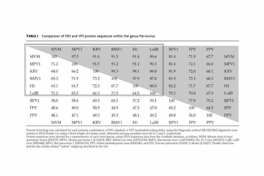
TABLE I Comparison of NS1 and VP2 protein sequences within the genus Parvovirus
Percent homology was calculated for each pairwise combination of NS1 (shaded) or VP2 (unshaded) polypeptides, using the Diagonals method (BLOSUM62 alignment scorematrix) in DNA Strider 1.4, using a block length of 6 amino acids. Mismatch and gap penalties were set to 1 and 2, respectively.Protein sequences were derived for a representative of each virus species, using DNA sequences data from the GenBank database, as follows: MVM, Minute virus of mice(prototype strain) [J02275]; MPV1, Mouse parvovirus 1 [U12469]; KRV, Kilham rat virus [AF321230]; RMV1, Rat minute virus 1 [AF332882]; H1, H-1 virus [X01457]; LuIII, LuIIIvirus [M81888]; RPV1, Rat parvovirus 1 [AF036710]; FPV, Feline panleukopenia virus [M38246]; and PPV, Porcine parvovirus (NADL-2 strain) [L23427]. Double-lined boxdenotes the closely related ‘‘rodent’’ subgroup described in the text.
188 Susan F. Cotmore and Peter Tattersall
immune response, but latency often ensues. In their natural host someviruses, most notably members of the rodent groups, are clinically silent,and can establish persistent infections associated with prolonged virusrelease from reservoirs that are currently unknown.
II. STRUCTURE OF A UNIQUELY DENSE ANDCOMPACT VIRION
Infectious parvoviral virions are nonenveloped, �260 A in diameter, andcontain a single-stranded, linear DNA genome of �5 kb. They comprisebetween 70% and 80% protein, with the remainder being DNA, andare uniquely dense and compact, with molecular masses in the order of5.5–6.2 � 106, sedimentation coefficients of 110S–122S, and buoyant den-sities of 1.39–1.43 g/cm3 in cesium chloride. Mature virions are stable inthe presence of lipid solvents or on exposure to pH 3–9. They are histori-cally reported to survive prolonged incubation at 56 �C, although thischaracteristic applies only to concentrated suspensions of particles or insituations where they are protected by animal tissue, since in dilutesolution they are metastable, undergoing an inactivating conformationaltransition in response to heat or denaturants. However, under naturalconditions, infectious virions are exceptionally durable, surviving forweeks or months at room temperature or for several years at 4 �C.
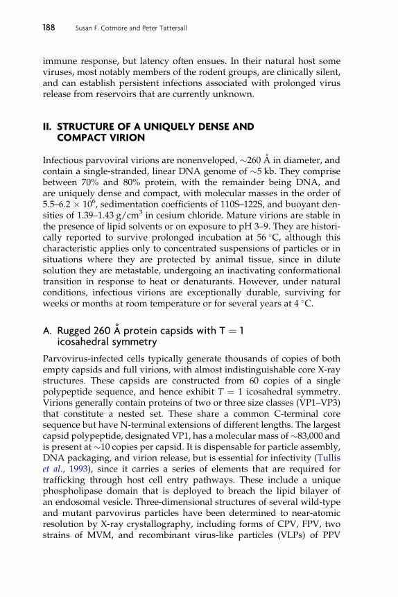
A. Rugged 260 A protein capsids with T ¼ 1icosahedral symmetry
Parvovirus-infected cells typically generate thousands of copies of bothempty capsids and full virions, with almost indistinguishable core X-raystructures. These capsids are constructed from 60 copies of a singlepolypeptide sequence, and hence exhibit T ¼ 1 icosahedral symmetry.Virions generally contain proteins of two or three size classes (VP1–VP3)that constitute a nested set. These share a common C-terminal coresequence but have N-terminal extensions of different lengths. The largestcapsid polypeptide, designated VP1, has a molecular mass of�83,000 andis present at�10 copies per capsid. It is dispensable for particle assembly,DNA packaging, and virion release, but is essential for infectivity (Tulliset al., 1993), since it carries a series of elements that are required fortrafficking through host cell entry pathways. These include a uniquephospholipase domain that is deployed to breach the lipid bilayer ofan endosomal vesicle. Three-dimensional structures of several wild-typeand mutant parvovirus particles have been determined to near-atomicresolution by X-ray crystallography, including forms of CPV, FPV, twostrains of MVM, and recombinant virus-like particles (VLPs) of PPV
Parvoviral Host Range and Cell Entry Mechanisms 189
(reviewed in Chapman and Agbandje-McKenna, 2006). Core structure isbased on a classic eight-stranded antiparallel b-barrel, but in parvovirusesthese b-strands are connected by elaborate and highly variable loops,which make up most of the viral surface (Chapman and Rossmann,1993). The N-terminal peptide domains of the larger proteins are submo-lar and disordered, so their disposition cannot be deduced from X-raydata.
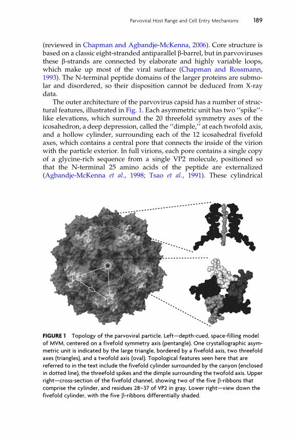
The outer architecture of the parvovirus capsid has a number of struc-tural features, illustrated in Fig. 1. Each asymmetric unit has two ‘‘spike’’-like elevations, which surround the 20 threefold symmetry axes of theicosahedron, a deep depression, called the ‘‘dimple,’’ at each twofold axis,and a hollow cylinder, surrounding each of the 12 icosahedral fivefoldaxes, which contains a central pore that connects the inside of the virionwith the particle exterior. In full virions, each pore contains a single copyof a glycine-rich sequence from a single VP2 molecule, positioned sothat the N-terminal 25 amino acids of the peptide are externalized(Agbandje-McKenna et al., 1998; Tsao et al., 1991). These cylindrical
FIGURE 1 Topology of the parvoviral particle. Left—depth-cued, space-filling model
of MVM, centered on a fivefold symmetry axis (pentangle). One crystallographic asym-
metric unit is indicated by the large triangle, bordered by a fivefold axis, two threefold
axes (triangles), and a twofold axis (oval). Topological features seen here that are
referred to in the text include the fivefold cylinder surrounded by the canyon (enclosed
in dotted line), the threefold spikes and the dimple surrounding the twofold axis. Upper
right—cross-section of the fivefold channel, showing two of the five b-ribbons thatcomprise the cylinder, and residues 28–37 of VP2 in gray. Lower right—view down the
fivefold cylinder, with the five b-ribbons differentially shaded.
190 Susan F. Cotmore and Peter Tattersall
struct ure s are themse lves encirc led on the outer viri on surfac e by a deep,canyon -like depressio n with highly conserved amino acid sequen ce, butunk nown function . Neutra lizing antibody bind ing sites general ly mapto the threef old spike or to its sho ulders, as do pro tein recepto r contac tsfor those serot ypes in which such interac tions have been iden tified.Sequ ences that det ermine viral tissue specif icity and oligos accharidereco gnition lie in the twofold dim ple and up the adjace nt edge of thethr eefold spike .
B. Linear single-stranded DNA genomes withpalin dromic telomeres
Matur e virion s of most species in this genu s contain a single 5-kb DNAstran d that is nega tive sens e with respec t to transcrip tion, wh ile one viru s,LuI II, package s appr oximatel y equimolar positive - and neg ative-se nsestran ds. This remar kable variabi lity illu minates the wh ole proces s ofstran d selectivity , sin ce it is caused by differen tial rates of initia tionfrom the two viral replicat ion origins rather than by any stran d-spec ificpac kaging sign al or mecha nism ( Cotmor e and Ta ttersall, 2005b ). Sincemo st, but not all, gen omes are neg ative sen se with regard to transcrip tion,a unifying co nvention has been adopt ed where by the 3 0 termin us of theneg ative stran d is rather cal led ‘‘the left’’ end and the 50 termin us of thisstran d ‘‘the rig ht’’ end. Within the viri ons, some of the single- strande dDN A displays icosah edral symmetr y, so that about a third of the geno mecan be visual ized by crysta llograph y, ab utting the particle shell. Thi sDN A has some limited nucleot ide specif icity, and is oriente d with itsbas es point ing outwar d, form ing a numbe r of co nserved pro tein–basehydrog en bon ds with the inner sur face of the capsid (Agban dje-McKe nna et al., 1 998 ; Xie and Chapm an, 1996). Rem arkabl y, not all ofthe genome is contain ed with in the particle . DNA pac kaging proce eds ina 30 -to-5 0 directi on, but the 50 en d of the stran d is left project ing throughthe cap sid wall a t an unknow n location so that � 24 nu cleotides (nts),cal led the ‘‘ tether’’ sequen ce, are left outsid e the particl e, covalentlyattac hed, at its 50 end, to a single molec ule of the viral replicat ion init iatorpro tein, NS 1 ( Cotmore and Tattersal l, 1989 ).
At bot h termin i of the linear, nonpe rmuted geno me there are esse ntialpalindr omic sequen ces that can fold into self-p riming duplex ‘‘hairpin ’’telomeres, as illustrated at the top of Fig. 2, which are diagnostic featuresof this virus family. These provide most of the cis-acting informationneeded for both viral DNA replication and encapsidation. In virusesfrom the genus Parvovirus, these two terminal hairpins differ from oneanother in both sequence and predicted secondary structure. This dispa-rity allows differential initiation and encapsidation of the two strands,and typically means that infected cells only receive negative-sense DNA.
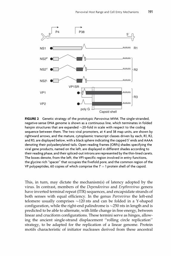
P4 P38
NS1 R1
VP1SR
poly GCapsid shell
AAAAA
AAAAA
AAAAA
AAAAA
AAAAA
AAAAA
R2
R3
NS2P
NS2Y
NS2L
VP1
VP2
3� 5�
FIGURE 2 Genetic strategy of the prototypic Parvovirus MVM. The single-stranded,
negative-sense DNA genome is shown as a continuous line, which terminates in folded
hairpin structures that are expanded �20-fold in scale with respect to the coding
sequence between them. The two viral promoters, at 4 and 38 map units, are shown by
rightward arrows, and the mature, cytoplasmic transcript classes driven by each, R1, R2,
and R3, are displayed below, with a black sphere indicating the capped 50 ends and AAAA
denoting their polyadenylated tails. Open reading frames (ORFs) shades specifying the
viral gene products, named on the left, are displayed in different shades according to
their reading phase, and their spliced-out introns are represented by the thin-lined carets.
The boxes denote, from the left, the VP1-specific region involved in entry functions,
the glycine rich ‘‘spacer’’ that occupies the fivefold pore, and the common region of the
VP polypeptides, 60 copies of which comprise the T ¼ 1 protein shell of the capsid.
Parvoviral Host Range and Cell Entry Mechanisms 191
This, in turn, may dictate the mechanism(s) of latency adopted by thevirus. In contrast, members of the Dependovirus and Erythrovirus generahave inverted terminal repeat (ITR) sequences, and encapsidate strands ofboth senses with equal efficiency. In the genus Parvovirus the left-endtelomere usually comprises �120 nts and can be folded in a Y-shapedconfiguration, while the right-end palindrome is�250 nts in length and ispredicted to be able to alternate, with little change in free energy, betweenlinear and cruciform configurations. These termini serve as hinges, allow-ing the ancient single-strand displacement ‘‘rolling circle replication’’strategy, to be adapted for the replication of a linear genome. Proteinmotifs characteristic of initiator nucleases derived from these ancestral
192 Susan F. Cotmore and Peter Tattersall
replicons are conserved in the viral genome, and its modified replicationscheme is termed ‘‘rolling hairpin replication.’’
C. Creating and expressing transcription templates
When the cell enters S-phase, the viral 30 hairpin acts as a primer forcomplementary-strand DNA synthesis, generating a duplex unit-lengthreplicative intermediate that can support viral transcription. This containstwo mRNA transcription units, with promoters at map units 4 and 38and a single functional polyadenylation site at the extreme right-hand end(reviewed in Qiu et al., 2006). These two promoters, P4 and P38, driveexpression of a nonstructural gene (NS), encoded in the left half of thegenome, and a capsid gene (VP), encoded in the right half, respectively.Alternative splicing events orchestrate gene expression, as shown forMVM in Fig. 2 (Cotmore and Tattersall, 1990; Jongeneel et al., 1986;Morgan and Ward, 1986; Pintel et al., 1983). The R1 transcripts, synthe-sized from the P4 promoter, contain a single contiguous open readingframe (ORF) that encodes the 83-kDa multifunctional replication initiatorprotein, NS1, located upstream of a complex alternately spliced smallintron region. In a further set of P4-derived transcripts, R2, the NS1 ORFis spliced into an alternate reading frame by removal of the major intron,and these transcripts encode, in order of abundance, NS2P, NS2Y, andNS2L, the extreme C-termini of which are different due to the use oftwo pairs of alternative 50 and 30 splice sites bordering the small intron.In contrast, members of the FPV serotype express a single, shorter NS2species, whose second exon is encoded in, and terminates within, thealternative reading frame, some 15 codons upstream of the small intron(Wang et al., 1998).
One function of NS1 is to upregulate the P4 promoter itself, and thispositive feedback loop appears to be a part of the ‘‘hard-wiring’’ of infec-tion that ensures rapid viral takeover of the cell. As infection progresses,the second promoter, at 38 map units, is transactivated by NS1 (Clemensand Pintel, 1988) and drives synthesis of the R3 transcripts, which use thesame pair of alternative 50 and 30 splice sites present in the small intronregion to regulate synthesis of the two primary capsid proteins, VP1 andVP2. In this case, a transcript that uses the downstream 50 and 30 splicesites encodes the minor VP1 polypeptide, translation of which initiates ata methionine codon between the two alternate 50 splice sites. In the moreabundant transcripts, which employ the upstream 50 splice site, thisinitiation codon is spliced out, and translation of the major coat proteinVP2 initiates from a start codon nearly 400 nts further downstream of thesplice. Thus, the two primary translation products from the structuralgene, VP1 (�83 kDa) and VP2 (�63 kDa), are expressed at a �1:5 ratio.A third,more-truncated formof theVP2polypeptide, calledVP3 (�60 kDa),
Parvoviral Host Range and Cell Entry Mechanisms 193
is generated in full, but not in empty, particles by proteolytic cleavageof some 22–25 amino acids from the N-termini of the VP2 polypeptides,following their exposure on the particle surface.
While all parvoviruses encode both NS1 and one or more forms ofNS2, only NS1, the replication initiator protein, is absolutely required forvirus growth in all cell types (Cater and Pintel, 1992; Naeger et al., 1990).NS1 functions in replication as an ATP-dependent, site-specific DNA-binding protein with DNA nicking and helicase activities, which allowsinitiation of DNA synthesis at specific viral origin sequences by introdu-cing a site-specific single-strand nick. This provides a base-paired 30 nt toserve as a primer for successive rounds of strand displacement DNAsynthesis (reviewed in Cotmore and Tattersall, 2006a), while the transes-terification reaction that creates the nick leaves NS1 covalently attached tothe 50 nt, where it is thought to recruit additional NS1 molecules to formthe 30-to-50 replicative helicase.
However, parvoviral replication initiators have evolved into highlypleiotropic proteins, playing multiple roles in the viral life cycle. As men-tioned above, in addition to their site-specific nicking function, they act aspotent transactivators of viral gene transcription, binding to their recog-nition sequences in viral promoters and activating transcription throughacidic C-terminal domains (Legendre and Rommelaere, 1994). In the MVMgenome, NS1 binding sites are reiterated so frequently that any sequence of100 base pairs or more contains a site, and some carry multiple tandem andinverted reiterations (Cotmore et al., 1995). This suggests that NS1 mightplay a significant role in viral chromatin structure and/or progeny strandpackaging. In contrast, NS2 polypeptides play indirect roles in supportingthe MVM life cycle, modifying the cells of their natural murine host tosupport viral replication and mediate efficient capsid assembly. Advancesin our knowledge of parvoviral DNA replication and packaging mecha-nisms have been reviewed extensively elsewhere (Cotmore and Tattersall,2006a,b).
III. RECOGNIZING THE TARGET: CELL SURFACE RECEPTORSAND VIRAL HOST RANGE
Parvovirus particles are extraordinarily rugged, remaining viable at roomtemperature for months, or years, and resisting desiccation or exposure tochaotropic agents. However, they also serve as covert delivery vehicles,able to gain access to the host cell cytosol and penetrate into its nucleus,where they lie in wait for it to initiate DNA synthesis as part of its ownnormal cell cycle. This reliance on the cell’s unchecked transit into S-phasetherefore suggests that the processes of parvovirus entry and latencyremain largely undetected by their host’s innate defense mechanisms.
194 Susan F. Cotmore and Peter Tattersall
This report focuses on both host range and cell entry mechanisms,since these topics are often intimately linked and informed by each other.Infection initiates through capsid-mediated binding to one or moreglycosylated receptor molecule on the cell surface and is followed byvirion uptake into the cell via receptor-mediated endocytosis. Transferacross the delimiting lipid bilayer of the entry vesicle into the cytoplasm isthen affected by a capsid-borne phospholipase, and this is followed bydelivery to, and entry into, the nucleus, where the viral genome is finallyreleased from its protective shell. Thus, parvovirus genomes remainassociated with their intact capsid throughout the entire entry process,and possibly even in primary viral transcription complexes, so that hostcell–specific interactions with the viral particle could potentially impingeat multiple stages during the initiation of infection. While some parvo-viruses exhibit narrowly restricted host ranges, others infect multiple hostspecies and/or many tissues. Although such specificity can operate bydisparate mechanisms, and be mediated either during entry or by celltype–specific differences in viral metabolism, two quite distinct patternsof capsid-controlled host range control have arisen in the genusParvovirus,one exemplified by MVM, and the other by the FPV/CPV serotype.Whether these operate by similar mechanisms or even at the same stagein the entry process still remains to be determined.
Rather than interacting with a single cell surface receptor, many virusfamilies employ two more-or-less separate classes of molecules: ‘‘attach-ment’’ receptors, or coreceptors, which simply accumulate virus in thevicinity of the cell surface; and infectious-entry receptors, which criticallymediate genome transfer into the cell cytoplasm. Some members of theParvovirinae are known to bind to a number of different cell surfacemolecules in ways that potentiate infection, although the extent to whichthey rely on multiple interactions appears to vary from species to species,and within a species from host cell to host cell, so that few general rulesare apparent. Within the genus Parvovirus, members of the FPV serotypecommonly bind to neuraminidase-sensitive N-glycolyl neuraminic acidside chains on some host cell types, but these presumably only functionas attachment receptors, since infectious entry is insensitive to neuramin-idase and is specifically mediated by binding to host species–specificprotein domains on cell surface transferrin receptor (TfR) molecules(Parker et al., 2001; reviewed in Hueffer and Parrish, 2003). In contrast,MVM binds to sialoglycoprotein receptor(s) present at about 5 � 105
copies per cell on murine fibroblasts, and both binding and infection areneuraminidase sensitive, indicating a critical role for specific oligosaccha-ride side chains in both of these steps. However, at present it is not clearwhether one specific cell surface molecule mediates MVM entry, whileothers effect attachment, or if all 5 � 105 receptors are equipotent.
Parvoviral Host Range and Cell Entry Mechanisms 195
The clearest example of a receptor interaction dictating parvovirus hostrange is seen for FPV and its canine-tropic variant CPV, in Chinese hamsterovary (CHO)-derivedTRVb cells,which lack any formof TfR. If feline TfR isexpressed by transfection in these cells it allows efficient binding of CPVand FPV, leading to infection. In contrast, transfected canine TfR bindsCPV capsids poorly, and FPV capsids not at all, and only allows infectionby CPV (Hueffer et al., 2003a). In this case, binding is specified by proteindeterminants on the receptor and involves several critical capsid residuesthat are arranged some 20–30 A apart around the threefold spike, suggest-ing a broad region of receptor–capsid interaction. Remarkably, for CPV thisinteraction appears to be restricted to as few as one site per capsid ratherthan occurring at every 60-fold-related position (Hafenstein et al., 2007;Palermo et al., 2006). In contrast, MVM entry does not rely on interactionswith the TfR, since MVM infects CHO TRVb cells efficiently without TfRtransfection (Cotmore, S. F., and Tattersall, P., unpublished observations),but whether it establishes comparable interactions with other cell surfaceglycoprotein species is currently unknown. Irrespective of any suchprotein-mediated interaction, MVM host range is critically regulated by subtle, celltype–specific, interactions with sialic acid-containing oligosaccharides,which bind into the dimple-like depression at the capsid’s icosahedraltwofold axis. Below, we review details of what is known about receptorbinding and host range constraints in these two disparate examples.
A. The MVM model: Glycan-specific interactions around thetwofold symmetry axes
MVM exhibits subtle strain-specific variations that allow different isolatesto grow productively in murine cells of dissimilar differentiated pheno-types. Two independently isolated strains, termed allotropic variants,were initially identified: the prototype strain, MVMp, which grows pro-ductively in culture in fibroblasts such as the A9 cell line; and the hema-totropic strain, MVMi, which replicates productively in T lymphocytesand hematopoietic precursors (McMaster et al., 1981; Segovia et al., 1991;Spalholz and Tattersall, 1983). Despite sharing 97% sequence identity andbeing serologically indistinguishable, these viruses are reciprocally res-tricted for growth in each other’s host cell type (Tattersall and Bratton,1983). In nonpermissive cells infection is restricted prior to viral geneexpression (Antonietti et al., 1988; Gardiner and Tattersall, 1988a), butboth virus strains are known to compete for specific binding sites on thesurfaces of both cell types (Spalholz and Tattersall, 1983), estimated to bepresent at 5 � 105 copies per cell on mouse A9 fibroblasts (Linser et al.,1977; Spalholz and Tattersall, 1983). Following intranasal inoculation intonewbornmice, MVMp is asymptomatic, and the virus remains confined to

196 Susan F. Cotmore and Peter Tattersall
the oropharynx (Kimsey et al., 1986), while MVMi causes a generalizedinfection in which the main targets are endothelial cells, lymphocytes, andhepatic erythropoietic precursors, but where the pathological outcomevaries with host genotype (Brownstein et al., 1992).
The ability of MVMp to grow in fibroblasts was mapped in vitro using aselective plaque assay to two specific amino acids at positions 317 and 321 inthe VP2 capsid protein sequence (Ball-Goodrich and Tattersall, 1992;Gardiner and Tattersall, 1988b). These lie at or near the particle surface,adjacent to the dimple-like depression that spans the icosahedral twofoldaxis of the virion (Agbandje-McKenna et al., 1998). When a restriction frag-ment fromMVMpdiffering at only these twoVP2 residues (T317 andG321)was substituted into an infectious plasmid clone ofMVMi (A317 andD321),the resulting virus was found to be >100-fold better at infecting fibroblaststhan its parent (Gardiner and Tattersall, 1988b). In contrast, when eithersingle change was introduced into MVMi separately, the resulting virusesshowed at most a twofold increase in their ability to replicate in fibroblasts(Ball-Goodrich and Tattersall, 1992). This restriction, in turn, allowedthe selection of second site mutants that could complement either of thesechanges (Agbandje-McKenna et al., 1998; Lopez-Bueno et al., 2007). For eachof the single mutants, multiple alternative second site mutations wereidentified, all affecting residues surrounding or extending down the sidesof the twofold-related dimple. Surprisingly, if the MVMi backbone alreadycarried the A317T mutation, complementing mutations in D321 were notselected, but instead the additional mutations D399G, D399A, V551A, orD553Nwere each found to effectively confer fibrotropism. In contrast,whenthe MVMi backbone already carried the D321G mutation, four of the sixsecond-site mutants identified carried the MVMp A317T change, while inthe other two, the coordinated mutations were S460A and Y558H. Thus,in anMVMi backbone, fibrotropism can be conferred by switching the sidechains of a number of different residues that surround the twofold depres-sion, suggesting that structural changes in this depression may mediateMVM cell type specificity. While little is know about the control of tissuespecificity for most other parvoviruses, it is clear that amino acid changesinvolved in determining both PPV cell type specificity and virulence arealso localized in this depression (Simpson et al., 2002).
Lack of a lymphocyte plaque assay prevented the equivalent analysisof MVMp host range mutants in culture, but this has been effectivelyaccomplished in vivo using adult immunodeficient SCID mice (Rubioet al., 2005). Following intravenous injection of MVMp into such mice,this normally apathogenic virus strain was found to evolve through atleast two distinct steps, the first of which conferred enhanced virulence,while the second generated complex shifts in host cell specificity andpathogenicity. During the early weeks of subclinical infection, injectedMVMp viruses consistently segregated variants that showed altered,
Parvoviral Host Range and Cell Entry Mechanisms 197
large-plaque, phenotypes when tested in vitro, but retained the fibrotropicMVMp host range. However, unlike wild-type MVMp, when these var-iants were reinoculated into SCIDmice via the oronasal route, they spreadsystemically from the oronasal cavity and were able to access, and repli-cate in, various major organs such as the brain, kidney, and liver. Geneticanalysis of 48 of these clones consistently showed one of three singlechanges in the VP2 gene, V325M, I362S, or K368R. Both MVMp and therecombinant viruses could be detected in the bloodstream 1- to 2-daypostoronasal inoculation and remained infectious when adsorbed toblood cells in vitro. However, wild-type MVMp was cleared from thecirculation within a few days, while the viremia caused by the mutantviruses was sustained for life, leading to their being described as havinghigher ‘‘virulence.’’ Significantly, attachment of bothmutant and wild-typeviruses to an abundant receptor on primary mouse kidney epithelial cellscould be quantitatively competed by wild-typeMVMp capsids, suggestingthat this enhanced virulence was not associated with major differences inreceptor usage in the target tissues. However, productive adsorption ofvariants carrying any of the three mutations showed increased sensitivityto neuraminidase, when compared to wild-type virus, suggesting that theparticles had a lower affinity for the sialic acid component of the receptor.This diminished affinity for sialic acid–bearing oligosaccharide chainswas later confirmed by plasmon surface resonance studies, discussedbelow. This suggests that the selection of capsids with lower affinity fortheir cell surface receptors favors systemic infection, which may be a majorevolutionary process in the adaptation of parvoviruses to new hosts.
As illustrated in Fig. 3, two of these virulence determinants, residuesI362 and K368, are located on the wall of the dimple recess surroundingthe icosahedral twofold symmetry axis, while V325 is positioned �22 Aaway in a threefold-related monomer, near the top of the depression.Consistent with this, the X-ray crystal structure of MVMp capsids soakedwith sialic acid (N-acetyl neuraminic acid) showed the sugar positionedin this depression, immediately adjacent to residues I362 and K368. Thus,this likely identifies the position of the terminal sugar in the infectiousreceptor attachment site on the viral capsid. However, the equivalentphenotype seen in mutants carrying the V325M mutation suggests thatthis residue also modulates sialic acid binding in a manner similar to I362and K368, even though it is physically somewhat distant (Lopez-Buenoet al., 2006). The depression at the twofold icosahedral axis of MVMp doesextend toward the loop containing V325 from a threefold-related mono-mer, which interdigitates with the reference monomer, as shown inFig. 3C. These observations therefore suggest that although sialic acid isan essential component of the receptor for MVMp infection, and itbinds to capsid residues in the icosahedral twofold depression, the carbo-hydrate component of the surface receptor recognized by the virus may
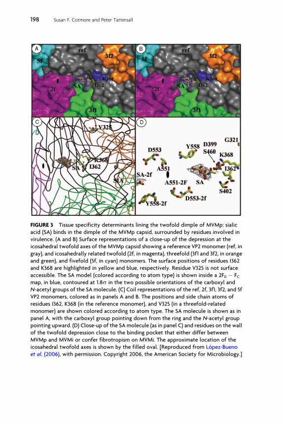
FIGURE 3 Tissue specificity determinants lining the twofold dimple of MVMp: sialic
acid (SA) binds in the dimple of the MVMp capsid, surrounded by residues involved in
virulence. (A and B) Surface representations of a close-up of the depression at the
icosahedral twofold axes of the MVMp capsid showing a reference VP2 monomer (ref, in
gray), and icosahedrally related twofold (2f, in magenta), threefold (3f1 and 3f2, in orange
and green), and fivefold (5f, in cyan) monomers. The surface positions of residues I362
and K368 are highlighted in yellow and blue, respectively. Residue V325 is not surface
accessible. The SA model (colored according to atom type) is shown inside a 2F0 � FCmap, in blue, contoured at 1.8s in the two possible orientations of the carboxyl and
N-acetyl groups of the SA molecule. (C) Coil representations of the ref, 2f, 3f1, 3f2, and 5f
VP2 monomers, colored as in panels A and B. The positions and side chain atoms of
residues I362, K368 (in the reference monomer), and V325 (in a threefold-related
monomer) are shown colored according to atom type. The SA molecule is shown as in
panel A, with the carboxyl group pointing down from the ring and the N-acetyl group
pointing upward. (D) Close-up of the SA molecule (as in panel C) and residues on the wall
of the twofold depression close to the binding pocket that either differ between
MVMp and MVMi or confer fibrotropism on MVMi. The approximate location of the
icosahedral twofold axes is shown by the filled oval. [Reproduced from Lopez-Bueno
et al. (2006), with permission. Copyright 2006, the American Society for Microbiology.]
198 Susan F. Cotmore and Peter Tattersall
Parvoviral Host Range and Cell Entry Mechanisms 199
be larger than a single sialic acid residue. Accordingly, a longer oligosac-charide might show additional contacts both within the dimple andadjacent to the loop carrying V325 at the top of this depression.
Evidence for enhanced interactions with longer, sialic acid–bearingoligosaccharides comes from glycan array and surface plasmon resonancestudies (Nam et al., 2006). Thesemonitored the interactions of baculovirus-derived VLPs harboring the VP2 protein of MVMi, MVMp, the high-virulence MVMp mutants I362S, and K368R, or the double mutantI362S/K368R, with 180 different glycans. All of the particles bound spe-cifically to oligosaccharide chains carrying terminal sialic acid residueslinked 2–3 to a common Gal 1–4GlcNAc moiety. However, binding onlyoccurred when the chains contained at least five saccharide residues andthe binding affinity generally increased as a function of chain length.None of the VLPs recognized oligosaccharides with NeuAc a2–6 linkedsialic acids, while MVMi was unique in binding efficiently to the fourmultisialylated glycans with a2–8 linkages that were present in the array,although the MVMp-derived K368R mutant also bound to one of thesewith lower affinity. This therefore supports a model in which the slightdifferences in topology and side chain interactions of specific residueslining the dimple, which can be seen in comparisons of the three-dimensional structures of MVMp and MVMi, reflect differences in theabilities of this cleft in each virus to accommodate somewhat differentcarbohydrate arrangements.
When reintroduced into SCID mice, these high-virulence MVMpmutants subsequently underwent pathogenic tissue-specific evolution,which again involved residues that map to the dimple (Lopez-Buenoet al., 2007). In this case, MVMp viruses carrying the I362S or K368Rvirulence changes, inoculated via the oronasal route, induced a lethalleukopenia after a 14–18 week delay, reflecting the pattern of diseasetypically found for MVMi infections within 7 weeks of infection. Sequenc-ing the emerging MVM populations in these leukopenic mice prior tocloning identified consensus sequence changes at G321E and A551V inthe I362S infections and at V575A and A551V in the K368R infections.Notably, changes at dimple residues 321 and 551 (indicated in Fig. 3) wereamong those previously identified in fibrotropic switch mutants selectedby plaquing MVMi on mouse fibroblast monolayers. However, clonalanalysis of the mutant populations from SCID mice revealed geneticheterogeneity at specific capsid residues, and only a few of these clonalisolates, which retained the parental G321 and V575 residues, were infec-tious in vitro. Rather, consensus genotypes were poorly infectious inculture, even in 324 K cells, an SV40-transformed human cell line thatsupports both lymphotropic and fibrotropic MVM variants, althoughvirions could be generated following transfection of cloned genomesinto these cells, indicating that later stages in the viral life cycle were

200 Susan F. Cotmore and Peter Tattersall
conserved. Virions from one such mutant, carrying the consensus muta-tions A551V and V575A, while unable to initiate infection in culture in avariety of different cell lines, rapidly induced lethal leukopenia whengiven to SCID mice, suggesting that in vivo this virus may exploit a subtlydifferent allotropic interaction. This all suggests that theMVMdimple canbe finely adapted to accommodate a range of different oligosaccharidesand that, by changing the side chains and interactions of a small numberof surface residues, the virus appears to be able to infect diverse reper-toires of differentiated host cell types.
Other aspects of the viral life cycle clearly influence MVM’s remark-able ability to switch its tissue specificity. In particular, the speed andefficiency with which heterogeneous virus populations are generatedduring parvoviral disease depend on high viral mutation rates, andresemble the generation of quasispecies typically encountered duringthe expansion of RNA viruses. Thus, for example, Lopez-Bueno et al.(2003) observed that when MVMi-infected SCID mice received passiveimmunotherapy with a neutralizing monoclonal anti-capsid antibody,escape mutants, harboring single radical amino acid changes at tip ofthe threefold spike, emerged at high frequency (2.8 � 0.5 � 10�5). Suchheterogeneity had not been previously expected for this DNA virus,which replicates using the normally high-fidelity DNA synthetic machin-ery of its host cell. However, similar mutation rates have now beenobserved for several members of the Parvovirinae (Badgett et al., 2002;Shackelton and Holmes, 2006; Shackelton et al., 2006), although the under-lying causes remain conjectural. Thus, during a productive MVM infec-tion, where high mutation rates are coupled with rapid virus expansion,generating up to 108 infectious particles per infected mouse, specific virusstrains may evolve rapidly, giving rise to host range mutants that arepotentially able to infect an alternative set of differentiated cell types.
For MVM there is even further latitude for phenotypic expansion,since the ability of host range mutants to thrive in their new host cellcan depend on the sequence, or even the expression level, of NS2, theminor viral nonstructural protein. As discussed above, when MVMi isadapted for growth in fibroblasts, the host range switch typically involvestwo coordinate mutations in the vicinity of the dimple. However, twohost range switch mutants have been characterized that carry a singlecoding mutation at residue D399 in VP2, to alanine or glycine, togetherwith a second, noncoding, guanine-to-adenine change at nucleotides 1970or 1967, which influence the splicing patterns of the viral transcripts(D’Abramo et al., 2005). When reconstructed into an infectious mole-cular clone of MVMi, all single mutants failed to replicate productivelyin fibroblasts, but viruses carrying a pair of mutations, with one of eachtype, were highly infectious. Specifically, the single D399 mutationsallowed viruses to initiate infection in fibroblasts, but NS2 expression
Parvoviral Host Range and Cell Entry Mechanisms 201
was low, which led to poor accumulation and release of progeny virus.Mutations at 1967 or 1970 restored the MVMp splicing pattern, enhancedNS2 accumulation, and allowed efficient progeny production and release.Conversely, the D399 mutations destroyed the viruses’ ability to initiateinfection in EL4 lymphocytes. However, in lymphocyte infections, NS2was expressed at high ratios even in the absence of upstream mutations,and progeny accumulation was efficient. Choi et al. (2005) showed thatthis requirement for different splicing signals to achieve optimal MVMNS2 levels reflects cell type–specific differences in RNA processing,which can thus impact host range. Exactly why high NS2 levels arerequired for efficient progeny virus production remains uncertain, andis probably multifactorial, but, in part, it appears to reflect a defect incapsid assembly seen in NS2 depleted cells (Cotmore et al., 1997). Thismay suggest that it is difficult to assemble the single D399 mutants, butthat either a second local capsid modification, such as A317T, or a boost inNS2 levels, eases this constraint. While wild-type NS2 is known to interactwith the cellular nuclear export protein, Crm1 (Bodendorf et al., 1999),remarkably, a mutation that promotes higher affinity Crm1 binding thanwild type was also able to reverse this progeny production defect, so thateven low-level expression of NS2 led to efficient virus expansion (Choiet al., 2005). The high-affinity Crm1 binding mutant used in this study andseveral other similar mutations were first identified in SCIDmice that hadbeen infected with MVMi and exposed to neutralizing polyclonal anti-sera, in an attempt to protect the mice from leukopenic disease. Thesesingle or double amino acids changes in the NS2 Crm1 binding domainincreased its ability to sequester Crm1 in a perinuclear locale, leading toan accelerated viral life cycle that somehow allowed the virus to circum-vent the effects of neutralizing antibody (Lopez-Bueno et al., 2004).Taken together, this data indicates that mutations in NS2 that promoteits efficient interaction with Crm1 can effectively modulate viral hostrange, by allowing a productive viral cycle to proceed in cells thatwould normally be nonproductive due to inadequate NS2 expression.Clearly, this provides a second example of how the virus’s capacity forrapid evolutionary change can permit shifts in host range in vivo. Againstthis evolutionary force is ranged the extreme conservatism of thisintensely compact virus, since most random mutations, or combinationsthereof, appear to be incompatible with overall viral viability.
B. The FPV/CPV model: Engaging the transferrin receptor at thethreefold symmetry axes
In sharp contrast to the situation in MVM, where research has focused onanalyzing changes in specificity for differentiated murine cell types, forviruses of the FPV serotype most attention has been directed at

202 Susan F. Cotmore and Peter Tattersall
understandinghow the virus switched frombeing able to infect a number ofcarnivore species, excluding dogs, to being a potent canine pathogen. Thisevent appears to have occurred early in the 1970s, when a complex virusmutant emerged and spread rapidly through the global dog population,erupting to pandemic status in 1978. This virus, called CPV-2, had lost theability to infect cats. However, in 1979 an antigenic variant emerged, calledCPV-2a, which can infect both host species and has since globally replacedthe original virus in both domestic and wild dog populations. Phylogeneticanalysis of DNA sequences suggests that all CPV isolates from dogs arederived from a single common ancestor, which only differs by a few nucleo-tides, some 0.4% of the genome, from the most recent common ancestoramong the FPV-like viruses. Most of these changes have been conserved inthe CPV variants emerging since 1978. All of the viruses from either cats ordogs replicate efficiently in feline cells in culture, but only CPV isolatesinfect dogs and cultured dog cells (Truyen and Parrish, 1992). The hostrange properties of CPV and FPV for both dogs and cats are controlledby multiple residues that map to disparate locales on or around the three-fold spike, as shown in Fig. 4. Primary control of canine host range isdetermined by residues at VP2 positions 93 and 323, which must beswitched coordinately (Chang et al., 1992; Horiuchi et al., 1994; Huefferet al., 2003b; Llamas-Saiz et al., 1996; Parker and Parrish, 1997; Strassheimet al., 1994). Certain changes at residues 299 and 300 block the ability ofthe virus to infect dog cells, and changes in that region also appear to controlthe in vivo feline host range of CPV (Truyen et al., 1994). The CPV-2a variantthat emerged in 1979, which infects both host species, has additionalchanges at VP2 residues 87, 101, 300, and 305 (Parrish and Carmichael,1986; Parrish et al., 1988, 1991), and several other single mutations in CPV-2a have become widely distributed in vivo since 1980, including an N426Dmutation that is present in the antigenic variant designated CPV-2b, whichshares the CPV-2a host range (Strassheim et al., 1994; Truyen et al., 1995).
Some of the host range constraints of CPV and FPV seen in animals arereflected in tissue culture, where it is now clear that the block to infection byFPV in dog cells is primarily due to lack of a functional cell surface receptor.FPV and CPV both bind the feline TfR and use it to infect cat cells, butonly CPV can bind to canine TfR. However, although CPV-2, CPV-2a, andCPV-2b all bind the canine TfR and infect dog cells, CPV-2 capsids bind tofeline and canine cells much more efficiently and to higher levels than doCPV-2a or CPV-2b capsids, suggesting that CPV-2 forms different interac-tions with the TfR or binds to additional receptors on those cells (Huefferet al., 2003a). Thus, while VP2 residues 93 and 323 together control virusbinding to the canine TfR (Hueffer et al., 2003a), changes at VP2 residues 87,300, and 305 in CPV-2a reduce receptor affinity and improve, in some way,the ability of the virus to use this receptor for infection (Hueffer et al., 2003a;Palermo et al., 2006).

FIGURE 4 Distribution of host-range determinant residues on the surface of CPV.
(A) Surface-rendered model of the CPV capsid, viewed from above the dimple that
surrounds the twofold symmetry axis, located on the bottom side of the triangle
representing a single asymmetric unit, halfway between the threefold spikes. Toward the
apex of the triangle lies the canyon surrounding the fivefold cylinder. A model of the
ectodomain human transferrin receptor is shown at the same scale to indicate the
relative size of the virus and its ligand on feline or canine cells. (B) A road map
determined by the method of Rossmann and Palmenberg (1988) showing the surface
exposure of VP2 residues in one asymmetric unit of the CPV type 2 capsid. The region
shown comprises several symmetry-related VP2 subunits. Residues mentioned in the
text that affect receptor binding or host range, and which differ naturally between FPV
and CPV strains, are shaded. [Modified from Hueffer et al. (2003b) with permission.
Copyright 2003, the American Society for Microbiology.]
Parvoviral Host Range and Cell Entry Mechanisms 203
TfR is a type II membrane protein that protrudes about 30 A from thecell surface. The structures of canine and feline TfR have yet to be deter-mined, but structural information is available for the human TfR, which is79% identical to feline TfR at the amino acid sequence level. The humanTfR consists of a large, butterfly-shaped, dimeric molecule with a span ofabout 100 A and a molecular weight of 180 kDa. Each monomer has anapical domain, a helical domain, and a carboxypeptidase-like domain(Lawrence et al., 1999), and mutagenesis of feline and canine TfRs indi-cates that both CPV and FPV bind to the apical domain. In confirmationof results from the in vitro cell binding assays, both FPV and CPV capsidswere found to bind strongly to a recombinant form of the feline TfR ecto-domain, while CPV-2b capsids bound much more weakly. In contrast,FPV capsids failed to bind at all to recombinant canine TfR (Palermo et al.,2006), and while CPV-2 capsids bound the canine receptor, they did soonly to very low levels, and CPV-2b binding was essentially undetectable.This binding pattern reflects the weak interaction seen in culture when thesame receptor was expressed by transfection on otherwise TfR-negative
204 Susan F. Cotmore and Peter Tattersall
CHO cells, which nevertheless was sufficient to allow CPV-2b to be takeninto and infect the cells. This low level of binding between canine TfR andCPV-2 or CPV-2b capsids, and its inability to bind FPV are in large partdetermined byminimal differences in the TfR apical domain, since simplychanging residues 383 and 385 in canine TfR to their feline TfR counter-parts allowed the mutant receptor to bind FPV to levels similar to thoseseen for the feline TfR, and likewise increased binding of CPV capsids.Residues 383 and 385 create a potential glycosylation site on canine TfR,which appears to be occupied in vivo, but the increased binding seen forthe mutant is probably due to protein sequence, rather than oligosaccha-ride, changes, since enzymatic removal of N-linked glycans from thecanine receptor did not lead to increased binding (Palermo et al., 2006).
The specific binding of CPV to canine TfR is thus controlled by severalresidues, positioned 20–30 A apart on the ‘‘high ground’’ around thethreefold spike, suggesting that a broad surface of the capsid interactswith the receptor (Govindasamy et al., 2003; Hueffer et al., 2003a,b). Whileless is known about the capsid residues that are involved in feline TfRbinding, capsid mutations reciprocal to those which in CPV preventedcanine TfR binding, at positions 93 and 323, did not appear to alter thebinding of FPV to the feline TfR expressed on CHO TRVb cells, indicatingthat the canine and feline receptors make somewhat different contactswith these viruses (Hueffer et al., 2003a).
Asymmetric cryo-electron microscopic (cryo-EM) reconstructions,supported by quantitative in vitro binding studies, suggest a model inwhich the canine TfR ectodomain can bind to only one, or a few, of the60 icosahedrally equivalent sites on empty CPV capsids, suggesting thatthese either have inherent asymmetry or that binding to their receptorinduces asymmetry (Hafenstein et al., 2007). When a difference map,calculated by comparing the virus-receptor complex with the nativevirus, was superimposed on a stereographic projection of the icosahedralCPV surface structure, the known crystal structure of the human TfRectodomain dimer (Lawrence et al., 1999) could be modeled into theadditional cryo-EM density such that one of its two apical domains wasin contact with the shoulder of one of the CPV spikes. In this model, theprojected contact sites on the virus included residues that are known tocontrol specific binding to canine TfR (Hueffer et al., 2003a).
Possibly, the restricted binding observed for the CPV–canine TfRinteraction is due to inherent asymmetry in the empty particle, withone, or a few, distinct sites that have a conformation capable of bindingTfR, whereas the other icosahedrally equivalent sites are slightly differ-ent. If so, this asymmetry must exist prior to genome encapsidation, andmight indeed direct that process. This might happen if assembly isinitiated around a special icosahedral fivefold vertex, which is the site ofsubsequent genome entry and exit, as is believed to occur in tailed
Parvoviral Host Range and Cell Entry Mechanisms 205
bacteriophage (Morais et al., 2003). Alternatively, the final subunits toassemble may be sterically hindered from perfectly finishing the icosahe-dron, thus creating an asymmetric structural element at the capsid sur-face. However, it is also possible that TfR binding itself might induceasymmetry in an initially icosahedral particle, perhaps priming it forsubsequent conformational shifts destined to occur during cell entry, asdiscussed below.
Thus it appears that after first adapting to dogs by acquiring changesthat allowed it to bind canine TfR in a productive way, CPV has continuedto evolve in vivo, acquiring additional mutations that lower its affinity forthis receptor but enhance its ability to infect cells. Use of the TfR as thecellular receptor for these viruses also correlates well with the patterns oftissue specificity seen in vivo, as this receptor is highly expressed on cryptcells in the intestinal epithelium and on hematopoietic cells, which are themain target cells of CPV and FPV in animals (Parrish, 1995). However,TfR acts as more than a simple tether, dragging the capsid into thecell, since the precise interactions are important for successful cell infec-tion, and some mutational changes in either the virus or the receptorallow capsid binding and cell uptake without leading to infection(Hueffer et al., 2003b; Palermo et al., 2003).
Transfer of the viral genome across the limiting lipid bilayer of itsprospective host cell is one of the most challenging steps encounteredduring cell entry, and for many nonenveloped viruses this maneuver isso finely orchestrated that critical interactions required with cell surfacereceptor molecules play a major role in determining viral host cell speci-ficity. To date, it is not clear whether any parvoviruses employ a receptor-orchestrated transfer mechanism of this type, but it is clear that theymust undergo a specific structural transition after endocytosis, but beforebilayer penetration, which leads to exposure of their VP1-specific ‘‘entry’’peptide, VP1SR, and that inappropriate exposure of this peptide leadsto their inactivation. Thus, it is tempting to speculate that interactionswith specific receptors could modulate host range by allowing this tran-sition to occur in a controlled way or in a favored locale that would becompatible with the transfer of a viable particle across the lipid bilayer, aswe will now discuss.
IV. BREACHING THE OUTER BARRIER: TO THECYTOSOL AND BEYOND
Viral particles must function as rugged containers that protect the genomefrom environmental assaults encountered during transmission, but mustalso recognize and respond to a succession of specific cellular signals thatallow them to navigate the complex entry portals of their host cell, and
206 Susan F. Cotmore and Peter Tattersall
ultimately deliver their nucleic acid to the appropriate replication com-partment. Since parvoviral virions lack any accessory proteins, the com-ponent polypeptides of the nonenveloped capsid are the sole mediators ofentry. While the capsid shell itself directs certain interactions, many othernecessary contacts with cellular pathways rely on signal-rich N-terminalextensions present on VP1 and VP2 molecules. These are initially seques-tered within the particle but are sequentially deployed at the virionsurface during the cell exit and entry processes by a series of concertedconformational shifts in the capsid structure. Aspects of parvoviral entryhave been reviewed by others in recent years (Vihinen-Ranta and Parrish,2006; Vihinen-Ranta et al., 2004), but in this section we attempt to integratedata from a broader range of analyses. Specifically, we will examine thestructural flexibility and transitions that viral particles are able to undergoin vitro, explore the structure of the VP1SR entry peptide, and finallyconsider vesicle trafficking and deployment of the entry peptide in vivo.This overview suggests that each step in the program of intracellulartranslocation of the intact particle to the cell nucleus is catalyzed bysuccessively revealed motifs built into the capsid structure itself.
A. Structural transitions in the virion induced in vitro
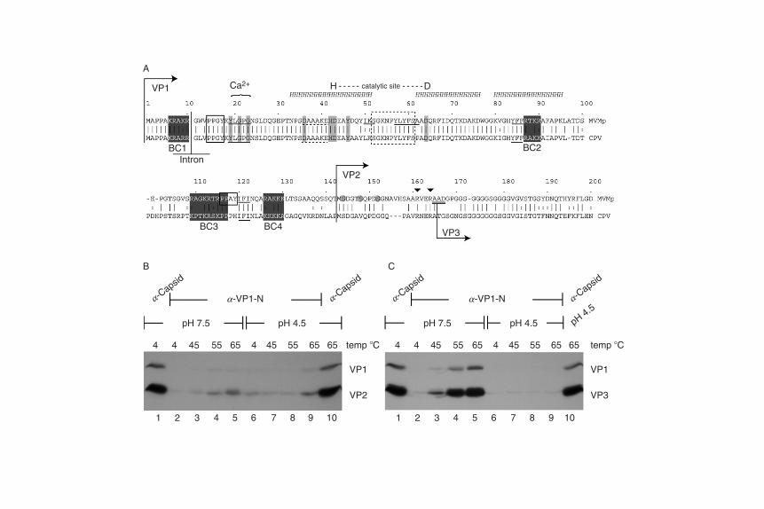
Empty parvovirus capsids are constructed from 60 copies of the capsidpolypeptides, comprising, on average, 50 copies of VP2 and 10 copies ofVP1. As diagrammed in Fig. 2, VP1 contains all of the VP2 sequence buthas an extra, basically charged, 142-amino acid N-terminal extension,termed the VP1-specific region, VP1SR. The VP1 extension, shown indetail in Fig. 5A, is dispensable for both capsid assembly and DNApackaging, but is absolutely required for infectious entry, since it carriesa phospholipase A2 (PLA2) active site essential for endosomal exit, aswell as various clusters of basic amino acids and signaling motifs thatmay function at subsequent steps during nuclear localization. However,in MVM, only 547 amino acids from the C-terminus of the VP polypep-tides are ordered, and therefore visible in the crystal structure, while thesignal-rich N-terminal extensions, of 39 residues for VP2 and 181 residuesfor VP1, resist 60-fold averaging. These N-terminal regions are seques-tered within the empty particle, but become sequentially externalized atspecific steps in its life cycle, to modulate particle stability and to mediatesuccessive interactions with the host cell.
In the viral particle, a cylindrical projection surrounds each of the12 fivefold symmetry axes, and is itself encircled by a 15 A-deep exteriordepression, of unknown function, called the canyon. The cylinder iscreated by the juxtaposition of antiparallel b-hairpins from each of thefivefold-related capsid proteins. These b-hairpins are not interdigitatedwithin the upper part of the resulting ‘‘turret’’ and so are potentially
Parvoviral Host Range and Cell Entry Mechanisms 207
flexible, and their organization in the crystal structure creates a narrow,8 A, central pore that penetrates through the virion shell to the particleinterior. The tightest constriction in this pore is formed at its inner endby the juxtaposition of leucine side chains from VP2 residue 172 of fiveindependent VP2 molecules. The phenotypic analysis of a complete set ofamino acid substitution mutants at this highly conserved residue stronglysuggests that L172 modulates the extrusion of VP1 N-termini (VP1NT)(Farr and Tattersall, 2004). All but one of these mutants produced DNA-containing virions, but only two, L172V and L172I, were infectious, theothers being blocked for assembly, packaging, or viral entry. Several ofthe mutants were significantly defective for assembly at 39 �C, but not at32 �C, and, while tryptic cleavage of their VP2 N-termini was normal, VP1was degraded during in vitro proteolysis of mutant, but not wild-type,virions. The L172W substitution, while not significantly affecting assem-bly, effectively abrogated genome encapsidation, contributing to theemerging genetic evidence for both the Parvovirus and Dependovirusgenera suggesting that one of these fivefold pores mediates encapsidationof the viral genome late in infection. For this step, the presumptive portalacts in concert with a viral helicase complex, which has been shown forAAV to be a small Rep protein, but, for the autonomous parvoviruses,is derived from NS1 in an unknown manner. It is currently not clearwhether the packaging portal is physically distinct from the other11 cylinders prior to being selected as the encapsidation point.
X-ray crystallography of MVM virions revealed ordered structurebeginning at VP2 residue 40, which is on the inside of the shell, formingpart of the basal structure that supports the cylinder. In full virions, butnot in empty particles, the pore contains additional weak density, intowhich has beenmodeled a single copy of a conserved glycine-rich peptidethat spans VP2 residues 28–38 (VP2 residue 28-GGSGGGGSGGG-38),shown in Figs. 1 and 5A. Additional density, corresponding to residues36–39 from the remaining capsid proteins, extends back into the particleinterior. Since, in the crystal structure, each pore accommodates a singleglycine-rich peptide, only one of the five locally available VP N-terminican be externalized at any time. However, almost all of the VP2N-terminalpeptides become surface-exposed during entry, or during proteolyticdigestion in vitro, suggesting that there are dynamic fluctuations in porestructure. Since the pore is only 8 A in diameter, but must accommodatethe passage of amino acids with bulky side chains during these extrusionevents, this implies that the cylinder is an inherently dynamic structure.Indeed, one function of the canyon might be to provide space for theb-hairpins of the cylinder to move outward, thus allowing the pore toexpand.
Viral genomes are packaged into some sort of preassembled emptyparticle, but evidence from AAV2 suggests that such particles are
VP1
A
B C
VP2
VP3
BC1
BC3
pH 7.5
4 4 45 55 65 4 45 55 65 65
1 2 3 4 5 6 7 8 9 10 1 2 3 4 5 6 7 8 9 10
4 4 45 55 65 4 45 55 65 65temp �C
VP1
a-VP1-Na-Capsid
a-Capsid
a-Capsid
a-Capsid
pH 4.5a-VP1-N
VP2
VP1
VP3
temp �C
pH 4.5 pH 7.5 pH 4.5
BC4
IntronBC2
Ca2+ H Dcatalytic site
Parvoviral Host Range and Cell Entry Mechanisms 209
somewhat specialized since they have to be assembled in the presence ofthe Rep proteins, which are the functional equivalent of the MVM NS1polypeptide (Wu et al., 2000). Both VP1 and VP2 N-termini are completelysequestered inside these empty capsids, but a structural shift occurs inthe packaging complex prior to, or concomitant with, the beginning ofDNA translocation, which allows a cohort of VP2 N-terminal peptidesto emerge at the virion surface (Cotmore and Tattersall, 2005a). Whetherthese termini play a role in the packaging process remains uncertain, butthey do appear to stabilize the final structure, as discussed below. TheseN-terminal extensions carry phosphoserine-rich export signals, which insome cell types direct packaged virions to be trafficked out of the nucleusprior to cell lysis (Maroto et al., 2004). Full particles are thus releasedfrom the parental cell with all of their VP2 N-termini intact, but a thirdstructural protein, VP3, is subsequently generated from most VP2 mole-cules by a proteolytic cleavage that removes 22–25 amino acids from itsN-terminus. VP2 to VP3 cleavage can occur in the extracellular environ-ment following release, but, if not, invariably occurs during entry into anew host cell (Clinton and Hayashi, 1975; Paradiso, 1984; Ros et al., 2002).This cleavage can be mimicked in vitro by incubating virions with a broadvariety of proteases, but the cleavage site appears flexible, and veryaccessible, so that it has been essentially impossible to totally ablatecleavage in MVM by mutagenesis or to stop it occurring in vivo usingcombinations of protease inhibitors (Clinton and Hayashi, 1975; Tulliset al., 1992; Farr, G. A., Cotmore, S. F., and Tattersall, P., unpublishedresults). Since each pore can only accommodate one N-terminal peptide ata time, it is suggested that following proteolytic cleavage the residual
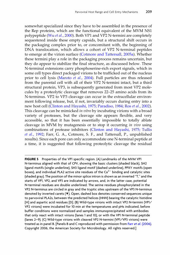
FIGURE 5 Properties of the VP1 specific region. (A) Landmarks of the MVM VP1
N-terminus aligned with that of CPV, showing the basic clusters (shaded black), SH2
ligand motifs (single underline), SH3 ligand motif (dashed underline), PPXY motifs (open
boxes), and individual PLA2 active site residues of the Ca2þ binding and catalytic sites
(shaded gray). The position of the minor splice intron is shown as an inverted ‘‘T,’’ and the
starts of VP1, VP2, and VP3 are indicated by arrows, and, in the latter case, potential
N-terminal residues are double underlined. The serine residues phosphorylated in the
VP2 N-terminus are circled in gray and the tryptic sites upstream of the VP3 N-terminus
denoted by inverted carets (▼). Open, dashed box denotes conserved sequences unique
to parvoviral PLA2s, between the predicted helices (HHH) bearing the catalytic histidine
[H] and aspartic acid residues [D]. (B) Wild-type virions with intact VP2 N-termini (VP1/
VP2 virions) were incubated for 10 min at the temperatures and pHs indicated, before
buffer conditions were normalized and samples immunoprecipitated with antibodies
that only react with intact virions (lanes 1 and 10), or with the VP1 N-terminal peptide
(lanes 2–9). (C) Wild-type virions with cleaved VP2 N-termini (VP1/VP3 virions) were
treated as in panel B. [Panels B and C reproduced with permission from Farr et al. (2006).
Copyright 2006, the American Society for Microbiology. All rights reserved.]

210 Susan F. Cotmore and Peter Tattersall
glycine-rich sequence that is left in the pore is in some way retracted intothe particle interior, and replaced by the intact terminus of a fivefold-related VP2. However, as mentioned above, the fivefold pores are quitenarrow, and could not accommodate the bulky side chains that wouldneed to be threaded through the cylinder from the particle interior, sug-gesting that each cylinder may be metastable. Remarkably, MVM virionscarrying the single point mutations V40A, N149A, N170A, L172F, orL172T, located in the base of the cylinder, are stable as long as their VP2N-termini remain intact, but become unstable when their VP2 N-terminiare cleaved, disgorging their VP1SRs and genomic DNA at neutral pH(Farr et al., 2006; S. F. C. and P. T., unpublished results). This suggests amodel in which the exposed VP2 N-termini act as ‘‘guy-ropes,’’ stabiliz-ing the virion by preventing the metastable cylinder from undergoing amajor structural rearrangement that is required for VP1SR extrusion, andwhich normally occurs at a later stage in entry. These point mutationsapparently promote instability by lowering the activation energy requiredfor this final transition. In this model, externally tethered VP2 N-terminalpeptides stabilize the full virion, but cleavage of the resident cohortresults in a transient conformational instability that allows concertedreplacement of the cleaved peptides by a subsequent cohort of intactVP2 N-termini, which in turn restabilize the virion. Thus, the MVMstructure would undergo several successive waves of destabilizationand restabilization, until all of the available VP2 N-termini were cleaved,at which point the cylinders would exist permanently in the metastablestate, poised to undergo the more drastic rearrangement that leads toextrusion of the VP1SR.
Although VP1 contains the same proteolytic cleavage site that is foundin VP2, this is not accessible to digestion, and the VP1SR remains totallysequestered within the capsid during the early stages of entry. However,in vitro, the particle is capable of undergoing its second, more-extensive,rearrangement in response to controlled heating, discussed above, whichallows exposure of the VP1SR without causing virion disassembly(Cotmore et al., 1999; Vihinen-Ranta et al., 2000; Weichert et al., 1998). Inaccord with the ‘‘guy-rope’’ model, freshly harvested, VP2-intact, virionsare substantially refractory to this transition, but it is greatly facilitated,and rendered almost quantitative at neutral pH, by extensive proteolysisof VP2 N-termin i to yield VP3, as docum ented in Figs. 5B and C, res pec-tively, where transitioned particles are quantified by precipitation withantibodies directed against the VP1SR. Remarkably, this VP2 cleavagealso renders the capsid transition highly pH dependent, so that it isimpossible to induce under acidic conditions, at least just by heating.However, such pH-induced stabilization is entirely reversible, becauseonce returned to a neutral environment, particles transition in response to
Parvoviral Host Range and Cell Entry Mechanisms 211
heat as if they had never experienced low pH (Farr et al., 2006). The VP2cleavage thus resembles an activation cleavage step seen in a number ofother nonenveloped virus families, where a previously stable virion ispotentially compromised by a specific proteolytic event that facilitatessubsequent exposure of a protein known to be essential for membranepenetration (Bubeck et al., 2005; Chandran et al., 2003). This allows theparticle to exist in a metastable state, where the lowest energy form ofthe cleaved product is sequestered by the energy barrier between the twoforms (Hogle, 2002). During entry, such viruses encounter some form ofcatalyst, such as low pH or an interaction with a specific receptor, whichreleases the metastable configuration, allowing the de novo exposure ofsequences required for membrane penetration. Extensive proteolysisof the VP2 N-termini thus appears to play a comparable global role forMVM, in that it has a major effect on the stability of most particles in thepopulation, strongly suggesting that it is likely part of a programmedentry mechanism. However, this cleavage has an unexpected outcome: itrenders subsequent exposure of the entry peptide highly pH dependent,such that it occurs readily at neutral pH, but is effectively, but transiently,suspended in acidic environments. The structural basis for this enhancedstability at low pH remains to be detailed, and it may be that in vivo it isconstrained by, for example, receptor interactions. Otherwise, it appearsto indicate that the virion must access a neutral locale before it canundergo the type of programmed transition that is needed to expose itsbilayer-penetrating PLA2 activity, and that this occurs as part of anauthentic, and highly controlled, unfolding process, ultimately leadingto productive infection.
In support of this model, heat-induced transition in vitro typicallyresults in exposure of both the VP1SR and the viral genome (Cotmoreet al., 1999; Farr et al., 2006; Vihinen-Ranta et al., 2002; Weichert et al.,1998), either of which would be irreversibly damaged within an obligatelate endosomal/lysosomal entry compartment by exposure to hydrolasesor depurinating acidic conditions. Enhanced virion stability at low pHcould thus serve to protect these sensitive elements as the particle istrafficked through hazardous entry compartments into a more favorablevacuolar microenvironment. Alternatively, although apparently closelylinked in vitro, exposure of the VP1SR and viral genome might be part of amultistep process in vivo, triggered sequentially by different stimuli in theentry pathway.
Suikkanen et al. (2003b) drew substantially different conclusions con-cerning the significance of particle acidification during CPV entry. Theyobserved that CPV particles exposed to pH 4–6 in vitro developed mea-surable PLA2 activity, which persisted when virions were returnedto neutral pH. Accordingly, they suggested that low pH could provide
212 Susan F. Cotmore and Peter Tattersall
an essential activation step in virion maturation preparatory to cyto-plasmic entry, which correlated with immunofluorescence studies ofvirion uptake, discussed later, that show exposure of VP1NT in a cellularlysosome-like compartment. However, the study does not report whatproportion of CPV particles became structurally rearranged, or whetherthey remained infectious. It is possible, therefore, that this observationcorresponds to the enhanced VP1 accessibility seen for a small proportionof MVM VP2-intact virions following exposure to pH 4.5 (compare lanes2 and 6 of Fig. 5B), and which is not seen in VP2-cleaved particles(compare lanes 2 and 6 of Fig. 5C). According to the alternative, ‘‘lowpH-stabilization model,’’ developed here, any particles in which thesesequences were exposed prematurely would be unlikely to progresscorrectly through the rest of the programmed sequence, and any particlein which they became exposed in an acidic environment, would, in anycase, be inactivated. Such low-pH-induced activation would also be sur-prising, and counterintuitive, in any virus that, like CPV, transits throughthe gastrointestinal tract of its host. However, further experiments will beneeded to clarify whether these disparate findings represent a significantbiological difference between CPV and MVM.
Ultimately, the genomemaywell be extruded in vivo, as it is in vitro, butstill remain attached to, and possibly sequestered by, the particle. Pro-longed storage of VP2-cleavedMVM virions at 4 �C does lead to exposureof both VP1SR and the genome in an increasing proportion of otherwiseintact particles. However, strong interactions between the left-end hairpinof theDNAand the transitioned particle keep these two elements together.Attempts to recapitulate this type ofmeasured transition in vitro, just usingheating steps, have proven equivocal, but it is possible to bind the left-endhairpins of MVM to intact particles in vitro (Willwand and Hirt, 1991), sothat perhaps physiologically induced transitions might preserve suchinteractions.
B. Essential elements in the VP1-specific entry peptide
During infection, VP1 molecules are transported into the nucleus aspart of a trimeric assembly intermediate, comprising one VP1 and twoVP2 molecules, which are then further assembled into empty particles(Riolobos et al., 2006; Valle et al., 2006). However, whether these hetero-trimers are distributed throughout the particle so that there is one VP1SRat 10 of the 12 fivefold symmetry axes, or are clustered in some other way,remains uncertain. The sequence of the MVM and CPV VP1 N-terminalregions are shown in Fig. 5A, with the positions of the VP2 start sites andthe predominant VP2-to-VP3 cleavage sites indicated. The 142-amino acidVP1SR contains at least three distinct elements: (1) a short N-terminalpeptide that contains a consensus nuclear localization sequence (NLS)
Parvoviral Host Range and Cell Entry Mechanisms 213
dubbed BC1, (2) a PLA2 domain of around 70–80 amino acids that ishighly conserved among the Parvoviridae, and (3) a second stretch ofsome 70 amino acids, which carries a series of basic amino acid clusters(BC2–BC4) that resemble conventional NLS, and, in MVM, also contains aPPXY motif that is essential for infectious entry. As can be seen in Fig. 5A,the VP1SR also contains several putative src homology (SH) interactiondomains, to which no function has yet been ascribed. Unfortunately, todate we have no structural data for the VP1SR positioned either inside theparticle or following its extrusion to the virion surface. It is quite likelythat this peptide domain may need to unfold and refold during transit, tonavigate its exit portal, and while the conserved PLA2 module is clearlyessential for infection, the exact limits of this functional unit have not beendetermined. It is thus possible that the inboard �70-amino acid peptide,which spaces the PLA2 sequences from the VP core, may also play astructural role in the folding and disposition of this essential enzyme, ormay function as a ‘‘stem’’ to position the PLA2 active site at an optimalorientation and distance from the virion surface.
1. The PLA2 domainThe conserved PLA2 domain, containing a sequence of �60 amino acidsthat can be modeled into a characteristic PLA2 helical fold, is presentin most Parvoviridae, generally occupying a region near the extremeN-terminus of VP1. First identified by Zadori et al. (2001), this elementis expressed in seven out of the nine genera in the family Parvoviridae,while no other virus families are currently known to possess such anactivity (Tijssen et al., 2006). The exceptions within the Parvoviridae areAleutian mink disease virus, the single member of the genus Amdovirus, andmembers of the Brevidensovirus genus of insect parvoviruses. Phospholi-pases are classified according to the position of the ester bond theyhydrolyze in the glycerol backbone of their phospholipid substrate, withPLA2 enzymes cleaving fatty acids at the sn-2 position. Parvovirus PLA2srequire millimolar Ca2þconcentrations for catalysis, which groups themwith a large class of extracellular or secretory enzymes (sPLA2s) ratherthan with intracellular species. Parvoviral PLA2s comprise a novel sub-family, Type XIII, of the secreted PLA2 (sPLA2) superfamily (Balsindeet al., 2002; Brown et al., 2003), which contain a YxGxG Ca2þ binding siteand a histidine/aspartic acid active site, as shown in Fig. 5A. Wherestructural details are known, the active site H and D residues in sPLA2sare situated on apposing a-helices, which are usually held in a parallelorientation by a number of disulfide bonds (Berg et al., 2001). Indeed,these small proteins are remarkable for the number of cysteine residuesthey contain—that is, except for the parvoviral enzymes, which containnone. It seems likely that this absence of disulfide bonds reflects theextraordinary requirement for parvoviral PLA2s to be translocated from
214 Susan F. Cotmore and Peter Tattersall
the inside to the outside of the virion. The viral enzymes are also distin-guished by being more compact than other subtypes, particularly in theloop between the two a-helices carrying the active site residues, whichis normally 20–40 residues long, but is truncated to �10 residues in theparvoviral PLA2s, with several of these being highly conserved acrossthe parvoviral genera. Remarkably, PLA2s from different parvovirusgenera can vary in specific activity by 1000-fold, but all exhibit resistanceto most specific sPLA2 inhibitors and low phospholipid polar headgroup specificity (Canaan et al., 2004), perhaps as a consequence of therelative lack of rigidity predicted from the absence of disulfide cross-links. Accordingly, parvoviral PLA2s exhibit broad substrate specificityin vitro, hydrolyzing phosphatidyl-glycerol, phosphatidyl-choline, andphosphatidic acid with high efficiency, phosphatidyl-ethanolamine andphosphatidyl serine somewhat less well, and phosphatidyl-inositolpoorly. These enzymes can therefore attack the outer leaflet of mamma-lian cell bilayers (Tijssen et al., 2006). They have pH optima between 6.0and 7.0, and require concentrations of calcium that are typically �10,000times those found in the cytosol (the CaKd for the PPV enzyme is 1 mM),suggesting that they are unlikely to function in this environment. Howthe apparently globular viral PLA2 polypeptide transits an �8A channelin order to function in endosomal escape remains enigmatic. Given thatthese enzymes lack disulfide bridges, it may be that this feat is achievedby both the directional unfolding and refolding of the enzyme, as wellas by the opening of the pore at the fivefold vertex. This would allowthe bulkier side groups of the random coil form of the polypeptide toreach the exterior of the virion, where it could refold to an enzymaticallyactive form.
2. Nuclear localization motifs, basic clusters, and PPXY motifsCapsid proteins must be transported into the cell nucleus twice duringthe viral life cycle, first as trimeric assembly intermediates followingsynthesis, and then again during cell entry, to traffic the incoming viralgenome into the nucleus. Lombardo et al. (2002) identified four clusters ofbasic amino acids in the VP1SR of MVM that conform to conventionalNLS sequences, as shown in Fig. 5A, and showed that two of these, BC1and BC2, as well as a nonconventional structural domain in VP2 referredto as a nuclear localization motif (NLM), were able to target individuallyexpressed VP proteins to the nucleus. Peptides containing the BC1 equiv-alent from CPV (6-KRARR-10) could also transport foreign proteins intothe cell nucleus, while changing individual basic residues to glycine,impaired such transport (Vihinen-Ranta et al., 1997). Introducing thesemutations into an infectious plasmid clone gave virus with diminishedinfectivity, suggesting that BC1 might also be involved in transporting
Parvoviral Host Range and Cell Entry Mechanisms 215
incoming CPV virions to the nuclear pore (Vihinen-Ranta et al., 2002).However, direct associations with members of the cellular karyopherinfamily of shuttling transport factors, which would be expected to mediatesuch processes, have yet to be demonstrated, and the BC1 motif is posi-tioned immediately next to conserved PLA2 sequences, so that majorsubstitutions in the MVM BC1 do compromise PLA2 activity (Farr, G.and Tattersall, P., unpublished results), and hence impair virion infectiv-ity for a different reason. Thus, at present, the trafficking role of BC1during virion entry remains uncertain. In contrast, while the entire regionbetween BC2 and BC4 could not be deleted without destroying infectivity(Lombardo et al., 2002), BC3 and BC4 did not behave like NLS as part ofmicroinjected peptides (Vihinen-Ranta et al., 1997), and did not showtransport activity for VP1 either expressed alone or in the context of theMVM genome (Lombardo et al., 2002), so that their role in the viral lifecycle remains obscure.
Comparisons with the VP1 and VP2 N-termini of AAV2, whichtogether constitute a region equivalent to the parvovirus VP1SR, highlightthe complex nature of this region. Thus, while AAV2 similarly deploysthese sequences to mediate virion entry, the peptides have no NLS activ-ity in the position of BC1 (Sonntag et al., 2006). However, they do retainboth the PLA2 module and a 70-amino acid sequence containing threebasic clusters in approximately the positions of the parvovirus BCs 2through four motifs. Notably, the last two motifs are represented in thevirion both as part of approximately five PLA2-bearing VP1NT, and alsoas part of five VP2 N-termini, which, like VP1NT, can become exposed atthe virion surface in vitro during a heat-induced transition (Grieger et al.,2006; Sonntag et al., 2006). While VP2 forms are not absolutely essential forAAV2 infectivity, the expression of these two basic motifs on a discreteextension suggests that the sequence does not merely serve as scaffoldingfor the PLA2 domain. Rather, it appears to perform some specific func-tion, as it would if, for example, it provided additional signals thatenhance VP1-mediated nuclear trafficking. Alanine scanning mutagene-sis directed at the three basic motifs effectively impaired the infectivityof the resulting particles (Wu et al., 2000), as did substitution of glutamicacid for three of the basic residues in each motif (Sonntag et al., 2006),but substituting asparagine for two basic residues in the first two motifsgave infectious virus, indicating that these two motifs may not indi-vidually constitute critical nuclear homing signals. In contrast the thirdmotif (166-PARKRLNF-173) in AAV2, called BR3 by Grieger et al. (2006),was inactivated by the double asparagine substitution. Significantly, thismutant retained functional PLA2 activity, and simply substituting theBC1 NLS from CPV (4-PAKRARR-10) for the mutated sequence, restoredinfectivity, suggesting that this cluster might well be implicated in virion
216 Susan F. Cotmore and Peter Tattersall
trafficking. However, Sonntag et al. (2006) could not detect transportactivity associated with this peptide or with the longer sequence,160-GKAGQQPARKRLNF-173, following microinjection, although pep-tides representing the first two basic clusters did mediate nuclear trans-port. Thus the available evidence is conflicted, but the strong conservationof spaced basic clusters, together with the negative effects of mutationsand deletions in this complex region, indicate that it has some sort ofessential role(s) in entry, possibly involving nuclear trafficking.
In MVM there are also two PPXY motifs in the VP1SR, one(6-KRAKRGWVPPGY-17) positioned just downstream of BC1 and theother (109-RAGKRTRPPAY-119) overlapping BC3. PPXY motifs couldpotentially influence trafficking because this sequence binds a subgroupof cellular ‘‘WW’’ domain-containing proteins. WW domains form alarge family of interaction modules, which mediate a wide range ofprotein–protein interactions in complex regulatory networks in the cell.For example, PPXY motifs control the trafficking of some cell surfacereceptors following endocytosis and orchestrate interactions with theclass E vacuolar protein sorting machinery, which directs trafficking tothe multivesicular body. This motif can also mediate ‘‘late domain’’functions in enveloped viruses, directing the final pinching-off step dur-ing progeny virus budding. Such interactions are generally inactivated bymutating the PPXY tyrosine to alanine, and when introduced into eitherof the MVM sites this mutation severely impaired virion infectivity (Farr,G. and Tattersall, P., in preparation). However, the BC1-proximal muta-tion also inactivated the viral phospholipase so that its significance forother steps in entry is hard to assess. In contrast, the BC3-proximal mutantretained PLA2 activity, and could complement PLA2 knockout virions forentry, but could not itself be complemented, even by wild-type virus. Thissuggests that it is likely required in cis with the incoming virion andoperates at a point in the entry process that is downstream of the PLA2function. All other members of the genus Parvovirus lack a perfect VP1SRPPXY motif in the BC3-equivalent position, for instance, in both FPV andCPV this sequence is 117-PPPH-120. However, the exact sequence con-straints that operate on the consensus remain uncertain, so that this regionof the VP1SR may also play motif-driven trafficking or interaction rolesin other parvovirus entry pathways.
C. Endocytosis, vacuolar trafficking, and structuraltransitions in vivo
Relatively little is known about parvovirus infectious entry pathways,in part because productive and nonproductive routes are difficult todifferentiate. Particle-to-infectivity ratios are in the order of 300:1 for

Parvoviral Host Range and Cell Entry Mechanisms 217
MVMp and �1000:1 for CPV, with most of the incoming particles failingto navigate the entry compartments successfully, so that ultimate translo-cation into the nucleus is a rare event. Vesicle trafficking is complex, andits study is further complicated by the observation that particles aredelivered to many different cell locations, including both recycling anddegradative compartments. Many studies typically compound these pro-blems by using high multiplicities of input virus to facilitate signal detec-tion, but trafficking pathways within the cell can be modified by cargooverload or drug treatments. Consequently, much of the internalizedvirus appears to enter dead-end pathways that cannot provide thegenome access to the nucleus, perhaps becoming structurally modifiedand/or inactivated en route. In such situations, comparison with mutantviruses that have specific, known, entry defects can be illuminating.For example, Fig. 6 shows by immunofluorescence confocal microscopythat 8 h after binding the intracellular distribution of wild-type MVMpappears identical to that of a PLA2 mutant known to be unable toexit from its vacuolar entry portal. Thus, essentially all observable wild-type viruses appear to be retained within the cell’s vacuolar network, and
FIGURE 6 Intracellular distribution of incoming virions. A9 cells infected with 500,000
wild-type (A), H42R (C), or DVP1 (D) virions per cell, fixed 8 h postinfection and stained
with a monoclonal antibody specific for intact capsids. Cells shown in panel B were
infected with wild type as in panel A, except that Type V neuraminidase (100 mg/ml) was
added to the medium during infection. Images (1-mm sections) were acquired on a Zeiss
LSM 510 laser scanning confocal microscope. (Reproduced with permission from Farr,
Ph. D. thesis, 2005.)
218 Susan F. Cotmore and Peter Tattersall
could be earmarked for recycling or degradation rather than infection. Tobe biologically significant, entry steps must be seen to lead to productiveinfection, but for parvoviruses the first readily measurable indicator ofsuccessful initiation is the expression of NS1, a significantly late event thatrequires prior viral DNA synthesis and transcription. Moreover, for theseviruses to initiate infection, the host cell must enter S-phase of its ownvolition so that any experimental intervention that slows or inhibitsprogress through the cell cycle may artifactually appear to interrupt theentry process. Accordingly, studies involving inhibitory drugs that aremore-or-less specific for particular cellular interactions, or the delivery ofmutant or overexpressed cellular control proteins, represent an area ofconsiderable interpretive challenge.
Following receptor binding, all parvoviruses are rapidly internalizedfrom the cell surface by receptor-mediated endocytosis, predominantlyvia clathrin-coated pits, and enter an endosome compartment that issensitive to lysosomotropic agents such as bafilomycin A, indicating thatlow endosomal pH is somehow essential for infection. However, mem-bers of the genus Parvovirus remain sensitive to bafilomycin A for manyhours after internalization, indicating that the required trafficking schememay be complex and/or the penetration process inefficient. Such expo-sure to low pH during entry might be required because it induces essen-tial conformational changes in the virion, because the viruses specificallyneed to transit a hydrolase-rich late endosomal/lysosomal compartmentto accomplish an essential cleavage event, or because the ability to gener-ate low pH compartments is absolutely required for the cell to sustain therequired endosomal trafficking patterns. How many of these possibil-ities pertain is currently unclear and could vary between viral species.For MVM, endosomal proteases are known to generate VP3 polypeptidesfrom VP2 molecules following engulfment (Mani et al., 2006; Ros andKempf, 2004), which is likely important because it both removes thenuclear export signals in the VP2 N-termini (Maroto et al., 2004) andprimes the virion for its subsequent conformational transition (Farret al., 2006). The need for this modification thus supports immunofluores-cence analysis of internalized particles and studies with inhibitory drugs(Ros et al., 2002; Suikkanen et al., 2002) in suggesting that infectious entryprobably occurs via a late endosomal or lysosomal route, since these com-partments are rich in proteases and nucleases. Such exposure would alsoexplain how genomes lose their covalently linked NS1 molecules, and thenucleotides of the ‘‘tether’’ DNA sequence, prior to arrival in the nucleus.Exposure to acidic conditions in vitro also influences particle stability,as discussed in detail in Section IV.A , perhaps protecting essential acidor hydrolase-sensitive viral structures within an obligate late entry com-partment or mediating other required rearrangements. Finally, vesicletrafficking is a protracted and potentially flexible process that leads to

Parvoviral Host Range and Cell Entry Mechanisms 219
particle delivery to many different cell locations. This complexity is illu-strated by studies with CPV, which appears to remain physically asso-ciated with its receptor, TfR, for at least 4 h after internalization, sinceinfectious entry can be blocked throughout this period by intracytoplas-mic injection of antibodies directed against the cytoplasmic tail of thereceptor (Parker et al., 2001). The normal cellular uptake and complexrecycling patterns of TfRs have been well characterized, and are known todepend on the presence of a YTRF (Tyr-Thr-Arg-Phe) motif in its cyto-plasmic tail. However, when these sequences were deleted or mutated, orpolar residues introduced into the TfR transmembrane domain, whichvastly increased receptor degradation, virus infection efficiency was unaf-fected (Hueffer et al., 2004), suggesting that infectious entry for CPV mayinvolve a minority of TfRs that take-or are induced by bound virionsto take-a rare pathway.
For MVM, the kinetics of intracellular VP2-to-VP3 cleavage, and ofVP1SR and DNA exposure for the bulk particle population, have beentracked within cellular entry vesicles by immunofluorescent staining andin situ hybridization (Mani et al., 2006). These changes were not conspicu-ously triggered by interactions with cell surface receptors, but becamedetectable, apparently simultaneously, within minutes of internalizationin early endosomes, and could be blocked by preventing endosomal acid-ification with chloroquine or bafilomycin A. Remarkably, these authorsobserved VP1SR extrusion from both empty and full virus populations,occurring with similar kinetics. Since such VP1SR exposure is never seenif empty particles are heated in vitro, this might suggest that prior interac-tionswith the cell hadmodified their structure, or led to their fragmentation.Suikkanen et al. (2003b) observed that VP1SR exposure from CPV virionsincreased with time between 1- and 8-h postinfection, and such formscolocalized with intact capsids in perinuclear lysosomes, whether or notthe cells were treated with acidification-blocking drugs. While relativelyfewTfRswere detected in lysosomal vesicles in uninfected cells, throughoutthe course of CPV infection intact virions colocalized with TfRs, first invesicles that resembled recycling endosomes, but later, by 8-h postinfection,in perinuclear lysosomes, perhaps suggesting that the virus had modifiedthe cycling pathway of its receptor. Signal from exposed MVM genomesalso colocalizedwith intact capsids in successive endosomal compartments,progressively accumulating for around 8 h after internalization, but by 21 h,although perinuclear vesicles remained loaded with intact capsid particles,little exposed viral DNA remained. Unfortunately, the fraction of the totalendosomal virion population that contributes to the colocalizing signalscannot be assessed in this kind of microscopic analysis, and may be quitelimited. It also seems likely that, given the relative inefficiency with whichinfection is initiated, the great majority of observed shifts in particle struc-ture may reflect their degradation rather than their participation in a
220 Susan F. Cotmore and Peter Tattersall
productive infectious entry pathway. Nevertheless, such observations doillustrate that some transitions that have been documented in vitro, do alsooccur in vivo.
Within a few hours of internalization, both infectious virions and entry-defective mutants or empty particles are similarly trafficked to large, extra-nuclear, crescent-shaped clusters of vesicles that are focused on one side ofthe cell nucleus, as seen inFig. 6. These resemble, and likely are,microtubuleorganizing centers since many of the vesicles appear to be lysosomes, andthe processing of early endosomes to late endosomes/lysosomes requirestheir movement along microtubule networks. Accordingly, Parker andParrish (2000) showed that overexpression of a dominant interferingmutant of dynamin altered trafficking of CPV-containing vesicles suchthat the concentration of input virus in perinuclear vesicles was signifi-cantly inhibited. Likewise, Vihinen-Ranta and colleagues (Suikkanenet al., 2002; Vihinen-Ranta et al., 1998), showed that the microtubule-depolymerizing drug nocodazole inhibits productive infection and leavesvesicular structures containing CPV near the cell periphery. In a classicstudy of transcytosolic vacuole trafficking, Heuser showed that shiftingthe extracellular medium of fibroblasts from pH 7.5 to 6.8 caused manyperinuclear late endosomes/lysosomes to return to the cell periphery, in anocodazole-dependent reaction, that could be reversed, mediated again bymicrotubules, by returning the cells to neutral pH (Heuser, 1989). Similarly,we have found that perinuclear clusters of MVM virions can be disruptedand scattered toward the cell periphery by exposing cells to low pH, in areaction that can be blocked by nocodazole, but these return rapidly to theiroriginal location if the extracellular pH is returned to neutral, again in anocodazole-dependent fashion. Thus many of the virus-filled vacuoles thatoccupy these perinuclear crescents appear to be typical late endosomes/lysosomes, pursuing their normal trafficking pathways. Microinjection ofantibodies to dynein caused CPV vesicles to remain peripheral, supportinga model in which this motor protein drives microtubular transport of CPVentry vesicles.
MVM virions in perinuclear crescents disperse with time and muchinternalized virus is recycled back to the cell surface, where it can bereleased by the receptor-destroying enzyme, neuraminidase. Thus, forexample, in one quantitative multiplex PCR analysis, populations of syn-chronized A9 cells infected for 6 h at 37 �C with 500 genomes per cell ofeither wild-type or a PLA2-negative mutant MVM, and then incubated inneuraminidase for an hour to ensure removal of all cell surface–boundvirus, subsequently recycled approximately two-third of the remaining,intracellular, genomes back into the neuraminidase-supplementedmediumduring an overnight incubation, without evidence of accompany-ing cell death (S. F. C. and P. T., unpublished results). Clearly, these virusparticles had failed to navigate the necessary infectious entry pathway.

Parvoviral Host Range and Cell Entry Mechanisms 221
Exactly where infecting viruses penetrate the endosomal bilayer isuncertain, but CPV infectivity can be blocked by the intracytoplasmicinjection of antibodies directed against structural epitopes on the capsidor VP1SR-specific sequences, indicating that there must be an essential,capsid-associated, cytoplasmic phase, and that exposure of the VP1SRmust accompany or precede infectious entry into the cytoplasm (Vihinen-Ranta et al., 2000, 2002). Labeled dextrans with molecular radii of �3000were progressively released into the cytosol 8–20 h after they are codeliv-ered to the cell with CPV virions, while dextrans of Mr 10,000 wereretained in vesicles. This may suggest that CPV infection does not leadto disruption of the endosomal vesicles, but does induce a permeabilitychange in their membranes (Suikkanen et al., 2003b). Thus, although theeffects of co-uptake with PLA2-defective virions were not explored in thisstudy, the observed permeability increase might reflect viral enzymeactivity. Complementation analysis between wild-type and mutant parti-cles has been used to show that bilayer penetration does require deploy-ment of this lipolytic PLA2 function (Farr et al., 2005). These studies usedan MVM mutant with a single H42D amino acid substitution in its PLA2active site, which severely impaired its enzymatic activity and abrogatedits infectivity. However, the mutant phenotype could be complementedin trans by coinfection with wild-type or mutant virions, provided theyexpressed functional PLA2, but not by wild-type empty particles, eventhough these carry sequestered VP1SR sequences. The H42R mutant wasalso complemented by polyethyleneimine-induced endosome rupture orby coinfection with adenovirus, as long as uptake of the two viruses wassimultaneous and the adenovirus was capable of deploying pVI, a capsidprotein with endosomolytic activity. Thus MVM, and likely other mem-bers of the genus, appears to use its capsid-tethered phospholipaseactivity to penetrate the endosomal membrane. If this event is successful,infection with the H42R mutant then proceeds normally, suggesting that,for MVM at least, transiting the endosomal membrane is the only stepduring infection that requires such potent phospholipase activity. How-ever, since the PLA2 activity of themutant virus was compromised, ratherthan destroyed, it remains possible that this diminished activity could besufficient to support additional roles for the enzyme in the viral life cycle.
D. From cytosol to nucleus
Since CPV infectivity can be blocked by the intracytoplasmic injectionof antibodies directed against both intact capsids and the VP1-specificsequen ces ( Vihin en-Ran ta et al., 2000 , 2002), genome s associa ted withsuch forms must at least enter the cytoplasm during the normal courseof infection. For AAV2 it has also been shown that monoclonal antibodieswith equivalent specificities injected into the cell nucleus similarly block

222 Susan F. Cotmore and Peter Tattersall
infection, providing the first functional evidence that, at least for thisvirus, a transitioned capsid is present throughout the cytoplasmic andnuclear translocation phases, and is implicated in nuclear functions(Sonntag et al., 2006). In support of this interpretation, CPV virions micro-injected into the cytoplasm were found to translocate into the nucleusintact, as demonstrated by their reactivity with structure specific anti-bodies, where they successfully initiated NS1 expression within 24 h(Suikkanen et al., 2003a; Vihinen-Ranta et al., 2000). Microinjected emptycapsids were similarly transported, but whether any of this movementwas VP1SR-driven remains uncertain. Notably, whereas karyopherin-mediated entry via the nuclear pores is typically rapid, viral transport inthe CPV studies was slow, with few particles entering the nucleuswithin 3 h of cytoplasmic injection, although these became apparent in40–50% of injected cells by 6 h. However, the injected particles did notinitially carry exposed VP1SR, suggesting that they had to undergo pro-tracted structural rearrangements before they were recognized as cargo(Vihinen-Ranta et al., 2000).
Although very few viral capsids are ever observed in the nuclei ofinfected cells (Mani et al., 2006; Suikkanen et al., 2003a), microinjectionof full virions into the cytoplasm allows potential transport mechanismsto be explored. Thus, microtubule-depolymerizing drugs have beenshown to block the nuclear transport of injected CPV virions, as haveanti-dynein antibodies, suggesting that such free particles may be trans-ported along microtubules. Electron micrographs of cells taken 10–12 hafter infection with CPV, in which the capsids were detected by immu-nolabeling with nanogold particles, identified virus lying next to, and insome cases apparently associated with, the nuclear membrane, whichappeared intact (Suikkanen et al., 2003a). However, whether these virionswere associated with nuclear pores is unclear. The�260 A diameter of thevirion means that it could, theoretically, be transported, Trojan horse-like,into nuclei via the nuclear pores, using normal cellular traffickingmechanisms, and the potential for karyopherin-mediated interactionswith motifs in the VP1SR has been already been discussed at length.However, compelling evidence for such transport is lacking, and analternate nuclear entry strategy, involving partial disruption of thenuclear membrane, has been proposed (Cohen and Pante, 2005; Cohenet al., 2006). These authors showed that between 1 and 4 h after infectionwith MVM there were dramatic changes in the shape and morphology ofA9 cell nuclei, alterations in nuclear lamin immunostaining, and breaks inthe nuclear envelope that increased in severity with time (Cohen et al.,2006). Addition of bafilomycin at hourly intervals following similar infec-tions (Ros et al., 2002; S. F. C. and P. T., unpublished results) suggests thatby 4-h postinfection many potentially infecting virions must still remaininside acidified vesicles, so that it will be interesting to see if damage to
Parvoviral Host Range and Cell Entry Mechanisms 223
the nuclear membrane becomes even more pronounced at later timepoints. Nevertheless, by 4 h, the lamin changes are reported to haveoccurred in �20% of infected cells, although whether such changesheralded productive infection or cell death remains uncertain, and willbe important to assess. Theoretically, it is difficult to envision how suchmechanisms could be compatible with the subsequent unchecked entry ofthese damaged cells into S-phase.
Finally, both MVM and CPV infections are reported to be disruptedby various proteosome inhibitors, such as MG132, lactacystin, or epoxomi-cin (Ros and Kempf, 2004; Ros et al., 2002), although analysis of capsidproteins during internalization in these studies showed no evidence ofparticle ubiquitination or degradation. Specifically, the chymotrypsin-like,but not the trypsin-like, activity of the proteosome appeared necessary, butwhether this operates in the cytoplasm or nucleus, or what step in infectionit might influence, remains to be determined.
However, as discussed earlier, there is a major caveat that must beconsidered when interpreting experiments involving drugs or other treat-ments that appear to interfere with parvovirus entry. Specifically, untilmethods are developed for directly demonstrating the arrival of thegenome in the nucleus, such experiments inevitably rely on NS1 expres-sion or the replication of viral DNA as the earliest indicator(s) of success-ful infection, but these events depend on the infected cell entering S-phaseas part of its own replicative program. Thus, it follows that any interven-tion that delays or arrests the cell cycle will score as one that interfereswith virus entry, whether or not it really does. Thus it is of paramountimportance, for the correct interpretation of parvoviral entry experiments,to determine that the experimental approach does not itself perturb thenormal cell cycle.
E. Waiting for S-phase: Cryptic versus productive infection
Once inside the nucleus, parvoviruses must wait for the cell to enterS-phase before they can commandeer its synthetic machinery for theirown preferential replication. Moreover, protracted latency occurs inparvovirus-infected animals and in noncycling cells in culture, but thelocation and physical state of the viral genome during this phase of the lifecycle is uncertain. However, several lines of evidence suggest that it mayremain sequestered within its intact particle. As discussed previously,heat-induced transitions that expose the VP1SR also expose the 30 end ofthe viral DNA to polymerases, so that it is possible that the genome isultimately extracted from the particle by the progress of the fork duringcomplementary-strand DNA synthesis, leaving it physically attached tothe capsid via interactions involving the left-end hairpin telomere. In vitroreplicating DNA is not released from the particle until the rolling-hairpin

224 Susan F. Cotmore and Peter Tattersall
mechanism proceeds through a dimer intermediate, which cannot occurin the absence of the major virally coded nonstructural protein, NS1, sincethis mediates the necessary hairpin transitions. Thus, capsid-associatedduplexes may even serve as the initial viral transcription templates,providing the NS1 necessary for their own subsequent release. Initialtranscription of MVM also depends on the availability of the host tran-scription factor E2F, which activates its P4 promoter (Deleu et al., 1999) sothat viral transcription is optimized for expression during early S-phase.
While little is known of the latency strategies employed by membersof the genus Parvovirus, AAV persistence has been explored in greaterdepth, and is known to involve several alternative mechanisms. In cyclinghuman cells, but in the absence of a helper virus, genomes capable ofexpressing competent Rep proteins can integrate into a specific site onchromosome 13qter, although this appears to be a rare event in vivo(Schnepp et al., 2005). Recombinant AAV (rAAV) vectors, in which ge-nomes with viral hairpins flanking a foreign promoter-driven transgeneare packaged into virions, provide additional insight into possiblemechanisms of persistence in the absence of Rep expression. Becausethese vectors generally have identical ITRs, they give rise to virion popu-lations with equal numbers of plus- and minus-sense genomes. Whendelivered in vivo to postmitotic cell populations at high input multiplicity,some of these genomes are able to escape from their capsids and integrateinto the host genome in a site-independent manner, predominantly atthe position of preexisting double-strand breaks (Miller et al., 2004).More commonly, the genomes appear to emerge as unit-length episomalduplexes, perhaps by progressive annealing between complementarystrands or by some sort of extensive DNA repair-driven pathway, andtheir ITRs then undergo intramolecular recombination, generating duplexcircles (Duan et al., 1998; Nakai et al., 2000). These can concatamerize withtime, possibly due to the recombinogenic characteristics of their ITRs, gen-erating stablemultimeric episomes.When formed in vivo from rAAVvectorgenomes, such circles can support transcription over extended periods,since they typically contain constitutive promoters and express nontoxicproducts.
Whether similar patterns of episomal stabilization and maintenancecan occur during the life cycle of members of the Parvovirus genus isunknown, but seems unlikely for several reasons. First, the autonomouslyreplicating parvoviruses almost invariably package predominantly onestrand, therefore cannot give rise to duplexes in the absence of significantDNA replication. Second, each of their termini is distinct from the other,both in sequence and predicted structure, making it much less likelythat they would readily undergo intramolecular recombination to formstable circular episomes. Third, the AAV2 P5 promoter drives expressionof the Rep proteins, which in the absence of the helper adenovirus E1A
Parvoviral Host Range and Cell Entry Mechanisms 225
protein, downregulate P5, resulting in a negative feedback loop. In con-trast, the parvovirus P4 promoter upregulates expression of its cytocidalproduct, NS1. Thus, for the parvoviruses, it appears likely that only cellsarrested somewhere in the cell cycle, presumably mostly in G1, couldsustain viral persistence without succumbing to the cytocidal effects ofinfection. This type of persistence has been termed cryptic infection(Tattersall and Gardiner, 1990) in order to distinguish it from the typesof latent infection enjoyed by AAV, described above. Since infected,quiescent cell populations are difficult to maintain as such under cultureconditions and cannot, by definition, be expanded, this aspect of the virallife cycle has proven difficult to explore, but it has been possible to showthat autonomously replicating parvoviruses will persist in noncyclingcells in vitro, emerging again once quiescence is broken (Paul et al., 1979;Tattersall, 1972). The presence of unreplicated, single-stranded DNA inthe nucleus, even in quiescent cells, would be expected to stronglyactivate DNA damage responses through the ATM–ATR pathway, lead-ing in normal cells to the suspension of subsequent entry into S-phase.The simplest solution to this conundrum would be for the virus to persistin the nucleus in a capsid-sequestered form, but this has yet to beexplored experimentally. Once cryptically infected cells enter S-phase,however, viral DNA could be uncoated and converted to a transcription-ally competent duplex form, allowing viral gene expression to beunleashed. Since NS1 expression is concomitant with the cessation ofhost cell DNA synthesis, its further progress through the cell cycle mustthen be impeded, leaving the cell’s DNA synthetic machinery at thedisposal of the replicating invader.
ACKNOWLEDGMENTS
The authors would like to acknowledge the members of our laboratory, past and present,for their contributions to many of the studies described in this chapter. We are also indebtedto many collaborators and colleagues within the parvovirus research community whoprovided us with encouragement, preprints, and unpublished results during the gestationperiod of this review. The work carried out in the authors’ laboratory described in thischapter was supported by USPHS Grants CA29303 and AI26109 from the National Institutesof Health.
REFERENCES
Agbandje-Mckenna, M., and Chapman, M. S. (2006). Correlating structure with function inthe viral capsid. In ‘‘The Parvoviruses’’ ( J. Kerr, S. F. Cotmore, M. E. Bloom, R.M. Linden,and C. R. Parrish, eds.), Chap. 10, pp. 125–140. Hodder Arnold, London.
Agbandje-McKenna, M., Llamas-Saiz, A. L., Wang, F., Tattersall, P., and Rossmann, M. G.(1998). Functional implications of the structure of the murine parvovirus, minute virus ofmice. Structure 6:1369–1381.
226 Susan F. Cotmore and Peter Tattersall
Antonietti, J. P., Sahli, R., Beard, P., and Hirt, B. (1988). Characterization of the cell type-specific determinant in the genome of minute virus of mice. J. Virol. 62:552–557.
Badgett, M. R., Auer, A., Carmichael, L. E., Parrish, C. R., and Bull, J. J. (2002). Evolutionarydynamics of viral attenuation. J. Virol. 76:10524–10529.
Ball-Goodrich, L. J., and Tattersall, P. (1992). Two amino acid substitutions within the capsidare coordinately required for acquisition of fibrotropism by the lymphotropic strain ofminute virus of mice. J. Virol. 66:3415–3423.
Balsinde, J., Winstead, M. V., and Dennis, E. A. (2002). Phospholipase A2 regulation ofarachidonic acid mobilization. FEBS Lett. 531:2–6.
Berg, O. G., Gelb, M. H., Tsai, M. D., and Jain, M. K. (2001). Interfacial enzymology: Thesecreted phospholipase. Chem. Rev. 101:2613–2653.
Bodendorf, U., Cziepluch, C., Jauniaux, J. C., Rommelaere, J., and Salome, N. (1999). Nuclearexport factor CRM1 interacts with nonstructural proteins NS2 from parvovirus minutevirus of mice. J. Virol. 73:7769–7779.
Brown, W. J., Chambers, K., and Doody, A. (2003). Phospholipase A2 (PLA2) enzymes inmembrane trafficking: Mediators of membrane shape and function. Traffic 4:214–221.
Brownstein, D. G., Smith, A. L., Johnson, E. A., Pintel, D. J., Naeger, L. K., and Tattersall, P.(1992). The pathogenesis of infection with minute virus of mice depends on expression ofthe small nonstructural protein NS2 and on the genotype of the allotropic determinantsVP1 and VP2. J. Virol. 66:3118–3124.
Bubeck, D., Filman, D. J., Cheng, N., Steven, A. C., Hogle, J. M., and Belnap, D. M. (2005).The structure of the poliovirus 135S cell entry intermediate at 10-angstrom resolutionreveals the location of an externalized polypeptide that binds to membranes. J. Virol.79:7745–7755.
Canaan, S., Zadori, Z., Ghomashchi, F., Bollinger, J., Sadilek, M., Moreau, M. E., Tijssen, P.,and Gelb, M. H. (2004). Interfacial enzymology of parvovirus phospholipases A2. J. Biol.Chem. 279:14502–14508.
Cater, J. E., and Pintel, D. J. (1992). The small non-structural protein NS2 of the autonomousparvovirus minute virus of mice is required for virus growth in murine cells. J. Gen. Virol.73:1839–1843.
Chandran, K., Parker, J. S., Ehrlich, M., Kirchhausen, T., and Nibert, M. L. (2003). The deltaregion of outer-capsid protein micro 1 undergoes conformational change and releasefrom reovirus particles during cell entry. J. Virol. 77:13361–13375.
Chang, S. F., Sgro, J. Y., and Parrish, C. R. (1992). Multiple amino acids in the capsid structureof canine parvovirus coordinately determine the canine host range and specific antigenicand hemagglutination properties. J. Virol. 66:6858–6867.
Chapman, M. S., and Agbandje-McKenna, M. (2006). Atomic structure of viral particles. In‘‘The Parvoviruses’’ (J. Kerr, S. F. Cotmore, M. E. Bloom, R. M. Linden, and C. R. Parrish,eds.), Chap. 9, pp. 107–124. Hodder Arnold, London.
Chapman, M. S., and Rossmann, M. G. (1993). Structure, sequence, and function correlationsamong parvoviruses. Virology 194:491–508.
Choi, E. Y., Newman, A. E., Burger, L., and Pintel, D. (2005). Replication of minute virus ofmice DNA is critically dependent on accumulated levels of NS2. J. Virol. 79:12375–12381.
Clemens, K. E., and Pintel, D. J. (1988). The two transcription units of the autonomousparvovirus minute virus of mice are transcribed in a temporal order. J. Virol. 62:
1448–1451.Clinton, G. M., and Hayashi, M. (1975). The parvovirus MVM: Particles with altered struc-
tural proteins. Virology 66:261–263.Cohen, S., and Pante, N. (2005). Pushing the envelope: Microinjection of minute virus of
mice into Xenopus oocytes causes damage to the nuclear envelope. J. Gen. Virol.86:3243–3252.
Parvoviral Host Range and Cell Entry Mechanisms 227
Cohen, S., Behzad, A. R., Carroll, J. B., and Pante, N. (2006). Parvoviral nuclear import:Bypassing the host nuclear-transport machinery. J. Gen. Virol. 87:3209–3213.
Cotmore, S. F., and Tattersall, P. (1989). A genome-linked copy of the NS-1 polypeptide islocated on the outside of infectious parvovirus particles. J. Virol. 63:3902–3911.
Cotmore, S. F., and Tattersall, P. (1990). Alternate splicing in a parvoviral nonstructural genelinks a common amino-terminal sequence to downstream domains which confer radi-cally different localization and turnover characteristics. Virology 177:477–487.
Cotmore, S. F., and Tattersall, P. (2005a). Encapsidation of minute virus of mice DNA:Aspects of the translocation mechanism revealed by the structure of partially packagedgenomes. Virology 336:100–112.
Cotmore, S. F., and Tattersall, P. (2005b). Genome packaging sense is controlled by theefficiency of the nick site in the right-end replication origin of parvoviruses minutevirus of mice and LuIII. J. Virol. 79:2287–2300.
Cotmore, S. F., and Tattersall, P. (2006a). Parvoviruses. In ‘‘DNA Replication and HumanDisease’’ (M. L. DePamphilis, ed.), pp. 593–608. Cold Spring Harbor Laboratory Press,Cold Spring Harbor, New York.
Cotmore, S. F., and Tattersall, P. (2006b). A rolling hairpin strategy: Basic mechanisms ofDNA replication in the parvoviruses. In ‘‘The Parvoviruses’’ ( J. Kerr, S. F. Cotmore,M. E. Bloom, R. M. Linden, and C. R. Parrish, eds.), Chap. 14, pp. 5–16. Hodder Arnold,London.
Cotmore, S. F., Christensen, J., Nuesch, J. P., and Tattersall, P. (1995). The NS1 polypeptide ofthe murine parvovirus minute virus of mice binds to DNA sequences containing themotif (ACCA)2–3. J. Virol. 69:1652–1660.
Cotmore, S. F., D’Abramo, A. M., Jr., Carbonell, L. F., Bratton, J., and Tattersall, P. (1997). TheNS2 polypeptide of parvovirus MVM is required for capsid assembly in murine cells.Virology 231:267–280.
Cotmore, S. F., D’Abramo, A. M., Jr., Ticknor, C. M., and Tattersall, P. (1999). Controlledconformational transitions in the MVM virion expose the VP1 N-terminus and viralgenome without particle disassembly. Virology 254:169–181.
D’Abramo, A. M., Jr., Ali, A. A., Wang, F., Cotmore, S. F., and Tattersall, P. (2005). Host rangemutants of minute virus of Mice with a single VP2 amino acid change require additionalsilent mutations that regulate NS2 accumulation. Virology 340:143–154.
Deleu, L., Pujol, A., Faisst, S., and Rommelaere, J. (1999). Activation of promoter P4 of theautonomous parvovirus minute virus of mice at early S phase is required for productiveinfection. J. Virol. 73:3877–3885.
Duan, D., Sharma, P., Yang, J., Yue, Y., Dudus, L., Zhang, Y., Fisher, K. J., andEngelhardt, J. F. (1998). Circular intermediates of recombinant adeno-associated virushave defined structural characteristics responsible for long-term episomal persistence inmuscle tissue. J. Virol. 72:8568–8577.
Farr, G. A., and Tattersall, P. (2004). A conserved leucine that constricts the pore throughthe capsid fivefold cylinder plays a central role in parvoviral infection. Virology
323:243–256.Farr, G. A. (2005). The capsid five-fold cylinder and the VP1 N-terminal unique region are
critical components of the parvoviral entry machine. PhD Thesis, Yale University.Farr, G. A., Zhang, L. G., and Tattersall, P. (2005). Parvoviral virions deploy a capsid-tethered
lipolytic enzyme to breach the endosomal membrane during cell entry. Proc. Natl. Acad.Sci. USA 102:17148–17153.
Farr, G. A., Cotmore, S. F., and Tattersall, P. (2006). VP2 cleavage and the leucine ring at thebase of the fivefold cylinder control pH-dependent externalization of both the VP1 Nterminus and the genome of minute virus of mice. J. Virol. 80:161–171.
228 Susan F. Cotmore and Peter Tattersall
Gardiner, E. M., and Tattersall, P. (1988a). Evidence that developmentally regulated controlof gene expression by a parvoviral allotropic determinant is particle mediated. J. Virol.62:1713–1722.
Gardiner, E. M., and Tattersall, P. (1988b). Mapping of the fibrotropic and lymphotropic hostrange determinants of the parvovirus minute virus of mice. J. Virol. 62:2605–2613.
Govindasamy, L., Hueffer, K., Parrish, C. R., and Agbandje-McKenna, M. (2003). Structuresof host range-controlling regions of the capsids of canine and feline parvoviruses andmutants. J. Virol. 77:12211–12221.
Grieger, J. C., Snowdy, S., and Samulski, R. J. (2006). Separate basic region motifs within theadeno-associated virus capsid proteins are essential for infectivity and assembly. J. Virol.80:5199–5210.
Hafenstein, S., Palermo, L. M., Kostyuchenko, V. A., Xiao, C., Morais, M. C., Nelson, C. D. S.,Bowman, V. D., Battisti, A. J., Chipman, P. R., Parrish, C. R., and Rossmann, M. G. (2007).Asymmetric binding of transferrin receptor to parvovirus capsids. Proc. Natl. Acad. Sci.
USA 104(16):6585–6589.Heuser, J. (1989). Changes in lysosome shape and distribution correlated with changes in
cytoplasmic pH. J. Cell Biol. 108:855–864.Hogle, J. M. (2002). Poliovirus cell entry: Common structural themes in viral cell entry
pathways. Annu. Rev. Microbiol. 56:677–702.Horiuchi, M., Goto, H., Ishiguro, N., and Shinagawa, M. (1994). Mapping of determinants of
the host range for canine cells in the genome of canine parvovirus using canine parvovi-rus/mink enteritis virus chimeric viruses. J. Gen. Virol. 75:1319–1328.
Hueffer, K., and Parrish, C. R. (2003). Parvovirus host range, cell tropism and evolution.Curr.Opin. Microbiol. 6:392–398.
Hueffer, K., Govindasamy, L., Agbandje-McKenna, M., and Parrish, C. R. (2003a). Combina-tions of two capsid regions controlling canine host range determine canine transferrinreceptor binding by canine and feline parvoviruses. J. Virol. 77:10099–10105.
Hueffer, K., Parker, J. S., Weichert, W. S., Geisel, R. E., Sgro, J. Y., and Parrish, C. R. (2003b).The natural host range shift and subsequent evolution of canine parvovirus resulted fromvirus-specific binding to the canine transferrin receptor. J. Virol. 77:1718–1726.
Hueffer, K., Palermo, L. M., and Parrish, C. R. (2004). Parvovirus infection of cells by usingvariants of the feline transferrin receptor altering clathrin-mediated endocytosis, mem-brane domain localization, and capsid-binding domains. J. Virol. 78:5601–5611.
Jongeneel, C. V., Sahli, R., McMaster, G. K., and Hirt, B. (1986). A precise map of splicejunctions in the mRNAs of minute virus of mice, an autonomous parvovirus. J. Virol.59:564–573.
Kimsey, P. B., Engers, H. D., Hirt, B., and Jongeneel, C. V. (1986). Pathogenicity of fibroblast-and lymphocyte-specific variants of minute virus of mice. J. Virol. 59:8–13.
Lawrence, C. M., Ray, S., Babyonyshev, M., Galluser, R., Borhani, D. W., and Harrison, S. C.(1999). Crystal structure of the ectodomain of human transferrin receptor. Science
286:779–782.Legendre, D., and Rommelaere, J. (1994). Targeting of promoters for trans activation by
a carboxy-terminal domain of the NS-1 protein of the parvovirus minute virus of mice.J. Virol. 68:7974–7985.
Linser, P., Bruning, H., and Armentrout, R. W. (1977). Specific binding sites for a parvovirus,minute virus of mice, on cultured mouse cells. J. Virol. 24:211–221.
Llamas-Saiz, A. L., Agbandje-McKenna, M., Parker, J. S., Wahid, A. T., Parrish, C. R., andRossmann, M. G. (1996). Structural analysis of a mutation in canine parvovirus whichcontrols antigenicity and host range. Virology 225:65–71.
Lombardo, E., Ramirez, J. C., Garcia, J., and Almendral, J. M. (2002). Complementary roles ofmultiple nuclear targeting signals in the capsid proteins of the parvovirusminute virus ofmice during assembly and onset of infection. J. Virol. 76:7049–7059.
Parvoviral Host Range and Cell Entry Mechanisms 229
Lopez-Bueno, A., Mateu, M. G., and Almendral, J. M. (2003). High mutant frequency inpopulations of a DNA virus allows evasion from antibody therapy in an immunode-ficient host. J. Virol. 77:2701–2708.
Lopez-Bueno, A., Valle, N., Gallego, J. M., Perez, J., and Almendral, J. M. (2004). Enhancedcytoplasmic sequestration of the nuclear export receptor CRM1 by NS2 mutations devel-oped in the host regulates parvovirus fitness. J. Virol. 78:10674–10684.
Lopez-Bueno, A., Rubio, M. P., Bryant, N., McKenna, R., Agbandje-McKenna, M., andAlmendral, J. M. (2006). Host-selected amino acid changes at the sialic acid bindingpocket of the parvovirus capsid modulate cell binding affinity and determine virulence.J. Virol. 80:1563–1573.
Lopez-Bueno, A., Segovia, J. C., Bueren, J. A., O’Sullivan, G. M., Wang, F., Tattersall, P., andAlmendral, J. M. (2007). Rapid evolution to pathogenicity of a DNA virus in an immuno-deficient host targets tropism determinant residues in the capsid. (In preparation).
Mani, B., Baltzer, C., Valle, N., Almendral, J. M., Kempf, C., and Ros, C. (2006). Low pH-dependent endosomal processing of the incoming parvovirusminute virus of mice virionleads to externalization of the VP1 N-terminal sequence (N-VP1), N-VP2 cleavage, anduncoating of the full-length genome. J. Virol. 80:1015–1024.
Maroto, B., Valle, N., Saffrich, R., and Almendral, J. M. (2004). Nuclear export of the non-enveloped parvovirus virion is directed by an unordered protein signal exposed on thecapsid surface. J. Virol. 78:10685–10694.
McMaster, G. K., Beard, P., Engers, H. D., and Hirt, B. (1981). Characterization of animmunosuppressive parvovirus related to the minute virus of mice. J. Virol. 38:317–326.
Miller, D. G., Petek, L. M., and Russell, D. W. (2004). Adeno-associated virus vectorsintegrate at chromosome breakage sites. Nat. Genet. 36:767–773.
Morais, M. C., Kanamaru, S., Badasso, M. O., Koti, J. S., Owen, B. A., McMurray, C. T.,Anderson, D. L., and Rossmann, M. G. (2003). Bacteriophage phi29 scaffolding proteingp7 before and after prohead assembly. Nat. Struct. Biol. 10:572–576.
Morgan, W. R., and Ward, D. C. (1986). Three splicing patterns are used to excise the smallintron common to all minute virus of mice RNAs. J. Virol. 60:1170–1174.
Naeger, L. K., Cater, J., and Pintel, D. J. (1990). The small nonstructural protein (NS2) of theparvovirus minute virus of mice is required for efficient DNA replication and infectiousvirus production in a cell-type-specific manner. J. Virol. 64:6166–6175.
Nakai, H., Storm, T. A., and Kay, M. A. (2000). Recruitment of single-stranded recombinantadeno-associated virus vector genomes and intermolecular recombination are responsi-ble for stable transduction of liver in vivo. J. Virol. 74:9451–9463.
Nam, H. J., Gurda-Whitaker, B., Yee Gan, W., Ilaria, S., McKenna, R., Mehta, P.,Alvarez, R. A., and Agbandje-McKenna, M. (2006). Identification of the sialic acid struc-tures recognized by minute virus of mice and the role of binding affinity in virulenceadaptation. J. Biol. Chem. 281:25670–25677.
Palermo, L. M., Hueffer, K., and Parrish, C. R. (2003). Residues in the apical domain of thefeline and canine transferrin receptors control host-specific binding and cell infection ofcanine and feline parvoviruses. J. Virol. 77:8915–8923.
Palermo, L. M., Hafenstein, S. L., and Parrish, C. R. (2006). Purified feline and caninetransferrin receptors reveal complex interactions with the capsids of canine and felineparvoviruses that correspond to their host ranges. J. Virol. 80:8482–8492.
Paradiso, P. R. (1984). Identification of multiple forms of the noncapsid parvovirus proteinNCVP1 in H-1 parvovirus-infected cells. J. Virol. 52:82–87.
Parker, J. S. L., and Parrish, C. R. (1997). Canine parvovirus host range is determined by thespecific conformation of an additional region of the capsid. J. Virol. 71:9214–9222.
Parker, J. S. L., and Parrish, C. R. (2000). Cellular uptake and infection by canine parvovirusinvolves rapid dynamin-regulated clathrin-mediated endocytosis, followed by slowerintracellular trafficking. J. Virol. 74:1919–1930.

230 Susan F. Cotmore and Peter Tattersall
Parker, J. S. L., Murphy, W. J., Wang, D., O’Brien, S. J., and Parrish, C. R. (2001). Canine andfeline parvoviruses can use human or feline transferrin receptors to bind, enter, and infectcells. J. Virol. 75:3896–3902.
Parrish, C. R. (1995). Pathogenesis of feline panleukopenia virus and canine parvovirus.Baillieres Clin. Haematol. 8:57–71.
Parrish, C. R., and Carmichael, L. E. (1986). Characterization and recombination mapping ofan antigenic and host range mutation of canine parvovirus. Virology 148:121–132.
Parrish, C. R., Aquadro, C. F., and Carmichael, L. E. (1988). Canine host range and a specificepitope map along with variant sequences in the capsid protein gene of canine parvovi-rus and related feline, mink, and raccoon parvoviruses. Virology 166:293–307.
Parrish, C. R., Aquadro, C. F., Strassheim, M. L., Evermann, J. F., Sgro, J. Y., andMohammed, H. O. (1991). Rapid antigenic-type replacement and DNA sequence evolu-tion of canine parvovirus. J. Virol. 65:6544–6552.
Paul, P. S., Mengeling,W. L., and Brown, T. T., Jr. (1979). Replication of porcine parvovirus inperipheral blood lymphocytes, monocytes, and peritoneal macrophages. Infect. Immun.
25:1003–1007.Pintel, D., Dadachanji, D., Astell, C. R., and Ward, D. C. (1983). The genome of minute virus
of mice, an autonomous parvovirus, encodes two overlapping transcription units.Nucleic
Acids Res. 11:1019–1038.Qiu, J., Yoto, Y., Tullis, G., and Pintel, D. J. (2006). Parvovirus RNA processing strategies. In
‘‘The Parvoviruses’’ ( J. Kerr, S. F. Cotmore, M. E. Bloom, R. M. Linden, and C. R. Parrish,eds.), Chap. 18, pp. 253–274. Hodder Arnold, London.
Riolobos, L., Reguera, J., Mateu, M. G., and Almendral, J. M. (2006). Nuclear transport oftrimeric assembly intermediates exerts a morphogenetic control on the icosahedralparvovirus capsid. J. Mol. Biol. 357:1026–1038.
Ros, C., and Kempf, C. (2004). The ubiquitin-proteasome machinery is essential for nucleartranslocation of incoming minute virus of mice. Virology 324:350–360.
Ros, C., Burckhardt, C. J., and Kempf, C. (2002). Cytoplasmic trafficking of minute virusof mice: Low-pH requirement, routing to late endosomes, and proteasome interaction.J. Virol. 76:12634–12645.
Rossmann, M. G., and Palmenberg, A. C. (1988). Conservation of the putative receptorattachment site in picornaviruses. Virology 164:373–382.
Rubio, M. P., Lopez-Bueno, A., and Almendral, J. M. (2005). Virulent variants emerging inmice infected with the apathogenic prototype strain of the parvovirus minute virus ofmice exhibit a capsid with low avidity for a primary receptor. J. Virol. 79:11280–11290.
Schnepp, B. C., Jensen, R. L., Chen, C. L., Johnson, P. R., and Clark, K. R. (2005). Characteri-zation of adeno-associated virus genomes isolated from human tissues. J. Virol.
79:14793–14803.Segovia, J. C., Real, A., Bueren, J. A., and Almendral, J. M. (1991). In vitro myelosuppressive
effects of the parvovirus minute virus of mice (MVMi) on hematopoietic stem andcommitted progenitor cells. Blood 77:980–988.
Shackelton, L. A., and Holmes, E. C. (2006). Phylogenetic evidence for the rapid evolution ofhuman B19 erythrovirus. J. Virol. 80:3666–3669.
Shackelton, L. A., Parrish, C. R., and Holmes, E. C. (2006). Evolutionary basis of codon usageand nucleotide composition bias in vertebrate DNA viruses. J. Mol. Evol. 62:551–563.
Simpson, A. A., Hebert, B., Sullivan, G. M., Parrish, C. R., Zadori, Z., Tijssen, P., andRossmann, M. G. (2002). The structure of porcine parvovirus: Comparison with relatedviruses. J. Mol. Biol. 315:1189–1198.
Sonntag, F., Bleker, S., Leuchs, B., Fischer, R., and Kleinschmidt, J. A. (2006). Adeno-associated virus type 2 capsids with externalized VP1/VP2 trafficking domains aregenerated prior to passage through the cytoplasm and are maintained until uncoatingoccurs in the nucleus. J. Virol. 80:11040–11054.
Parvoviral Host Range and Cell Entry Mechanisms 231
Spalholz, B. A., and Tattersall, P. (1983). Interaction of minute virus of mice with differen-tiated cells: Strain-dependent target cell specificity is mediated by intracellular factors.J. Virol. 46:937–943.
Strassheim, M. L., Gruenberg, A., Veijalainen, P., Sgro, J. Y., and Parrish, C. R. (1994). Twodominant neutralizing antigenic determinants of canine parvovirus are found on thethreefold spike of the virus capsid. Virology 198:175–184.
Suikkanen, S., Saajarvi, K., Hirsimaki, J., Valilehto, O., Reunanen, H., Vihinen-Ranta, M., andVuento, M. (2002). Role of recycling endosomes and lysosomes in dynein-dependententry of canine parvovirus. J. Virol. 76:4401–4411.
Suikkanen, S., Aaltonen, T., Nevalainen, M., Valilehto, O., Lindholm, L., Vuento, M., andVihinen-Ranta, M. (2003a). Exploitation of microtubule cytoskeleton and dynein duringparvoviral traffic toward the nucleus. J. Virol. 77:10270–10279.
Suikkanen, S., Antila, M., Jaatinen, A., Vihinen-Ranta, M., and Vuento, M. (2003b). Release ofcanine parvovirus from endocytic vesicles. Virology 316:267–280.
Tattersall, P. (1972). Replication of the parvovirus MVM I. Dependence of virus multiplica-tion and plaque formation on cell growth. J. Virol. 10:586–590.
Tattersall, P., and Bratton, J. (1983). Reciprocal productive and restrictive virus-cell interac-tions of immunosuppressive and prototype strains of minute virus of mice. J. Virol.46:944–955.
Tattersall, P., and Gardiner, E. M. (1990). Autonomous parvovirus-host cell interactions. In‘‘Handbook of Parvoviruses; Volume I’’ (P. Tjissen, ed.), pp. 111–122. CRC Press, BocaRaton, FL.
Tattersall, P., Bergoin, M., Bloom, M. E., Brown, K. E., Linden, R. M., Muzyczka, N.,Parrish, C. R., and Tijssen, P. (2005). Parvoviridae. In ‘‘Virus Taxonomy, VIIIth Reportof the ICTV’’ (C. M. Fauquet, M. A. Mayo, J. Maniloff, U. Desselberger, and L. A. Ball,eds.), pp. 353–369. Elsevier/Academic Press, London.
Tijssen, P., Szelei, J., and Zadori, Z. (2006). Phospholipase A2 domains in structural proteinsof parvoviruses. In ‘‘The Parvoviruses’’ ( J. Kerr, S. F. Cotmore, M. E. Bloom,R. M. Linden, and C. R. Parrish, eds.), Chap. 8, pp. 95–105. Hodder Arnold, London.
Truyen, U., and Parrish, C. R. (1992). Canine and feline host ranges of canine parvovirus andfeline panleukopenia virus: Distinct host cell tropisms of each virus in vitro and in vivo.J. Virol. 66:5399–5408.
Truyen, U., Agbandje, M., and Parrish, C. R. (1994). Characterization of the feline host rangeand a specific epitope of feline panleukopenia virus. Virology 200:494–503.
Truyen, U., Gruenberg, A., Chang, S. F., Obermaier, B., Veijalainen, P., and Parrish, C. R.(1995). Evolution of the feline-subgroup parvoviruses and the control of canine hostrange in vivo. J. Virol. 69:4702–4710.
Tsao, J., Chapman, M. S., Agbandje, M., Keller, W., Smith, K., Wu, H., Luo, M., Smith, T. J.,Rossmann, M. G., Compans, R. W., and Parrish, C. R. (1991). The three-dimensionalstructure of canine parvovirus and its functional implications. Science 251:1456–1464.
Tullis, G. E., Burger, L. R., and Pintel, D. J. (1992). The trypsin-sensitive RVER domain in thecapsid proteins of minute virus of mice is required for efficient cell binding and viralinfection but not for proteolytic processing in vivo. Virology 191:846–857.
Tullis, G. E., Burger, L. R., and Pintel, D. J. (1993). The minor capsid protein VP1 of theautonomous parvovirusminute virus of mice is dispensable for encapsidation of progenysingle-stranded DNA but is required for infectivity. J. Virol. 67:131–141.
Valle, N., Riolobos, L., and Almendral, J. M. (2006). Synthesis, post-translationalmodification and trafficking of the parvovirus structural polypeptides. In ‘‘The Parvo-viruses’’ ( J. Kerr, S. F. Cotmore, M. E. Bloom, R. M. Linden, and C. R. Parrish, eds.),Chap. 20, pp. 291–304. Hodder Arnold, London.
232 Susan F. Cotmore and Peter Tattersall
Vihinen-Ranta, M., and Parrish, C. R. (2006). Cell infection processes of the autonomousparvoviruses. In ‘‘The Parvoviruses’’ ( J. Kerr, S. F. Cotmore, M. E. Bloom, R. M. Linden,and C. R. Parrish, eds.), Chap. 12, pp. 157–163. Hodder Arnold, London.
Vihinen-Ranta, M., Kakkola, L., Kalela, A., Vilja, P., and Vuento, M. (1997). Characterizationof a nuclear localization signal of canine parvovirus capsid proteins. Eur. J. Biochem.
250:389–394.Vihinen-Ranta, M., Kalela, A., Makinen, P., Kakkola, L., Marjomaki, V., and Vuento, M.
(1998). Intracellular route of canine parvovirus entry. J. Virol. 72:802–806.Vihinen-Ranta, M., Yuan, W., and Parrish, C. R. (2000). Cytoplasmic trafficking of the canine
parvovirus capsid and its role in infection and nuclear transport. J. Virol. 74:4853–4859.Vihinen-Ranta, M., Wang, D., Weichert, W. S., and Parrish, C. R. (2002). The VP1 N-terminal
sequence of canine parvovirus affects nuclear transport of capsids and efficient cellinfection. J. Virol. 76:1884–1891.
Vihinen-Ranta, M., Suikkanen, S., and Parrish, C. R. (2004). Pathways of cell infection byparvoviruses and adeno-associated viruses. J. Virol. 78:6709–6714.
Wang, D., Yuan, W., Davis, I., and Parrish, C. R. (1998). Nonstructural protein-2 and thereplication of canine parvovirus. Virology 240:273–281.
Weichert, W. S., Parker, J. S., Wahid, A. T., Chang, S. F., Meier, E., and Parrish, C. R. (1998).Assaying for structural variation in the parvovirus capsid and its role in infection.Virology 250:106–117.
Willwand, K., and Hirt, B. (1991). The minute virus of mice capsid specifically recognizes the30 hairpin structure of the viral replicative-form DNA: Mapping of the binding site byhydroxyl radical footprinting. J. Virol. 65:4629–4635.
Wu, P., Xiao, W., Conlon, T., Hughes, J., Agbandje-McKenna, M., Ferkol, T., Flotte, T., andMuzyczka, N. (2000). Mutational analysis of the adeno-associated virus type 2 (AAV2)capsid gene and construction of AAV2 vectorswith altered tropism. J. Virol. 74:8635–8647.
Xie, Q., and Chapman, M. S. (1996). Canine parvovirus capsid structure, analysed at 2.9 Aresolution. J. Mol. Biol. 264: 497–520.
Zadori, Z., Szelei, J., Lacoste, M. C., Li, Y., Gariepy, S., Raymond, P., Allaire, M., Nabi, I. R.,and Tijssen, P. (2001). A viral phospholipase A2 is required for parvovirus infectivity.Dev. Cell 1:291–302.