Embed Size (px)
Citation preview

a SpringerOpen Journal
Senba et al. SpringerPlus 2013, 2:419http://www.springerplus.com/content/2/1/419
RESEARCH Open Access
Passive carriage of rabies virus by dendritic cellsKazuyo Senba1,2, Takashi Matsumoto1, Kentaro Yamada1, Seiji Shiota1, Hidekatsu Iha1, Yukari Date3,Motoaki Ohtsubo2 and Akira Nishizono1*
Abstract
The rabies virus (RABV) is highly neurotropic and it uses evasive strategies to successfully evade the host immunesystem. Because rabies is often fatal, understanding the basic processes of the virus-host interactions, particularly inthe initial events of infection, is critical for the design of new therapeutic approaches to target RABV. Here, weexamined the possible role of dendritic cells (DCs) in the transmission of RABV to neural cells at peripheral site ofexposure. Viral replication only occurred at a low level in the DC cell line, JAWS II, after its infection with eitherpathogenic RABV (CVS strain) or low-pathogenic RABV (ERA strain), and no progeny viruses were produced in theculture supernatants. However, both viral genomic RNAs were retained in the long term after infection andmaintained their infectivity. The biggest difference between CVS and ERA was in their ability to induce type Iinterferons. Although the ERA-infected JAWS II cells exhibited cytopathic effect and were apparently killed bynormal spleen cells in vitro, the CVS-infected JAWS II cells showed milder cytopathic effect and less lysis whencocultured with spleen cells. Strongly increased expression of major histocompatibility complex classes I,costimulatory molecules (CD80 and CD86), type I interferons and Toll- like receptor 3, and was observed only in theERA-inoculated JAWS II cells and not in those inoculated with CVS. During the silencing of the cellular immuneresponse in the DCs, the pathogenic CVS strain cryptically maintained an infectious viral genome and was capableof transmitting infectious RABV to permissive neural cells. These findings demonstrate that DCs may play a role inthe passive carriage of RABV during natural rabies infections.
Keywords: Rabies virus; Dendritic cells; Immune invasion
BackgroundInnate immunity is the first line of defense against invad-ing pathogens and encompasses several reactions that tar-get the initial phases of viral infections. Dendritic cells(DCs) are the most efficient type of antigen-presenting celland play a pivotal role in orchestrating both the innateand adaptive immune responses during viral infections(Steinman 1991). Upon the uptake of certain viral anti-gens, DCs undergo maturation, which involves the up-regulation of surface major histocompatibility complex(MHC) molecules and the production of type I interferon(IFN), including IFN-α and –β (Barchet et al. 2002; Chenget al. 2004; Dhib-Jalbut and Cowan 1993). During viralinfection, the activation of retinoic-acid-inducible gene Iprotein (RIG1), a cellular DExD/H box RNA helicase thatinduces the production of antiviral factors, following viral
* Correspondence: [email protected] of Microbiology, Faculty of Medicine, Oita University, 1-1Idaigaoka, Hasama-machi, Yufu-City, Oita 879-5593, JapanFull list of author information is available at the end of the article
© 2013 Senba et al.; licensee Springer. This is aAttribution License (http://creativecommons.orin any medium, provided the original work is p
RNA detection, leads to increased type I IFN production(Hornung et al. 2006; Imaizumi et al. 2004; Rothenfusseret al. 2005; Sakaki et al. 2005). However, several virusestend to elicit only weak immune responses or immune re-sponses that are insufficient to eliminate the invadingpathogen, and it has been suggested that certain RNA vi-ruses specifically target RIG1 to allow immune evasion(Leung et al. 2012).Rabies, although not preeminent among current infec-
tious diseases, continues to afflict humans, with as manyas 55,000 deaths annually. The fatality rate remains thehighest among infectious diseases, and medical treatmentshave proven ineffective (Jackson 2013). Rabies virus(RABV) is a highly neurotropic negative-stranded RNAvirus that spreads along the neural pathway and invadesthe central nervous system (CNS), where it causes an acuteand often fatal infection (Finke and Conzelmann 2005).One of the characteristic features of RABV is its ability toreach the CNS in spite of inducing an immune responsethat can eliminate the virus (Feder et al. 2012). This ability
n Open Access article distributed under the terms of the Creative Commonsg/licenses/by/2.0), which permits unrestricted use, distribution, and reproductionroperly cited.
Senba et al. SpringerPlus 2013, 2:419 Page 2 of 12http://www.springerplus.com/content/2/1/419
is considered particularly important during the initialstages of peripheral infection following an animal bite, themost common route of transmission, as the virus movesfrom the muscle or subcutaneous tissue to the peripheralneuronal cells. Although reaching the CNS represents acritical stage in the establishment of the infection, thepathogenesis and detailed mechanism of host evasion andthe preservation of RABV during these early stages are notwell understood.Several types of cells directly interact with RABV in the
early stages of infection, at the site of initial exposure. Arecent study has shown that vesicular stomatitis virus,which belongs to the same genus Rhabdoviridae, gainsaccess to the CNS through the peripheral nerves inmacrophage-depleted lymph nodes (Iannacone et al.2010). RABV particles that are injected into the skin ormuscles can slowly replicate in these tissues before theyreach the CNS (Charlton et al. 1997; Murphy et al. 1973).It is unclear whether RABV directly invades neural cellsor is sequestered by nonpermissive cells that subsequentlymigrate to neural cells. Recently, Li et al. demonstratedthat infection with a pathogenic strain of RABV led toweak DC maturation, whereas a low-pathogenic straincaused the up-regulation of the NF- κB signaling pathway,resulting in high levels of IFN-α mRNA in immature DCs(Li et al. 2008). RABVs are generally classified into two cat-egories: field-isolated virus (street viruses) and laboratory-adapted viruses (fixed strains). The former are known to bemore pathogenic after peripheral infections and highlyneuropathogenic. In contrast, the latter generally lose theirinfectivity after peripheral inoculation, despite the preser-vation of their neuropathogenicity (Lepine 1938). Amongthe fixed RABVs, representative low-pathogenic strains, in-cluding Evelyn-Rokitnicki-Abelseth (ERA), Street AlabamaDufferin and Vnukovo-32, also trigger apoptosis in bothneural and nonneural cells, which acts as a potent activatorof the immune response, leading to the effective eli-mination of RABV from the CNS (Préhaud et al. 2003;Thoulouze et al. 1997). In contrast, pathogenic RABVstrains, such as the challenge virus standard (CVS), arepostulated to evade the host immune surveillance systemand to reach neural cells of the CNS by two differentmechanisms (Lafon 2011; Rieder and Conzelmann 2011).Here, we hypothesize that immature DCs harboring infec-tious viruses are present for a long time at the site of initialexposure to RABV. In this study, we sought to clarify thepossible role of RABV-infected DCs at the initial infectionsite in the early phase of RABV infection.
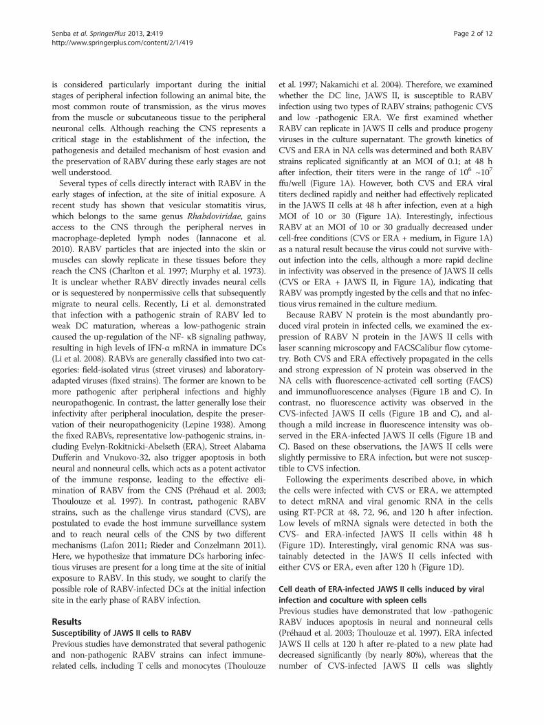
ResultsSusceptibility of JAWS II cells to RABVPrevious studies have demonstrated that several pathogenicand non-pathogenic RABV strains can infect immune-related cells, including T cells and monocytes (Thoulouze
et al. 1997; Nakamichi et al. 2004). Therefore, we examinedwhether the DC line, JAWS II, is susceptible to RABVinfection using two types of RABV strains; pathogenic CVSand low -pathogenic ERA. We first examined whetherRABV can replicate in JAWS II cells and produce progenyviruses in the culture supernatant. The growth kinetics ofCVS and ERA in NA cells was determined and both RABVstrains replicated significantly at an MOI of 0.1; at 48 hafter infection, their titers were in the range of 106 ~107
ffu/well (Figure 1A). However, both CVS and ERA viraltiters declined rapidly and neither had effectively replicatedin the JAWS II cells at 48 h after infection, even at a highMOI of 10 or 30 (Figure 1A). Interestingly, infectiousRABV at an MOI of 10 or 30 gradually decreased undercell-free conditions (CVS or ERA + medium, in Figure 1A)as a natural result because the virus could not survive with-out infection into the cells, although a more rapid declinein infectivity was observed in the presence of JAWS II cells(CVS or ERA + JAWS II, in Figure 1A), indicating thatRABV was promptly ingested by the cells and that no infec-tious virus remained in the culture medium.Because RABV N protein is the most abundantly pro-
duced viral protein in infected cells, we examined the ex-pression of RABV N protein in the JAWS II cells withlaser scanning microscopy and FACSCalibur flow cytome-try. Both CVS and ERA effectively propagated in the cellsand strong expression of N protein was observed in theNA cells with fluorescence-activated cell sorting (FACS)and immunofluorescence analyses (Figure 1B and C). Incontrast, no fluorescence activity was observed in theCVS-infected JAWS II cells (Figure 1B and C), and al-though a mild increase in fluorescence intensity was ob-served in the ERA-infected JAWS II cells (Figure 1B andC). Based on these observations, the JAWS II cells wereslightly permissive to ERA infection, but were not suscep-tible to CVS infection.Following the experiments described above, in which
the cells were infected with CVS or ERA, we attemptedto detect mRNA and viral genomic RNA in the cellsusing RT-PCR at 48, 72, 96, and 120 h after infection.Low levels of mRNA signals were detected in both theCVS- and ERA-infected JAWS II cells within 48 h(Figure 1D). Interestingly, viral genomic RNA was sus-tainably detected in the JAWS II cells infected witheither CVS or ERA, even after 120 h (Figure 1D).
Cell death of ERA-infected JAWS II cells induced by viralinfection and coculture with spleen cellsPrevious studies have demonstrated that low -pathogenicRABV induces apoptosis in neural and nonneural cells(Préhaud et al. 2003; Thoulouze et al. 1997). ERA infectedJAWS II cells at 120 h after re-plated to a new plate haddecreased significantly (by nearly 80%), whereas that thenumber of CVS-infected JAWS II cells was slightly
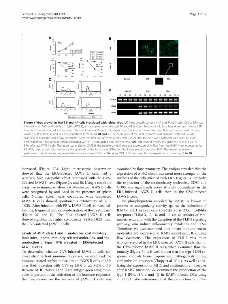
Figure 1 Virus growth in JAWS II and NA cells inoculated with rabies virus. (A) Virus growth curves in NA and JAWS II cells. CVS or ERA wasinfected at an MOI of 0.1 (NA) or 10,30 (JAWS II), and samples were collected 24 and 48 h after infection. n = 3. Error bars represent mean ± SEM.The black line and dotted line represent the viral titers of CVS and ERA, respectively. Decline in the infected viral titer was determined by usingJAWS II cells (+JAWS II) and cell-free conditions (+medium). (B and C) The expression of the viral N protein was analyzed with both a laserscanning microscope and a flow cytometer. After the infection of JAWS II cells with CVS or ERA, the cells were permeabilized with IntraPrepPermeabilization Reagent, and then incubated with FITC-conjugated anti-RABV N MAb. (D) Detection of mRNA and genomic RNA in CVS- orERA-infected JAWS II cells. The upper panel shows GAPDH, the middle panel shows the expression of mRNA from the RABV N gene detected byRT–PCR, using a poly (T)15 primer for the synthesis of the first-strand cDNA and the lower panel shows viral RNA. The experiments wereperformed three times and representative data are shown. CVS or ERA at an MOI of 10 was used for the experiments shown in (B to D).
Senba et al. SpringerPlus 2013, 2:419 Page 3 of 12http://www.springerplus.com/content/2/1/419
increased (Figure 2A). Light microscopic observationshowed that the ERA-infected JAWS II cells had arelatively high cytopathic effect compared with the CVS-infected JAWS II cells (Figure 2A and B). Using a cocultureassay, we examined whether RABV-infected JAWS II cellswere recognized by and lysed in the presence of spleencells. Normal spleen cells cocultured with uninfectedJAWS II cells showed spontaneous cytotoxicity of 36 ±4.03%. After infection with ERA, JAWS II cells showed bal-looning, fragmentation, or condensation of their cytoplasm(Figure 2C and D). The ERA-infected JAWS II cellsshowed significantly higher cytotoxicity (92.5 ± 6.02%) thanthe CVS-infected JAWS II cells.
Levels of MHC class I and II molecules costimulatorymolecules, innate-immunity-related molecules, and theproduction of type I IFNs elevated in ERA-infectedJAWS II cellsTo determine whether CVS-infected JAWS II cells canavoid eliciting host immune responses, we examined theimmune-related surface molecules on JAWS II cells at 48 hafter their infection with CVS or ERA at an MOI of 10.Because MHC classes I and II are antigen-presenting mole-cules important in the activation of the immune responses,their expression on the surfaces of JAWS II cells was
examined by flow cytometry. The analysis revealed that theexpression of MHC class I increased more strongly on thesurfaces of the cells infected with ERA (Figure 3). Similarly,the expression of the costimulatory molecules, CD80 andCD86 was significantly more strongly upregulated in theERA-infected JAWS II cells than in the CVS-infectedJAWS II cells.The phosphoprotein encoded by RABV is known to
possess an antagonizing activity against the induction ofIFN by RIG1 in host cells (Brzózka et al. 2006). Toll-likereceptors (TLRs)-3, -7, -8, and −9 act as sensors of viralnucleic acids and, with the exception of the TLR-3 signalingpathway, also induce inflammatory cytokines and IFNs.Therefore, we also examined how innate immune sensormolecules are expressed in RABV-inoculated DCs, usingflow cytometry. The expression of TLR-3 was morestrongly elevated in the ERA-infected JAWS II cells than inthe CVS-infected JAWS II cells, when examined flow cy-tometry (Figure 3). It is well known that the type I IFN re-sponse controls tissue tropism and pathogenicity duringviral infection processes (Chopy et al. 2011). As well as ana-lyzing the expression of MHC and costimulatory moleculesafter RABV infection, we examined the production of thetype I IFNs, IFN-α and -β, in RABV-infected DCs usingan ELISA. We determined that the production of IFN-α
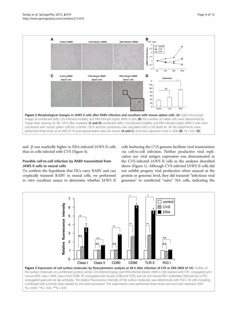
Figure 2 Morphological changes in JAWS II cells after RABV infection and coculture with mouse spleen cells. (A) Light-microscopicimages of uninfected (left), CVS-infected (middle), and ERA-infected (right) JAWS II cells; (B) the numbers of viable cells were determined byTrypan blue staining 24, 48, 120 h after re-plated. (C and D) uninfected (left), CVS-infected (middle), and ERA-infected (right) JAWS II cells werecocultured with mouse spleen cells for a further 120 h and the cytotoxicity was calculated with a cell death kit. All the experiments wereperformed three times at an MOI of 10 and representative data are shown (A and C). Error bars represent mean ± SEM (B). *p < 0.01 (D).
Senba et al. SpringerPlus 2013, 2:419 Page 4 of 12http://www.springerplus.com/content/2/1/419
and -β was markedly higher in ERA-infected JAWS II cellsthan in cells infected with CVS (Figure 4).
Possible cell-to-cell infection by RABV transmitted fromJAWS II cells to neural cellsTo confirm the hypothesis that DCs carry RABV and cancryptically transmit RABV to neural cells, we performedin vitro coculture assays to determine whether JAWS II
Figure 3 Expression of cell-surface molecules by flowcytometric analythe surface molecules on uninfected (control; white), CVS-infected (gray), amouse MHC class I, MHC class II and CD80, PE conjugated anti-mouse CD8conjugated goat anti-rat IgG antibody. The relative fluorescence intensity ouninfected cells (control) were stained by the same procedure. The experim*p < 0.001, **p < 0.05, ***p < 0.01.
cells harboring the CVS genome facilitate viral transmissionvia cell-to-cell infection. Neither productive viral repli-cation nor viral antigen expression was demonstrated inthe CVS-infected JAWS II cells in the analyses describedabove (Figure 1). Although CVS-infected JAWS II cells didnot exhibit progeny viral production when assayed at theprotein or genomic level, they did transmit “infectious viralgenomes” to uninfected “naïve” NA cells, indicating the
sis at 48 h after infection of CVS or ERA (MOI of 10). Profiles ofnd ERA-infected (black) JAWS II cells reacted with FITC conjugated anti-6 and TLR3, and rat anti-mouse RIG1 antibodies, followed by a FITC-f the surface molecules was determined with FACS. All cells includingents were performed three times and error bars represent SEM.
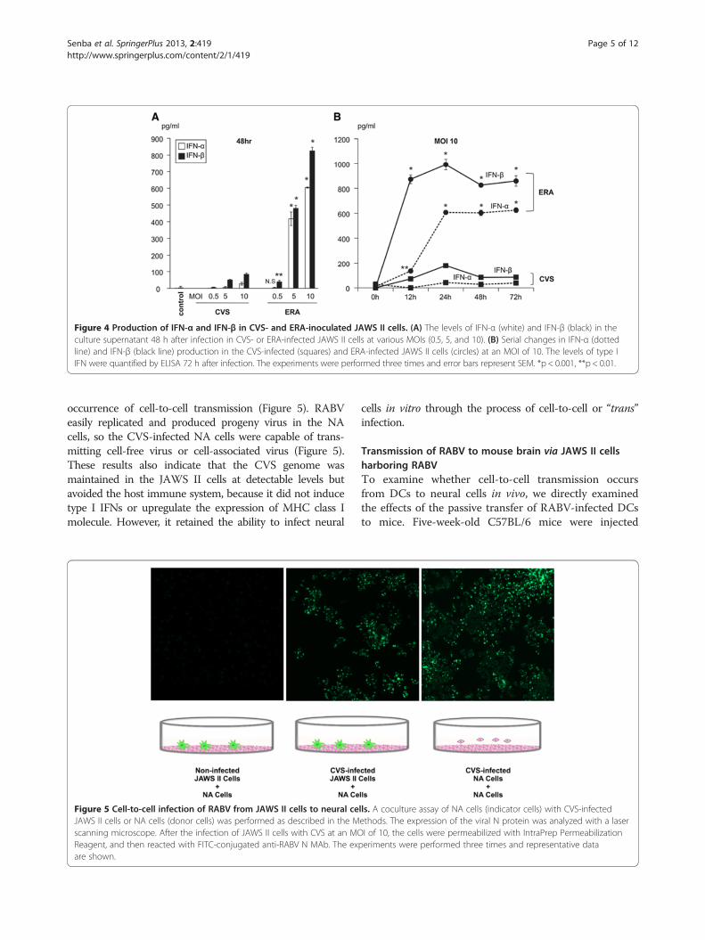
Figure 4 Production of IFN-α and IFN-β in CVS- and ERA-inoculated JAWS II cells. (A) The levels of IFN-α (white) and IFN-β (black) in theculture supernatant 48 h after infection in CVS- or ERA-infected JAWS II cells at various MOIs (0.5, 5, and 10). (B) Serial changes in IFN-α (dottedline) and IFN-β (black line) production in the CVS-infected (squares) and ERA-infected JAWS II cells (circles) at an MOI of 10. The levels of type IIFN were quantified by ELISA 72 h after infection. The experiments were performed three times and error bars represent SEM. *p < 0.001, **p < 0.01.
Senba et al. SpringerPlus 2013, 2:419 Page 5 of 12http://www.springerplus.com/content/2/1/419
occurrence of cell-to-cell transmission (Figure 5). RABVeasily replicated and produced progeny virus in the NAcells, so the CVS-infected NA cells were capable of trans-mitting cell-free virus or cell-associated virus (Figure 5).These results also indicate that the CVS genome wasmaintained in the JAWS II cells at detectable levels butavoided the host immune system, because it did not inducetype I IFNs or upregulate the expression of MHC class Imolecule. However, it retained the ability to infect neural
Figure 5 Cell-to-cell infection of RABV from JAWS II cells to neural ceJAWS II cells or NA cells (donor cells) was performed as described in the Mscanning microscope. After the infection of JAWS II cells with CVS at an MOReagent, and then reacted with FITC-conjugated anti-RABV N MAb. The exare shown.
cells in vitro through the process of cell-to-cell or “trans”infection.
Transmission of RABV to mouse brain via JAWS II cellsharboring RABVTo examine whether cell-to-cell transmission occursfrom DCs to neural cells in vivo, we directly examinedthe effects of the passive transfer of RABV-infected DCsto mice. Five-week-old C57BL/6 mice were injected
lls. A coculture assay of NA cells (indicator cells) with CVS-infectedethods. The expression of the viral N protein was analyzed with a laserI of 10, the cells were permeabilized with IntraPrep Permeabilization
periments were performed three times and representative data
Senba et al. SpringerPlus 2013, 2:419 Page 6 of 12http://www.springerplus.com/content/2/1/419
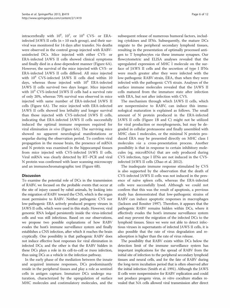
intracerebrally with 106, 105, or 104 CVS- or ERA-infected JAWS II cells (n = 10 each group), and their sur-vival was monitored for 14 days after transfer. No deathswere observed in the control group injected with RABV-uninfected DCs. Mice injected with either CVS- orERA-infected JAWS II cells showed clinical symptomsand finally died in a dose-dependent manner (Figure 6A).However, the survival of the mice injected with CVS- orERA-infected JAWS II cells differed. All mice injectedwith 106 CVS-infected JAWS II cells died within 10days, whereas those injected with 106 ERA-infectedJAWS II cells survived two days longer. Mice injectedwith 104 CVS-infected JAWS II cells had a survival rateof only 20%, whereas 70% survival was observed in miceinjected with same number of ERA-infected JAWS IIcells (Figure 6A). The mice injected with ERA-infectedJAWS II cells showed less lethality and longer survivalthan those injected with CVS-infected JAWS II cells,indicating that ERA-infected JAWS II cells successfullyinduced the optimal immune responses required forviral elimination in vivo (Figure 6A). The surviving miceshowed no apparent neurological manifestations orsequelae during the observation period. To confirm viralpropagation in the mouse brain, the presence of mRNAand N protein was examined in the hippocampal tissuesfrom mice injected with CVS-infected JAWS II cells.Viral mRNA was clearly detected by RT–PCR and viralN protein was confirmed with laser scanning microscopyand an immunochromatographic test (Figure 6B).
DiscussionTo examine the potential role of DCs in the transmissionof RABV, we focused on the probable events that occur atthe site of injury caused by rabid animals, by looking intothe migration of RABV toward the CNS, which is the organmost permissive to RABV. Neither pathogenic CVS norlow-pathogenic ERA actively produced progeny viruses inJAWS II cells, which were used in this study. However, viralgenomic RNA lodged persistently inside the virus-infectedcells and was still infectious. Based on our observations,we propose two possible explanations of how RABVevades the host’s immune surveillance system and finallyestablishes a CNS infection, after which it reaches the braincryptically. One possibility is that pathogenic RABV doesnot induce effective host responses for viral elimination ininfected DCs; and the other is that the RABV hidden inthese DCs plays a role in its cell-to-cell or trans infection,thus using DCs as a vehicle in the infection pathway.In the early phase of the mediation between the innate
and acquired immune responses, DCs predominantlyreside in the peripheral tissues and play a role as sentinelcells in antigen capture. Immature DCs undergo ma-turation, characterized by the upregulation of surfaceMHC molecules and costimulatory molecules, and the
subsequent release of numerous humoral factors, includ-ing cytokines and IFNs. Subsequently, the mature DCsmigrate to the peripheral secondary lymphoid tissues,resulting in the presentation of optimally processed anti-gen to T lymphocytes via these immune synapses. Ourflowcytometric and ELISA analyses revealed that theupregulated expression of MHC I molecule on the sur-face of JAWS II cells and the secretion of type I IFNswere much greater after they were infected with thelow-pathogenic RABV strain, ERA, than when they wereinfected with the pathogenic CVS strain. Analyses of thesurface immune molecules revealed that the JAWS IIcells matured from the immature state after infectionwith ERA, but not after infection with CVS.The mechanism through which JAWS II cells, which
are nonpermissive to RABV, can induce this immu-nological maturation is explained as follows. The smallamount of N protein produced in the ERA-infectedJAWS II cells (Figure 1B and C) might not be utilizedfor viral production or morphogenesis, but may be de-graded in cellular proteasome and finally assembled withMHC class I molecules, or the minimal N protein pro-duced ERA may be presented directly on MHC class Imolecules via a cross-presentation process. Anotherpossibility is that in response to certain inhibitory mole-cules (eg. microRNAs) that are only produced duringCVS infection, type I IFNs are not induced in the CVS-infected JAWS II cells (Zhao et al. 2012).The inadequate immune response stimulated by CVS
is also supported by the observation that the death ofCVS-infected JAWS II cells was not induced in the pres-ence of naïve spleen cells, whereas the ERA-infectedcells were successfully lysed. Although we could notconfirm that this was the result of apoptosis, a previousstudy has demonstrated that low-pathogenic strains ofRABV can induce apoptotic responses in macrophages(Jackson and Rossiter 1997). Therefore, it appears that thepathogenic RABV remains hidden within DCs, where iteffectively evades the host’s immune surveillance systemand may prevent the migration of the infected DCs to thelymphoid tissues. Since we were not able to detect infec-tious viruses in supernatants of infected JAWS II cells, it isalso possible that the rate of virus degradation and re-adsorption is higher than the rate of virus release.The possibility that RABV exists within DCs below the
detection limit of the immune surveillance system hasimportant implications for the spread of RABV from theinitial site of infection to the peripheral secondary lymphoidtissues and neural cells, and for the fate of RABV duringthe long-term incubation period that is often observed afterthe initial infection (Smith et al. 1991). Although the JAWSII cells were nonpermissive for RABV replication and couldnot produce progeny virus, in vitro coculture studies re-vealed that NA cells allowed viral transmission after direct
Figure 6 (See legend on next page.)
Senba et al. SpringerPlus 2013, 2:419 Page 7 of 12http://www.springerplus.com/content/2/1/419
(See figure on previous page.)Figure 6 Transmission of RABV to mouse brain via JAWS II cells harboring RABV. (A) Survival rates of five-week-old C57BL/6 mice injectedintracerebrally with 106, 105, or 104 CVS- or ERA-infected JAWS II cells (n = 10 each group) at MOI of 30. Their survival was observed for 14 daysafter the transfer of the cells. The black and dotted lines represent the survival of the mice injected with CVS- and ERA-infected JAWS II cells,respectively. The circles indicate the survival of mice injected with 106 CVS- or ERA-infected JAWS II cells. The triangles indicate the survival ofmice injected with 105 CVS- or ERA-infected JAWS II cells. The crosses indicate the survival of mice injected with 104 CVS- or ERA-inoculated JAWSII cells. The experiments were performed three times. The Kaplan-Meier method was used to analyze mouse survival. Statistical analyses wereperformed by log-rank test. (p < 0.01; 106 CVS: 106 ERA, 105 CVS: 105 ERA. p <0.05; 104 CVS: 104 ERA) (B) Half the mice from each group weresacrificed after seven days, and the presence of viral N mRNA and RABV N protein in the hippocampal tissues was determined by RT–PCR (mRNA)and a RABV N detection kit with FITC staining, respectively. The arrow indicates the positive band of N protein on the immunochromatographictest. The upper band corresponds to nonspecific control. The experiments were performed three times as shown in Figure 1B and representativepictures are shown.
Senba et al. SpringerPlus 2013, 2:419 Page 8 of 12http://www.springerplus.com/content/2/1/419
cell-to-cell contact with CVS–infected JAWS II cells. Thisphenomenon is supported by the fact that the exposure ofCVS- infected JAWS II cells to monoclonal antibodiesdirected against a RABV surface glycoprotein (G) 100%blocked the transmission of RABV to NA (KS and AN, un-published observation). The relevance of viral transmissionby a cell-to-cell contact process was demonstrated by theinjection of CVS-infected JAWS II cells into the brains ofmice, which led to the rapid spread of the virus and 100%mortality. In the establishment of immunodeficiency virus1 (HIV-1) infection, the uptake of HIV-1 by immature DCsat the mucosal site occurs when it binds to either DC-SIGN or syndecan-3 (de Witte et al. 2007; Geijtenbeeket al. 2000). Following the migration of HIV-1 from the ini-tial mucosal invasion site, DCs may carry the virus to thedraining lymph nodes. It has been demonstrated that dur-ing the antigen presentation process, DCs cluster with Tcells in the secondary lymphoid tissues (de Witte et al.2007; Geijtenbeek et al. 2000; Lore and Larsson 2003). It iswell known that abundant nerve-end fibers innervate theperipheral lymphoid tissue. Other studies have suggestedthat prions initially accumulate on follicular dendritic cellsin the lymphoid tissues and subsequently spread via theperipheral nervous system to the brain (Raymond andMabbott 2007; Sethi et al. 2007). Recently, it has beenshown that micro RNAs secreted by Epstein-Barr virus-infected cells are transferred to uninfected cells viaexosome structures (Pegtel et al. 2010). Our results alsosuggest that a mechanism similar to that discussed aboveis involved in the transmission of RABV from DCs to CNS.DCs are heterogeneous cells and comprise several sub-
sets characterized by unique morphological shapes andfunctions. Conventional or myeloid-type DCs (mDCs)have a dendritic shape, exhibit typical DC functions,such as antigen uptake, processing, and presentation,and are characterized as CD11c+, CD11b+, and B220-.In contrast, plasmacytoid DCs, which are nondendriticround cells, are defined as CD11low, CD11b-, and B220+
cells, and lack the ability to produce costimulatory mole-cules (Siegemund et al. 2009). Whereas mDC subsetsmigrate to the lymph nodes from peripheral tissue,plasmacytoid DCs directly enter the lymph nodes from
the blood by crossing high endothelial venules (HEVs)(Banchereau and Steinman 1998; Shortman and Liu2002) and are able to transform into mDCs in responseto the surrounding conditions (Fukao et al. 2000; Fukaoet al. 2001; Zuniga et al. 2004). When the cell-surfacemarkers on JAWS II cells were analyzed by flow cytometry,CD11c and CD11b were detected, whereas CD8α was not,and the expression of costimulatory molecules was up-regulated by stimulation with viral antigens. These resultsindicate that JAWS II cells are mDCs, which is consistentwith a previous report (Otsu et al. 2006). It has also beenspeculated that the administration of RABV vaccine via anintradermal route in humans, as well as via the routineintramuscular route, induce adequate levels of viral neu-tralizing antibody (Shiota et al. 2008), indicating that mDCsubset are recruited to the vaccination site and migrate tothe lymph nodes for effective antigen presentation to naïveT cells. Therefore, we speculate that mDCs may play a crit-ical role in RABV infection and pathogenesis.Several limitations of our study warrant mention. Our
attempts to determine whether RABV particles could bevisualized in CVS-infected JAWS II cells by immunogoldelectron microscopy were unsuccessful. We were also un-able to confirm the occurrence of apoptosis in the presenceof spleen lymphocytes. However, high level of IFN wasdetected, indicating that cellular immunity was fully func-tional after the infection of JAWS II cells with ERA.We have provided several lines of evidence that DCs
allow RABV to escape the host immune surveillance sys-tem and facilitate the transmission of the virus to theneural cells. Following the uptake of pathogenic CVS, im-mature DCs do not properly process the viral antigens, asindicated by their lack of MHC class I and II expression.Because the RABV genome persists undetected in DCsand does not replicate or produce progeny virus, wepropose that RABV uses these nonpermissive cells as avehicle to reach the peripheral neural cells, where the viruscan then replicate and spread to the CNS. This is the firstreport to demonstrate that DCs can transmit RABV toneural cells by cell-to-cell or trans infection, and shedsnew light on the immune evasion strategies used by thisdeadly virus.
Senba et al. SpringerPlus 2013, 2:419 Page 9 of 12http://www.springerplus.com/content/2/1/419
MethodsCellsThe DC cell line, JAWS II, and neuroblastoma (NA) cellswere purchased from the American Type Culture Collec-tion (Manassas, VA). JAWS II cells (CD11b+, CD11c+,CD8α-, TLR7+, and RIG-1+) were originally isolated frombone-marrow cultures from p53-deficient C57BL/6 mice.The cells were grown in a complete culture mediumconsisting of RPMI 1640 medium with GlutaMAX™ (GibcoBRL, Grand Island, NY) and supplemented with 10% fetalbovine serum (FBS), 1% penicillin-streptomycin, 50 μm2-mercaptoethanol, and 5 ng/mL recombinant mousegranulocyte-macrophage colony-stimulating factor (BDBiosciences, San Jose, CA). Murine NA cells were grown inEagle’s minimal essential medium (Gibco BRL, GrandIsland, NY) containing 10% FBS and 1% penicillin-streptomycin.
VirusesA pathogenic strain of RABV (CVS-11) and a low- patho-genic strain (ERA) were propagated in NA cells. The vi-ruses in the culture supernatants of the RABV-inoculatedNA cells were collected and centrifuged at 1200 × g for 10min at 4°C (TOMY EX-125, TS-38, TOMY, Tokyo, Japan),and then purified by ultrafiltration through a 0.45-μm filter.The infection of NA or JAWS II cells with RABV wasperformed at 37°C at various multiplicities of infection(MOIs).
AntibodiesFluorescein isothiocyanate (FITC)-conjugated goat anti-rat IgG antibody was obtained from Organon TeknikaCorp. (Durham, NC). FITC-conjugated anti-mouse MHCclass I, MHC class II, and CD80 antibodies, and phyco-erythrin (PE)-conjugated anti-mouse CD86 antibody werepurchased from eBioscience Inc. (San Diego, CA). PE-conjugated anti-mouse CD283 (TLR3) antibody and puri-fied rat anti RIG1 antibody were obtained from BioLegendInc. (San Diego, CA). FITC-conjugated anti-RABV Nmonoclonal antibody (MAb) was obtained from FujirebioDiagnostics, Inc. (Malvern, PA).
Measurement of viral replicationNA and JAWS II cells were seeded in 48-well cultureplates (105 cells/well) and infected with CVS or ERA atthe appropriate MOI. The culture supernatants obtainedfrom the RABV-infected NA or JAWS II cells were thenseparated by centrifugation at 1200 × g for 10 min 0, 24,or 48 h after infection. The virions in the supernatantwere purified by ultrafiltration through a 0.45 μm filterand then titrated. The viruses in the culture superna-tants were titrated with a focus assay on confluentmonolayers of NA cells in 24-well plates, according topreviously reported procedures (Yamada et al. 2012).
Flow cytometry and confocal analysisThe expression of cell-surface molecules was measuredby immunofluorescence flow-cytometric analysis usinga FACSCalibur flow cytometer (BD ImmunocytometrySystems, San Jose, CA) and CellQuest software. For theanalysis, NA and JAWS II cells were first seeded in six-well culture plates (106 cells/well) and infected withCVS or ERA at an MOI of 10. After 48 h at 37°C, thecells were collected and washed in phosphate-bufferedsaline (PBS), and then incubated on ice for 30 minwith a FITC-conjugated anti-mouse MHC class I (1:50),MHC class II (1:50), or CD80 (1:50) antibody, orPE-conjugated anti-mouse CD86 antibody (1:50). Theexpression of the intracellular proteins, TLR-3 andRIG1 was also measured by immunofluorescence flow-cytometric analysis using a FACSCalibur flow cytometerand CellQuest software. After the infection of JAWS IIcells with CVS or ERA (as described above), the cellswere permeabilized with Intra Prep PermeabilizationReagent (Immunotech, Marseille Cedex, France), accordingto the manufacturer’s protocol, and then reacted withPE-conjugated anti-mouse TLR3 (1:50) or rat anti-mouse RIG-1 (1:50) antibody. An FITC-conjugated goatanti-rat IgG (1:200) was used as the secondary antibodyto detected RIG1.The expression of the viral N protein was analyzed with
both a laser scanning microscope (Carl Zeiss Micro-Imaging, LSM 510; Carl Zeiss, Jena, Germany) and aFACSCalibur flow cytometer. After the infection of JAWSII cells with CVS or ERA (as described above), the cellswere permeabilized with IntraPrep Permeabilization Re-agent, and then reacted with FITC-conjugated anti-RV NMAb (1:50).
Enzyme-linked immunosorbent assays (ELISAs)To measure the quantities of IFN-α and IFN-β secreted bythe RABV-infected JAWS II cells, JAWS II cells seeded insix-well plates (106 cells/well) were inoculated with CVS orERA at an MOI of 10 and the culture supernatants werethen harvested at 0, 12, 24, 48, or 72 h after infection. Theconcentrations of IFN-α and IFN-β in the culture superna-tants were assessed with sandwich ELISA kits (PBL Bio-medical Laboratories, Piscataway, NJ), according to themanufacturer’s instructions.
Viral RNA extraction and reverse transcription (RT)-PCRJAWS II cells in 12-well culture plates (5 × 10⁵ cells/well) were infected with CVS or ERA at an MOI of 10.Following incubation for 48, 72, 96, or 120 h, the cellswere collected and washed three times with PBS. The totalRNA was then extracted from the cells using the acid-guanidinium thiocyanate -phenol -chloroform method(TRIzol®, Gibco BRL, Gaithersburg, MD). To detect theviral genomic RNA (negative polarity) or viral mRNA
Senba et al. SpringerPlus 2013, 2:419 Page 10 of 12http://www.springerplus.com/content/2/1/419
amplification, a partial sequence of the N gene was ampli-fied using the sense primer NF2850 (nucleotides 28–50,5′-ACAGACAGCGTCAATGGCAGAGC-3′) and anti-sense primer N660 (R) (nucleotides 660–676, 5′-GTTT#GGTATAGTACTCC-3′) (Nishizono et al. 2002). Theprimer positions are given according to the N gene ofCVS (GenBank accession number DQ286762). Total RNA(1 μg) was reverse transcribed to synthesize the first-strandcDNA of the viral genome RNA using the primer N660 (R)or the viral mRNA using the primer NF2850 and Moloneymurine leukemia virus reverse transcriptase (Gibco BRL) at37°C for 2 h. The first PCR reaction was performed withthe following reaction mixture: 1 μg of sample cDNA, 50mM KCl, 10 mM Tris–HCl (pH 8.4), 1.5 mM MgCl2, 20mM each of primers NF2850 and N660 (R), 200 μMdNTPs, and 2 U of Taq DNA polymerase (Promega Corp.,Madison, WI). The reaction mixture was subjected to 35cycles of denaturation at 95°C for 30 s, annealing at 50°Cfor 30 s, and extension at 72°C for 90 s. The resultingamplicons were resolved electrophoresed on 1% agarosegels and stained with ethidium bromide. As the internalcontrol mRNA, glyceraldehyde phosphate dehydrogenase(GAPDH) transcripts were amplified under the sameconditions, with the sense primer 5′-TTCACCACCATGGAGAAGGC-3′ and the antisense primer 5′-GGCATGGACTGTGGTCATGA-3′.
Effect of naïve spleen cells on RABV-infected JAWS II cellsJAWS II cells were seeded in six-well culture plates (106
cells/well) and infected with CVS or ERA at an MOI of 10.The cells were collected after 48h incubation, washed threetimes in PBS, and then plated in a new six-well cultureplate (105 cells/well). Spleen cells were obtained from thespleens of normal C57BL/6 mice (Otsu et al. 2006) (CharlesRiver Japan Inc., Yokohama, Japan) and then cultured withor without the RABV-infected JAWS II cells (106 cells/well;final E/T ratio = 10:1). After 120 h of coculture, the cellswere observed microscopically (Olympus CK30, Olympus,Tokyo, Japan) and a cytotoxicity assay was performed withthe Cyto Tox 96® Non-Radioactive Cytotoxicity Assay(Promega Corp.). Cytotoxicity was calculated according tothe formula: % cytotoxicity = (Experimental LDH release -Effector Spontaneous LDH release - Target SpontaneousLDH release / Target Maximum LDH release - TargetSpontaneous LDH release × 100).
Cell-to-cell transmission of RABV from JAWS II cells toneural cells in vitroJAWS II cells or NA cells in six-well culture plates (106
cells/well) were infected with CVS at an MOI of 10. Afterincubation for 48 h at 37°C, the cells were collected andwashed thoroughly three times with PBS. A virus titrationassay was performed to confirm that there were noremaining viruses in the supernatant, as described above.
The cells were then added to a 24-well plate containingmonolayers of cultured NA cells. After 48 h of coculture,the NA cells were stained with FITC-conjugated anti-RABV N protein MAb and observed under a laser scan-ning microscope.
Injection of CVS- or ERA-inoculated JAWS II cells intomouse brainsJAWS II cells in six-well culture plates (106 cells/well)were infected with CVS or ERA at an MOI of 30. Follow-ing incubation for 48 h, the cells were collected andwashed three times with PBS. A virus titration assay wasperformed to confirm that no viruses remained in thesupernatant, as described above. Specific pathogen-freefive-week-old female C57BL/6 mice (Seac Yoshitomi,Fukuoka, Japan) were injected intracerebrally with 106,105, or 104 CVS- or ERA-infected JAWS II cells in 0.03mL with a 23-gauge needle. Half the mice in each injectedgroup (n = 10) were observed for 2 weeks and theirsurvival was recorded daily. The remaining mice weresacrificed on day 7 and the tissue samples taken from thehippocampus were analyzed for viral N protein using alaser scanning microscope, for viral mRNA and animmunochromatographic test that had been developedpreviously by us (Nishizono et al. 2008). All animal proce-dures conformed to animal care guidelines approved byEthics Committee in Oita University.
AbbreviationsDC: Dendritic cells; MHC: Major histocompatibility complex; IFN: Interferon;RIG 1: Retinoic-acid-inducible gene I; RABV: Rabies virus; CNS: Central nervoussystem; ERA: Evelyn-Rokitnicki-Abelseth; FACS: Fluorescence activated cellsorting; TLR: Toll-like recepetor; HIV: Human immunodeficiency virus;MDC: Myeloid-type DC; HEV: High endothelial venules; FITC: Fluoresceinisothiocyanate; PE: Phycoerythrin; MAb: Monoclonal antibody;GADPH: Glyceraldehyde phosphate dehydrogenase.
Competing interestsThe authors declare they have no competing interests in relation to this article.
Authors’ contributionsKS and AN conceive of the study and participated in its design and coordinationand draft the manuscript. KS, TM and MO carried out the PCR and ELISAexperiments. KS, HI and YD carried out the confocal microscopy and FACSanalysis. KS, KY and SS performed animal studies. All authors participated in thepreparation of the manuscript, and read and approved the final manuscript.
Authors’ informationKazuyo Senba: DDS, PhDKagoshima University Graduate School of Medical and Dental SciencesArea of specialization: Virology, Immunology, BacteriologyTakashi Matsumoto: PhDOita University Graduate School of Medical SciencesArea of specialization: Virology, Immunology, BacteriologyKentaro Yamada: DVM, PhDUnited Graduate School of Veterinary SciencesArea of specialization: VirologySeiji Shiota: MD, PhDOita University Graduate School of Medical SciencesArea of specialization: Immunology,Hidekatsu Iha: PhDThe Graduate University for Advanced StudiesArea of specialization: Cell biology
Senba et al. SpringerPlus 2013, 2:419 Page 11 of 12http://www.springerplus.com/content/2/1/419
Yukari Date: MD, PhDMiyazaki University Graduate School of Medical SciencesArea of specialization: BiochemistryMotoaki Ohtsubo: PhDGraduate School of Medical Sciences, Kyushu UniversityArea of specialization: Cell biologyAkira Nishizono: MD, PhDOita University Graduate School of Medical SciencesArea of specialization: Virology, Immunology, Bacteriology
AcknowledgementsWe thank Kunimitsu Inoue and Mika Okamoto for technical assistance andhelpful discussion. This work was supported by the Grant-in-Aid for ScientificResearch from the Health and Labour Science Research Grants of Japan(Research on International and Cooperation in Medical Science, H23-, H24-KOKUI-SHITEI-003). This work was in part supported by The Naito foundation.
Author details1Department of Microbiology, Faculty of Medicine, Oita University, 1-1Idaigaoka, Hasama-machi, Yufu-City, Oita 879-5593, Japan. 2Faculty of FoodScience and Nutrition, Beppu University, Beppu, Oita, Japan. 3Frontier ScienceResearch Center, Faculty of Medicine, Miyazaki University, Miyazaki, Japan.
Received: 28 May 2013 Accepted: 23 August 2013Published: 29 August 2013
ReferencesBanchereau J, Steinman RM (1998) Dendritic cells and the control of immunity.
Nature 392:245–252Barchet W, Cella M, Odermatt B, Asselin-Paturel C, Colonna M, Kalinke U (2002)
Virus-induced interferon alpha production by a dendritic cell subset in theabsence of feedback signaling in vivo. J Exp Med 195:507–516
Brzózka K, Finke S, Conzelmann KK (2006) Inhibition of interferon signaling by rabiesvirus phosphoprotein P: activation-dependent binding of STAT1 and STAT2. J Virol80:2675–2683 extracellular signal-regulated kinases 1 and 2. J Virol 78:9376–9388
Charlton KM, Nadin-Davis S, Casey GA, Wandeler AI (1997) The long incubationperiod in rabies: delayed progression of infection in muscle at the site ofexposure. Acta Neuropathol 94:73–77
Cheng Y, King NJ, Kesson AM (2004) Major histocompatibility complex class I (MHC-I)induction by West Nile virus: involvement of 2 signaling pathways in MHC-I up-regulation. J Infect Dis 189:658–668
Chopy D, Pothlichet J, Lafage M, Mégret F, Fiette L, Si-Tahar M, Lafon M (2011)Ambivalent role of the innate immune response in rabies virus pathogenesis.J Virol 85:6657–6668
de Witte L, Bobardt M, Chatterji U, Degeest G, David G, Geijtenbeek TB, Gallay P(2007) Syndecan-3 is a dendritic cell-specific attachment receptor for HIV-1.Proc Natl Acad Sci USA 104:19464–19469
Dhib-Jalbut SS, Cowan EP (1993) Direct evidence that interferon-beta mediatesenhanced HLA-class I expression in measles virus-infected cells. J Immunol151:6248–6258
Feder HM Jr, Petersen BW, Robertson KL, Rupprecht CE (2012) Rabies: still a uniformlyfatal disease? Historical occurrence, epidemiological trends, and paradigm shifts.Curr Infect Dis Rep 4:408–422
Finke S, Conzelmann KK (2005) Replication strategies of rabies virus. Virus Res111(2):120–131
Fukao T, Matsuda S, Koyasu S (2000) Synergistic effects of IL-4 and IL-18 on IL-12-dependent IFN-gamma production by dendritic cells. J Immunol 164:64–71
Fukao T, Frucht DM, Yap G, Gadina M, O’Shea JJ, Koyasu S (2001) Inducible expressionof Stat4 in dendritic cells and macrophages and its critical role in innate andadaptive immune responses. J Immunol 166:4446–4455
Geijtenbeek TB, Kwon DS, Torensma R, van Vliet SJ, van Duijnhoven GCF, Middel J,Cornelissen ILMHA, Nottet HSLM, KewalRamani VN, Littman DR, Figdor CG, vanKooyk Y (2000) DC-SIGN, a dendritic cell-specific HIV-1-binding protein thatenhances trans-Infection of T cells. Cell 100:587–597
Hornung V, Ellegast J, Kim S, Brzózka K, Jung A, Kato H, Poeck H, Akira S, ConzelmannK, Schlee M, Endres S, Hartmann G (2006) 5′-Triphosphate RNA is the ligand forRIG-I. Science 314:994–997
Iannacone M, Moseman EA, Tonti E, Bosurgi L, Junt T, Henrickson SE, Whelan SP,Guidotti LG, von Andrian UH (2010) Sub capsular sinus macrophages preventCNS invasion on peripheral infection with a neurotropic virus. Nature465:1079–1083
Imaizumi T, Hatakeyama M, Yamashita K, Yoshida H, Ishikawa A, Taima K, Satoh K,Mori F, Wakabayashi K (2004) Interferon-gamma induces retinoic acid-inducible gene-I in endothelial cells. Endothelium 11:169–173
Jackson AC (2013) Current and future approaches to the therapy of human rabies.Antiviral Res 99(1):61–67
Jackson AC, Rossiter JP (1997) Apoptosis plays an important role in experimentalrabies virus infection. J Virol 71:5603–5607
Lafon M (2011) Evasive strategies in rabies virus infection. Adv Virus Res 79:33–53Lepine P (1938) On the evolution of fixed strains of rabies virus. J Hyg (Lond)
38:180–184Leung DW, Basler CF, Amarasinghe GK (2012) Molecular mechanisms of viral inhibitors
of RIG-I like receptors. Trends Microbiol 20(3):139–146Li J, McGettigan JP, Faber M, Schnell MJ, Dietzschold B (2008) Infection of
monocytes or immature dendritic cells (DCs) with an attenuated rabies virusresults in DC maturation and a strong activation of the NFκB signalingpathway. Vaccine 17:419–426
Lore K, Larsson M (2003) The role of dendritic cells in the pathogenesis of HIV-infection. APMIS 111:776–788
Murphy FA, Baur SP, Harrison AK, Winn WC (1973) Comparative pathogenesis ofrabies and rabies-like viruses. Lab Invest 28:361–376
Nakamichi K, Inoue S, Takasaki T, Morimoto K, Kurane I (2004) Rabies virusstimulates nitric oxide production and CXC chemokine ligand 10 expressionin macrophages through activation of extracellular signal-regulated kinases 1and 2. J Virol 78:9376–9388
Nishizono A, Mannen K, Elio-Villa LP, Tanaka S, Li KS, Mifune K, Arca BF, Cabanban A,Martinez B, Rodriguez A, Atienza VC, Camba R, Resontoc N (2002) Genetic analysisof rabies virus isolates in the Philippines. Microbiol Immunol 46:413–417
Nishizono A, Khawplod P, Ahmed K, Goto K, Shiota S, Mifune K, Yasui T, Takayama K,Kobayashi Y, Mannen K, Tepsumethanon V, Mitmoonpitak C, Inoue S, MorimotoK (2008) A simple and rapid immunochromatographic test kit for rabiesdiagnosis. Microbiol Immunol 52:243–249
Otsu S, Gotoh K, Yamashiro T, Yamagata J, Shin K, Fujioka T, Nishizono A (2006)Transfer of antigen-pulsed dendritic cells induces specific T-cell proliferation and atherapeutic effect against long-term Helicobacter pylori infection in mice. InfectImmun 74:984–993
Pegtel DM, Cosmopoulos K, Thorly-Lawson DA, van Eijndhoven MA, Hospmans ES,Lindenberg JL, de Gruijl TD, Wurdinger T, Middeldorp JM (2010) Functionaldelivery of viral miRNAs via exosomes. Proc Natl Acad Sci USA 107:6328–6333
Préhaud C, Lay S, Dietzschold B, Lafon M (2003) Glycoprotein of nonpathogenic rabiesviruses is a key determinant of human cell apoptosis. J Virol 77:10537–10547
Raymond CR, Mabbott NA (2007) Assessing the involvement of migratorydendritic cells in the transfer of the scrapie agent from the immune toperipheral nervous systems. J Neuroimmunol 187:114–125
Rieder M, Conzelmann KK (2011) Interferon in rabies virus infection. Adv Virus Res79:91–114
Rothenfusser S, Goutagny N, DiPerna G, Gong M, Monks BG, Schoenemeyer A,Yamamoto M, Akira S, Fitzgerald KA (2005) The RNA helicase Lgp2 inhibitsTLR-independent sensing of viral replication by retinoic acid-inducible gene-I.J Immunol 175:5260–5268
Sakaki H, Imaizumi T, Matsumiya T, Kusumi A, Nakagawa H, Kubota K, Nishi N,Nakamura T, Hirashima M, Satoh K, Kimura H (2005) Retinoic acid-induciblegene-I is induced by interleukin-1β in cultured human gingival fibroblasts.Oral Microbiol Immunol 20:47–50
Sethi S, Kerksiek KM, Brocker T, Kretzschmar H (2007) Role of the CD8+ dendriticcell subset in transmission of prions. J Virol 81:4877–4880
Shiota S, Khawplod P, Ahmed K, Mifune K, Nishizono A (2008) A pilot study onintradermal vaccination of Japanese rabies vaccine for pre-exposureimmunization. Vaccine 26:6441–6444
Shortman K, Liu YJ (2002) Mouse and human dendritic cell subtypes. Nat Rev Immunol2:151–161
Siegemund S, Hartl A, von Buttlar H, Dautel F, Raue R, Freudenberg MA, FejerG, Būttner M, Köhler G, Kirschning CJ, Sparwasser T, Alber G (2009)Conventional bone marrow-derived dendritic cells contribute to toll-likereceptor-independent production of alpha/beta interferon in response toinactivated parapoxvirus ovis. J Virol 83:9411–9422
Smith JS, Fishbein DB, Rupprecht CE, Clark K (1991) Unexplained rabies in threeimmigrants in the United States. A virologic investigation. N Engl J Med 324:205–211
Steinman RM (1991) The dendritic cell system and its role in immunogenicity.Annu Rev Immunol 9:271–296
Thoulouze MI, Lafage M, Montano-Hirose JA, Lafon M (1997) Rabies virus infectsmouse and human lymphocytes and induces apoptosis. J Virol 71:7372–7380
Senba et al. SpringerPlus 2013, 2:419 Page 12 of 12http://www.springerplus.com/content/2/1/419
Yamada K, Park CH, Noguchi K, Kojima D, Kubo T, Komiya N, Matsumoto T,Mitui MT, Ahmed K, Morimoto K, Inoue S, Nishizono A (2012) Serialpassage of a street rabies virus in mouse Neuroblastoma cells resultedin attenuation: potential role of the additional N-glycosylation of a viralglycoprotein in the reduced pathogenicity of street rabies virus. VirusRes 165:34–45
Zhao P, Ahao L, Zhang K, Feng H, Wang H, Wang T, Xu T, Feng N, Wang C, Gao Y,Huang G, Qin C, Yang S, Xia X (2012) Infection with street strain rabies virusinduces modulation of the microRNA profile of the mouse brain. Virol J 9:159–171
Zuniga EI, McGavern DB, Pruneda-Paz JL, Teng C, Oldstone MB (2004) Bone marrowplasmacytoid dendritic cells can differentiate into myeloid dendritic cells uponvirus infection. Nat Immunol 5:1227–1234
doi:10.1186/2193-1801-2-419Cite this article as: Senba et al.: Passive carriage of rabies virus bydendritic cells. SpringerPlus 2013 2:419.
Submit your manuscript to a journal and benefi t from:
7 Convenient online submission
7 Rigorous peer review
7 Immediate publication on acceptance
7 Open access: articles freely available online
7 High visibility within the fi eld
7 Retaining the copyright to your article
Submit your next manuscript at 7 springeropen.com