Embed Size (px)
Citation preview
Research ArticlePassive Immunoprophylaxis for the Protection ofthe Mother and Her Baby: Insights from In Vivo Models ofAntibody Transport
Yanqun Xu,1 Iftekhar Mahmood,2 Lilin Zhong,1 Pei Zhang,1 and Evi B. Struble1
1Division of Plasma Protein Therapeutics, Office of Tissues and Advanced Therapies, CBER/FDA, Plasma Derivatives Branch,Silver Spring, MD, USA2Division of Clinical Evaluation and Pharmacology/Toxicology, Office of Tissues and Advanced Therapies, CBER/FDA, Silver Spring,MD, USA
Correspondence should be addressed to Evi B. Struble; [email protected]
Received 26 August 2016; Accepted 21 November 2016; Published 11 January 2017
Academic Editor: Roberta A. Diotti
Copyright © 2017 Yanqun Xu et al. This is an open access article distributed under the Creative Commons Attribution License,which permits unrestricted use, distribution, and reproduction in any medium, provided the original work is properly cited.
Pregnant women are at high risk for infection by pathogens. Vertical transmission of infectious agents, such as Zika, hepatitis B,and cytomegalovirus during pregnancy, remains a public health problem, associated with dire outcomes for the neonate. Thus,a safe prophylactic and therapeutic approach for protecting the mother and the neonate from infections remains a high priority.Our work is focused on better understanding the safety and efficacy determinants of IgG antibody preparations when used duringpregnancy to benefit the mother and her baby. Using pregnant guinea pigs, we demonstrated that biodistribution of administeredIgG to the fetus increases with gestation and results in lower maternal and higher fetal antibody concentrations as pregnancyprogresses. Data suggests that partition of antibody immunotherapy to the fetal compartment may contribute to a lower maternalexposure (as measured by the AUC) and a shorter mean residence time of the IgG therapeutic at the end of pregnancy comparedto nonpregnant age-matched controls, irrespective of the administered dose. Our studies provide insights on the importance ofselecting an efficacious dose in pregnancy that takes into account IgG biodistribution to the fetus. The use of appropriate animalmodels of placental transfer and infectious disease during pregnancy would facilitate pharmacokineticmodeling to derive a startingdose in clinical trials.
1. Introduction
Infectious diseases are a significant contributor to pregnancyrelated maternal morbidity and mortality [1] accounting formore than 10% of pregnancy related deaths in the US [2].Changes in immune status during pregnancy render womenmore susceptible to infections and, when infected, proneto more severe disease [3, 4]. Infections in pregnancy areassociated with poor outcomes for the newborn, rangingfrom premature birth to congenital abnormalities and death[4–8]. Maternal immunity to pathogens improves outcomes;thus a significant emphasis has recently been placed onimmunization of pregnant women in the US [9]. For vaccinesthat are contraindicated or not recommended during preg-nancy, and for pathogens for which there are no approved
vaccines, passive immunization with hyperimmune antibodypreparations can be an alternative during pregnancy as thereare no known risks to the fetus from such preparations [10].However, in the few clinical studies where IgG was adminis-tered during pregnancy time-concentration data have oftennot been collected [11, 12]. Such information is critical, asthe efficacy of IgG preparations has been shown to correlatewith the dose [13] and the elevation of IgG trough levelsis associated with reduced incidence of infections such aspneumonia [14].
Because intact IgG molecules can pass the placenta ina receptor-mediated fashion [15], passive immunization ofthe pregnant woman during pregnancy is believed to benefitnot only the mother but also her baby [16] and it has beenproposed or is being studied for CMV [12], HBV [17, 18],
HindawiJournal of Immunology ResearchVolume 2017, Article ID 7373196, 8 pageshttps://doi.org/10.1155/2017/7373196
2 Journal of Immunology Research
rubella [19], and other infections, with mixed results. Gapsremain in our knowledge of the efficacious dose, frequencyof administration during pregnancy, and the determinantsof protection in preventing mother-to-child transmission. Inaddition, not all IgG subclasses traverse the placenta at thesame rate [20], and the magnitude of the clinical benefit maydepend on the isotype of the neutralizing antibodies for aspecific pathogen.
It is clear there is a need formore data, and, until such gapsare bridged, animal studies can inform decisions regardingstarting dose and frequency of administration in clinicalstudies. In pregnant guinea pigs we have demonstrated thatpharmacokinetic properties of IgG therapeutics administeredto animals at the end of pregnancy differ from those innonpregnant controls and that these changes may correlatewith the transplacental transfer to the fetus which increaseswith gestation.
2. Materials and Methods
2.1. Animal Studies. All animal procedures were performedin accordance with protocols approved by the CBER AnimalCare andUse Committee as previously described [21]. Briefly,Hartley Albino (Crl:HA) guinea pigs were purchased fromcommercial sources and mated to produce timed pregnan-cies. For the pharmacokinetic study, a total of ten pregnantguinea pigs (𝑛 = 5/group) on day 65 ± 2 of pregnancy wereweighed and a polyclonal commercial human IgG purifiedfrom pooled plasma of healthy donors with high titers ofantibodies against Hepatitis B, HepaGam� (Emergent Bioso-lutions, 549 IU/mL and 41mg/mL) was administered intra-venously at a dose 50 or 100 IU/kg (∼3.5 or ∼7mg/kg). Dosewas chosen to correspond with the approved dose for infantsborn tomothers testing positive for hepatitis B [22].Maternalblood samples for pharmacokinetic (PK) studywere collectedat 10, 30, and 60 minutes and then every day until delivery.All pregnant guinea pigs gave birth 2–6 days after test articleadministration. An additional ten age-matched nonpregnantcontrols (𝑛 = 5/group) received the same IgG doses; bloodsamples for the PK studywere collected 10, 30, and 60minutesafter administration and then daily for 5 days. Blood wasstored overnight at 4∘C to coagulate and then spun in abenchtop centrifuge at 1500×g for 5 minutes. Serum was col-lected, transferred into fresh tubes, and then frozen at −80∘Cfor storage.
For the IgG trough levels at different gestation ages study,five groups of pregnant sows, one for each gestation age,𝑛 = 4–7/group, were used. On gestation days (GD) 22 ± 1(𝑛 = 6), 30 ± 1 (𝑛 = 6), 40 ± 1 (𝑛 = 7), 50 ± 1 (𝑛 = 7), and60±1 (𝑛 = 4), approximately corresponding to the end of firsttrimester, middle and end of second trimester, and mid-dle and end of the third trimester, the animals wereweighed and HepaGam (Emergent Biosolutions, 549 IU/mLand 41mg/mL) was administered intravenously at a dose100 IU/kg (0.182mL/kg or ∼7mg/kg). Five days after injec-tion, blood samples were collected from all dams, five ofthe litters on GD45, and all the litters of GD55 and 65 viacardio- or cordocentesis; whole fetuses were collected fromall the remaining animals. Five days after injection was
used as the sampling point for multiple reasons that havebeen addressed before [21] and included lack of anti-humanantibody response. In addition, results from this and previouspharmacokinetic studies [23] indicated that five days follow-ing HepaGam administration in guinea pigs is approximately1.5 times the half-life of human IgG in this species andthus can be considered equivalent to the time point whenCmin or trough antibody levels are achieved during IGIVtherapy.
Fetuses were carefully separated from the placenta,cleaned with cold PBS, weighed, flash-frozen individually,and then homogenized by placing 50% tissue : PBS w : v mix-ture on icewith anOMNITHapparatus (Omni International,Kennesaw, GA). The mixture was centrifuged at 10,000×gfor 10 minutes at 4∘C and the supernate frozen at −80∘C forstorage until use. Human IgG and anti-HBsAg neutralizingactivity in the serum and tissue homogenates were deter-mined with a human IgG ELISA kit (Assaypro, St. Charles,MO) and ETI-AB-AUK PLUS (DiaSorin, Saluggia, Italy),respectively. IgG subclasses were measured with a humanIgG subclasses kit (Cell Sciences, Newburyport, MA). Allsamples were measured in duplicates; data points out of datafitting range or with CV > 15% were excluded from analysisand repeated measurements taken, if possible. The kits didnot cross-react with guinea pig serum or homogenates fromcontrols that did not receive human IgG.
2.2. Data Transformation and Analysis. Absorbance valuesfromELISAwere transformed intomaternal and fetal humanIgG concentration or anti-HBs international units by fittingthem to an equation derived from a five-parameter fit ofthe standard curve (SoftMax Pro, Molecular Devices). Theassumption was made that IgG was distributed equallyin fetal tissue and serum, and no adjustment was madefor the concentration measured in total body homogenatesversus serum. Human IgG concentrations (𝜇g/mL) fromall litter-mates were averaged to obtain a litter average;the fetal : maternal concentration ratios were calculated bydividing the litter averages by human IgG concentration fromthe respective dam. The litter was used as the unit for sta-tistical analysis. One-way ANOVA with Bonferroni post hocanalysis was used to compare gestation dependent humanIgG concentrations or fetal : maternal concentration ratios(GraphPad Software, San Diego, CA). Two-way ANOVAwas used to compare the concentrations or fetal : maternalconcentration ratios of IgG subclasses in different gestations;𝑝 values < 0.05 were considered significant.
Maternal and fetal human IgG concentrations were testedfor correlation; Pearson two-tailed test was used to lookfor presence of a linear relationship; 𝑝 value < 0.05 wasconsidered significant.
2.3. Pharmacokinetic (PK) Analysis. PK parameters fromserum concentration-time data in pregnant and nonpregnantguinea pigs were estimated by noncompartmental analysis.These PK parameters were estimated as follows.
Journal of Immunology Research 3
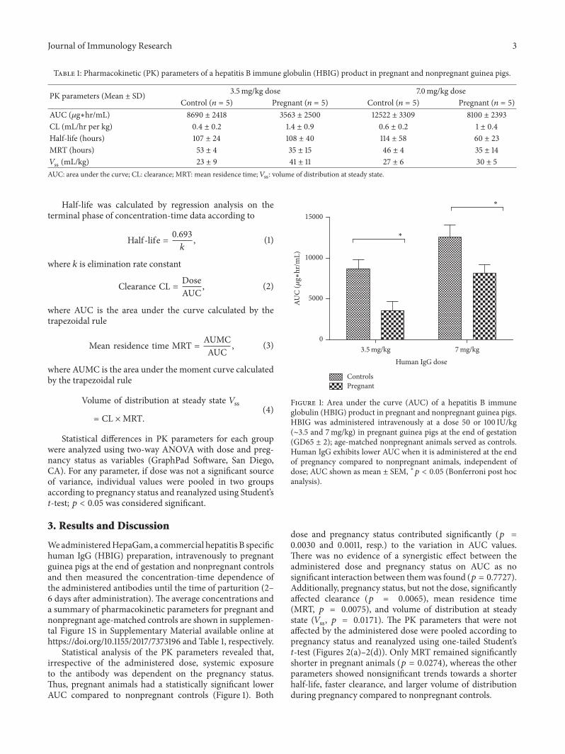
Table 1: Pharmacokinetic (PK) parameters of a hepatitis B immune globulin (HBIG) product in pregnant and nonpregnant guinea pigs.
PK parameters (Mean ± SD) 3.5mg/kg dose 7.0mg/kg doseControl (𝑛 = 5) Pregnant (𝑛 = 5) Control (𝑛 = 5) Pregnant (𝑛 = 5)
AUC (𝜇g∗hr/mL) 8690 ± 2418 3563 ± 2500 12522 ± 3309 8100 ± 2393CL (mL/hr per kg) 0.4 ± 0.2 1.4 ± 0.9 0.6 ± 0.2 1 ± 0.4Half-life (hours) 107 ± 24 108 ± 40 114 ± 58 60 ± 23MRT (hours) 53 ± 4 35 ± 15 46 ± 4 35 ± 14𝑉ss (mL/kg) 23 ± 9 41 ± 11 27 ± 6 30 ± 5AUC: area under the curve; CL: clearance; MRT: mean residence time; 𝑉ss: volume of distribution at steady state.
Half-life was calculated by regression analysis on theterminal phase of concentration-time data according to
Half-life = 0.693𝑘, (1)
where 𝑘 is elimination rate constant
Clearance CL = DoseAUC, (2)
where AUC is the area under the curve calculated by thetrapezoidal rule
Mean residence time MRT = AUMCAUC, (3)
where AUMC is the area under the moment curve calculatedby the trapezoidal rule
Volume of distribution at steady state 𝑉ss
= CL ×MRT.(4)
Statistical differences in PK parameters for each groupwere analyzed using two-way ANOVA with dose and preg-nancy status as variables (GraphPad Software, San Diego,CA). For any parameter, if dose was not a significant sourceof variance, individual values were pooled in two groupsaccording to pregnancy status and reanalyzed using Student’st-test; 𝑝 < 0.05 was considered significant.
3. Results and Discussion
WeadministeredHepaGam, a commercial hepatitis B specifichuman IgG (HBIG) preparation, intravenously to pregnantguinea pigs at the end of gestation and nonpregnant controlsand then measured the concentration-time dependence ofthe administered antibodies until the time of parturition (2–6 days after administration). The average concentrations anda summary of pharmacokinetic parameters for pregnant andnonpregnant age-matched controls are shown in supplemen-tal Figure 1S in Supplementary Material available online athttps://doi.org/10.1155/2017/7373196 and Table 1, respectively.
Statistical analysis of the PK parameters revealed that,irrespective of the administered dose, systemic exposureto the antibody was dependent on the pregnancy status.Thus, pregnant animals had a statistically significant lowerAUC compared to nonpregnant controls (Figure 1). Both
Human IgG dose
ControlsPregnant
∗
∗
3.5mg/kg 7mg/kg0
5000
10000
15000
AUC
(𝜇g∗
hr/m
L)
Figure 1: Area under the curve (AUC) of a hepatitis B immuneglobulin (HBIG) product in pregnant and nonpregnant guinea pigs.HBIG was administered intravenously at a dose 50 or 100 IU/kg(∼3.5 and 7mg/kg) in pregnant guinea pigs at the end of gestation(GD65 ± 2); age-matched nonpregnant animals served as controls.Human IgG exhibits lower AUC when it is administered at the endof pregnancy compared to nonpregnant animals, independent ofdose; AUC shown as mean ± SEM, ∗𝑝 < 0.05 (Bonferroni post hocanalysis).
dose and pregnancy status contributed significantly (𝑝 =0.0030 and 0.0011, resp.) to the variation in AUC values.There was no evidence of a synergistic effect between theadministered dose and pregnancy status on AUC as nosignificant interaction between themwas found (𝑝 = 0.7727).Additionally, pregnancy status, but not the dose, significantlyaffected clearance (𝑝 = 0.0065), mean residence time(MRT, 𝑝 = 0.0075), and volume of distribution at steadystate (𝑉ss, 𝑝 = 0.0171). The PK parameters that were notaffected by the administered dose were pooled according topregnancy status and reanalyzed using one-tailed Student’st-test (Figures 2(a)–2(d)). Only MRT remained significantlyshorter in pregnant animals (𝑝 = 0.0274), whereas the otherparameters showed nonsignificant trends towards a shorterhalf-life, faster clearance, and larger volume of distributionduring pregnancy compared to nonpregnant controls.
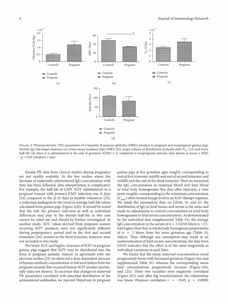
4 Journal of Immunology Research
×10−3
ControlsPregnant
PregnantControls0
0.5
1.0
1.5
2.0Cl
eara
nce (
L/hr
/kg)
(a)
∗
PregnantControls0
20
40
60
MRT
(Hrs
)
ControlsPregnant
(b)
×10−2
PregnantControls0
1
2
3
4
5
Vss
(L/k
g)
ControlsPregnant
(c)
PregnantControls0
50
100
150H
alf-li
fe (H
rs)
ControlsPregnant
(d)
Figure 2: Pharmacokinetic (PK) parameters of a hepatitis B immune globulin (HBIG) product in pregnant and nonpregnant guinea pigs.Human IgG has larger clearance (a), lower mean residence time (MRT, (b)), larger volume of distribution at steady state (𝑉ss, (c)), and lowerhalf-life (d) when it is administered at the end of gestation (GD65 ± 2) compared to nonpregnant animals; data shown as mean ± SEM,∗𝑝 < 0.05 (Student’s t-test).
Similar PK data from clinical studies during pregnancyare not readily available. In the few studies where thedecrease of maternally administered IgG concentration withtime has been followed, data interpretation is complicated.For example, the half-life of CMV IGIV administered to apregnant woman with primary CMV infection was 11 days[24] compared to the 21-22 days in healthy volunteers [25],a reduction analogous to the trend in average half-life valuescalculated from guinea pigs (Figure 2(d)). It should be notedthat the role the primary infection as well as individualdifferences may play in the shorter half-life in this casecannot be ruled out and should be further investigated. Inanother study, AUC values derived from pregnant womenreceiving IGIV products were not significantly differentduring prepregnancy period and in the first and secondtrimesters [26]; women in the third trimester, however, werenot included in this study.
The lower AUC and higher clearance of IGIV in pregnantguinea pigs suggest that IGIV may be distributed into thefetus in pregnant animals. Indeed, in agreement with ourprevious studies [23] we observed a dose dependent increaseof human antibody concentration in full term babies from thepregnant animals that received human IGIV product perina-tally (data not shown). To ascertain that changes in maternalPK parameters correlated with placental distribution of theadministered antibodies, we injected HepaGam in pregnant
guinea pigs at five gestation ages roughly corresponding toend of first trimester,middle and end of second trimester, andmiddle and the end of the third trimester. Then we measuredthe IgG concentration in maternal blood and fetal bloodor total body homogenates five days after injection, a timepoint roughly corresponding to the minimum concentration(Cmin) often termed trough level in an IGIV therapy regimen.We made the assumption that, on GD26, 35, and 45, thedistribution of IgG in fetal tissues and serum is the same andmade no adjustments to convert concentration in total bodyhomogenates to fetal serum concentrations. As demonstratedby the individual data (supplemental Table 1S), the averageIgG concentration in the serum of 𝑛 = 5GD45 litters is ∼2.5-fold higher than that in whole body homogenate preparationsof 𝑛 = 2 litters from the same gestation age (Table 1S,italics). Thus, although our assumption may result in anunderestimation of fetal serum concentrations, the data fromGD45 indicates that the effect is of the same magnitude asindividual variations in each litter.
We found that the mean maternal concentrations trendprogressively lower with increased gestation (Figure 3(a) andsupplemental Table S1) whereas the corresponding meanfetal concentrations progressively increase (Figure 3(b),and [21]). These two variables were negatively correlated(Figure 3(f)) and, after log transformation, the relationshipwas linear (Pearson correlation 𝑟 = −0.60, 𝑝 = 0.0008,
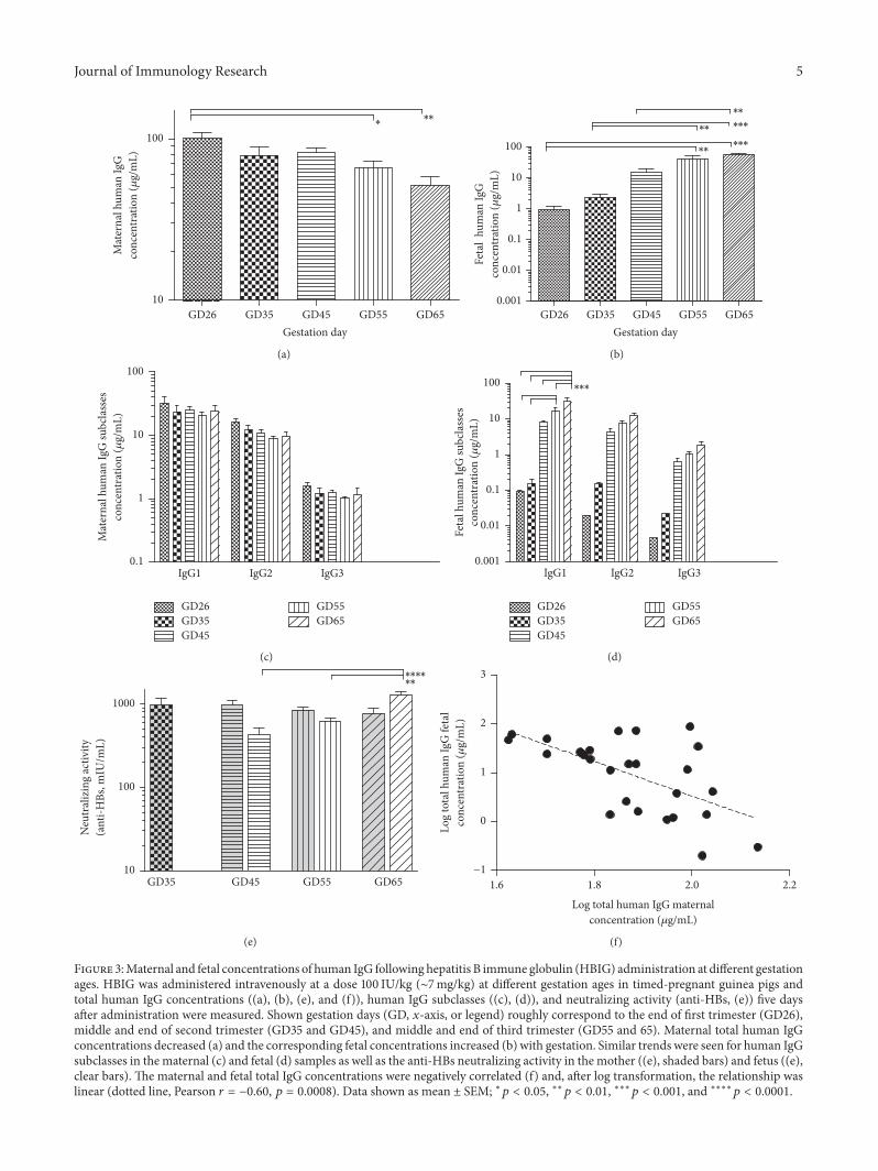
Journal of Immunology Research 5
GD35 GD45 GD55 GD65GD26Gestation day
∗ ∗∗
10
100
Mat
erna
l hum
an Ig
Gco
ncen
trat
ion
(𝜇g/
mL)
(a)
∗∗
∗∗
∗∗
∗∗∗
∗∗∗
0.001
0.01
0.1
1
10
100
GD35 GD45 GD55 GD65GD26Gestation day
Feta
lhu
man
IgG
conc
entr
atio
n (𝜇
g/m
L)
(b)
0.1
1
10
100
IgG3IgG2IgG1
Mat
erna
l hum
an Ig
G su
bcla
sses
conc
entr
atio
n (𝜇
g/m
L)
GD65GD55
GD45GD35GD26
(c)
GD65GD55
GD45GD35GD26
IgG3IgG2IgG1
∗∗∗
Feta
l hum
an Ig
G su
bcla
sses
conc
entr
atio
n (𝜇
g/m
L)
0.001
0.01
0.1
1
10
100
(d)∗∗∗∗∗∗
GD65GD55GD45GD3510
100
1000
Neu
tral
izin
g ac
tivity
(ant
i-HBs
, mIU
/mL)
(e)
Log
tota
l hum
an Ig
G fe
tal
conc
entr
atio
n (𝜇
g/m
L)
−1
0
1
2
3
1.8 2.0 2.21.6
Log total human IgG maternalconcentration (𝜇g/mL)
(f)
Figure 3:Maternal and fetal concentrations of human IgG following hepatitis B immune globulin (HBIG) administration at different gestationages. HBIG was administered intravenously at a dose 100 IU/kg (∼7mg/kg) at different gestation ages in timed-pregnant guinea pigs andtotal human IgG concentrations ((a), (b), (e), and (f)), human IgG subclasses ((c), (d)), and neutralizing activity (anti-HBs, (e)) five daysafter administration were measured. Shown gestation days (GD, 𝑥-axis, or legend) roughly correspond to the end of first trimester (GD26),middle and end of second trimester (GD35 and GD45), and middle and end of third trimester (GD55 and 65). Maternal total human IgGconcentrations decreased (a) and the corresponding fetal concentrations increased (b) with gestation. Similar trends were seen for human IgGsubclasses in the maternal (c) and fetal (d) samples as well as the anti-HBs neutralizing activity in the mother ((e), shaded bars) and fetus ((e),clear bars). The maternal and fetal total IgG concentrations were negatively correlated (f) and, after log transformation, the relationship waslinear (dotted line, Pearson 𝑟 = −0.60, 𝑝 = 0.0008). Data shown as mean ± SEM; ∗𝑝 < 0.05, ∗∗𝑝 < 0.01, ∗∗∗𝑝 < 0.001, and ∗∗∗∗𝑝 < 0.0001.
6 Journal of Immunology Research
confidence interval −0.80 to −0.29). One data point fromGD35 was excluded from this calculation (supplementalTable 1S, gray font and italics) due to maternal concentrationbeing a clear outlier compared to all the other points.
While the negative correlation does not prove causality,we suggest that transplacental transfer of antibody from themother to the baby may be a significant contributor to thedecreased AUC and mean residence time we observed at theend of gestation.We further noted that not only arematernal-fetal concentration changes with increased gestation corre-lated, but the magnitude of changes follows the same trend.Thus, trough maternal human IgG levels are significantlylower at the middle and the end of third trimester (GD54and 65, respectively, Figure 3(a)), the same time points wherestatistical significant concentration increases were seen in thefetal samples (Figure 3(b)).
We also measured concentration of all human IgG sub-classes and neutralizing activity (anti-HBs levels) in bothmaternal and fetal samples. Given that IgG4 constitutessmall percentage of HepaGam (and all other plasma derivedpolyclonal IgG products as well as human serum) bothmaternal and fetal concentrations for this subclasswere belowthe detection limit in the majority of the collected samples.Nevertheless, we were able to detect IgG4 in some of the fetalsamples in the third trimester, but most of the values werebelow the level of quantitation (data not shown). For the othersubclasses, we observed the same general trend of decreasingmaternal concentrations with increased gestation (two-wayANOVA 𝑝 = 0.0636, Figure 3(c)), a significant increase offetal concentrations with progression of pregnancy (two-way ANOVA 𝑝 < 0.0001, Figure 3(d)), and a significantinteraction between subclass concentrations in the fetus andgestation age (𝑝 = 0.0004). Post hoc analyses revealed thatonly IgG1 increases in GD55 and GD65 are statisticallysignificant; the concentration increases in other subclasses donot reach significance.
We obtained similar results for the neutralizing activity(Figure 3(e)), with fetal anti-HBs levels on GD45, 55, and 65one to two orders of magnitude higher than 10mIU/mL, theaccepted serological level of protection [27], and neutralizingantibody levels GD35 fetal blood below quantification limit.
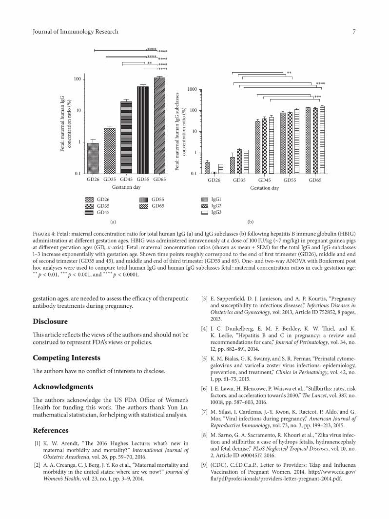
Previously we showed that pregnant guinea pigs arean appropriate animal model for studying human antibodytransfer during pregnancy [21, 23]. The additional experi-mental data we present here enabled us to more preciselymeasure placental transfer and further demonstrate that thismodel recapitulates well the time course of the placentaltransfer of IgG in pregnant women.Thus, 17–22-week humanfetuses have circulating concentrations of IgG that are only5–10% of maternal levels [28] but they significantly increaseduring the third trimester [20, 29], often surpassing levelsfound in their mother. Our results show that, in the pregnantguinea pig, fetal : maternal ratios for administered humanantibodies are ∼3 and 20% in the middle and end of secondtrimester (GD35 and 45), respectively, but increase to ∼60and 110% in the middle and end of the third trimester(Figure 4(a)). Similarly to what we previously found, thefetal : maternal IgG concentration ratios with increased ges-tation fit an exponential growth curve (𝑅2 = 0.87, not
shown). Thus, the pharmacokinetic changes we observe inthe guinea pig animal model may closely match what canbe observed in women receiving IGIV during pregnancy. Weshould note that even though themean fetal : maternal valuesfor each gestation differ somewhat from what we previouslycalculated [21], the differences are due to an increased samplesize and do not change any of the previous conclusions. Allfetal : placental ratios lie within one standard deviation ofpreviously calculated means [21] and the new group averages(Figure 4(a)) can be considered more precise point estimatesof the gestation dependent placental transfer.
In a similar fashion to the total human IgG, thefetal : maternal concentration ratios for IgG subclasses 1–3increased exponentially with gestation age (two-wayANOVA𝑝 < 0.0001) but, unlike the concentration changes, weresimilar to each other at all time points (Figure 4(b)). Posthoc analyses revealed that fetal : maternal ratios for all threeIgG subclasses analyzed increased significantly in the thirdtrimester (GD55 and 65) compared to earlier in gestation.Unlike the increases in the fetal IgG subclass concentrations(Figure 3(d)), an interaction between gestation age and sub-class effects in the variance of fetal : maternal concentrationratios was not observed (𝑝 = 0.85).
Under our experimental conditions, we did not find anydifference in placental transfer propensity for human IgGsubclasses 1–3 in the pregnant guinea pig. The same has notbeen reported in human pregnancy [28, 30] where thereis a clear difference between the fetal : maternal ratios forIgG subclasses. Some of these differences may be related topopulation level polymorphism in the sequence of Fc [31],but other factors have also been proposed [16]. More studiesare needed to better understand the differences in placentaltransfer of IgG subclasses aiming to optimize the efficacy ofantibody therapeutics during pregnancy.
4. Conclusions
Our studies in an animal model of human pregnancy showthat intact human IgGmolecules of all subclasses traverse theplacenta at increasing levels with progression of pregnancy.This transplacental distribution can have dual implications:it may contribute to a reduction of maternal exposure to theadministered antibodies compared to nonpregnant controls(Figures 1 and 3) and it can expose the fetus to progressivelyhigher levels of therapeutic IgG with increased gestation(Figure 3). Fetal partition of the IgG, at least for HBIGand depending on the dose, may result in fetal neutralizingactivity (anti-HBs levels) at time points starting with theend of second trimester (GD45) that reach and surpassthe accepted serological level of protection for children andadults (Figure 3(e), [23, 27]). However, it is unknown if andat what levels neutralizing antibodies in the fetus can preventfetal viral infections and what the effects of reduced mater-nal exposure to administered antibody therapy would be,especially in the presence of maternal infection. The clinicalscenariomay be further complicated by changes in immunityand other pregnancy related changes [7].Thus, well-designedclinical studies and careful dosing considerations, especiallyin light of changes in biodistribution to the fetus at different
Journal of Immunology Research 7
GD35 GD45 GD55 GD65GD26Gestation day
Feta
l: m
ater
nal h
uman
IgG
conc
entr
atio
n ra
tio (%
)
0.1
1
10
100
∗∗∗∗
∗∗∗∗∗∗∗∗∗∗∗∗
∗∗∗∗
∗∗∗∗
∗∗
GD26GD35GD45
GD55GD65
(a)
IgG1IgG2IgG3
Feta
l: m
ater
nal h
uman
IgG
subc
lass
esco
ncen
trat
ion
ratio
(%)
0.1
1
10
100
1000
GD35 GD45 GD55 GD65GD26Gestation day
∗∗∗
∗∗∗∗
∗∗
(b)
Figure 4: Fetal : maternal concentration ratio for total human IgG (a) and IgG subclasses (b) following hepatitis B immune globulin (HBIG)administration at different gestation ages. HBIG was administered intravenously at a dose of 100 IU/kg (∼7mg/kg) in pregnant guinea pigsat different gestation ages (GD, 𝑥-axis). Fetal : maternal concentration ratios (shown as mean ± SEM) for the total IgG and IgG subclasses1–3 increase exponentially with gestation age. Shown time points roughly correspond to the end of first trimester (GD26), middle and endof second trimester (GD35 and 45), and middle and end of third trimester (GD55 and 65). One- and two-way ANOVA with Bonferroni posthoc analyses were used to compare total human IgG and human IgG subclasses fetal : maternal concentration ratios in each gestation age;∗∗𝑝 < 0.01, ∗∗∗𝑝 < 0.001, and ∗∗∗∗𝑝 < 0.0001.
gestation ages, are needed to assess the efficacy of therapeuticantibody treatments during pregnancy.
Disclosure
This article reflects the views of the authors and should not beconstrued to represent FDA’s views or policies.
Competing Interests
The authors have no conflict of interests to disclose.
Acknowledgments
The authors acknowledge the US FDA Office of Women’sHealth for funding this work. The authors thank Yun Lu,mathematical statistician, for helpingwith statistical analysis.
References
[1] K. W. Arendt, “The 2016 Hughes Lecture: what’s new inmaternal morbidity and mortality?” International Journal ofObstetric Anesthesia, vol. 26, pp. 59–70, 2016.
[2] A. A. Creanga, C. J. Berg, J. Y. Ko et al., “Maternal mortality andmorbidity in the united states: where are we now?” Journal ofWomen’s Health, vol. 23, no. 1, pp. 3–9, 2014.
[3] E. Sappenfield, D. J. Jamieson, and A. P. Kourtis, “Pregnancyand susceptibility to infectious diseases,” Infectious Diseases inObstetrics and Gynecology, vol. 2013, Article ID 752852, 8 pages,2013.
[4] J. C. Dunkelberg, E. M. F. Berkley, K. W. Thiel, and K.K. Leslie, “Hepatitis B and C in pregnancy: a review andrecommendations for care,” Journal of Perinatology, vol. 34, no.12, pp. 882–891, 2014.
[5] K. M. Bialas, G. K. Swamy, and S. R. Permar, “Perinatal cytome-galovirus and varicella zoster virus infections: epidemiology,prevention, and treatment,” Clinics in Perinatology, vol. 42, no.1, pp. 61–75, 2015.
[6] J. E. Lawn, H. Blencowe, P. Waiswa et al., “Stillbirths: rates, riskfactors, and acceleration towards 2030,”The Lancet, vol. 387, no.10018, pp. 587–603, 2016.
[7] M. Silasi, I. Cardenas, J.-Y. Kwon, K. Racicot, P. Aldo, and G.Mor, “Viral infections during pregnancy,” American Journal ofReproductive Immunology, vol. 73, no. 3, pp. 199–213, 2015.
[8] M. Sarno, G. A. Sacramento, R. Khouri et al., “Zika virus infec-tion and stillbirths: a case of hydrops fetalis, hydranencephalyand fetal demise,” PLoS Neglected Tropical Diseases, vol. 10, no.2, Article ID e0004517, 2016.
[9] (CDC), C.f.D.C.a.P., Letter to Providers: Tdap and InfluenzaVaccination of Pregnant Women, 2014, http://www.cdc.gov/flu/pdf/professionals/providers-letter-pregnant-2014.pdf.
8 Journal of Immunology Research
[10] CDC, “General recommendations on immunization—recom-mendations of the Advisory Committee on ImmunizationPractices (ACIP),”MMWR Recommendations and Reports, vol.60, no. 2, pp. 1–64, 2011.
[11] Z. Shi, X. Li, L. Ma, and Y. Yang, “Hepatitis B immunoglobulininjection in pregnancy to interrupt hepatitis B virus mother-to-child transmission-a meta-analysis,” International Journal ofInfectious Diseases, vol. 14, no. 7, pp. e622–e634, 2010.
[12] M. G. Revello, T. Lazzarotto, B. Guerra et al., “A random-ized trial of hyperimmune globulin to prevent congenitalcytomegalovirus,” The New England Journal of Medicine, vol.370, no. 14, pp. 1316–1326, 2014.
[13] R. S. Shapiro, R. L. Wasserman, V. Bonagura, and S. Gupta,“Emerging paradigm of primary immunodeficiency disease:individualizing immunoglobulin dose and delivery to enhanceoutcomes,” Journal of Clinical Immunology, 2014.
[14] J. S. Orange, W. J. Grossman, R. J. Navickis, and M. M. Wilkes,“Impact of trough IgG on pneumonia incidence in primaryimmunodeficiency: a meta-analysis of clinical studies,” ClinicalImmunology, vol. 137, no. 1, pp. 21–30, 2010.
[15] M. Firan, R. Bawdon, C. Radu et al., “The MHC class I-related receptor, FcRn, plays an essential role in thematernofetaltransfer of 𝛾-globulin in humans,” International Immunology,vol. 13, no. 8, pp. 993–1002, 2001.
[16] P. Palmeira, C. Quinello, A. L. Silveira-Lessa, C. A. Zago,and M. Carneiro-Sampaio, “IgG placental transfer in healthyand pathological pregnancies,” Clinical and DevelopmentalImmunology, vol. 2012, Article ID 985646, 13 pages, 2012.
[17] X. Lin, Y. Guo, A. Zhou et al., “Immunoprophylaxis failureagainst vertical transmission of hepatitis B virus in the Chinesepopulation: a hospital-based study and a meta-analysis,” ThePediatric Infectious Disease Journal, vol. 33, no. 9, pp. 897–903,2014.
[18] L. Ma, N. R. Alla, X. Li, O. A. Mynbaev, and Z. Shi, “Mother-to-child transmission of HBV: review of current clinical manage-ment and prevention strategies,” Reviews in Medical Virology,vol. 24, no. 6, pp. 396–406, 2014.
[19] M. K. Young, A. W. Cripps, G. R. Nimmo, and M. L. van Driel,“Post-exposure passive immunisation for preventing rubellaand congenital rubella syndrome,” The Cochrane Database ofSystematic Reviews, vol. 9, Article ID CD010586, 2015.
[20] A. Malek, R. Sager, and H. Schneider, “Maternal-fetal transportof immunoglobulin G and its subclasses during the thirdtrimester of human pregnancy,” American Journal of Reproduc-tive Immunology, vol. 32, no. 1, pp. 8–14, 1994.
[21] Y. Xu, L. Ma, M. G. Norton et al., “Gestation age dependenttransfer of human immunoglobulins across placenta in timed-pregnant Guinea pigs,” Placenta, vol. 36, no. 12, pp. 1370–1377,2015.
[22] HepaGam B Prescribing Information, 2012, http://www.hepa-gamb.com/pdfs/HepaGamBPI.pdf.
[23] L. Ma, M. G. Norton, I. Mahmood et al., “Transplacental trans-fer of hepatitis B neutralizing antibody during pregnancy in ananimal model: implications for newborn and maternal health,”Hepatitis Research and Treatment, vol. 2014, Article ID 159206,7 pages, 2014.
[24] K. Hamprecht, K.-O. Kagan, and R. Goelz, “Hyperimmuneglobulin to prevent congenital CMV infection,” New EnglandJournal of Medicine, vol. 370, no. 26, p. 2543, 2014.
[25] P. A. Thurmann, C. Sonnenburg-Chatzopoulos, and R. Lissner,“Pharmacokinetic characteristics and tolerability of a novel
intravenous immunoglobulin preparation,” European Journal ofClinical Pharmacology, vol. 49, no. 3, pp. 237–242, 1995.
[26] M. H. H. Ensom andM. D. Stephenson, “A two-center study onthe pharmacokinetics of intravenous immunoglobulin beforeand during pregnancy in healthy women with poor obstetricalhistories,” Human Reproduction, vol. 26, no. 9, pp. 2283–2288,2011.
[27] E. E. Mast, H. S. Margolis, A. E. Fiore et al., “A comprehensiveimmunization strategy to eliminate transmission of hepatitis Bvirus infection in the United States: recommendations of theAdvisory Committee on Immunization Practices (ACIP) part1: immunization of infants, children, and adolescents,”MMWRRecommendations and Reports, vol. 54, no. 13, pp. 1–23, 2005.
[28] A. Malek, R. Sager, P. Kuhn, K. H. Nicolaides, and H. Schnei-der, “Evolution of maternofetal transport of immunoglobulinsduring human pregnancy,” American Journal of ReproductiveImmunology, vol. 36, no. 5, pp. 248–255, 1996.
[29] N. E. Simister, “Placental transport of immunoglobulin G,”Vaccine, vol. 21, no. 24, pp. 3365–3369, 2003.
[30] S. Hashira, S. Okitsu-Negishi, and K. Yoshino, “Placentaltransfer of IgG subclasses in a Japanese population,” PediatricsInternational, vol. 42, no. 4, pp. 337–342, 2000.
[31] H. Einarsdottir, Y. Ji, R. Visser et al., “H435-containingimmunoglobulin G3 allotypes are transported efficiently acrossthe human placenta: implications for alloantibody-mediateddiseases of the newborn,” Transfusion, vol. 54, no. 3, pp. 665–671, 2014.

Submit your manuscripts athttps://www.hindawi.com
Stem CellsInternational
Hindawi Publishing Corporationhttp://www.hindawi.com Volume 2014
Hindawi Publishing Corporationhttp://www.hindawi.com Volume 2014
MEDIATORSINFLAMMATION
of
Hindawi Publishing Corporationhttp://www.hindawi.com Volume 2014
Behavioural Neurology
EndocrinologyInternational Journal of
Hindawi Publishing Corporationhttp://www.hindawi.com Volume 2014
Hindawi Publishing Corporationhttp://www.hindawi.com Volume 2014
Disease Markers
Hindawi Publishing Corporationhttp://www.hindawi.com Volume 2014
BioMed Research International
OncologyJournal of
Hindawi Publishing Corporationhttp://www.hindawi.com Volume 2014
Hindawi Publishing Corporationhttp://www.hindawi.com Volume 2014
Oxidative Medicine and Cellular Longevity
Hindawi Publishing Corporationhttp://www.hindawi.com Volume 2014
PPAR Research
The Scientific World JournalHindawi Publishing Corporation http://www.hindawi.com Volume 2014
Immunology ResearchHindawi Publishing Corporationhttp://www.hindawi.com Volume 2014
Journal of
ObesityJournal of
Hindawi Publishing Corporationhttp://www.hindawi.com Volume 2014
Hindawi Publishing Corporationhttp://www.hindawi.com Volume 2014
Computational and Mathematical Methods in Medicine
OphthalmologyJournal of
Hindawi Publishing Corporationhttp://www.hindawi.com Volume 2014
Diabetes ResearchJournal of
Hindawi Publishing Corporationhttp://www.hindawi.com Volume 2014
Hindawi Publishing Corporationhttp://www.hindawi.com Volume 2014
Research and TreatmentAIDS
Hindawi Publishing Corporationhttp://www.hindawi.com Volume 2014
Gastroenterology Research and Practice
Hindawi Publishing Corporationhttp://www.hindawi.com Volume 2014
Parkinson’s Disease
Evidence-Based Complementary and Alternative Medicine
Volume 2014Hindawi Publishing Corporationhttp://www.hindawi.com


![INSTRUCTION MANUAL Copyright © 2007 · Maximum On-State Resistance [RDS-ON][Ohms] 0.29 0.13 0.10 0.05 0.29 0.23 0.05 0.6 0.35 0.8 0.55 INPUT SPECIFICATIONS (1) Description DC Control](https://img.pdfslide.net/doc/110x75/6147f36da830d0442101c478/instruction-manual-copyright-2007-maximum-on-state-resistance-rds-onohms.jpg)