Embed Size (px)
Citation preview
8/6/2019 Patho Pre Lab
http://slidepdf.com/reader/full/patho-pre-lab 1/4
1
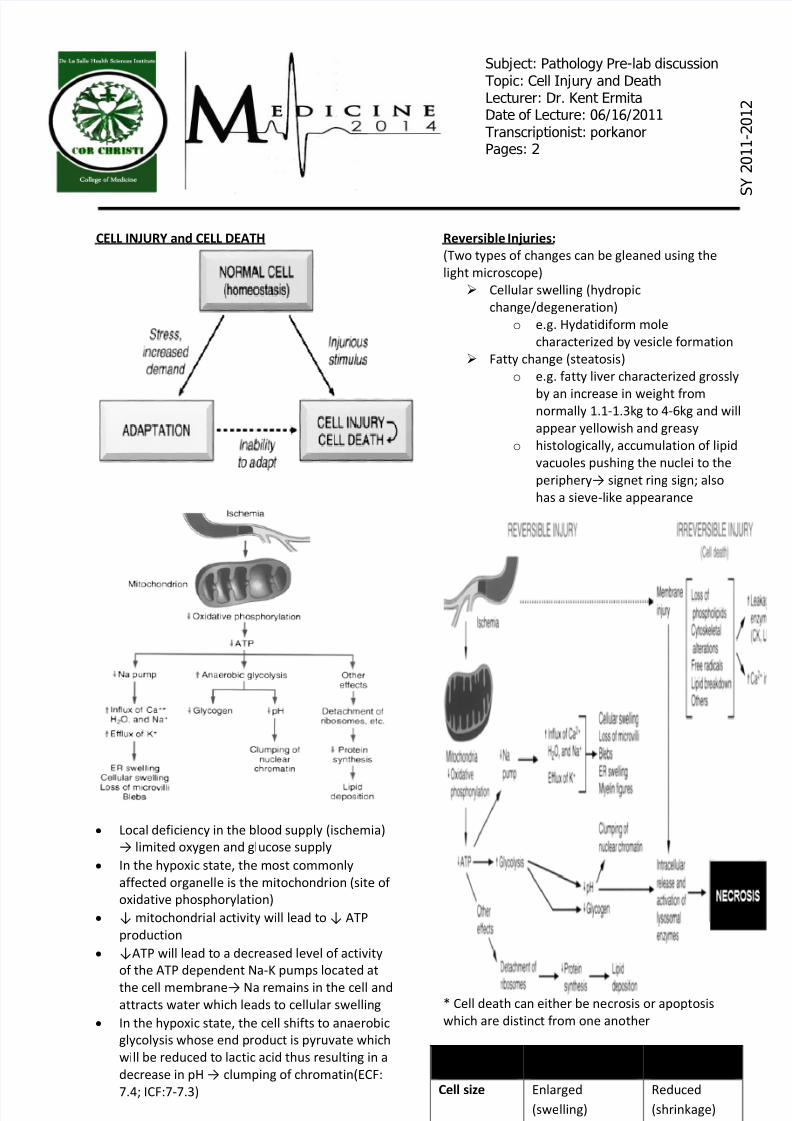
CELL INJURY and CELL DEATH
y Local deficiency in the blood supply (ischemia)
limited oxygen and glucose supply
y In the hypoxic state, the most commonly
affected organelle is the mitochondrion (site of
oxidative phosphorylation)
y mitochondrial activity will lead to ATP
production
y ATP will lead to a decreased level of activity
of the ATP dependent Na-K pumps located at
the cell membrane Na remains in the cell andattracts water which leads to cellular swelling
y In the hypoxic state, the cell shifts to anaerobic
glycolysis whose end product is pyruvate which
will be reduced to lactic acid thus resulting in a
decrease in pH clumping of chromatin(ECF:
7.4; ICF:7-7.3)
Reversible Injuries:
(Two types of changes can be gleaned using the
light microscope)
Cellular swelling (hydropic
change/degeneration)
o e.g. Hydatidif orm mole
characterized by vesicle f ormation
Fatty change (steatosis)
o e.g. f atty liver characterized grossly
by an increase in weight f romnormally 1.1-1.3kg to 4-6kg and will
appear yellowish and greasy
o histologically, accumulation of lipid
vacuoles pushing the nuclei to the
periphery signet ring sign; also
has a sieve-like appearance
* Cell death can either be necrosis or apoptosis
which are distinct f rom one another
Features Necrosis Apoptosis
Cell size Enlarged
(swelling)
Reduced
(shrinkage)
Subject: Pathology Pre-lab discussionTopic: Cell Injury and DeathLecturer: Dr. Kent ErmitaDate of Lecture: 06/16/2011Transcriptionist: porkanorPages: 2
S Y
2 0 1 1 - 2 0 1 2
8/6/2019 Patho Pre Lab
http://slidepdf.com/reader/full/patho-pre-lab 2/4
2
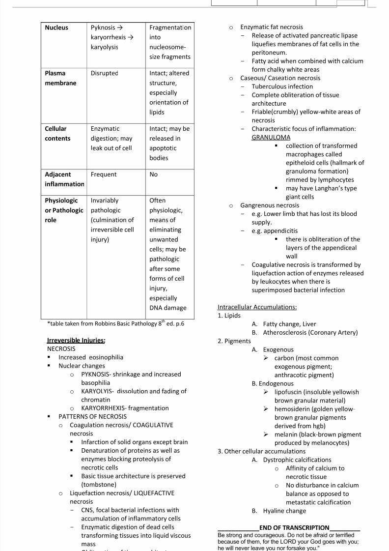
Nucleus Pyknosis
karyorrhexis
karyolysis
Fragmentation
into
nucleosome-
size f ragments
Plasma
membrane
Disrupted Intact; altered
structure,
especially
orientation of
lipids
Cellular
contents
Enzymatic
digestion; may
leak out of cell
Intact; may be
released in
apoptotic
bodies
Adjacent
inflammation
Frequent No
Physiologic
or Pathologic
role
Invariably
pathologic
(culmination of
irreversible cell
injury)
Often
physiologic,
means of
eliminating
unwanted
cells; may be
pathologic
after some
f orms of cell
injury,especially
DNA damage
*table taken f rom Robbins Basic Pathology 8th
ed. p.6
Irreversible Injuries:
NECROSIS
Increased eosinophilia
Nuclear changes
o PYKNOSIS- shrinkage and increased
basophilia
o KARYOLYIS- dissolution and f ading of
chromatin
o KARYORRHEXIS- f ragmentation
PATTERNS OF NECROSIS
o Coagulation necrosis/ COAGULATIVE
necrosis
Inf arction of solid organs except brain
Denaturation of proteins as well as
enzymes blocking proteolysis of
necrotic cells
Basic tissue architecture is preserved
(tombstone) o Liquef action necrosis/ LIQ UEFACTIVE
necrosis
- CNS, f ocal bacterial infections with
accumulation of inf lammatory cells
- Enzymatic digestion of dead cells
transf orming tissues into liquid viscous
mass
- Obliteration of tissue architecture
o Enzymatic f at necrosis
- Release of activated pancreatic lipase
liquefies membranes of f at cells in the
peritoneum.
- Fatty acid when combined with calcium
f orm chalky white areas
o Caseous/ Caseation necrosis
- Tuberculous infection
- Complete obliteration of tissuearchitecture
- Friable(crumbly) yellow-white areas of
necrosis
- Characteristic f ocus of inf lammation:
GRANULOMA
collection of transf ormed
macrophages called
epitheloid cells (hallmark of
granuloma f ormation)
rimmed by lymphocytes
may have Langhans type
giant cells
o Gangrenous necrosis
- e.g. Lower limb that has lost its blood
supply.
- e.g. appendicitis
there is obliteration of the
layers of the appendiceal
wall
- Coagulative necrosis is transf ormed by
liquef action action of enzymes released
by leukocytes when there is
superimposed bacterial infection
Intracellular Accumulations:
1. Lipids
A. Fatty change, Liver
B. Atherosclerosis (Coronary Artery)
2. Pigments
A. Exogenous
carbon (most common
exogenous pigment;
anthracotic pigment)
B. Endogenous lipofuscin (insoluble yellowish
brown granular material)
hemosiderin (golden yellow-
brown granular pigments
derived f rom hgb)
melanin (black-brown pigment
produced by melanocytes)
3. Other cellular accumulations
A. Dystrophic calcifications
o Affinity of calcium to
necrotic tissue
o No disturbance in calcium
balance as opposed to
metastatic calcification
B. Hyaline change
____________END OF TRANSCRIPTION _________ Be strong and courageous. Do not be afraid or terrifiedbecause of them, for the LORD your God goes with you;he will never leave you nor forsake you."
Deuteronomy 31:6

8/6/2019 Patho Pre Lab
http://slidepdf.com/reader/full/patho-pre-lab 3/4
3

8/6/2019 Patho Pre Lab
http://slidepdf.com/reader/full/patho-pre-lab 4/4
4