Embed Size (px)
Citation preview

1
Understanding
Pathogenaemia in Man:
A Proof of Concept Study
Robert Nipah
School of Environment and Life Sciences University of Salford, Salford, UK
A thesis submitted in partial fulfilment of the requirements of the degree
of Master of Philosophy, August
2015

2
Contents The Author ........................................................................................................................ 5
Abstract .............................................................................................................................. 6
List of Figures .................................................................................................................. 9
List of Tables .................................................................................................................. 10
Acknowledgements ....................................................................................................... 11
Abbreviations ................................................................................................................. 12
CHAPTER 1 ....................................................................................................................... 15
Background ..................................................................................................................... 16
Pathogens, infection and sepsis: A relationship explored .............................................. 16
Sepsis: a pathophysiological response to pathogenaemia .................................................... 19
Previous in vivo Models of Sepsis ....................................................................................... 21
Model of investigation........................................................................................................ 25
Introduction .......................................................................................................................... 25
Endourology ......................................................................................................................... 27
Endourology procedure - Ureteroscopy ............................................................................... 28
Endourology procedure - Percutaneous nephrolithotomy (PCNL) ..................................... 28
Antibiotic prophylaxis in urology procedures ..................................................................... 30
Tools for investigation ....................................................................................................... 32
Microbiological investigations – Blood Cultures ................................................................ 32
Limitation of blood cultures................................................................................................. 33

3
Molecular investigation – Nucleic acid based diagnostic technology ................................. 35
NAT assays for the detection and identification of pathogens from blood culture bottles .. 36
Direct molecular detection of pathogens in blood with PCR .............................................. 37
SeptiFast® assay ................................................................................................................... 39
Effect of antimicriobial administration pre testing on both microbiological and molecular
approaches............................................................................................................................ 41
Cytokine driven immune reponses ................................................................................... 44
Interleukin 6 ......................................................................................................................... 45
Interleukin 10 ....................................................................................................................... 47
Summary .......................................................................................................................... 48
Aim of study........................................................................................................................ 50
CHAPTER 2: ...................................................................................................................... 51
Participant recruitment ......................................................................................................... 52
Study design ......................................................................................................................... 52
Blood sampling .................................................................................................................... 54
Data collection ..................................................................................................................... 55
Laboratory analyses ............................................................................................................. 56
Analysis of pathogen DNA by SeptiFast®
PCR assay ......................................................... 56
Pathogen DNA extraction from blood ................................................................................. 57
SeptiFast® real-time PCR of pathogen DNA ....................................................................... 58
SeptiFast® - Identification of pathogen species and controls............................................... 59
Cytokine analysis ................................................................................................................. 61
IL-6 ELISA .......................................................................................................................... 61
IL-10 ELISA ........................................................................................................................ 63
Statistical analysis ................................................................................................................ 63
CHAPTER 3: ...................................................................................................................... 64
SeptiFast® assay coverage and historic data on positive cultures ........................................ 65

4
Study Population .................................................................................................................. 66
Pathogen species identified by SeptiFast®
assay and blood culture..................................... 68
PCR positive signal patients vs. PCR negative signal patients ............................................ 70
Relationship between pathogen isolate and clinical outcome .............................................. 72
Analysis of circulating IL-6 and IL-10 levels ...................................................................... 74
Pathogen detection and physiological response ................................................................... 76
Case specific findings - PCR positive patients .................................................................... 81
Case specific findings - PCR negative patients ................................................................... 81
Stone composition and pathogen detection.......................................................................... 82
CHAPTER 4 ....................................................................................................................... 83
Background of the study ...................................................................................................... 84
Key findings of the study ..................................................................................................... 87
SeptiFast® - Role in circulating pathogen DNA detection in infection ? ............................ 88
SeptiFast® - A place in clinical practice? ............................................................................. 89
Circulating pathogen DNA and the immune response ........................................................ 94
The urology patient: Host response, sepsis and antibiotic prophylaxis .............................. 96
Strength and weakness of the study ..................................................................................... 98
Future work .......................................................................................................................... 99
Conclusion ........................................................................................................................... 99
REFERENCES ................................................................................................................. 101

5
The Author
The author graduated from the Queen Mary and Westfield College, University of
London with a First Class Honours Bachelor of Science degree in Basic Medical
Science with Clinical Immunology in 1998. He qualified as a medical doctor from St
Bartholomew and Royal London School of Medicine and Dentistry, University of
London with MB BS in 2001. He passed his MRCP post graduate examination in 2006.
He commenced his research as an out of programme placement as an intensive care
registrar at Salford Royal Foundation Trust. He has completed his specialist registrar
training and is dual accredited in general medicine and nephrology. Since March 2013,
he has become a consultant in acute medicine at Salford Royal Foundation Trust.

6
Abstract
Introduction: The current understanding of the significance of circulating pathogen
DNA in infection is limited. Blood cultures are the current gold standard for pathogen
detection. The administration of antibiotics can confound pathogen detection by blood
cultures. Polymerase chain reaction assays of circulating pathogen DNA has the
potential ability for rapid diagnosis of infection and may be potentially useful in a
clinical setting. However, the use of this technology has only recently been used in the
study of sepsis.
Animal models for the study of sepsis have added to our understanding. The failure to
translate results from animals to humans has been attributed to the disease
characteristics of sepsis (complexity and heterogeneity), inappropriate clinical trials
(study of ineffective drugs, inadequate clinical trial designs), and animal models that do
not fully mimic human sepsis. Therefore, the finding of an easily reproducible in vivo
human model for pathogenaemia may provide a platform for exploring the host’s
immune response to circulating pathogen material.
Infection and urosepsis are common complications in diagnostic and therapeutic
urological procedures. Urological interventional procedures for the removal of renal
stones are commonly done in a controlled operating environment and may potentially
be an in vivo model for investigating the host immune response to detected pathogen.
Antibiotic prophylaxis is routinely given across the world for these urological
procedures even though the evidence for their use is weak. If the presence of pathogen
is seen to generate an immune response in these in vivo human models, it could be
argued that there may be a potential benefit to antibiotic prophylaxis.

7
Aim: To investigate interventional urological stone removal as a model of
pathogenaemia in man and, therefore, test the following hypotheses:
1. The detection of circulating pathogen DNA by SeptiFast® PCR is an indicator
for the infection associated with urological procedures.
2. The presence of circulating pathogen DNA correlates with the host immune and
physiological response, supporting the notion that antibiotics prophylaxis is
important in urological procedures.
Method: In a prospective group of patients undergoing stone removal, blood samples
were taken at five time points, peri-operatively to assess:
The presence of pathogen by blood culture and pathogen DNA by SeptiFast® PCR;
Circulating IL - 6 and IL - 10 levels to assess activation of host inflammatory
response.
Routine peri - operative observations were recorded throughout as measures of
physiological responses.
Results: Collected historical data on urological procedures by Salford Royal
Foundation Trust urology services show that the SeptiFast® assay gave full coverage for
pathogens seen in this clinical setting. While positive blood culture was rare, 50% of
patients tested positive for pathogen DNAemia and this was associated with increased
circulating IL-6 compared to patients with no circulating pathogen DNA. Linkage
between pathogen DNA positivity and patient outcome was not established.
Conclusion: These data provide novel evidence that pathogen DNAemia is a common
feature of routine urological procedures correlating with an increased systemic

8
inflammatory response. Endourological stone removal interventions may be a useful
model for understanding the role of pathogen DNA in triggering inflammatory
responses to infection in man.

9
List of Figures
Figure 1 - Simplified clinical course of sepsis (Doi et al 2009) ..................................... 20
Figure 2 - Percentage of kidney stones ........................................................................... 27
Figure 3 - PCNL (taken from www.uroinfo.ca) ............................................................. 29
Figure 4 - Identification and genotyping of microbes by PCR/ESI/mass (Ecker et al
2010) ............................................................................................................................... 36
Figure 5 - IL - 6 and sepsis - associated mortality (Hack et al 1989) ............................. 46
Figure 6 - Interleukin 10 and receptor (Kotenko et al 1997) .......................................... 47
Figure 7 - Study design .................................................................................................. 53
Figure 8 - Distribution of melting temperature and respective detection channels for all
microorganism and internal control in Septifast assay ( Lehmann et al 2008) ............... 60
Figure 9 - The principle of sandwich ELISA ( adapted from www.lenico.com) ........... 62
Figure 10 - Mean IL - 6, IL10, IL-6:IL-10 ratio in PCR - positive and PCR - negative
patients ............................................................................................................................ 75
Figure 11 - Variations of mean arterial blood pressures in PCR negative group ........... 77
Figure 12 - Variation of mean arterial blood pressure in PCR positive group ............... 77
Figure 13 - Mean MAP between PCR positive and PCR negative ................................ 78
Figure 14 - Variation in pulse rate in PCR negative group............................................. 79
Figure 15 - Variations in pulse rate in PCR positive groups .......................................... 80
Figure 16 - A comparison of the variation between mean pulse rate between PCR
negative patients and PCR positive patients ................................................................... 80

10
List of Tables
Table 1 - Diagnostic criteria for sepsis according to the International Sepsis Definitions
Conference (Levy et al , 2003). ...................................................................................... 18
Table 2 - Possible reasons for failure in sepsis trials (Riedemann et al, 2003) .............. 24
Table 3 Generally accepted risk factors for infectious complication ............................ 31
Table 4 - Needs and current status of methods to identify bloodstream infections
(Adapted from Ecker et al 2010) .................................................................................... 43
Table 5 – Timing schedule for blood sampling for RT-PCR analysis ............................ 54
Table 6 – Septifast® Pathogen Detection Panel .............................................................. 57
Table 7 - List of positive organism growth from mid-stream urine sample from all
patients who underwent renal stone removal surgery at Salford Royal Foundation Trust
between 2004 – 2011 ...................................................................................................... 65
Table 8 - Patient demographic and operation detail ....................................................... 67
Table 9 - Detection of pathogen DNA signal RT - PCR on blood samples at different
time points ....................................................................................................................... 69
Table 10 - Characteristic of PCR positive patients vs. PCR negative patients ............... 70
Table 11 - Correlation between PCR positivity and clinical outcome............................ 72
Table 12 - Correlation between PCR negativity and clinical outcome ........................... 74
Table 13 - Stone compostion and organism detection ................................................... 82

11
Acknowledgements
I wish to thank my principal supervisor, Professor Geoff Warhurst, and my co-
supervisor, Dr Paul Dark, for their support and advice throughout my MPhil project.
I would like to thank Ms Laura Derbyshire for assisting in data collection throughout the
project.
I would like to thank Mr Chris Betts for his input in the planning and preparation of the
project.
I would like to thank the Infection, Injury and Inflammation Research Group (3IRG),
urology services and intensive care unit at Salford Royal NHS Foundation Trust for
their support.
I would like to thank my family and friends for their encouragement, support and
patience throughout the project.

12
Abbreviations
ARDS Acute Respiratory Distress Syndrome
BC Blood culture
BSI Bacterial bloodstream infection
CLP Caecal ligation and puncture
CFU Colony Forming Units
CoNS Coagulase-negative Staphylococci
COX Cyclooxygenase
CRP C Reactive Protein
DNA Deoxyribonucleic acid
EAU European Association of Urology
EPR Electronic Patient Record
ESI Electro Spray Ionisation
ESIU EAU Section for Infection in Urology
ESWL Extracorporeal Shock Wave Lithotripsy
EWS Early Warning Score
FISH Fluorescence in-situ hybridization
HMGB-1 High-mobility group box-1
ICU Intensive Care Unit
IL Interleukin
IL - 6R Interleukin 6 receptor
IQR Interquartile range
ITS Internal transcribed spacer

13
kDA Kilodalton
LPS Lipopolysaccharide
mls Millilitres
mg Milligrams
MODS Multiple Organ Dysfunction Syndrome
MRSA Methicillin-resistant Staphylococcus aureus
NAT Nucleic acid based diagnostic technology
NFkB Nuclear factor kB
NLR NOD-like receptor
ng Nanogram
PAI-1 promoter Plasminogen activator inhibitor-1 promoter
PAMP Pathogen-associated molecular pattern
PCWP Pulmonary capillary wedge pressure
PCNL Percutaneous nephrolithotomy
PCR Polymerase Chain reaction
pDC Plasmacytoid dendritic cells
PG Peptidoglycan
PNA-FISH Peptidic nucleic acid fluorescent in-situ hybridisation
RCT Randomized Controlled Trial
RT PCR Real time polymerase chain reaction
RNA Ribonucleic acid
rRNA Ribosomal ribonucleic acid
SCC Society of Critical Medicine
SIRS Systemic Inflammatory Response Syndrome
sIL - 6R Soluble Interleukin 6 receptor
SOAP Sepsis Occurrence in Acutely ill Patients

14
SRFT Salford Royal Foundation Trust
SSC Surviving Sepsis Campaign
STARD Standards for Reporting of Diagnostic Accuracy
TNFA Tumour Necrosis Factor
TLR Toll like receptor
UNG Uracil-N-glycosylate
UTI Urinary Tract Infection

15
CHAPTER 1
INTRODUCTION

16
Background
Sepsis is one of the oldest syndromes in medicine. Sepsis has been described in literature
since the days of the ancient Greeks. Sepsis is still one of the top 10 causes of death today.
The clinical manifestations of sepsis are highly variable, depending on the initial site of
infection, the causative organism, the pattern of acute organ dysfunction, the underlying
health status of the patient, and the interval before the initiation of treatment (Angus et al,
2013). The 'Surviving Sepsis Campaign' has been instrumental in providing the nomenclature
to help physicians understand the pathophysiology of sepsis (Dellinger et al, 2008).
The interaction of infection and immune response plays a decisive role in the pathogenesis of
sepsis. Animal and human studies in sepsis have furthered our understanding on the effect of
pathogenaemia in sepsis, such as the role of pathogen – associated molecules pattern (PAMP)
in the host response in sepsis. The significance of circulating pathogen DNA, a recognised
PAMP, in the infection and sepsis process has not been fully defined in these studies. The
advancement in nucleic acid technology (NAT) could potentially provide techniques to
explore the role of pathogen DNA in the host response in sepsis. The aim of this proof of
concept study was to explore the feasible use of a novel in vivo human model of
pathogenaemia using microbiological, immunochemistry and NAT techniques.
Pathogens, infection and sepsis: A relationship explored
Infection is the invasion and multiplication of pathogens, such as bacteria, viruses, and fungi.
If pathogen in blood is present, this represents progression of the infection to pathogenaemia.
Patients’ responses to pathogenaemia differ and there is no clear answer to why some
individuals show only minor symptoms and others progress to a life threatening state of
17
sepsis (Rao et al, 1991, Mancini et al, 2010). Sepsis is defined as a suspected or proven
infection plus a systemic inflammatory response syndrome (e.g. fever, tachycardia,
tachypnoea, and leucocytosis) (Bone et al, 1992) Severe sepsis is defined as sepsis with organ
dysfunction (hypotension, hypoxemia, oliguria, metabolic acidosis, thrombocytopenia, or
obtundation). Septic shock is defined as severe sepsis with hypotension, despite adequate
fluid resuscitation.
Septic shock and multiorgan dysfunction are the most common causes of death in patients
with sepsis. The mortality rates associated with severe sepsis and septic shock are 25 to 30%
and 40 to 70%, respectively (Angus et al 2001, Vincent et al, 2006). There is an estimated
annual mortality of between 30 and 50 deaths per population of 100, 000. In Europe alone, an
estimated 135,000 patients die each year of sepsis – associated complications, with an overall
incidence of sepsis of three cases per 1000 individuals (Lever et al, 2007). In the US, the
calculated cost of sepsis is close to 20 billion pounds and is the most common cause of in
hospital mortality (Lagu, 2012).
There has been a strong international drive to provide guidelines for clinicians caring for a
patient with severe sepsis and septic shock. The on-going motivation for the international
drive is that severe sepsis and septic shock are major health care problems, affecting millions
of people around the world each year, killing one in four (and often more), and increasing in
incidence (Dellinger et al, 2013). In 2004, the surviving sepsis campaign, an international
collaboration of professional societies involved in critical care, treatment of infectious
diseases and emergency medicine, published the first internationally accepted guidelines to
improve outcomes for this group of patients (Dellinger et al, 2008). Although controversially
associated with potential conflicts of interest with the drug industry, it served as a nidus for
future transparency and guidance in a complex field of medical practice. The following table

18
(Table 1) highlights the diagnostic criteria for sepsis, severe sepsis and septic shock derived
from this group.
Table 1 - Diagnostic criteria for sepsis according to the International Sepsis Definitions Conference
(Levy et al, 2003).
Sepsis (documented or suspected infection plus ≥1 of the following)†
General variables
Fever (core temperature, >38.3°C)
Hypothermia (core temperature, <36°C)
Elevated heart rate (>90 beats per min or >2 SD above the upper limit of the normal range for age)
Tachypnoea
Altered mental status
Substantial oedema or positive fluid balance (>20 ml/kg of body weight over a 24-hr period)
Hyperglycaemia (plasma glucose, >120 mg/dl [6.7 mmol/litre]) in the absence of diabetes
Inflammatory variables
Leucocytosis (white-cell count, >12,000/mm3)
Leukopenia (white-cell count, <4000/mm3)
Normal white-cell count with >10% immature forms
Elevated plasma C-reactive protein (>2 SD above the upper limit of the normal range)
Elevated plasma procalcitonin (>2 SD above the upper limit of the normal range)
Hemodynamic variables
Arterial hypotension (systolic pressure, <90 mm Hg; mean arterial pressure, <70 mm Hg; or decrease in systolic pressure of
>40 mm Hg in adults or to >2 SD below the lower limit of the normal range for age)
Elevated mixed venous oxygen saturation (>70%)
Elevated cardiac index (>3.5 litres/min/square meter of body-surface area)
Organ-dysfunction variables
Arterial hypoxemia (ratio of the partial pressure of arterial oxygen to the fraction of inspired oxygen, <300)
Acute oliguria (urine output, <0.5 ml/kg/hr or 45 ml/hr for at least 2 hr)
Increase in creatinine level of >0.5 mg/dl (>44 μmol/litre)
Coagulation abnormalities (international normalized ratio, >1.5; or activated partial-thromboplastin time, >60 sec)
Paralytic ileus (absence of bowel sounds)
Thrombocytopenia (platelet count, <100,000/mm3)
Hyperbilirubinemia (plasma total bilirubin, >4 mg/dl [68 μmol/litre])
Tissue-perfusion variables
Hyperlactataemia (lactate, >1 mmol/litre)
Decreased capillary refill or mottling
Severe sepsis (sepsis plus organ dysfunction)
Septic shock (sepsis plus either hypotension [refractory to intravenous fluids] or hyperlactataemia)

19
These definitions give a coherent language to discuss the parameters seen in sepsis which is
used globally (Levy et al, 2003). A revised and updated edition of ‘Surviving Sepsis
Campaign: International Guidelines for management of severe sepsis and shock: 2012’ was
officially released at the Society of Critical Medicine’s (SCC) 42nd
Congress (Dellinger et al,
2013), including full disclosures of potential conflicts of interest of the authors. It has been
proposed that the host response to pathogenaemia must play a vital defining role in the septic
response. To further our understanding on the pathophysiology of sepsis, an in vivo model of
human pathogenaemia could prove beneficial.
Sepsis: a pathophysiological response to pathogenaemia
Sepsis is the culmination of complex interactions between the infecting microorganism and
the host immune, inflammatory, and coagulation responses (James et al, 2006). In the last 10
years, current knowledge of the host’s ability to recognise a pathogen has increased. The
expression of common structures known as pathogen – associated molecules pattern (PAMP)
are thought to be central to the host response in sepsis (Christaki et al, 2011). These
molecules have the potential to trigger a series of events via the immune system giving us the
signs and symptoms seen in sepsis and septic shock.
Examples of PAMPs are:
Lipopolysaccharide (LPS);
Endotoxins – typically expressed by Gram negative bacteria;
Peptidoglycans;
Lipoprotein;
Flagellin;
Bacterial DNA;

20
The use of nucleic acid based diagnostic technology (NAT) such as polymerase chain
reaction (PCR) assays has prompted significant interest in the role of bacterial DNA as
PAMP. NAT could potentially have clinical significance as a modern day technique for
pathogen detection and in the future supplement or even supersede blood cultures in the
management of infections and sepsis.
In a recent review (Doi et al, 2009), the course of human sepsis is described as being likely to
have an initial pro-inflammatory burst responsible for hypotension and organ dysfunction,
followed by a compensatory anti-inflammatory immune response that leads to an
immunocompromised state often called immune depression or immune dysfunction.
Figure 1 -Simplified clinical course of sepsis (Doi et al, 2009)
Figure 1. Progression of disease is complex, non-linear and varies from one patient to the
next. Shown is an outline of selected landmark events and processes that appear to be
common among patients and some animal models.

21
Although the progression of disease is complex and varies individually, from patient to
patient, there are a few common pivotal events seen in all septic patients. A frequent feature
in septic shock is a hyper dynamic circulation associated with diminished myocardial
function. Blood volume is continually lost into the interstitial space of tissue and intracellular
locations, and blood vessels become blocked by dysfunctional coagulation cascades
(disseminated intravascular coagulation). Infectious disease, septicaemia in particular, is the
most common clinical condition associated with disseminated intravascular coagulation
(DIC). Although, virtually all microorganisms can cause DIC, bacterial infection is most
frequently related to the development of the syndrome. Clinically onset DIC may occur in 30
to 50 percent of patients with gram negative sepsis (Levi et al, 1999). The mechanism of DIC
starts with the systemic activation of coagulation and leads to widespread intravascular
deposition of fibrin. Additionally, there is a depletion of platelets and coagulation factors. As
a result, thrombosis of small and mid-size vessels may occur, contributing to organ failure
and may lead to significant bleeding. Eventually, systolic hypotension and diffuse
vasoconstriction lead to a fatal, therapy-refractory ischemia of multiple organs and to organ
necrosis (Kruttgen et al, 2012).
It should be emphasized that the pro–inflammatory and anti–inflammatory 'stages' are not a
fixed sequence of events, nor is there an intermediate dissecting point in this diphasic
process. Inflammatory responses in sepsis are complex, dynamically evolving, pleiotropic,
synergistic and mutually reinforcing (Christaki et al, 2011). In this thesis, most of our work is
concerned with analysis of the pro-inflammatory stage/early stage of the host response to a
pathogen. In the next section, we will discuss the basis of most of the research into the
pathophysiology of sepsis through animal and human models of sepsis.
Previous in vivo Models of Sepsis
Animal models of sepsis can be divided into three categories:

22
Injection of an exogenous toxin such as lipopolysaccharide;
Alteration of animal’s endogenous protective barrier such as intestinal leaks;
Infusion or instillation of exogenous bacteria.
Animal studies initially gave credence to the theory that death from sepsis may be due to an
overstimulated immune system. A number of animal studies have used large doses of
endotoxin or bacteria, leading to situations where circulating levels of cytokines, such as
tumour necrosis factor, were exponentially higher in animals than they are in patients with
sepsis (Hotchkiss et al, 2003).
LPS infusion/ injection model has been widely used for sepsis research. LPS administration
induces systemic inflammation that mimics many of the initial clinical features of sepsis,
including increases in pro-inflammatory cytokines such as TNFA and IL-1, but without
bacteraemia (Witcherman et al, 1980, Remick et al, 2000, Michie et al, 1988). These results
prompt work into possible therapeutic interventions. Treatment of LPS-injected animals with
neutralizing antibody against TNFA or IL-1 resulted in improved outcomes for this model
(Tracey et al, 1987, McNamara et al, 1993). The most commonly used animal models of
sepsis in the last 10 - 20 years have been models which alter the animal's endogenous
protective barrier, such as intestinal leak. Caecal ligation and puncture is very straightforward
and is the most popular technique used. CLP-induced sepsis models show a cytokine profile
similar to that in human sepsis (Remick et al, 2000, Eskandari et al, 1992), and anti–TNFA
treatment fails to alleviate sepsis in CLP models as in human sepsis (Eskandari et al, 1992).
CLP-induced sepsis increased lymphocyte apoptosis, which mimics immunosuppression at
the later phase of human sepsis (Ayala et al, 1996, Dear et al, 2006). In this respect, CLP-
induced sepsis is completely different from LPS-induced sepsis and more closely mimics
human sepsis. There has been a great deal of disappointment in the inability to extrapolate

23
these findings to address sepsis in man. A major drawback with animal models is their
inability to reflect the complex clinical picture seen in humans. There are clear differences
between laboratory animals and patients. Mice and rats are housed in specific pathogen-free
areas, may often be inbred strains, have the same age and weight, and most importantly, do
not have comorbidities (such as diabetes, hypertension, and pre-existing immunosuppression
among others) seen in septic humans. Most humans with sepsis are >50 years old, and most
mice used in sepsis are <3 months old (with an average lifespan of 24 months). Furthermore,
the experimental models have a precisely known time period. In contrast, we encounter
patients of different ethnicities, ages, and weight, and most of the time, we are uncertain
when the symptoms first emerged. In addition, there are differences between rodents and
humans on the molecular level (Rittirsch et al, 2007). The predominant source of infection in
septic patients before the late 1980s was Gram negative bacteraemia. Lipopolysaccharide
(LPS), the main component of gram negative bacterial cell wall, was known to stimulate the
release of inflammatory mediators from various cell types and induce acute infection
symptoms in animals (Riedemann et al, 2003).
Administration of Gram negative bacterial LPS has been used as a model of severe infection
in man and has been shown to reliably induce a febrile systemic inflammatory response with
associated hormonal and cytokine changes (Agwunobi et al, 2000). Michie et al (Michie et al,
1988) measured plasma concentrations of circulating tumour necrosis factor alpha
(cachectin), interleukin-1 beta, and gamma interferon, together with physiologic and
hormonal responses, in 13 healthy men after intravenous administration of Escherichia coli
endotoxin (4ng per kilogram of body weight) and during a control period of saline
administration. The group showed high levels of plasma concentrations of circulating TNFA
after the infusion. Results such as these, along with animal studies, prompted the
investigation of anti – TNFA and anti-LPS interventions as possible treatments in those with

24
sepsis but further studies have not shown this. There are a number of plausible explanations
for the perceived failure in sepsis trials (Table 2).
Table 2 Possible reasons for failure in sepsis trials (Riedemann et al, 2003)
Development of sepsis model theory Possible reason for failure
Assumptions
1. Gram negative bacteria are cause of sepsis
2. Bacteria causing disease shed LPS
3.High levels of serum LPS achieved in
septic patients
1. Incorrect assumptions based on initiating
factors of disease
2. Incomplete clinical observation
Observation in animals
1. High level of TNFA achieved following
LPS infusion
1. Unsuitable animal not translatable to
humans
2. High serum level of TNFA not achieved in
humans during sepsis
Observation of intervention
1. Anti - TNFA antibodies increase survival
1. Unable to block all TNFA
2. Results incorrect
Clinical trials in humans in sepsis
1. Anti - TNFA antibodies not protective
1. Anti - TNFA antibodies not protective
2.Study design insufficient
3.Sepsis definition insufficient
4. Drug not working (not tested, etc.)
5. Wrong dose, time point, etc

25
However, LPS injection in humans is a valid model of endotoxemia and has been used as a
model to review stress responses and metabolic response seen in human subjects (Agwunobi
et al, 2000). For this thesis, it is proposed that urological interventional procedures involving
instrumentation could be a novel in vivo model of pathogenaemia in man and provide an
easily reproducible model assessing the host immune response to presence of pathogen.
Model of investigation
Introduction
Urology is a surgical speciality which has seen marked changes in the last few decades.
Surgical procedures have moved significantly from open to endoscopic and laparoscopic
procedures (Bootsma et al, 2008). However, it is well established in the field of urology that
there are significant levels of bacteraemia after invasive urological procedures. In one study
of 300 patients (Sullivan et al, 1972) the incidence of bacteraemia after urethral dilatation
without antibiotic prophylaxis was 24%. It is well known that in urological interventions such
as cystoscopy, ureteroscopy and percutaneous nephrolithotomy, the rate of positive blood
cultures detected up to 15% (Christanio et al, 2000, Knopf et al, 2003, Doğan et al, 2002
Turan et al, 2006, Rao et al 1991). The incidence of septic shock after endoscopic
manipulation for renal stones was about 1% but ‘less serious effects’ were more common
(Rao et al 1991). A recent study (Sohn et al, 2013) retrospectively reviewed the medical notes
of 531 patients who underwent ureteroscopy and ureteroscopic lithotripsy in their hospital
over a nine year period. A total of 20 patients (3.8%) contracted infectious complications
after various procedures in the upper urinary tract. The studies above have all been put
forward as evidence for the perceived benefit of prophylactic antibiotics before urological
procedures. It is thought the use of prophylactic antibiotics will limit post operation infection
and urosepsis, which is seen as a common complication of these procedures.
26
One of the most common urological interventions is the removal of renal stones stuck in the
renal tract that cause persistent symptoms. Therefore, more patients have their renal stones
removed by ureteroscopy with lasering of the stone or percutaneous nephrolithotomy
(PCNL). In PCNL, one third experience some peri-operative complications, the most
common being fever secondary to a urinary tract infection (Gutierrez et al, 2013) In two other
studies looking at infectious complications in PCNL (Michel et al, 2007, Draga et al, 2009)
21 - 39.8% of patients had a post-operative fever which was transient in most cases.
However, 0.3 - 9.3% patients developed sepsis. In both studies, prophylactic antibiotics were
used. Post op fevers are not uncommon and have caused people to suggest that this could be
an indicator of an early systemic response to infection. Rao et al (Rao et al, 1991) showed
that even though post-operative fever was seen in 74% of the patients who had PCNL, only
41% actually had endotoxemia.
There is a significant burden associated with urosepsis and urinary tract infection. Urinary
tract infections are the most common cause of hospital associated infections (nosocomial
infection). Approximately 80% of nosocomial UTI have been found to be associated with
indwelling urinary catheters. Genitourinary interventions appear to be the facilitating factor
in 5 – 10% of nosocomial UTI. Patients who have been exposed to some instrumentation are
at high risk of urinary tract infections (Turan et al, 2006). In the USA, a study from early
2000s showed that urinary tract infection accounts for 1 million emergency department visits,
resulting in 100,000 hospitalizations (Foxman et al, 2003).

27
Endourology
Endourology is the branch of urologic surgery concerned with closed procedures for
visualizing or manipulating the urinary tract. It has lately grown to include all urological
minimally invasive surgical procedures. Opposed to open surgery, endourology is performed
using small cameras and instruments inserted into the urinary tract. For the purpose of this
thesis, ureteroscopy and PCNL are the endourological procedures used and the indication for
all of these procedures was for the treatment of renal calculi.
Renal calculi develop from crystals that separate from urine within the urinary tract. The
chemical composition of renal stones, typically seen in clinical practice, is highlighted below
(Figure 2). There are well-recognised predisposing factors for stone formation which include
dehydration, lifestyle, geographical location (dry arid climate), and certain specific risk
factors.
Figure 2 - Percentage of kidney stone types
Figure 2. This figure shows the most common composition of
kidney stones. The most common type of kidney stones is
composed of calcium oxalate in about 75 to 80% of all stones.
About 10% of all stones are formed from uric acid.

28
These factors may include anatomical / structural abnormalities (e.g. ureteropelvic junction
obstruction, urinary diversion surgery, horseshoe kidney, calyceal diverticulum), and
underlying metabolic conditions (e.g. cystinuria, oxaluria, gout), certain drugs, and urease-
producing infective organisms (Tseng et al, 2011).
Endourology procedure - Ureteroscopy
An ureteroscopy is an examination or procedure using an ureteroscope. An ureteroscope, like
a cystoscope, is an instrument for examining the inside of the urinary tract. An ureteroscope
is longer and thinner than a cystoscope and is used to see beyond the bladder into the ureters.
There are two main types of ureteroscopes (1) flexible like a thin, long straw (2) rigid and
firm. Through the ureteroscope, the obstructing stone in the ureter can be visualised and then
removed via a small basket at the end of a wire inserted through an extra channel in the
ureteroscope. In addition, a separate way to treat urolithiasis through an ureteroscope is to
extend a flexible fibre through the scope up to the stone and then, with a laser beam shone
through the fibre, break the stone into smaller pieces that can then pass out of the body in the
urine. A stent is usually placed to keep ureter patent.
Endourology procedure - Percutaneous nephrolithotomy (PCNL)
Percutaneous nephrolithotomy is a surgical procedure for removing medium-sized or larger
renal stones from the patient's urinary tract by means of a nephroscope passed into the kidney
through a track created in the patient's back via a small puncture wound (up to about 1cm).
PCNL was first performed in Sweden in 1973 as a less invasive alternative to open surgery
on the kidneys. The term "percutaneous" means that the procedure is done through the skin.
Nephrolithotomy is a term formed from two Greek words that mean "kidney" and "removing
stones by cutting."

29
With a small 1cm incision in the loin, the percutaneous nephrolithotomy (PCNL) needle is
passed into the pelvis of the kidney. The position of the needle is confirmed by fluoroscopy.
A guide wire is passed through the needle into the pelvis. The needle is then withdrawn with
the guide wire still inside the pelvis. Over the guide wire the dilators are passed and a
working sheath is introduced (Fig 3).
Figure 3 - PCNL (taken from www.uroinfo.ca)
A nephroscope is then passed inside and small stones are taken out. A nephroscope is an
instrument with a fiberoptic light source and two additional channels for viewing the inside of
the kidney and irrigating (washing out) the area.
The surgeon may use a device with a basket on the end to grasp and remove smaller kidney
stones directly. Larger stones are broken up with an ultrasonic or electro hydraulic probe, or
via a laser beam. In case the stone is big it may first have to be crushed using ultrasound
Figure 3. This figure outlines the approach use for stone removal by percutaneous
nephrolithotomy (PCNL).

30
probes and then have the stone fragments removed. The procedure can take 1 - 2 hours.
(Wynberg et al, 2012). As mentioned earlier, prophylactic antibiotics are commonly used in
this procedure and, presently, there is little evidence for their use in the urological procedures
mentioned in this proof of concept study. It can be postulated that in our novel in vivo mode,
if there is a relationship between the presence of detected pathogen and a detected host
response to the pathogen, it could be argued that antibiotic prophylaxis may be beneficial in
this setting. In the following section, there will be a brief exploration on the current use of
antibiotic prophylaxis in urological procedures.
Antibiotic prophylaxis in urology procedures
In urology, the indication for antibiotic prophylactic use is to prevent post-operative
infections. A pan-European survey was carried out by the EAU Section for Infection in
Urology (ESIU) in a large number of European countries and found that ≥ 10-12% of patients
had a healthcare-associated UTI (Bjerklund et al, 2007). The current European Association
guidelines on peri-operative antibiotic prophylaxis state that there is no evidence for any
benefits of antibiotic prophylaxis in standard non-complicated endoscopic procedures and
shockwave lithotripsy (SWL), although it is recommended in complicated procedures and
patients with identified risk factors. The EAU guidelines reference papers which are
described later in this introduction (Fourcade et al, 1990, Knopf et al, 2003, Rao et al, 1991).
Of all urological surgical procedures, there is strong evidence for a role of antibiotic
prophylaxis in transrectal prostate biopsies and transurethral resection of the prostate
(Bootsma et al, 2008). However, antibiotic prophylaxis is still widely used with marked
differences in the regimens and choice of antibiotics used from one urology department to
another. With different approaches, there is the risk of antimicrobial resistance developing.

31
There are a number of clearly established risk factors for peri-procedural infectious
complications. These are highlighted in the table below (Table 3)
Table 3 Generally accepted risk factors for infectious complication (Grabe et al 2012)
General risk factors Special risk factors associated with an
increased bacterial load
Older age Long pre-operative hospital stay or recent
hospitalisation
Deficient nutritional status History of recurrent urogenital infections
Impaired immune response Surgery involving bowel segment
Diabetes mellitus Long term drainage
Smoking Urinary obstruction
Extreme weight Urinary stones
Coexisting infection in a remote site Colonisation with microorganisms
A pan-European study on nosocomial UTI (Bjerklund et al, 2007) has identified the three
most important risk factors for infectious complications as:
An indwelling catheter;
Previous urogenital infection;
Long preoperative hospital stay.
The risk of infection varies with the type of intervention undertaken.

32
There are few randomised control trials looking at antibiotic prophylaxis in ureteroscopy and
PNCL procedures. Knopf and his group (Knopf et al, 2003) studied 113 patients undergoing
uretoscopy for stone removal randomised to a single oral dose of levofloxacin versus no
antimicrobial. There was a significantly lower incidence of post-operative bacteriuria in those
who received the prophylactic antibiotic (1 patient [1.8%] vs. 7 patients [12.5%]) (p=0.026)).
Fourcade and his group (Fourcade et al, 1990) compared placebo with antibiotic prophylaxis
in both PCNL and ureteroscopy, with separate analysis performed for each intervention
group. With such small individual groups, no statistical significant difference was seen
between the groups.
Given these findings, the evidence for antibiotic prophylaxis is low to moderate in these
endourological procedures. It is proposed that our proof of concept model will identify
patients who show a peri-procedural inflammatory response associated with pathogen DNA.
This is potentially significant as it could be a first step towards developing targeted antibiotic
prophylaxis for urological procedures. This in turn may have both cost-effectiveness benefits
and reduce adverse events associated with unnecessary antibiotic use.
Tools for investigation
In this study to achieve our aims, blood cultures and polymerase chain reaction (PCR) assays
were used for pathogen and pathogen DNA detection respectively. In order to assess the host
immune response amongst the study population, serum interleukin 6 (IL-6) and interleukin 10
(IL - 10) levels were used as markers for the hosts’ immune response.
Microbiological investigations – Blood Cultures
The current gold standard of bloodstream microbial detection and identification is blood
culture analysis. Blood culture analysis involves the automatic, continuous monitoring of
33
liquid culture, followed by Gram stain, subculturing and use of phenotypic methods to
identify the organism and its susceptibilities. With blood culture analysis, it is very important
to differentiate between the presence of true pathogens in blood compared to detection of
contaminants. A study of 843 episodes of positive blood cultures in adult inpatients from
three hospitals in the US suggested that certain organisms should almost always be thought to
represent true bacteraemia or fungaemia when isolated from a blood culture rather than
contaminant (Weinstein et al, 1997). These organisms included Staphylococcus aureus,
Streptococcus pneumoniae, Escherichia coli and other Enterobacteriaceae, Pseudomonas
aeruginosa, and Candida albicans.
Certain organisms have been found to represent contamination in a significant proportion of
cases. These organisms include coagulase-negative staphylococci, Corynebacterium species,
Bacillus species other than Bacillus anthracis, Propionibacterium acnes, Micrococcus
species, viridans group streptococci, enterococci, and Clostridium perfringens (Weinstein et
al, 1997). However, it is crucial to recognize that each of these organisms can also represent
true bacteraemias with devastating consequences, particularly if untreated due to
misinterpretation as contaminants (Hall et al, 2006).
Limitation of blood cultures
Blood cultures have a central role in the detection of blood borne pathogens in patients with
evidence of a systemic inflammatory response (SIRS). SIRS defines a clinical response to a
non-specific insult of either infectious or non-infectious (e.g. ischaemia, trauma,
inflammation). The detection and identification of pathogens defines such patients as being
septic and along with the clinical presentation would prompt appropriate treatment with
antimicrobial therapy. However, there are a number of limitations associated with blood
cultures.

34
1. Timing issues - A major limitation to blood culture is the time required to complete the
process, which ranges from one to five days or more. (Ecker et al, 2010). After a positive
signal is given by the automated instrument (usually within 24 to 48 h of incubation), a Gram
stain is then performed (together with a preliminary evaluation of the antimicrobial
susceptibility) directly from the blood culture bottle.
The pathogen is then identified by biochemical tests. Rapid phenotypic tests may allow the
identification of a large percentage of pathogens commonly recovered from blood cultures
(usually within 18 to 24 h); however, more time is often needed for the final identification
and for antimicrobial susceptibility evaluation of a given isolate, especially when slow-
growing pathogens such as yeasts or anaerobes are present (Mancini et al, 2010).
2. Sensitivity and false positive - Sensitivity of blood cultures for slow-growing and fastidious
organisms can be poor. Blood cultures miss fastidious organisms that are difficult or
impossible to culture such as Legionella pneumophilia, Chlamydia pneumoniae, and
Mycoplasma pneumoniae (Socan et al, 1999).
The culture diagnosis of invasive fungal infections has low sensitivity and the results are not
usually available for many days in an important number of cases. From a clinical view point,
these infections are seen amongst neutropenic patients frequently and mortality from
untreated infection is high (Peters et al, 2004). Reportedly, more than 50% of blood cultures
are negative where true bacterial or fungal sepsis is believed to exist (Ecker et al, 2010). In
addition to this, as many as half of the cultures that are positive, represent contaminants
organisms inoculated from the skin into culture bottles at the time of sample collection. Such
results are false-positive blood cultures that can lead to unnecessary investigations and
treatments.

35
3. Low impact on clinical management - It has been shown that there are a number of medical
disorders where blood cultures have little influence on clinical management such as non –
severe community acquired pneumonia and cellulitis (Peters et al 2004). It has been shown
that the most therapeutic interventions occur immediately after collection of blood samples
for culture and that the number of intervention decreased rapidly with time (Munson et al,
2003).
Blood cultures remain central to care of septic patients; however, molecular techniques aimed
at complementing and negating the limitations of blood cultures are likely to be pivotal in the
future, especially for ‘time-critical’ decision making and diagnosis. Polymerase chain
reaction (PCR) assays of circulating pathogen DNA is seen as a potential technology which
can be utilised for the rapid detection of infection.
Molecular investigation – Nucleic acid based diagnostic technology
Molecular methods based on nucleic acid based diagnostic technology (NAT) have been
developed for the diagnosis of infection and pathogen identification. Polymerase chain
reaction (PCR) is an example of a nucleic acid based diagnostic technology (NAT). PCR is a
biochemical technology in molecular biology, which amplifies a single (or a few copies) of a
piece of DNA across several orders of magnitude, generating thousands to millions of copies
of a particular DNA sequence. Several pathogen-specific, broad range, and multiplex PCR-
based amplification strategies have been used for positive blood cultures (Mancini et al,
2010). An example of a more recent process used for the direct molecular detection of
pathogens is shown below (Fig 4)

36
Figure 4 - Identification and genotyping of microbes by PCR/ ESI/ mass (Ecker et al,
2010)
NAT applied to sepsis and detection of blood borne micro organisms can be divided into two
main categories which will be discussed next.
NAT assays for the detection and identification of pathogens from blood culture bottles
In the first main catergory, molecular detection and speciation after an initial growth in blood
culture medium occurs most easily with either hybridisation based or amplification based
techniques (Peters et al, 2004). After the initial growth in blood culture medium, the
hybridisation based technique provides identification of most pathogens within two hours. An
example is fluorescence in situ hybridization (FISH) with oligonucleotide probes targeting
Figure 4 – This schematic shows the stepwise processes involved in the identification
of microorganisms by PCR. ESI – electro spray ionisation

37
bacterial or fungal genes (typically rRNA genes). However, these methods can be technically
difficult and need advanced detection systems and also require skilled laboratory staff.
Pathogen-specific assays is limited due to the high variety of pathogens, potentially
responsible for blood borne infections. The main disadvantage of broad-range approaches are
that after the PCR amplification of a target sequence, further identification procedures are
necessary. The broad assay approach could be more useful for persistently negative blood
cultures in the presence of a strong clinical suspicion of bacteraemia and fungaemia, as in the
case of infective endocarditis (Mancini et al, 2010). Typically, blood cultures samples are
taken multiple times for improved detection of organsims which are located in regions with
little blood supply such as heart valves.
The risk of false-positive results due to environmental bacterial or fungal DNA contaminaton
on sampling bloods needs to be considered when these panbacterial or panfungal approaches
are used (Ng, 2006). The multiplex PCR approach targets different genes of those pathogens
most frequently isolated from blood stream infections.
After the amplification process, there is sequencing analysis of the microorganism. The above
methods of molecular identification of micro organisms on positive blood cultures are not
routinely practiced in microbiology labs as they do not show greater or parallel clinical
benefit or cost effectiveness compared with conventional methods (Peters et al, 2004).
Direct molecular detection of pathogens in blood with PCR
In 1993, two separate groups published the first use of pathogen specific PCR assay to detect
bacteria in blood. In the first study to be published (Song et al, 1993), a Salmonella typhi
PCR assays was described. The number of clinical samples was small. However, the
peripheral mononuclear cells from 11 of 12 patients with typhoid fever confirmed by blood

38
culture were positive for DNA fragment of the flagellin gene of Salmonella typhi, whereas 10
blood specimens of patients with other febrile diseases were negative. This shows that the
sensitivity of the PCR assay was 92% compared with blood cultures. In addition, Salmonella
typhi DNA were detected from blood specimens of four patients with suspected typhoid fever
on the basis of clinical features but with negative cultures. Interestingly, on the basis of the
results of the PCR, these patients were treated with ciprofloxacin for 14 days with an
excellent outcome.
There are four different molecular diagnostic approaches for detection of bacterial and fungal
DNA in whole blood samples that are currently approved for clinical use by European
regulatory authorities (Pletz et al, 2011):-
A multiplex real – time PCR that simultaneously detects a pre defined panel of the
most important sepsis pathogens by species- or genus-specific fluorescent probes (SeptiFast®;
Roche).
A eubacterial and panfungal real – time PCR that is able to detect nearly all known
bacterial and fungal pathogens by a 16S and 18S ribosomal RNA (rRNA) gene-based
universal PCR followed by sequencing of the amplification product for species identification
(SepsiTestTM
; Molzym). The term eubacteria is commonly used to describe 'true bacteria'
which includes all bacteria except archaebacteria.
A multiplex PCR that detects a predefined panel of the most important sepsis
pathogens by electrophoretic separation of target-specific amplicons. An amplicons is a piece
of DNA or RNA that is the source and/or product of natural or artificial amplification or
replication events. (VYOO®; SIRS Lab)

39
A eubacterial and panfungal PCR that is able to detect nearly all known bacterial and
fungal pathogens by genome – specific targets followed by mass spectrometry for species
identification (Plex-ID; Abbott).
The majority of clinical studies which compared PCR based diagnostics with conventional
blood culture showed that more pathogens were detected by PCR techniques (Pletz et al,
2011).
SeptiFast® assay
The Roche product SeptiFast® has been available longer than other molecular-based tests and
is the assay used in our proof of concept model. SeptiFast® uses real-time PCR in a
nonquantitative mode to identify ten bacteria at the species level, several more at the genus
level, as well as five Candida species and Aspergillus fumigatus. This assay reportedly
identifies the 25 organisms that account for more than 90% of the culturable pathogens
associated with sepsis (Ecker et al, 2010). No unculturable organisms are identified nor are
most of the highly fastidious organisms that are difficult to culture. Whilst the majority of
studies have reported that SeptiFast® has a higher pathogen detection rate compared with
blood culture analysis (Dierkes et al, 2009, Lehmann et al, 2010, Louie et al, 2008, Mancini
et al, 2008, Westh et al, 2009), other studies have disputed this (Bloos et al, 2010).
Furthermore, it has been reported that SeptiFast® is more sensitive in detecting fungal
pathogens, such as Candida species and Aspergillus fumigatus when compared with
conventional blood culture (Dierkes et al, 2009, Westh et al, 2009).
A number of studies have described the occasional incidence of negative PCR results
associated with a simultaneous positive blood culture, so-called ‘false’ negative results
(Lehmann et al 2010, Louie et al 2008, Yanagihara et al, 2010). This can occur because the
aetiologic organism is not found in the SeptiFast® Masterlist (which is an obvious limitation
40
of the assay), or because of a technical fault with the platform not detecting the presence of
the organism. Low concentration of the organisms in the blood sample can result in a ‘false’
negative result due to the limit of detection of SeptiFast® (Yanagihara et al, 2010). Excess
total DNA in the sample can lead to saturatation to the enzyme which interferes with the
amplification and signaling to produce a negative result (Louie et al, 2008).
As SeptiFast® has an analytical sensitivity of 3 to 30 CFU/ml (Lehmann et al, 2008, Pletz et
al, 2011), the diagnostic capabilities of the assay is limited to some extent, compared with the
theoretical sensitivity of one CFU per culture bottle in conventional blood culture after
inoculating approximately 10ml whole blood (Lehmann et al, 2010). This is potentially
problematic as quantitative blood culture studies have shown that the majority of clinically
significant bacteraemia in adults are characterised by circulating low numbers of bacteria
(Lehmann et al, 2010). There have been a number of studies which have looked at the clinical
application on the SeptiFast® assay in medical practice. Avolio et al (Avolio et al, 2010)
compared traditional blood cultures with SeptiFast®
in patients with suspected blood stream
infections (BSI) arriving at the emergency department of a regional Italian hospital. In this
study population, not all pathogens detected by blood cultures were also found by the PCR
technique used even when those pathogens are on the PCR panel profile. Of 144 blood
samples examined, 13 cases (24.5%) blood culture identifed organisms which were not
detected by real time PCR and similar findings have been seen in other studies.
In Avolio et al’s study (Avolio et al, 2010), the SeptiFast® PCR assay gave positive results
where the blood culture was negative in 10 cases. In these10 cases there was a microbiology
confirmation of infection by pathogen isolation from other sites with diagnoses ranging from
pneumonia (two cases), meningitis (two cases), aortic prosthesis infection (one case),
necrotising fasciitis (one case), or urosepsis (three cases). Therefore, in this population the

41
SeptiFast® results positively impacted the therapeutic choices and clinical outcome of the
patients (Avolio et al, 2010).
Effect of antimicriobial administration pre testing on both microbiological and
molecular approaches
The most recent international guidelines for sepsis (Dellinger et al, 2013), recommend
obtaining blood cultures before antimicrobial therapy is initiated if such cultures do not
significantly delay ( >45 minutes) the start of antimicrobial(s) administration. However, a
substantial number of blood cultures are taken from pretreated patients or patients developing
sepsis despite antibiotic prophylaxis, i.e. pre–operative or after solid organ transplants. A
percieved advantage of a DNA based detection system, compared to blood cultures, is that
the pathogen does not have to be viable at the time of sampling. A Danish study (Westh et al,
2009), compared SeptiFast® with blood cultures in a multicentre trial of patients with
suspected bacterial and fungal sepsis. 558 samples from 359 patients were evaluated. The rate
of positivity was 17% from blood cultures and 26% for SeptiFast®. The administration of
antibiotics did not affect the ability of DNA detection by the SeptiFast® assay and the
SeptiFast® assay had a better pick up rate for pathogen detection. This study highlights the
fact that blood culture positivity and pathogen DNA positivity are not equivalent measures of
infection.
There are situations where PCR detection occurs in those with negative cultures. It is
commonly known that our current gold standard for detecting pathogens in blood is blood
cultures. Blood cultures fail to identify more than 50% of the cases of sepsis, believed to be
caused by bacteria or fungi based on clinical and other criteria (Ecker et al, 2010). In
numerous studies, as mentioned previously, SeptiFast®
consistently identified more positive
specimens than blood cultures. These potential ‘false positives’ were frequently deemed
clinically significant based on chart reviews of clinical data, other analytical evidence of

42
infection or disease severity and were often subsequently confirmed after isolation of the
pathogen from relevant clinical samples from other sources (Ecker et al, 2010). SeptiFast®
-
positive / culture-negative results could conceivably come from non-viable organisms in the
blood (resulting from ongoing antibiotic treatment), cell-free DNA released from infected or
colonized remote infection sites, or antibiotic interference with culture. With this variation in
the potential role of circulating pathogen DNA, at present molecular PCR techniques would
have a role as an adjunct to blood cultures.
For the purpose of this proof of concept study, it was vital to choose a method of detecting
pathogen DNA in this model which would be robust enough to detect the organisms typical
seen in this study population. It is widely established that the SeptiFast® assay covers the
majority of bacteria and fungi seen in intensive care patients. It is also important that this
coverage is broad enough for the urology patient population in this study. At Salford Royal
Foundation Trust, where the study was undertaken, the urology service maintains a clinical
database comprising clinical infection and microbiological data on interventional renal stone
treatment dating from 2004 to 2011. The data establishes that SeptiFast® has suitable
coverage of microbial detection to make this a viable tool for investigating infection in this
study population.
Table 4 summarises the comparative advantages and disadvantages between blood culture
and molecular methods in identifying blood stream infection (CFU - colony forming unit).

43
Table 4 - Needs and current status of methods to identify bloodstream infections
(Adapted from Ecker et al, 2010)
NEED
CURRENT STATUS
FUTURE
OPPORTUNITIES
Culture Molecular methods Molecular methods
Identify all bacterial and
fungal infection
Identifies only
culturable
organisms
Varies with method
from 25 frequently
cultured organisms to
panbacterial; limited
or no fungal
Panbacterial and
panfungal identification
High sensitivity:
bloodstream infections
can be caused by less
than 10 CFU/ml in adults
Blood cultures
are negative in
>50% of
clinically sepsis
cases
Mixed results.
Blood culture is more
sensitive in some
cases, but molecular
methods identify
organisms missed by
culture; reported
sensitivities as low as
3 CFU/ml
Mixed results.
Blood culture is more
sensitive in some cases,
but molecular methods
identify organisms
missed by culture;
reported sensitivities as
low as 3 CFU/ml
Rapid identification;
mortality increases hourly
in the absence of
appropriate antimicrobial
therapy
Requires 1–5
days
Requires 1 day < 1 hr
Antimicrobial resistance
determination
Requires 1
additional day
after obtaining
culture isolate
A few major
resistance
determinants
measured
Significant opportunity
with future
understanding of
molecular mechanisms
Quantitative assessment
of pathogen load; load
correlates with disease
severity
Quantitative
culture methods
too difficult for
routine practice
Not quantitative Quantitative results
Low labour requirements, Labour intensive Labour intensive Fully automated

44
Cytokine driven immune reponses
The body has developed intricate pathways to protect and defend against pathogenic
organisms. Cytokines are one of the main protagonists in these processes. Cytokines are
small cell-signalling protein molecules that are secreted by numerous cells and used
extensively in intercellular communication. Cytokines can be proteins, peptides, or
glycoproteins; the term "cytokine" encompasses a large and diverse family of regulators
produced throughout the body by cells of diverse embryological origin (Kruttgen et al, 2012).
To understand the role of important cytokines such as IL - 6 and IL - 10, it is important to
understand the early stages of the host immune response to common pathogens such as
bacteria and fungi. The expression of common structures, known as PAMP, is thought to be
central to the host response in sepsis (see earlier). The release and increase in serum
concentration pro – inflammatory cytokines (cytokine storm) in sepsis has been shown to be
triggered by release of bacterial endotoxin (Hack, 1989, Krettgen et al, 2012).
In gram positive sepsis, a similar role is seen with lipoteichoic acid and peptidoglycans
(Schmidt et al, 2011). Both LPS and lipoteichoic acids bind to members of a family of PAMP
receptors. These receptors are known as the toll like receptors (TLR). The combination of the
receptor and its ligand alerts the innate immune response system to presence of invading
pathogen. The innate immune system comprises the cells and mechanisms that defend the
host from infection by other organisms in a non-specific manner and is the first line of the
host's defence .
TLRs trigger intracellular pathways involving the signaling molecules MyD88 or TRIF and
leading to activation of the transcription factors c – Jun N – terminal kinase and NF-kB,
thereby initiating the transcription of pro – inflammatory cytokine genes and production of
pro – inflammatory cytokines such as interleukin - 6 (IL-6) (Stearns – Kurosawa, 2011).

45
Interleukin 6
IL-6 is a cytokine that acts as both a pro – inflammatory and anti – inflammatory cytokine.
IL–6 is a cytokine linked to sepsis and is one of the major NF – kB target genes. IL-6 is a
member of 4 helical cytokine families, which signals via an 80 kDA cytokine receptor (IL –
6R). Once IL – 6 binds to IL – 6R, the resultant complex associates with the signalling
receptor subunit gp130. Il – 6 signals by two mechanisms (a) via the ubiquitous trans
membrane gp130: ‘classic’ signalling using membrane –bound IL – 6R (gp80) and (2) via
trans-signalling using soluble IL – 6R (sIL – 6R).
Research has shown that pro – inflammatory activities of IL – 6 are mainly driven by IL – 6
trans – signalling via the sIL – 6R, whereas anti – inflammatory or regenerative functions rely
on classic IL – 6 signalling via the membrane bound receptor (Scheller et al, 2011). In a study
(Waage et al, 1989), serum samples from patients with meningococcal disease were
examined for the presence of IL - 6, TNFA and LPS. Median serum concentration of IL - 6
was 1,000 times higher in patients with septic shock (189 ng/ml) than in patients with
bacteraemia or meningitis alone. This suggests that IL - 6 has an important role in the sepsis
process. It was concluded that a complex pattern of cytokine exists in patients with fatal
sepsis in those with meningococcal infection, and that the release of IL - 6 as well as
interleukin 1 (IL - 1) is associated with a fatal outcome.
For a number of years, IL - 6 has been established as a prognostic marker for mortality in
sepsis. In a study (Hack et al, 1989), a group measured levels of IL - 6 in plasma samples
from 37 patients with sepsis or septic shock obtained at the time of admission to the intensive
care unit and related these levels to hemodynamic and biochemical parameters as well as to
clinical outcome. In 32 of the 37 patients, increased levels of IL - 6 were found, occasionally
up to 7,500 times the normal level (Fig 5).

46
Figure 5 - Il – 6 and sepsis - associated mortality (Hack et al, 1989)
Importantly, IL - 6 on admission appeared to be of prognostic significance: levels were
higher in septic patients who subsequently died than in those who survived (P = .0003),
particularly when only patients with septic shock were considered (P less than .0001). All
nine septic patients with levels of less than 40 U/mL on admission survived, whereas 89% of
the nine patients with levels exceeding 7,500 U/mL died.
Research in the oncology field has raised a possible role for IL – 6 in the hemodynamic
response typically seen in the sepsis process. IL – 6 trans signalling was found to increase
endothelial permeability by phosphorylation of VE – cadherin (Kruttgen et al, 2012). This
process could lead to vascular leakage and may play a vital role in the life threatening
refractory drop in blood pressure seen in shocked patients.
Figure 5 – Mortality in 37 patients with sepsis as a function of IL- 6 levels on
admission. Patients were divided into four groups according to the IL- 6
levels as indicated

47
Interleukin 10
Interleukin 10 is an anti-inflammatory cytokine. Interleukin 10 levels are in-detectable in
healthy individuals. The IL – 10 protein is a homodimer; each of its subunits is 178 amino
acids long, as shown in the figure below (Figure 6).
Figure 6 – Interleukin 10 and receptor (Kotenko et al, 1997)
IL – 10 is a pleiotropic cytokine with important immunoregulatory functions whose actions
influence activities of many cell types in the immune system (Couper et al, 2008). IL – 10 is
capable of repressing synthesis of pro-inflammatory cytokines such as TNFA, IL – 2 and
interferon gamma made by macrophages and regulatory T – cells.
Figure 6 – This figure shows diagramatically the structure of IL – 10 and IL – 10 receptor and
the subsequent intercellular signalling from their interaction

48
IL-10 has the ability to suppress the antigen presentation capacity of antigen presenting cells
as well. Conversely, IL – 10 stimulates a number of inflammatory cells as well. IL – 10 has
been seen to be elevated in the state of sepsis with an association between its concentration,
severity of sepsis and death (Friedman et al, 1997, Giannodous et al, 2000).
Summary
Sepsis is the leading cause of death in critically ill patients. Sepsis has been described in
medical literature for over 2000 years but is still a leading cause of both economic burden and
patient morbidity and mortality. All episodes of sepsis are associated with infection and the
presence of pathogen in body tissue. The complex interaction between pathogen and the host
response has been extensively investigated for decades. However, the role of circulating
pathogen DNA in the early triggering of the host response to infection is not understood fully.
Blood culture is the current gold standard method for pathogen detection in blood but cannot
provide time – critical results that impact on the initial management of a patient (Dark et al,
2009). In addition, this method of pathogen detection is particularly susceptible to false
negative results following antimicrobial use. Molecular methods like PCR have the potential
ability to rapidly detect (or rule out) the presence of illness causing organisms. These
molecular methods could facilitate effective immediate management of the infection and
influence the subsequent clinical outcome. These molecular methods are not influenced by
antibiotic use. However, this expensive technology has a number of limitations.
In recent years, urology as a surgical speciality has moved away from open surgery, towards
endourological procedures for the treatment of renal stones. As with any procedure, these
interventions are not without complications. One of the most commonly seen complications
is infection and urosepsis. To combat this complication, it is common practice to give
prophylactic antibiotics. There is a long established association between bacteraemia and

49
invasive instrumentation during urological procedures. However, there is no clear evidence
on the use of prophylactic antibiotics in all urological procedures, particularly not on those
commonly used in stone removal.
Therefore, in this thesis, it is proposed that interventional urological stone removal can be a
model to establish if the detection of pathogen by PCR is an indicator for infection in man. In
this clinical setting, we can explore the patient's physiological response (peri-operative
observations) and immune response (serum IL - 6 and IL-10 levels) in those who have
pathogen detected by blood culture and more importantly, PCR assay. The PCR assay used in
this study was the SeptiFast® assay. SeptiFast
® is a multiplex RT - PCR assay that has a
broad detection coverage of the 25 most commonly detected pathogens in critically ill
patients seen in intensive care settings.

50
Aim of study
To appraise the presence and significance of blood borne microorganisms that may appear in
patients during interventional urological procedure involving urinary tract instrumentation
with:-
1. A multiplex real – time PCR that simultaneously detects a pre-defined panel of the most
important sepsis pathogens by species- or genus-specific fluorescent probes (SeptiFast®;
Roche);
2. Blood cultures.
To appraise the host immune responses and clinical relevance of urological interventional
procedures as a model of human pathogenaemia by:-
1. Measuring the serum levels of IL - 6 and IL – 10 at five time points;
2. Measurements of patient clinical observation both pre, during and after procedure.
To help understand the significance of the presence of antibiotics in the laboratory detection
of microorganisms using the complementary techniques of culture and microorganism DNA
or RNA analysis.
The hypotheses being tested were, therefore, that:
1. The detection of circulating pathogen DNA by SeptiFast® PCR is an indicator for infection
associated with urological procedures.
2. The presence of circulating pathogen DNA correlates with host immune and physiological
response supporting the notion that antibiotic prophylaxis is important in urological
procedures.

51
CHAPTER 2:
METHODS

52
Participant recruitment
All eligible participants were identified through either urology outpatient clinics, elective or
emergency admission under the Salford Royal Urology team. Potential participants were
provided with a written information sheet giving details of the research question and protocol
and were given at least 24 hours to decide on whether to participate in the study. The
potential eligible participants had been formally listed for either ureteroscopy or percutaneous
nephrolithotomy. Informed written consent were obtained from eligible participant (REC
reference 10/H1016/135)
The inclusion criteria were that all participants were older than 16, were due to be listed for
the aforementioned urological procedures for ureteric stones and showed capacity to consent
to participation in the study. Interpreter facilities were made available for potential
participants who were unable to understand English.
Study design
The following diagram shows the study design for this proof of concept study and highlights
when microbiological and immunochemical sampling occurred. In addition, the figure shows
when data was collected on the physiological status of the patient during the pre procedure,
peri procedure and post procedure phases (Figure 7).

53
Figure 7 Study Design
Induction of
anaesthesia
T1
T2
T3
T5
Post op
observation
2 week notes
review
Measured parameters
Blood pressure, Heart
rate, Temperature,
Clinical interventions
Timing of PCR
sampling (T)
Intra – operative
events
Post – operative
events
Measured parameters
Blood pressure, Heart
rate, Temperature,
Clinical interventions
Blood cultures taken
T4
EWS 0 - 6 hrs
EWS 6 – 12 hrs
EWS 24 – 48hrs
EWS 12 – 24hrs
Operative
stages
Interventions and
observations
Figure 8 - Study design
Figure 7 – The figure highlights the pivotal time points within the study

54
Blood sampling
From each participant, 5 mls of blood was taken at five distinct referenced time points for
analysis of pathogen DNA by SeptiFast® PCR assay. These five specific time points
represented times during the procedure which were proposed as the times when there would
be an increased likelihood of pathogen DNA material being shed into the participant's blood
stream. An additional 20mls of blood was taken at the point of maximal urological
instrumentation use (time point T3). This time point was postulated as the time where
maximal pathogen DNA shedding would likely occur and this 20mls was used for blood
culture analysis. The timing schedule for blood sampling for all participants in the study is
shown below (Table 5).
Table 5 – Timing schedule for blood sampling for RT-PCR analysis
Patient Timing (t)
T1 T2 T3 T4 T5
A Induction At visualisation of
renal stone
During laser On stenting Post op in
recovery
B Induction Puncture lithotripsy Suturing patient
for closure at end
of operation
Post op in
recovery
C Induction At visualisation of
renal stone
During laser On stenting Post op in
recovery
D Induction At visualisation of
renal stone
During laser On stenting Post op in
recovery
E Induction At visualisation of
renal stone
During laser On stenting Post op in
recovery
F Induction At visualisation of
renal stone
During laser On stenting Post op in
recovery
G Induction At visualisation of
renal stone
During laser On stenting Post op in
recovery
H Induction At visualisation of
renal stone
During laser On stenting Post op in
recovery
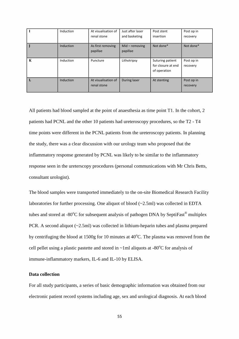
55
I Induction At visualisation of
renal stone
Just after laser
and basketing
Post stent
insertion
Post op in
recovery
J Induction As first removing
papillae
Mid – removing
papillae
Not done* Not done*
K Induction Puncture Lithotripsy Suturing patient
for closure at end
of operation
Post op in
recovery
L Induction At visualisation of
renal stone
During laser At stenting Post op in
recovery
All patients had blood sampled at the point of anaesthesia as time point T1. In the cohort, 2
patients had PCNL and the other 10 patients had ureteroscopy procedures, so the T2 - T4
time points were different in the PCNL patients from the ureteroscopy patients. In planning
the study, there was a clear discussion with our urology team who proposed that the
inflammatory response generated by PCNL was likely to be similar to the inflammatory
response seen in the ureterscopy procedures (personal communications with Mr Chris Betts,
consultant urologist).
The blood samples were transported immediately to the on-site Biomedical Research Facility
laboratories for further processing. One aliquot of blood (~2.5ml) was collected in EDTA
tubes and stored at -80oC for subsequent analysis of pathogen DNA by SeptiFast
® multiplex
PCR. A second aliquot (~2.5ml) was collected in lithium-heparin tubes and plasma prepared
by centrifuging the blood at 1500g for 10 minutes at 40oC. The plasma was removed from the
cell pellet using a plastic pastette and stored in ~1ml aliquots at -80oC for analysis of
immune-inflammatory markers, IL-6 and IL-10 by ELISA.
Data collection
For all study participants, a series of basic demographic information was obtained from our
electronic patient record systems including age, sex and urological diagnosis. At each blood

56
sampling time point, the following parameters were recorded: blood pressure, temperature,
pulse rate and clinical observations such as use of inotropic drugs and intravenous fluid
administration. Post operative, the early warning score (EWS) for each participant over a 48
hour period post procedure was recorded.
The EWS tool is used in Salford Royal Foundation Trust to assess the degree of illness of
patients. It is based on data derived from four physiological readings (systolic blood pressure,
heart rate, respiratory rate, body temperature) and one observation (level of consciousness,
AVPU). The resulting observations are compared to a normal range to generate a single
composite score with the higher the score the higher the level of medical care or risk of death.
This data was obtained by review of the electronic patient records (EPR) used in Salford
Royal Foundation trust. A subsequent review of the EPR system was done to see if any of the
study participants were readmitted within two weeks from the date of their urological
procedure. In addition, urine culture and stone debris results sent on the day of the
participant’s procedures were obtained and analysed. If no urine culture results from the day
of the procedure were obtained a retrospectively review for any recent mid-stream urine
results was performed.
Laboratory analyses
Analysis of pathogen DNA by SeptiFast® PCR assay
SeptiFast® is a CE - marked assay for detection of a panel of bacterial and fungal pathogens
in blood. The organisms covered by SeptiFast® represents > 95% of pathogens commonly
found in healthcare-associated blood stream infections (Table 6). The assay involves steps for
extraction of pathogen DNA and pathogen DNA analysis by real - time and was performed
according to the manufacturer's detailed protocol.

57
Table 6 - SeptiFast® Pathogen Detection Panel
Gram negative Gram positive Fungi
Escherichia coli
Klebsiella (pneumoniae/oxytoca)
Serratia marcescens
Enterobacter (cloacae/aerogenes)
Proteus mirabilis
Pseudomonas aeruginosa
Acinetobacter baumannii
Stenotrophomonas maltophila
Staphylococcus aureus
CoNS
Staphylococci *
Streptococcus pneumoniae
Streptococcus spp **
Enterococcus faecium
Enterococcus faecalis
Candida albicans
Candida tropicalis
Candida parapsilosis
Candida krusei
Candida glabrata
Aspergillus fumigatus
*S.epidermis, S. haemolytics ** S.pyogenes. S.agalactae, S.mitis
Pathogen DNA extraction from blood
In order to initiate DNA extraction, whole blood samples were mixed on a bottle roller for 30
minutes. 1.5ml of each whole blood specimen was placed in SeptiFast® Lys Kit MGRAD
tubes and subjected to mechanical lysis with ceramic beads in a MagNALyzer® instrument
(Roche Diagnostics GmbH, Mannheim, Germany) for70 sec at 7000 rpm and left to stand in
the MagNALyzer for 10 min.
SeptiFast® preparation M Grade kit (Roche Diagnostics GmbH, Mannheim, Germany) was
used to extract the DNA using the protocol provided by the manufacturers. 1ml of the lysed
specimen was vortexed with 150ul of proteinase K and an internal control (10ul; IC) and
1500ul of chaotropic lysis buffer (50% guanidinium, Tris-HCL buffer, thiocyanate and 20%
Triton X-100) were then added.
To promote the release of the DNA, this mixture was incubated for 15 min at 56°C with
gentle mixing at 500rpm. Following mixing with the binding buffer, the extract was

58
transferred onto spin columns designed to adsorb genomic DNA to the glass fibre matrix.
Unbound substances were removed by washing the spin columns sequentially with 1.8ml
inhibition removal buffer (containing 50% guanidinium HCL, 40% ethanol and Tris-HCl
buffer) and 1.6ml wash buffer (containing 0.2% sodium chloride, Tris-HCl buffer and 80%
ethanol). Finally, the adsorbed genomic DNA was eluted into1.5ml DNA-free reagent tubes
by incubation with 300µl of preheated (70°C) elution buffer (containing Tris-HCl) for 5 min
followed by centrifugation for 2 min at 4200xg. This extract contained pathogen DNA which
was then assayed by SeptiFast®.
SeptiFast® real-time PCR of pathogen DNA
Real-time PCR was carried out in 100µl glass capillary tubes using the LightCycler®2.0
instrument. Three capillary tubes were used per specimen for detection of Gram positive
bacteria, Gram negative bacteria and Fungi with appropriate controls. 50µl of ready-to-use
master mix (Roche Diagnostics) was added to 50µl of the target DNA.
The process of PCR amplification begins with a Pre-UNG incubation cycle at 40°C for 5
min. This cycle is carried out for the activation of Uracil-N-glycosylate that recognizes and
catalyses the destruction of DNA strands containing deoxyuridine, but not DNA
deoxythymidine. Since, PCR products contain uracil while genomic DNA contains
thymidine; this procedure reduces the risk of carry-over contamination in the assay.
PCR was initiated by a denaturation cycle at 95°C for 10 min followed by two amplification
programs (1-2) and a single melting curve profile (3).
1. 15 cycles of denaturation at 95°C for 15 sec, annealing at 58°C for 50 cycles and
extension at 72°C for 40 sec.
2. 30 cycles of denaturation at 95°C for 15 sec, annealing at 58°C for 50 sec and
extension at 72°C for 40 sec.

59
3. Denaturation at 95°C for 60 sec, annealing at 40°C for 60 sec and melting at 80°C at a
temperature change rate of 0.1°C/sec.
SeptiFast®- Identification of pathogen species and controls.
The measured fluorescence (emitted during the annealing phase when the fluorescent probe
hybridizes to the PCR amplicons) in four different detection channels was detected.
The assay is provided with specialised software (SeptiFast® Identification Software, SIS)
which facilitates automated identification of species and controls by analysing the melting
temperatures of the amplicons produced and a report for the pathogen status for each sample
is obtained. The analysis protocol adopted by SeptiFast® is based on the study by Lehmann et
al who elegantly showed that melting centre analysis permits rapid simultaneous detection of
25 different microorganisms using wide signal-to-signal melting peak differences (Lehmann
et al, 2008). The specific melting temperatures for the PCR products and probes derived from
the different organisms are shown in Figure 8 overleaf (Lehmann et al, 2008).

60
Figure 8 - Distribution of melting temperature and respective detection channels for all
microorganisms and internal control in the SeptiFast® assay (Lehmann et al, 2008)
Figure 8 – Distribution of melting temperature (Tm) and respective detection
channels for all microorganism and internal controls. A= Gram negative bacteria;
B= Gram positive bacteria; C=Fungi

61
Cytokine analysis
IL-6 ELISA
IL-6 was measured using a Pelikine compact human sandwich enzyme-linked
immunosorbent assay (ELISA) kit (Mast Diagnostics M1916). All solutions were made up
according to the manufacturer’s instructions.
A ninety six well plate was coated with the monoclonal antibody anti-human IL-6 coat
antibody diluted 1:100 in coating buffer and 100µl was added to each well. The covered plate
was incubated at room temperature (18-250) overnight. The plate was washed five times with
PBS using an automatic plate washer. 200µl of blocking buffer was added to each well and
the plate was incubated at room temperature for one hour. A standard curve was prepared as
per the specification sheet, and the plate was washed five times with wash buffer (PBS with
0.005% TWEEN 20).
The substrate blank wells were left empty and 100µl of standards and diluted samples were
added to the appropriate wells. The plate was re-covered and incubated at room temperature
for one hour, with continuous shaking at 700rpm. Biotinylated antibody was diluted 1:100 in
working-strength high performance ELISA dilution buffer (HPE). The plate was washed five
times in wash buffer, the substrate blank wells were left empty and 100µl diluted biotinylated
antibody was added to the remaining wells. The plate was re-covered and again incubated at
room temperature for one hour, whilst being shaken at 700rpm. The streptavidin-horseradish
peroxidise (HRP) conjugate was diluted 1:10 000 in working strength HPE dilution buffer.
The plate was washed a further five times in wash buffer, and 100µl streptavidin-HRP
conjugate was added to all wells except the substrate blank wells.

62
The covered plate was incubated at room temperature for 30 minutes, whilst being shaken at
700rpm. A suitable volume of TMB (Invitrogen 00-2023) was brought to room temperature
ensuring that it was not exposed to light. A stop solution (1.8M sulphuric acid) was also
prepared. The plate was re-washed five times and 100µl substrate was added to all wells. The
plate was incubated in the dark at room temperature for 30 minutes, whilst being shaken.
100µl of stop solution was added to all wells and the plate was read at 450nm using a Victor
multi-label counter (Wallac, Milton Keynes, UK). The cytokine concentrations were
determined using a four parameter curve fit algorithm (Delta Soft analysis software,
BioMetallics Inc, Princeton, NJ).
Figure 9 - The principle of sandwich ELISA (adapted from www.lenico.com)
IL-10 ELISA
Figure 9 –.An enzyme-linked immunosorbent assay (ELISA) is typically performed to
detect the presence and / or amount of a target protein

63
IL-10 ELISA
IL-10 was measured using a Pelikine compact human sandwich ELISA kit (Mast Diagnostics
M1910), following the same protocol as used for IL-6 (Fig 9). The sandwich ELISA process
initially starts with a plate being coated with capture antibody. Blocking buffer is added to
block remaining protein-binding sites on plate. A sample is added to plate and any antigen
present is bound by the capture antibody. A washing stage follows to remove any non-
binding antigen. The next step usually involves a labelling reagent which can be a labelled
detection antibody which when added to the plate binds to any antigen present and triggers an
enzymatic reaction to produce a detectable product for analysis.
Statistical analysis
In looking for an association between the detection of circulating pathogen DNA and
interleukin levels, I used a simple unpaired t test at each time point to compare those with
positive and negative pathogen DNA at each time point.

64
CHAPTER 3:
RESULTS
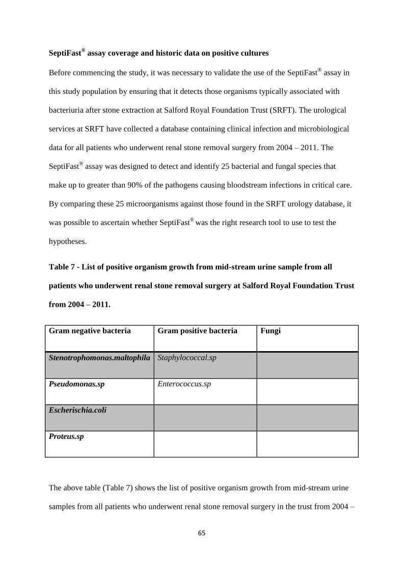
65
SeptiFast® assay coverage and historic data on positive cultures
Before commencing the study, it was necessary to validate the use of the SeptiFast® assay in
this study population by ensuring that it detects those organisms typically associated with
bacteriuria after stone extraction at Salford Royal Foundation Trust (SRFT). The urological
services at SRFT have collected a database containing clinical infection and microbiological
data for all patients who underwent renal stone removal surgery from 2004 – 2011. The
SeptiFast® assay was designed to detect and identify 25 bacterial and fungal species that
make up to greater than 90% of the pathogens causing bloodstream infections in critical care.
By comparing these 25 microorganisms against those found in the SRFT urology database, it
was possible to ascertain whether SeptiFast®
was the right research tool to use to test the
hypotheses.
Table 7 - List of positive organism growth from mid-stream urine sample from all
patients who underwent renal stone removal surgery at Salford Royal Foundation Trust
from 2004 – 2011.
Gram negative bacteria Gram positive bacteria Fungi
Stenotrophomonas.maltophila Staphylococcal.sp
Pseudomonas.sp Enterococcus.sp
Escherischia.coli
Proteus.sp
The above table (Table 7) shows the list of positive organism growth from mid-stream urine
samples from all patients who underwent renal stone removal surgery in the trust from 2004 –

66
2011. Comparing with the SeptiFast® Pathogen Detection Panel (Table 5), it can be seen that
the SeptiFast®
assay's coverage of organisms, has 100% overlap with the organisms detected
via mid-stream urine results from the historical data compiled by urology services of SRFT.
Study Population Twelve patients were consented for participation in the study. These 12 patients all
underwent a urological procedure between 27 September 2011 and 14 November 2011. Ten
patients (83%) underwent ureteroscopy with laser treatment to renal stones followed by
ureteric stent insertion and two of 12 patients underwent PCNL. The median age of the study
population was 54.5 years (interquartile range 24 - 61 yrs.).
The study population comprised of five male patients and seven female patients. For the
purpose of this proof of concept study, we assume that similar inflammatory reactions occur
with both described urological intervention as both procedures are using instrumentation in
removal of renal stones. The basic demographics data for the 12 patients included in the study
are shown in the following table (Table 8)

67
Table 8 - Patient demographic and operation detail
Patient identification Age Gender Operation
A 77 M Left ureteroscopy and
laser to ureteric stone,
basket extraction and
insertion of stent
B 20 F Left PCNL
C 59 M Left ureteroscopy and
laser to ureteric stone
extraction, insertion of
stent
D 62 M Right ureteroscopy and
laser to stone, insertion
of stent
E 61 F Left ureteroscopy and
laser to ureteric stone,
insertion of stent
F 50 M Left ureteroscopy and
laser to ureteric stone,
basket extraction of
fragments, insertion of
stent
G 61 F Right ureteroscopy,
removal of stent, laser
to stone, insertion of
stent
H 24 F Left ureteroscopy and
laser to ureteric stone,
basket extraction of
fragments, insertion of
stent
I 75 F Right ureteroscopy and
laser to stone, basket
extraction of fragments
and washout, insertion
of stent
J 25 F Left ureteroscopy and
laser to stone
K 20 F Left PCNL
L 32 M Left ureteroscopy,
removal of stent,
basket extraction of
fragments and laser to
stone, insertion of stent

68
Pathogen species identified by SeptiFast® assay and blood culture
Using the SeptiFast® assay, the detection of a PCR signal was either classified as positive
(signal present) or negative (signal absent). A positive blood culture result occurred in one
out of the 12 patients (9%). The organism detected was the yeast, Candida glabrata.
A total of six patients (50% of cohort) returned a positive signal for the presence of
circulating pathogen DNA. It can be argued from the data that the absence of PCR signal for
pathogen DNA is likely to be an indicator of the absence of infection. In the case of patient J,
there was a positive signal for circulating pathogen DNA and positive blood culture (Table
8). Post operatively, Patient J was transferred to a critical care setting for severe urosepsis.
Five out of the six patients with a positive signal from SeptiFast® assay had a detectable
signal at a single time point. One patient had a positive signal at anaesthetic induction
(Patient H, T1), another at the time of maximal instrumentation (Patient J, T3) whilst two
separate patients had positive signals in post recovery (Patients F & L, T5). Patient I was the
only patient to elicit positive DNA signals at two separate time points in the procedure (T3
and T4 respectively). These results are all highlighted in the next the table overleaf (Table 9).

69
Table 9 - Detection of pathogen DNA signal RT - PCR on blood samples at different
time points
Patient Frequency
of sampling
for PCR
analysis
detected (n)
Blood
Culture
positive
PCR
signal
T1
PCR
signal
T2
PCR
signal
T3
PCR
signal
T4
PCR
signal
T5
A 5 No Negative Negative Negative Negative Negative
B 5 No Negative Negative Negative Negative Negative
C 5 No Negative Negative Negative Negative Negative
D 5 No Negative Negative Negative Negative Negative
E 5 No Negative Negative Negative Negative Negative
F 5 No Negative Negative Negative Negative Positive
G 5 No Negative Negative Negative Negative Negative
H 5 No Positive Negative Negative Negative Negative
I 5 No Negative Negative Positive Positive Negative
J 3 Yes Negative Negative Positive Not
applicable
Not
applicable
K 5 No Negative Positive Negative Negative Negative
L 5 No Negative Negative Negative Negative Positive

70
PCR positive signal patients vs. PCR negative signal patients
The background characteristics between those where the SeptiFast® PCR assay detected
circulating DNA compared to those with a negative signal from the PCR assay was
compared. The PCR negative group were our control group in the study (Table 10).
Table 10 - Characteristic of PCR positive patients vs. PCR negative patients
Characteristic PCR signal detection
PCR positive PCR negative
Female : Male 4 : 2 3 : 3
Median age, range (yrs.) 28.5 years (20 - 75) 61 years, (20 - 77)
Percutaneous
nephrolithotomy ( PCNL)
1 /6 1/6
Vasopressor use in
procedure
1/6 2/6
Gentamicin administered
at T1
6/6a
6/6
Tazocin administered at T1 1/6b
1/6b
Augmentin administered at
T1
2/6
0/6
Mean LOS for readmission
directly related to
infectious complication of
procedures
7 days 4 days
Key:
a - Patient J received 300mg compared to 120mg given to the other 11 patients.
b - given to PCNL patients

71
There are more females than males in the PCR positive signal group compared to the PCR
negative group. The mean age in the positive signal group was 38 years compared to 57 years
in the negative signal group. The proportion of PCNL to ureteroscopy/ laser treatment/
ureteric stent insertion procedures was identical in both groups.
In both groups all patients received gentamicin. All patients received 120 mg gentamicin,
apart from patient J who received 300 mg gentamicin and was clinically unwell throughout
the procedure. In the two patients who had PCNL, each were also given two doses of
Piperacillin/ tazobactam 4.5 grams as prophylaxis. Piperacillin/ tazobactam is a combination
broad spectrum antibiotic containing the extended-spectrum penicillin antibiotic piperacillin
and the β-lactamase inhibitor tazobactam. In addition, two of those in the PCR positive group
were also given a dose of Augmentin 1.2g in the T1 stage. These results highlight that the
antibiotic strategies used in these procedures are not fixed and universal.

72
Relationship between pathogen isolate and clinical outcome
In the group with a positive signal detected on PCR, three patients were admitted with
infective episodes within 30 days of their initial procedures (table 11),
Table 11 - Correlation between PCR positivity and clinical outcome
Patient Organism detected
by blood culture
Organism detected
by SeptiFast®
analysis
Post - operative
event
F Nil detected Streptococcal sp
S.pneumoniae
Had subsequent
EWSL. 2 weeks later
stent removal then
Readmitted next day
with clinical episode
of urosepsis - within
30 days of initial
procedure
H Not performed K.pneumoniae /
oxytoca
No clinical episodes
of urosepsis
I Nil detected E. Faecalis No clinical episodes
of urosepsis
J Candida glabrata Candida glabrata Clinical episode of
urosepsis – stent
removal, post op
admission
K Nil detected E.coli Subsequent
admission with
pyelonephritis –
required readmission
L Nil detected S.aureus No clinical episodes
of urosepsis
In the PCR positive group, Patient F was readmitted with urosepsis. After the patient's initial
interventional procedure, the patient underwent a non-invasive urological procedure for stone
removal, Extracorporeal Shock Wave Lithotripsy (EWSL) and subsequently had the stent
removed. The day after removal of stent the patient was admitted with urosepsis.

73
Urine samples taken at the time of the EWSL and on admission with urosepsis showed only
pyuria. If the urine culture had grown streptococcal sp and/or S.pneumoniae, it would have
provided evidence that the interventional procedure may have directly contributed to the
episode of urosepsis.
Within the PCR positive group, patient K was admitted with a clinical diagnosis of
pyelonephritis directly related to the urological intervention. This patient was readmitted 12
days post procedure. The patient had a positive PCR signal for E.coli but on readmission had
no growth in blood culture. Urine cultures from readmission episodes were positive for skin
flora and pyuria.
The third patient, patient J, had a clinical diagnosis of urosepsis. The patient developed septic
shock post procedure and required admission to intensive care unit. In this patient, Candida
glabrata was detected on both SeptiFast® assay and blood cultures. Regarding the length of
stay seen, the total extra in-patient bed days attributed to infection in the PCR positive group
was 14 days with the mean stay in the two patients with an infection episode being seven
days.
In the PCR negative group (table 12 overleaf), one patient was re-admitted for urosepsis 12
days post procedure. Urine was sent from two separate sites, a urostomy sample and a sample
from the patient's urinary catheter. E.coli was grown from a sample sent from urostomy
whilst the urinary catheter sample showed pyuria only. In the PCR negative group, the length
of stay in the only patient with a clinical episode of infection was four days. These results
suggest that PCR positive patients have a poorer clinical outcome regarding increased
frequency of infections and longer length of stay.

74
Table 12 - Correlation between PCR negativity and clinical outcome
Patient Organism detected
by blood culture
Organism detected
by SeptiFast®
analysis
Post - operative
event
A Nil detected Nil detected No clinical episode
of urosepsis
B Nil detected Nil detected Clinical episode of
urosepsis - antibiotics
and subsequent
repeat PCNL.
C Nil detected Nil detected No clinical episode
of urosepsis
D Nil detected Nil detected No clinical episode
of urosepsis
E Nil detected Nil detected No clinical episode
of urosepsis
G Nil detected Nil detected No clinical episode
of urosepsis
Analysis of circulating IL-6 and IL-10 levels
Immunochemical analysis was performed to establish if there was any detectable relationship
between the presence of pathogen DNA and the level of plasma IL-6 and IL-10 in patients
undergoing these interventional urological procedures. Plasma IL - 6 and plasma IL - 10
levels were measured in the T1 - T5 samples and direct comparisons were made between the
PCR positive group and PCR negative group (Fig 10).

75
Figure 10 - Mean IL – 6 and IL-10 in PCR positive and PCR negative patients
The mean level of IL - 6 in PCR - positive group was higher by a factor of five at all time
points compared to the level found in PCR - negative group. This effect was statistically
significant (p<0.05) at all time points except for T5 (Figure 12) A different pattern was
observed with IL-10 (Figure 12). There was no statistic difference (p > 0.05) between the
Figure 10 - This figure clearly shows that at each time point the detected level of IL – 6 is
higher in those with a positive signal compared to those where no signal was detected.
This pattern was not seen with IL – 10.
76
mean plasma levels of IL - 10 between the PCR - positive and PCR - negative group at any
time points, although there was some qualitative evidence of higher levels at T3 and T4 in the
PCR - positive groups.
From these results, there seems to be an association between the detection of circulating
pathogen DNA and elevated pro inflammatory cytokine levels. The results demonstrate a
likely link between the presence of circulating pathogen DNA and the host immune response
in this study population.
Pathogen detection and physiological response
All patients had bedside observations and this helped to assess if there was an association
between the peri procedure observations such as temperature, pulse and blood pressure
(markers of physiological response) and PCR positivity. Blood pressure was expressed as the
mean arterial blood pressure (MAP). The MAP is a measure of the perfusion pressure to vital
organs of the body.
MAP is derived from the formula:
Mean arterial pressure = diastolic blood pressure + 0.33 (systolic blood pressure -
diastolic blood pressure) in mmHg
The measured pulse rate is a measure of the heart's response during the procedures and is
expressed in beats per minute. There was no clear pattern in the physiological responses seen
in the PCR positive or PCR negative group. There seems to be no clear difference in the
witnessed variation in the MAP across the time points when comparing those who were PCR
positive with those who were PCR negative (Fig 11 and 12). When the mean MAP across
each study group is compared at all time points, there was no clear difference seen (Fig 13).

77
Figure 11 - Variations of mean arterial blood pressures in PCR negative group
Figure 12 - Variation of mean arterial blood pressure in PCR positive group
0
20
40
60
80
100
120
140
T1 T2 T3 T4 T5
MA
P (
mm
Hg)
Time points
Mean arterial blood pressure (MAP) in PCR negative patients
Patient A
Patient B
Patient C
Patient D
Patient E
Pattient G
0
20
40
60
80
100
120
T1 T2 T3 T4 T5
MA
P (
mm
Hg)
Time points
Mean arterial blood pressures variations in PCR - positive patients
Patient F
Patient H
Patient I
Patient J
Patient K
Patient L
Figure 11 – In the 6 patients who were PCR signal negative, there is no clear variation in MAP
with the time points.
Figure 12 – The PCR signal positive patients have differing MAPs across the different time
points with no clear pattern of change across the timepoints for each patient.

78
Figure 13 - Comparison of the variation of mean MAP between PCR positive patient
group and PCR negative patient group
The cardiovascular system response can be assessed by both MAP and heart rates. In the PCR
negative group, there was a generalised high starting pulse rate at T1 amongst the PCR
negative patients with a lower pulse rate recorded at T5 in these patients but not to a point of
bradycardia (pulse rate below 60 beats per minute).
0
10
20
30
40
50
60
70
80
90
T1 T2 T3 T4 T5
MA
P (
mm
Hg)
Time point
Mean MAP between PCR positive vs PCR negative patients
PCR negative
PCR positive
Figure 13 – The mean MAP at each time point showed no difference between patients with a
positive signal and those patients with no signal detected.

79
Figure 14 - Variations in pulse rate in PCR negative group
In the PCR positive group, 3/6 patients exhibited a rise in their pulse rate with two patients
exhibiting tachycardia (pulse rate higher than 100 beats per minute). However, there was no
uniform or observed pattern to the change in pulse rate for either the PCR positive or PCR
negative patients (Fig 16). This is highlighted when, from these results, there is no marked
difference between the blood pressure and pulse responses seen between the PCR positive
and the PCR negative group through the procedures.
0
20
40
60
80
100
120
140
T1 T2 T3 T4 T5
Pu
lse
rta
te (
be
ats
pe
r m
inu
te)
Time points
Pulse rate variations in the PCR negative group
Patient A
Patient B
Patient C
Patient D
Patient E
Patient G
Figure14 – There was no consistent pulse variations in those who do not have DNA detected
by PCR across the 5 time points.

80
Figure 15 - Variations in pulse rate in PCR positive groups
Figure 16 - A comparison of the variation between mean pulse rate between PCR
negative patients and PCR positive patients
0
20
40
60
80
100
120
140
T1 T2 T3 T4 T5
Pu
lse
rat
e(b
eat
s p
er
min
ute
)e
Time points
Pulse rate variations in the PCR positive patients
Patient F
Patient H
Patient I
Patient J
Patient K
Patient L
0
20
40
60
80
100
120
T1 T2 T3 T4 T5
Pu
lse
rat
e (
bp
m)e
Timepoints
Mean pulse variation between PCR negative vs PCR positive
PCR negative
PCR positive
Figure 15 – In the 6 patients who had a PCR positive signal, there was no observed pattern
across the patients in the response of their pulse rates across the different time points.
Figure 16 – This figure shows that there is no difference between the mean pulse rates at
different time points between the two groups

81
Case specific findings - PCR positive patients
Case J had a positive blood culture and positive DNA signal at time point T3 and
subsequently had a significant rise in temperature and pulse rate in recovery room post
procedure. The patient became more unwell and deteriorated leading to sample at T4 and T5
not being taken. This patient progressed rapidly from a SIRS response to septic shock, and
required admission to intensive care for organ support. Case J needed aggressive fluid
resuscitation throughout the procedure and had a total length of stay in hospital of eight days.
Case F had Streptococcal sp and S pneumoniae detected with the SeptiFast®
assay. This
patient experienced a steady drop in blood pressure across the five time points associated
with a similar drop in pulse rate as well but no recorded bradycardia (pulse rate less than 60
beats per minute). Case H had a positive signal for K.pneumoniae /oxytoca by the SeptiFast®
assay. Case F was administered metaraminol at T1 stage. It is a well-known phenomenon for
blood pressure to be low due to the anaesthetics. Metaraminol is an alpha 1 adrenergic
receptor agonist and is used by anaesthetists for this clinical indication.
Case specific findings - PCR negative patients
Patient A was hemodynamically unstable during the procedure. Patient A had a tachycardia
(120 beats per minute) and a reduced MAP (56.7). At each of the first four time points, the
patient received vasopressor therapy - ephedrine (T1 and T3) and metaraminol (T2 and T4).
This patient, however, had no post operative complications and did not require hospital
admission. The patient was both PCR and blood culture negative. This hemodynamic
instability was not associated with an inflammatory response to a pathogen.
Patient B had a clinical episode of urosepsis and was readmitted for IV antibiotics treatment
12 days after the procedure. The patient had six units of blood loss through the procedure and
required four litres of intravenous fluids. No vasopressor medication was administered during
the procedure and the patient’s total length of stay on readmission was four days.

82
Stone composition and pathogen detection
There is evidence in the literature that stones of certain compositions, such as struvite stones
are associated with pathogens such as Klebsiella and Proteus. (Tseng et al, 2011)
These urease producing bacteria are well-recognised predisposing factors for stone formation
(Tseng et al 2011). In our study there was a clear association between the detection of
circulating pathogen DNA and the presence of calcium oxalate stones (Table 13). There was
no clear evidence of any growth from the stone particles sent for analysis (Table 13). With
the study population being so small, care needs to be taken from drawing conclusions and
larger study would further prove if there is an association between stone composition and
pathogen detection.
Table 13 - Stone composition and organism detection
Patient Stone composition
(percentage)
Record of
culture from
stone
Organism
detected by
blood culture
Organism detected
by SeptiFast®
analysis
A Uric acid (99%) No Nil detected Nil detected
B Calcium phosphate
(47%) Magnesium
ammonium phosphate
(53%)
No Nil detected Nil detected
D Calcium oxalate
(98%)
No Nil detected Nil detected
E Calcium oxalate
monohydrate (96%)
No Nil detected Nil detected
F Calcium oxalate
(98%)
No Nil detected Streptococcal sp
S.pneumoniae
I Calcium oxalate
(97%)
No Nil detected E. Faecalis
L Calcium oxalate
(21%) Urate (79%)
No Nil detected S.aureus

83
CHAPTER 4
DISCUSSION

84
Background of the study
This is the first proof of concept study to propose that urological procedures involving
instrumentation could be a reproducible model to study pathogenaemia in man. If there was a
better understanding of the role pathogens play in triggering the host immune response, we
would be able to further our knowledge on the pathogenesis of infection and sepsis. Sepsis is
clinically defined when a patient shows at least two of the criteria for systemic inflammatory
response (SIRS) with evidence of systemic infection (Mancini et al, 2010).
The systemic inflammatory response (SIRS) is activated in patients in response to either a
non-infectious (e.g. tissue injury. trauma) or an infectious aetiology; however, it is not
uncommon for both forms of aetiology to co-exist. The difficulty from a diagnostic point of
view is determining when the inflammatory response is caused by infection (i.e. sepsis).
Blood cultures are the current gold standard for microbial detection in the blood stream.
Blood culture analysis is often unreliable, particularly where patients have received
antimicrobial therapy. Recent advances in molecular diagnostics allow the measurement of
DNA from pathogens in clinical samples (i.e. blood). These approaches are less likely to be
affected by antibiotic use and may have great benefit in the diagnostic process.
Currently, the relationship between circulating pathogen DNA, the presence of infection and
the impact on the systemic inflammatory response is not fully understood. There have been a
number of both animal and human models of sepsis described in the literature. In this thesis, I
propose that an easily reproducible model for exploring the relationship between circulating
pathogen DNA and the subsequent inflammatory response could be urological procedures
involving instrumentation.
It was initially thought that, endourological surgery were considered clean contaminated
procedures and did not require antibiotic coverage. A clean-contaminated surgical site is seen

85
when the operative procedure enters into a colonized viscus or cavity of the body, but under
elective and controlled circumstances. However, evidence in the literature dating back
decades shows that urological instrumentation is associated with increased incidence of
urinary tract infection and bacteraemia (Alsaywid et al, 2013).
Infections and urosepsis is one of the most common complications seen in urological
procedures. The rate of infection complication in all urological procedures in the literature is
as high as 12 - 14 % in the literature (Bjerklund et al 2007). Following ureteroscopy, the
reported incidences of UTI range between 3.9% and 25%. (Rao et al, 1991, Puppo et al, 1991,
Hendrickx et al, 1999).
A number of studies have previously highlighted the significant inflammatory response in
patients undergoing endourological manipulations for urinary stones. A frequently referenced
study on this was published by Rao et al in 1991 (Rao et al, 1991). In an effort to predict
septicaemia following endourological manipulation for stones in the upper urinary tract, 117
patients were studied and classified according to the procedure performed ( Rao et al, 1991).
In this study, fever was used as a proxy for infection and 74% of the PCNL patients in their
study developed post-operative fever; however, only 41% had endotoxemia. The only patient
in the Rao's study to develop septic shock had a PCNL.
Antibiotic prophylaxis is commonly used perioperative in urological surgery and procedures
to alleviate the occurrence of infection. Antibiotic prophylaxis aims to prevent healthcare-
associated infections that result from diagnostic and therapeutic procedures (Grabe et al,
2012). I have highlighted the prevalence and incidence of infection and sepsis in those
undergoing endourological procedures however regarding evidence of countering infection
with antibiotic prophylaxis, there is no clear-cut evidence that exists on the use of antibiotic
prophylaxis in this population (Knopf et al, 2003, Fourcade et al, 1990).

86
In this study, ureteroscopy and PCNL are the two urological interventions which may be
useful in vivo models for investigating pathogenaemia in man. If this model shows that there
is a relationship the presence of a detectable level of circulating pathogen DNA and a
witnessed immune and physiological response, this would suggest a possible beneficial need
for the use of prophylactic antibiotics . In this study, there was evidence of a relationship
between the levels of cytokines particularly IL - 6 and the presence of circulating pathogen
DNA , however in this study the only patient to develop severe sepsis (to prompt an
admission to ICU) did not receive antimicrobial to cover the causative organism i.e. fungi. As
a result of our study I therefore propose that a randomised controlled trial on antimicrobial
prophylaxis in this population is warranted.
Current international guidelines state that it is important to distinguish urological
interventions into low-risk procedures (e.g. simple diagnostic and distal stone treatment) and
higher-risk procedures (e.g. treatment of proximal impacted stones and intrarenal
interventions) when considering prophylactic antibiotics (Grabe et al, 2012). In higher-risk
procedures, there would be more of an indication to use antibiotic prophylaxis.
In this study, all patients received some form of antibiotic prophylaxis; however, the
antibiotic regimen was variable 25% of the study population (3/12 patients) went on to
develop an infective episode despite the use of prophylactic antibiotics. However, it is hard to
provide strong arguments against prophylactic antibiotics in a study of this size with
difference in the choice of antibiotics, doses and time of administration of antibiotics.
In a recent worldwide study, (Gutierrez et al, 2013) 5803 consecutive PCNL patients were
enrolled from 96 centres over 12 months to participate in the PCNL Global Study. The
presence of a post-operative fever of ≥38.5°C was used as a proxy for infection in this study

87
and the main finding was that a10% of PCNL-treated patients developed fever in the post-
operative period despite receiving antibiotic prophylaxis.
In this proof of concept study, my aim was:-
1. To appraise the presence and significance of blood borne microorganisms that may
appear in patients during interventional urological surgery involving urinary tract
instrumentation by (blood culture and by multiplex real time polymerase chain
reaction assay (SeptiFast® Roche).
2. To appraise the host immune response and physiological response in the study
population to the presence of circulating pathogen DNA and if this provides evidence
for the use of antibiotics prophylactically in these patients.
On-going research using PCR technology in an in vivo human model of pathogenaemia could
help determine if PCR technology has a role in the detection of circulating pathogen DNA in
infective and septic patients. PCR technology could supersede the current gold standard of
pathogen detection in blood in clinical settings and provide rapid detection of pathogen
regardless of whether antibiotics have been administered.
Key findings of the study
The key findings from the study are that the SeptiFast® assay had broad pathogen coverage
for this study population. In the group of patients with circulating pathogen DNA detected by
SeptiFast®, there was a clear immune response elicited (elevated cytokine levels) but no
witnessed physiological response in this group, when compared with patients with no
detectable circulating pathogen DNA.

88
SeptiFast® - Role in circulating pathogen DNA detection in infection ?
The SeptiFast® assay was chosen as the assay to test the study hypotheses. The SeptiFast
®
assay has been available longer than any other molecular-based tests. SeptiFast® uses real-
time PCR in a non-quantitative mode to identify ten bacteria at the species level, several more
at the genus level, as well as five Candida species and Aspergillus fumigates. This assay
reportedly identifies the 25 organisms that account for more than 90% of the culturable
pathogens associated with sepsis (Ecker et al, 2010).
Prior to commencing this study, a review of the collected historical data on urological
procedures as Salford Royal Foundation Trust urology services show that the SeptiFast®
assay has complete coverage of all the pathogens present in urine cultures commonly seen in
the urology patients treated at Salford Royal. This confirms the suitability of this potential
diagnostic tool in this study population. Notably, the SeptiFast® assay did not detect any
circulating pathogen DNA from a species not normally seen in this clinical setting.
In our study, 1 patient (9%) had a positive growth from blood culture and the organism
detected was Candida glabrata. This figure is well within the range for rate of positive
cultures associated with urological surgery in the literature (Knopf et al, 2003, Dogan et al,
2002). This patient was admitted to intensive care for organ support due to urosepsis. In a
number of papers looking specifically at those with urosepsis after endourological
intervention for stones, blood cultures tend not to be positive and sepsis rates are 0.25 - 1.5 %
(Demirtas et al, 2012, Dogan et al, 2002). Interestingly, with studies looking specifically at
PCNL, between 0.3 and 9.3% of patients can develop potentially life-threatening sepsis
(Michel et al, 2007, Draga et al, 2009). In this proof of concept study, we have assumed that
PCNL and ureteroscopy produce similar results and further work would need to be done to
establish if this assumption is valid.

89
In our thesis, there is a low level of blood culture positivity compared to PCR positivity from
the SeptiFast® assay and there are a number of possible explanations for this observation. A
proposed reason for this finding could relate to the administration of antibiotics pre procedure
which can affect the detection of pathogen by blood cultures. In our study, out of the four
patients in the study who had infective and / or septic episodes post procedure, 50% were
PCR positive blood culture negative, 25% were PCR positive blood culture positive. Blood
cultures are reported to be negative in more than 50% of the cases where true bacterial or
fungal sepsis is believed to exist (Ecker et al, 2010). A theoretical explanation between the
difference in circulating pathogen DNA detection (by SeptiFast®
) and intact pathogen
detection (by blood cultures) could relate directly to the urological procedure. In these
endourological procedures, the use of instrumentation could lead to the introduction of
fragments of pathogen DNA particles into the blood stream rather than intact pathogen.
SeptiFast® - A place in clinical practice?
There are no described studies in the literature on the use of the SeptiFast® assay in the
setting of urological surgery. There have been a number of studies looking at the use of
SeptiFast®
assay in other clinical settings. In a recent Danish study (Westh et al, 2009)
SeptiFast® was compared directly against blood cultures in patients with suspected bacterial
and fungal sepsis. The aim of the study was to observe if non viable pathogen detection by
SeptiFast® would aid in the clinical decision making process. The group postulated that one
advantage of PCR assays over blood cultures is that DNA based detection systems can detect
pathogens causing sepsis at the time of sampling if they are viable or non viable.
558 samples from 359 patients were evaluated. The rate of positivity was 17% from blood
cultures (BC) and 26% from SeptiFast®
assay. 96 microorganisms were isolated with BC, and
186 microorganisms were identified with SeptiFast®; 231 microorganisms were found by
combining the two tests. Of the 96 isolates identified with blood culture, 22 isolates were

90
considered to be contaminants. Of the remaining 74 non-contaminant BC isolates available
for comparison with SeptiFast®, 50 were identified as a species identical to the species
identified with SeptiFast®
in the paired sample. Of the remaining 24 BC isolates for which
the species, identified in the BC, could not be detected in the paired Septifast®
sample. 18 BC
isolates were identified as a species included in the SeptiFast®
masterlist, and six blood
culture isolate were not organism detected by SeptiFast®
. With SeptiFast®, 186
microorganisms were identified, 12 of which were considered to be contaminants. Of the 174
clinically relevant microorganisms identified with Septifast®, 50 (29%) were detected by BC.
More than half of the remaining microorganisms identified with SeptiFast® (but not isolated
after BC) were also found in routine cultures of other relevant samples taken from the
patients.
Dierkes et al (Dierkes et al, 2009) performed a retrospective analysis of PCR results on the
clinical management of patients with presumed sepsis. Of the 101 blood samples from 77
patients with presumed sepsis, 39 samples had pathogens identified from either the use of
blood cultures or the SeptiFast® assay. From the samples, 63 (62%) yielded concordant
negative results, 14 (13 %) concordant positive and 9 (9%) were blood culture positive only.
In 14 (13%) samples pathogen was detected only by PCR assay, resulting in adjustment to
therapy in five patients. In three samples a treatment adjustment would have been made
earlier resulting in a total of 8 adjustment in all 101 samples (8%). In this single centre study
with small number, the group have suggested that rapid molecular diagnostic tests may lead
to a higher rate of early adequate antibiotic therapy in approximately 8% of patients with
suspected sepsis. This could have had a potential therapeutic impact on clinical outcomes.
The SeptiFast® assay ability for pathogen detection has also been used in onco-
haematological patients (Mancini et al, 2008). These patients are susceptible to neutropenia.
The term 'neutropenia' describes low levels of neutrophils in the blood. Neutrophils are

91
important in fighting bacterial infections. Among the 103 blood samples, 35 (34 %) samples
were positive by at least one of the two methods. Twenty-one (20.4 %) samples were positive
by blood culture and 34 (33 %) with SeptiFast®. The analysis of concordance evidenced a low
correlation between the two approaches. When comparing the six hour turnaround time for
the SeptiFast® assay with blood cultures, even in the two observed cases of fast-growing
Escherichia coli, a mean of 10 h of incubation was needed, with definitive identification after
an additional 36 h (Mancini et al, 2008). As evident from these three separate studies, there is
potential in the utilisation of the SeptiFast® assay in the management of infection. In this
thesis, one of our main hypotheses was to ascertain if the detection of circulating DNA by the
SeptiFast® assay was an indicator of infection.
In this study, two out of the three patients who went on to have an infective or septic episode
only had an organism detected by the SeptiFast® assay alone and not by blood cultures. Only
one patient who was PCR signal and blood culture negative, went on to have a clinical
episode of urosepsis. Half of the patients in the study with a positive signal for an organism
detected by the SeptiFast® assay did not go on to develop an infective or septic episode. In
this small study, there was a low level of blood culture positivity and I was unable to do a
direct assessment of concordance between PCR assay and blood cultures as diagnostic tools.
In the study, the one patient who had a fulminant fungal septicaemia gave a positive signal
via the SeptiFast® assay and had a positive BC.
In this study, the presence of detectable DNA seem to stimulate an inflammatory response.
The clinical relevance of this is that potentially we have a technology which will greatly
speed up the identification of pathogenic organisms and faciltate the prompt and targeted
treatment in patients with sepsis and infection.

92
Using blood culture as a reference ‘gold standard’ to compare molecular methods has been
shown to be problematic in the literature. It is well recognised that blood culture fails to
identify more than 50% of the cases of sepsis believed to be caused by bacteria or fungi based
on clinical and other criteria (Ecker et al, 2010). In most studies, SeptiFast® consistently
identified more positive specimens than blood culture methods (Pletz et al, 2011) and this
was seen in this study. An obvious limitation of the SeptiFast® as a diagnostic tool is that the
assay does not provide information about antimicrobial susceptibilities of the organisms
detected.
In this study, half of the cases with positive PCR signal can be classed as 'false' positive
results. The definition of PCR ‘false’ positives is those cases in which the presence of
pathogen DNA is associated with no growth in simultaneous blood cultures. In this study, our
controls were those who were negative for DNA signal. The lack of normal control group
makes it difficult to assess the SeptiFast®
assay with respect to false positive outcomes. There
has however been a recent study (Warhurst et al, 2015) which has set out to determine the
accuracy of SeptiFast®
real-time PCR for the detection of health care associated BSI against
standard microbiological culture.
Of 1006 new patient episodes of systemic inflammation in 853 patients, 922 (92%) met the
inclusion criteria and provided sufficient information for analysis. Adult patients had been
exposed to a median of 8 days (interquartile range 4–16 days) of hospital care, had high
levels of organ support activities and recent antibiotic exposure. SeptiFast®
real-time PCR,
when compared with culture-proven bloodstream infection at species/genus level, had better
specificity (85.8%, 95% CI 83.3% to 88.1%) than sensitivity (50%, 95% CI 39.1% to 60.8%).
When compared with pooled diagnostic metrics derived from their systematic review, their
clinical study revealed lower test accuracy of SeptiFast® real-time PCR, mainly as a result of
low diagnostic sensitivity. There was a low prevalence of BC-proven pathogens in these

93
patients (9.2%, 95% CI 7.4% to 11.2%) such that the post-test probabilities of both a positive
(26.3%, 95% CI 19.8% to 33.7%) and a negative SeptiFast® test (5.6%, 95% CI 4.1% to
7.4%) indicate the potential limitations of this technology in the diagnosis of bloodstream
infection. Using this analysis approach, the sensitivity of the SeptiFast® test was low but also
appeared significantly better than BC. Blood samples identified as positive by either culture
or SeptiFast® real-time PCR were associated with a high probability (> 95%) of infection,
indicating higher diagnostic rule-in utility than was apparent using conventional analyses of
diagnostic accuracy. In my study, all of the urology patients were subject to sterile injury, had
evidence of systemic inflammation and had prophylactic antibiotics administered so the data
from the aforementioned study (Warhurst et al, 2015) data could be applied to our population.
The presence of false positive patients poses more questions than answers regarding the
detecting of circulating DNA by the SeptiFast® assay as a potential indicator of infection.
Avolio et al (Avolio et al, 2010) compared traditional blood cultures with SeptiFast® in cases
suspected to have blood borne infection. There were 10 patients who had an organism
detected by SeptiFast® but not by blood culture that subsequently had microbiology
confirmation from other sites. It would be important, therefore, to differentiate between DNA
associated with intact pathogens from free pathogen DNA detected by PCR, if pathogen
DNAemia is to be a marker of infection. PCR analysis may be a sensitive method of
detecting pathogen DNA released in to circulation at levels which would:
1. Not be detected by sampling volumes obtained for blood cultures;
2. From sites with restricted or poor blood flow.
Importantly, PCR assays are not affected by antimicrobials given (Ecker et al, 2010, Mancini
et al, 2010).

94
A separate issue raised by false positive cases is that, presently, we cannot ascertain through
the assays if the circulating pathogen DNA detected is associated with viable bacteria rather
than non-viable bacteria destroyed by host immune system +/- antimicrobial therapy. So we
cannot, in this study, definitely say that the detected circulating pathogen in the case which
was PCR signal positive and blood culture negative was acting as a pathogen.
In the study amongst all the patients who were PCR signal positive, a positive signal for
pathogen DNA was detected at all time points. However, it was often noted that it was only
present transiently in each of the six PCR positive cases. There are a number of likely
possibilities for these findings. Firstly, there is the possibility that pathogen DNA is only
found fleetingly in circulation. A second viable explanation is that circulating pathogen DNA
levels may be low or close to limits of detection by the SeptiFast® assay so that in some of
the cases DNA may not have been detected even though present. Thirdly, sampling may be
an issue and a larger study population may help to explain this.
SeptiFast® consistently identified more positive specimens than blood culture methods;
however, the seminal paper on SeptiFast® (Lehmann et al, 2008) showed that culture
consistently identified some organisms that were not identified by SeptiFast®. There are a
number of possible explanations such as the larger volume of blood analysed by culture and
the lower limit of SeptiFast® detection of approximately 3–30 CFU/ml. Ideally, we would
want to use as sensitive a PCR assay as possible and, presently, the most sensitive is the
SeptiFast® assay.
Circulating pathogen DNA and the immune response
In this study, there was a strong correlation between the detection of circulating pathogen
DNA and increased level of IL - 6. These results suggest that in these urology patients there
is a positive association between the presence of circulating pathogen DNA and the witnessed

95
immune response. However, there was no direct association between circulating DNA and
the physiological response in these patients.
It has been shown that bacterial - derived short DNA fragments are able to stimulate immune
cells to promote the release of IL-6 from human mono nuclear cells (Schindler et al, 2004). In
this study, we have shown an association between the presence of circulating pathogen DNA
and increased IL- 6 detected levels during urological procedures in this study population. In
our study, cytokine levels were reviewed over the period of the procedure and in post
operation (Mokart et al, 2002). In this study, serial blood samples were collected from 30
consecutive patients for determination of serum cytokine levels. Healthy volunteers were
used as the control group. Eleven patients developed no complications (group 1), 14
developed sepsis or severe sepsis (group 2), and five developed septic shock (group 3).
After operation IL-6 levels in group 1 were increased in comparison with day 0 but
normalized with time, suggesting that surgical trauma induced the immediate and temporary
postoperative increase in IL-6 concentration. However, on day 1, patients in groups 2 and 3
had IL-6 levels that were respectively, two and six times higher than those in group 1
patients. In addition, they remained high during the course of the study. These high levels of
IL-6 were independently associated with postoperative septic events and correlated with post-
operative morbidity (length of ICU stay and duration of mechanical ventilation). As septic
events always occurred after day 2 the group it suggested that, IL-6 is probably an early
indicator of post-operative infection following the trauma of major oncological surgery
(Mokart et al, 2002). It would be interesting to use our study population to confirm these
findings.
The findings in this thesis suggest that the detectable presence of pathogen DNA could
stimulate an inflammatory response. In the literature, there have been advances in our

96
knowledge on how the cells of our immune system detect microbial pathogens. The ability of
the host's immune system to sense nucleic acid is one such mechanism which has been
extensively studied. For detecting microbial DNA, toll - like receptor 6 (TLR6) in endosomes
and numerous cytoplasmic DNA binding proteins have been discovered (Holm et al, 2013).
Toll-like receptors (TLRs) are a class of pattern recognition molecules that play a unique
function in the innate immune system. This system is the first line of defence against
microorganisms that initiate cellular signal in response to pathogen-associated molecular
patterns (PAMPs) and induce expression of genes involved in the inflammatory process,
therefore, it plays a crucial role initiating and directing the adaptive immune system
(Jahantigh et al, 2013).
Ten functional TLR members (TLR1–TLR13) have been identified in humans. TLR9, an
endosomal localized receptor on B cells, plasmacytoid dendritic cells (pDCs), and
monocytes/ macrophages, recognizes unmethylated nucleic acid motifs, especially Cytosine-
phosphate-Guanine motifs, in bacterial DNA and it is one of the most important receptors in
the initiation of protective immunity against intracellular pathogens by activation signalling
cascade of intracellular receptor signalling (Jahantigh et al, 2013). From our study, we
detected similar DNA levels in the patients with a positive signal from the SeptiFast® study;
however, we do not currently know whether this level of pathogen DNA could activate the
TLR9.
The urology patient: Host response, sepsis and antibiotic prophylaxis
This study is not the first to look at the host inflammatory response and sepsis in urology
patients. Rao et al’s (Rao et al 1991) study is one of the most cited studies in urology that
looked directly at systemic inflammatory responses and sepsis in patients undergoing

97
urological manipulation. In the individuals in this study who had PCNL, despite use of a
prophylactic antibiotic, postoperative bacteraemia and fever are reported as 37% and 74%.
Antibiotic prophylaxis aims to prevent healthcare associated infections that result from
diagnostic and therapeutic procedures. Antibiotic prophylaxis is only one of several measures
to prevent infections and can never compensate for poor hygiene and operative technique.
The principle of antibiotic prophylaxis is to provide protection to the patient with creating an
environment to promote antibiotic resistance (Grabe et al, 2012). Unfortunately, the benefit
of antibiotic prophylaxis for most modern urological procedures has not yet been established
by well-designed intervention studies (Grabe et al, 2012). Antibiotic prophylaxis in urology
has been controversial for many years. In the literature regarding PCNL case studies, there is
conflicting evidence for the role of prophylactic antibiotics s evidence in PCNL procedures.
Charton et al (Charton et al, 1986) reviewed 126 cases of percutaneous extraction of renal
stones by PCNL that no major septic complication was observed without prophylaxis. Only
10% of the patients were exposed to fever and 35% had bacteriuria. Mariappan et al
(Mariappan et al, 2006) presented results showing that treating patients who have dilated
pelvicalyceal systems and / or stones of ≥ 20 mm before PCNL with ciprofloxacin 250 mg
twice daily for 1 week significantly reduces the risk of upper UTI and urosepsis. Eighteen of
46 (39%) patients in the control group developed SIRS, whereas only 7 of 52 (13.4%)
patients in the treatment group developed SIRS. Mariappan's group reported that one-week
administration of ciprofloxacin prophylaxis decreased positivity of pelvicalyceal culture by
three times, stone culture positivity by two times, and risk of developing SIRS by three
times.
In this study, circulating pathogen DNA is definitely seen to stimulate a significant
inflammatory response in this urology population. Within the study, a quarter of our patients

98
went on to develop an infective episode despite the use of prophylactic antibiotics. So, in this
proof of concept study, even though we have been able to appraise the host immune response
to the presence of circulating pathogen DNA the study cannot put forward evidence for the
use of antibiotics prophylactically in his population. This study has not provided concrete
evidence to support the use of prophylactic antibiotics in this group but has stimulated
enough questions to suggest the need to design a RCT in this population. The model may be a
useful system to explore this but we would need to look in a larger study population with set
antibiotic protocol regarding antibiotic use, dose administered and time of antibiotic
administration during the procedure.
It is widely appreciated that the presence of urinary stones is a risk factor for infectious
complication in urological procedures and is associated with increased bacterial load (Grabe
et al, 2012). There has also been literature which has looked at the association of the
composition of the stones and infection (Tseng et al, 2011). In our study, there was no
association between stone composition and the circulating pathogen DNAemia.
Strength and weakness of the study
The major strength of this proof of concept study is that even with this small number of
patients we have been able to address the hypotheses above and have provided results which
suggest that this study should be scaled up in regards to study number to find if these results
are reproducible in larger cohorts. Compared to animal study, this in vivo study gives us
results in the clinical setting which is a strength of the study.
There were limitations in this study. In obtaining the ethics for this study, our study size was
targeted to a small number of 12. The assay cost are high and not in routine use and it was
decided when obtaining our ethics approval that a first in man proof of concept study of this

99
size was deemed acceptable. However, with such a small number, it can be difficult to
ascertain the true significance from the data obtained in our study.
In our group of 12, there were two patients who had PCNL, not out of design, but due to the
nature of the cases encountered. Prior to the study commencing discussions were had with the
urology department (personal communication with Mr Chris Betts, consultant urologist) and
it was felt that the similar surgical insult between the two urological procedures would likely
produce a comparable inflammatory response. If the study was scaled up, it would be ideal to
compare these procedures head to head to prove this postulation.
In regards to other weaknesses to the study, it would have been ideal to compare urinary
samples for growth to see if there was any concordance with Septifast@
assay. The fact that a
complete database of results was not present on all patients slightly weakens the findings
presented.
Future work
This proof of study has set up the potential for further work.
It is important to take this work forward by using the same study design but to recruit enough
patients so the study is well powered to:
1. Show that the findings in this study are reproducible;
2. Have sufficient numbers to show if the observed findings are truly statistically significant.
Hopefully, in future studies we could test if there is concordance between the detection of
circulating pathogen DNA and other microbiological samples (e.g. mid-stream urine samples
peri - operatively or pathogens in stone debris). In future testing, it would be good to explore
the use of prophylactic antibiotics in this model by having patients split into an arm having a
prophylactic antibiotics pre procedure and an arm which does not receive prophylactic

100
antibiotics. It would show if there is any effect of the antibiotics on pathogen detection by
blood culture and SeptiFast® and if the immune response effects seen are different. This
would add further evidence on the role of prophylactic antibiotics in this population.
Conclusion
This was a proof of concept study looking to see if urology patients having instrumental stone
removal procedures could potentially be an in vivo model for exploring pathogenaemia in
humans. The data from this study provides novel evidence that presence of pathogen
DNAemia correlates with an increased systemic inflammatory response. This may be a useful
model system for understanding the role of pathogen DNA in triggering inflammatory
responses to infection in man. Additionally, further studies using this model may address the
issue of the role of antibiotic prophylaxis in endourological procedures. However, larger
studies are needed for this work to progress further.

101
REFERENCES

102
Agwunobi AO, Reid C, Maycock, P, Little RA, Carlson GL. Insulin resistance and substrate
utilization in human endotoxemia. Journal of Clinical Endocrinology & Metabolism 2000;
85 (10): 3770-3778.
Alsaywid B, Smith GHH. Antibiotic prophylaxis for transurethral urological surgeries:
Systematic review. Urol Ann 2013; 5(2): 61–74.
Angus DC, Linde-Zwirble WT, Lidicker J, Clermont G, Carcillo J, Pinsky MR.
Epidemiology of severe sepsis in the United States: analysis of incidence, outcome and
associated costs of care. Crit Care Med 2001; 29(7): 1303-1310.
Angus D, Van der Poll T. Severe Sepsis and Septic Shock. N Engl J Med 2013; 369: 840-
851
Avolio M, Diamante P, Zamparo S, Modolo ML, Grosso S, Zigante P, Tosoni N, De Rosa R,
Stano P, Camporese A. Molecular identification of blood stream pathogens in patients
presenting to the emergency department with suspected sepsis. Shock. 2010; 34 (1): 27 –30.
Ayala A, Chaudry IH. Immune dysfunction in murine polymicrobial sepsis: mediators,
macrophages, lymphocytes and apoptosis. Shock. 1996; 6(Suppl. 1): S27–S38
Bjerklund Johansen TE, Cek M, Naber K, Stratchounski, L, Svendsen, M, & Tenke, P.
Prevalence of hospital-acquired urinary tract infections in urology departments. European
urology, 2007; 51(4), 1100-1112.
Bloos F, Hinder F, Becker K, Sachse S, Dessap AM, Straube E, Cattoir V, Brun-Buisoon C,
Reinhart K, Peters G, Bauer M. A multicenter trial to compare blood culture with polymerase
chain with polymerase chain reaction in severe human sepsis. Intensive Care Med. 2010; 36
(2): 241-247.
103
Bone RC, Balk RA, Cerra FB, Dellinger RP, Fein AM, Knaus WA, Schein RM, Sibbald WJ.
Definition for sepsis and organ failure and guidelines for the use of innovative therapies in
sepsis. The ACCP/SCCM Consensus Conference Committee. American College of Chest
Physicians / Society of Critical Medicine. Chest. 1992; 101(6): 1644 – 1655.
Bootsma AM, Laguna Pes MP, Geerlings SE, Goossens A. Antibiotic Prophylaxis in
Urologic Procedures: A Systematic Review. European urology 2008; 54(6): 1270–1286.
Charton M, Vallancien G, Veillon B, Brisset JM. Urinary tract infection in percutaneous
surgery for renal calculi. J Urol 1986; 135(1): 15 – 7.
Christaki E, Anyfanti P, Opal SM. Immunomodulatory therapy for sepsis: an update. Expert
review of anti-infective therapy. 2011; 9 (11): 1013-1033.
Christiano AP, Hollowell CM, Kim H, Kim J, Patel R, Bales GT, Gerber GS. Double-blind
randomized comparison of single-dose ciprofloxacin versus intravenous cefazolin in patients
undergoing outpatient endourologic surgery. Urology. 2000; 55(2):182-5.
Coburn B, Morris AM, Tomlinson G, Detsky AS. Does This Adult Patient With Suspected
Bacteraemia Require Blood Cultures? JAMA. 2012; 308(5): 502-511.
Couper KN, Blount DG, Riley EM. IL-10: The Master Regulator of Immunity to Infection. J
Immunol 2008; 180 (9): 5771-5777.
Dark PM, Dean P, Warhurst G. Bench-to-bedside review: The promise of rapid infection
diagnosis during sepsis using polymerase chain reaction-based pathogen detection. Crit Care.
2009; 13(4): 217.

104
Dear JW, Yasuda H, Hieny S, Yuen PS, Hewtt SM, Sher A, Star RA. Sepsis-induced organ
failure is mediated by different pathways in the kidney and liver: acute renal failure is
dependent on MyD88 but not renal cell apoptosis. Kidney Int. 2006;69:832–836.
Demirtas A, Yildirim Y, Sofikerim M. Comparison of Infection and Urosepsis Rates of
Ciprofloxacin and Ceftriaxone Prophylaxis before Percutaneous Nephrolithotomy: A
Prospective and Randomised Study, The Scientific World Journal, 2012; 2012
Dierkes C, Ehrenstein B, Siebig S, Linde HJ, Reischl U, Salzberger B. Clinical impact of a
commercially available multiplex PCR system for rapid detection of pathogens in patients
with presumed sepsis. BMC Infect Dis. 2009; 11(9): 126.
Dellinger RP, Levy MM., Carlet JM., Bion J, Parker MM, Jaeschke R. Surviving Sepsis
Campaign: international guidelines for management of severe sepsis and septic shock:
Intensive care medicine 2008; 34(1): 17-60.
Dellinger RP, Levy MM, Rhodes A, Annane D, Gerlach H, Opal SM, Sevransky JE, Sprung
CL, Douglas IS, Jaeschke R. Surviving sepsis campaign: international guidelines for
management of severe sepsis and septic shock. Crit Care Med 2013; 41(2):580-637.
Doi K, Leelahavanichkul A, Yuen P, Star R. Animal models of sepsis and sepsis - induced
kidney injury. J Clin Invest. 2009; 119(10): 2868–2878.

105
Dogan H, Sahin A, Çetinkaya Y, Akdogan B, Özden E, Kendi S. (2002). Antibiotic
prophylaxis in percutaneous nephrolithotomy: prospective study in 81 patients. Journal of
endourology, 2002; 16(9): 649-653
Draga RO, Kok ET, Sorel MR, Bosch RJ, Lock TM. Percutaneous nephrolithotomy: factors
associated with fever after the first postoperative day and systemic inflammatory response J
Endourol l2009; 23 (6): 921.
Ecker DJ, Sampath R, Li H, Massire C, Matthews HE, Toleno D, Hall TA, Blyn LB, Eshoo
MW, Ranken R, Hofstadler SA, Tang YW. New technology for rapid molecular diagnosis of
bloodstream infections. Expert Rev. Mol. Diagn. 2010; 10(4): 399–415.
Esteve E, Castro A, Lopez-Bermejo A, Vendrell J, Ricart W, Fernandez-Real JM. Serum
interleukin-6 correlates with endothelial dysfunction in healthy men independently of insulin
sensitivity. Diabetes Care 2007; 30:939–945.
Eskandari M.K, Bolgos G, Miller C. Anti-tumor necrosis factor antibody therapy fails to
prevent lethality after cecal ligation and puncture or endotoxemia. J. Immunol. 1992; 148:
2724–2730
Fourcade RO. Antibiotic prophylaxis with cefotaxime in endoscopic extraction of upper
urinary tract stones: a randomized study. The Cefotaxime Cooperative Group. J Antimicrob
Chemother. 1990; 26(Suppl A): 77-83.
Foxman B. Epidemiology of urinary tract infections: incidence, morbidity, and economic
costs. Am J Med. 2002; 113(Suppl 1A): 5S-13S.
Fraser A, Paul M, Almanasreh N, Tacconelli E, Frank U, Cauda R, Borok S, Cohen M,
Andreassen S, Nielsen AD, Leibovici L, TREAT Study Group. Benefit of appropriate

106
empirical antibiotic treatment. Thirty day mortality and duration of hospital stay. Am J Med
2006; 119: 970 – 976.
Friedman G, Jankowski S, Marchant A, Goldman M, Kahn RJ. Blood interleukin 10 levels
parallel the severity of septic shock. J Crit Care 1997; 12: 183–187.
Giannoudis PV, Smith RM, Perry SL, Windsor AJ, Dickson RA. Immediate IL-10 expression
following major orthopaedic trauma: relationship to anti-inflammatory response and
subsequent development of sepsis. Intensive Care Med 2000; 26: 1076–1081.
Grabe, M , Bjerklund-Johansen TE, Botto H, Çek M, Naber KG, Pickard RS, Tenke P,
Wagenlehner F, Wull B. Guidelines Office. Guidelines on Urological Infection. European
Association of urology Guidelines 2012; 50 - 55
Gutierrez, J., Smith, A., Geavlete, P., Shah, H., Kural, A. R. Urinary tract infections and post-
operative fever in percutaneous nephrolithotomy. World Journal of Urology 2013; 31(5):
1135 - 1140
Hack CE, De Groot ER, Felt-Bersma RJ, Nuijens JH., Van Schijndel RS., Eerenberg-Belmer
AJ, Aarden LA. Increased plasma levels of interleukin-6 in sepsis. Blood 1989; 74(5): 1704-
1710.
Hall KK, Lyman JA. Updated review of blood culture contamination. Clin Microbiol Rev.
2006;19(4):788-802.
Hartemink KJ, Groeneveld AB. The hemodynamics of human septic shock relates to
circulating innate immunity factors. Immunol Invest 2010; 39(8): 849–862.

107
Hendrikx AJ, Strijbos WE, de Knijff DW, Kums JJ, Doesburg WH, Lemmens WA.
Treatment for extended-mid and distal ureteral stones: SWL or ureteroscopy? Results of a
multicenter study. J Endourol. 1999; 13:727–33.
Holm CK, Paludan SR, Fitzgerald KA. DNA recognition in immunity and disease Curr Opin
Immunol. 2013; 25(1):13-18.
Hotchkiss RS, Karl IE. The pathophysiology and treatment of sepsis. New England Journal of
Medicine, 2003; 348(2): 138-150.
Jahantigh D, Salimi S, Alavi-Nain Ri, Emamdad A, Owaysee Osquee H, Mashhadi F.
Association between TLR4 and TLR9 Gene Polymorphisms with Development of Pulmonary
Tuberculosis in Zahedan, Southeastern Iran. The Scientific World Journal 2013; 2013
James A, Russell MD. Management of Sepsis. NEJM 2006; 355: 1699 – 1713.
Kim B, Park S, Kim T, Kim J, Rim D, Choi T, Pai H, Kang J. Clinical Efficacy Evaluation of
Multi-parameter Real-time Polymerase Chain Reaction for the Central Venous Catheter-
related Blood Stream Infection. Infect Chemother. 2011; 43(3):240-244.
Knopf HJ, Graff HJ, Schulze H. Perioperative antibiotic prophylaxis in ureteroscopic stone
removal. Eur Urol. 2003; 44(1):115-8.
Kollef MH, Sherman G, Ward S, Fraser VJ. Inadequate antimicrobial treatment of infections:
a risk factor for hospital mortality among critically ill patients Chest. 1999; 115(2): 462 –
474.
Kotenko SV, Krause CD, Izotova LS, Pollack BP, Wu W, and Pestka S, Identification and
functional characterization of a second chain of the interleukin-10 receptor complex. The
EMBO Journal 1997; 16: 5894 – 5903.

108
Knolle P, Schlaak J, Uhrig A, Kempf P, Meyer zum Büschenfelde KH, Gerken G. Human
Kupffer cells secrete IL-10 in response to lipopolysaccharide (LPS) challenge. Journal of
Hepatology 1995; 22:226-229.
Krüttgen A, Rose-John, S. Interleukin - 6 in Sepsis and Capillary Leakage Syndrome Journal
of Interferon & Cytokine Research. 2012; 32(2): 60-65.
Kurosawa DJ, Osuchowski MF, Valentine C, Kurosawas, Remick DG. The pathogenesis of
sepsis. Annu Rev Pathol 2011; 6:19–48.
Lagu T, Rothberg MB, Shieh M-S, Pekow PS, Steingrub JS, Lindenauer PK.
Hospitalizations, Costs, and Outcomes of Severe Sepsis in the United States 2003 to 2007:
Crit Care Med 2012; 40: 754–61.
Lai CF, Ripperger J, Morella KK, Jurlander J, Hawley TS, Carson WE, Kordula T, Caligiuri
MA, Hawley RG, Fey GH, Baumann H. Receptors for Interleukin (IL)-10 and IL-6-type
Cytokines Use Similar Signalling Mechanisms for Inducing Transcription through IL-6
Response Elements. J. Biol. Chem. 1996; 271: 13968-13975.
Lee CC, Chen SY, Chang IJ, Chen SC, Wu SC. Comparison of clinical manifestations and
outcome of community-acquired bloodstream infections among the oldest old, elderly, and
adult patients. Medicine (Baltimore). 2007;86(3):138-144.
Lehmann LE, Hunfeld KP, Emrich T. A multiplex real-time PCR assay for rapid detection
and differentiation of 25 bacterial and fungal pathogens from whole blood samples. Med
Microbiol Immunol. 2008 197(3), 313–324.
Leone M, Bourgoin A, Cambon S, Dubuc M, Albanèse J, Martin C. Critical Care Medicine
Empirical antimicrobial therapy of septic shock patients: Adequacy and impact on the
outcome - Crit Care Med 2003; 31(2) 462-467.

109
Levi M, Hugo TC. Disseminated intravascular coagulation. New England Journal of
Medicine 1999; 341(8): 586-592.
Lever A, Mackenzie I. Sepsis: definition, epidemiology, and diagnosis. BMJ 2007; 335:879–
883.
Levy MM, Fink MP, Marshall JC, Abraham E, Angus D, Cook D, Cohen J, Opal SM,
Vincent JL, Ramsay G. 2001 SCCM/ESICM/ACCP/ATS/SIS International Sepsis
Definitions Conference. Intensive Care Med. 2003;29(4):530-538.
Louie RF, Tang Z, Albertson TE, Cohen S, Tran NK, Kost GJ. Multiplex polymerase chain
reaction detection enhancement of bacteraemia and fungaemia. Crit. Care Med.2008 36(5):
1487–1492.
Mancini N, Carletti S, Ghidoli N, Cichero P, Burioni R, Clementi M. The Era of Molecular
and Other Non-Culture-Based Methods in Diagnosis of Sepsis Clin Microbiol Rev. 2010;
23(1): 235-51.
Mancini N, Clerici D, Diotti R, Perotti M, Ghidoli N, De Marco D, Pizzorno B, Emrich T,
Burioni R, Ciceri F, Clementi M. Molecular diagnosis of sepsis in neutropenic patients with
haematological malignancies. J. Med. Microbiol 2008; 57:601–604.
Mariappan, P, Smith, G, Moussa, SA, Tolley, DA. One week of ciprofloxacin before
percutaneous nephrolithotomy significantly reduces upper tract infection and urosepsis: a
prospective controlled study. BJU International 2006; 98: 1075–1079.
McNamara MJ, Norton JA, Nauta RJ, Alexander HR. Interleukin-1 receptor antibody (IL-
1rab) protection and treatment against lethal endotoxemia in mice. J. Surg. Res. 1993;
54:316–321

110
Mokart D, Capo C, Blanche JL, Delpero JR, Houvenaeghel G, Martin C, Mege JL. Early
postoperative compensatory anti-inflammatory response syndrome is associated with septic
complications after major surgical trauma in patients with cancer. Br J Surg. 2002;
89(11):1450-6.
Munson EL, Diekema DJ, Beekmann SE, Chapin KC, Doern GV. Detection and treatment of
bloodstream infection: laboratory reporting and antimicrobial management J Clin Microbiol
2003; 41: 495 – 97.
Michel MS, Trojan L, Rassweiler JJ. Complications in percutaneous nephrolithotomy. Eur
Urol 2007; 51 (4):899 – 906
Michie H.R, Manogue KR, Spriggs DR, Revhaug A, O’Dwyer. Detection of circulating
tumor necrosis factor after endotoxin administration. New Engl. J. Med. 1988; 318:1481–
1486
Ng, PC, Lam HS. Diagnostic markers for neonatal sepsis. Curr. Opin. Pediatr. 2006; 18:125–
131.
Peters M, Müller AM, Rose-John S.Interleukin-6 and soluble interleukin-6 receptor: direct
stimulation of gp130 and haematopoiesis. Blood. 1998; 92(10): 3495-504.

111
Peters RP, van Agtmael MA, Danner SA, Savelkoul PH, Vandenbroucke-Grauls CM. New
developments in diagnosis of blood stream infections. Lancet Infect Dis 2004; 4(12): 751 –
760.
Pletz MW, Wellinghausen N, Welte T. Will polymerase chain reaction (PCR)-based
diagnostics improve outcome in septic patients? A clinical view. Intensive Care Med 2011;
37:1069–1076.
Puppo P, Ricciotti G, Bozzo W, Introini C. Primary endoscopic treatment of ureteric calculi.
A review of 378 cases. Eur Urol. 1999; 36:48–52.
Rao PN, Dube DA, Weightman NC, Oppenheim BA, Morris J. Prediction of septicaemia
following endourological manipulation for stones in the upper urinary tract. Journal of
Urology 1991; 146: 955–960.
Remick DG, Newcomb DE, Bolgos G.L, Call DR. Comparison of the mortality and
inflammatory response of two models of sepsis: lipopolysaccharide vs. caecal ligation and
puncture. Shock 2000; 13:110–116.
Riedemann N-C, Guo RF and Ward RA. The enigma of sepsis, Clin. Invest 2013; 112: 460-
46.
Rittirsch D, Hoesel LM, Ward PA. The disconnect between animal models of sepsis and
human sepsis. J. Leukoc. Biol. 2007 ;81: 137–143.
Scales CD, Curtis LH, Norris RD. Changing gender prevalence of stone disease. Journal of
Urology 2007; 177:979–982.
Scheller J, Chalaris A, Schmidt-Arras D, Rose-John S. The pro- and anti-inflammatory
properties of the cytokine interleukin-6. Biochim Biophys Acta 2011; 1813(5):878–888.
112
Schindler R, Beck W, Deppisch R, Aussieker M, Wilde A, Göhl, H, Frei, U. Short bacterial
DNA fragments: detection in dialysate and induction of cytokines. Journal of the American
Society of Nephrology 2004; 15 (12): 3207-3214.
Schmidt RR, Pedersen CM, Qiao Y, Zahringer U. Chemical synthesis of bacterial
lipoteichoic acids: an insight on its bio- logical significance. Org Biomol Chem 2011; 9
(7):2040–2052.
Segura JW, Preminger GM, Assinos DG, et al. Ureteral Stones Clinical Guidelines Panel
summary report on the management of ureteral calculi. J Urol 1997;158 (5):1915–1921.
Seifert H. The clinical importance of microbiological findings in the diagnosis and
management of bloodstream infections. Clin Infect 2009; 48(Suppl 4):S238–S245.
Skolarikos AA, Papatsoris AG, Mitsogiannis IC, Chatzidarellis L, Liakouras C, Deliveliotis
C. Current status of ureteroscopic treatment for urolithiasis. International Journal of Urology
2009; 16(9): 713-717.
Slade N. Bacteraemia and septicaemia after urological operations. Proceedings of the Royal
Society of Medicine 1958; 51 331-4
Socan M, Marinic-Fiser N, Kraigher A, Kotnik A, Logar M. Microbial aetiology of
community acquired pneumonia in hospitalised patients. Eur J Clin Microbiol Infect Disease
1999; 18; 777 – 82.
Song JH, Cho H, Park MY, Na DS, Moon HB, Pai CH. Detection of Salmonella typhi in the
blood of patients with typhoid fever by polymerase chain reaction. J Clin Microbiol. 1993
31(6): 1439–1

113
Sohn, D. W., Kim, S. W., Hong, C. G., Yoon, B. I., Ha, U. S., & Cho, Y. H. Risk factors of
infectious complication after ureteroscopic procedures of the upper urinary tract. Journal of
Infection and Chemotherapy 2013; 19(6): 1102-1108
Srirangam S, Darling R, Stopford M, and Neilson D. Contemporary Practice of Percutaneous
Nephrolithotomy: Review of Practice in a Single Region of the UK. Ann R Coll Surg Engl.
2008; 90(1): 40–44.
Stearns-Kurosawa DJ, Osuchowski MF, Valentine C, Kurosawa S, Remick DG. The
pathogenesis of sepsis. Annu Rev Pathol 2011; 6:19–48.
Sullivan NM, Sutter VL, Carter WT, Attebery HR, Finegold SM. Bacteraemia after
Genitourinary Tract Manipulation: Bacteriological Aspects and Evaluation of Various Blood
Culture Systems . Applied Biology 1972; 23(6): 1101-1106.Tseng T, Preminger G, Kidney
stones. BMJ Clinical Evidence. 2011.
Tracey KJ, Fong Y, Hesse DG. Anti-cachectin/TNF monoclonal antibodies prevent septic
shock during lethal bacteraemia. Nature. 1987; 330: 662–664.
Turan H, Balci U, Erdinc FS, Tulek N, Germiyanoglu C. Bacteruria, pyuria and bacteraemia
following cystoscopy. International Journal of Urology 2006; 13: 25 – 28.
Vincent JL, Sakr Y, Sprung CL, Ranieri VM, Reinhart K, Gerlach H, Moreno R, Carlet J, Le
Gall JR, Payen D. Sepsis in European intensive care units: results of the SOAP study. Crit
Care Med 2006; 34:344-353.
Waage A, Brandtzaeg P, Halstensen A, Kierulf P, Espevik T. The complex pattern of
cytokines in serum from patients with meningococcal septic shock. Association between
interleukin 6, interleukin 1, and fatal outcome. J Exp Med 1989; 169:333–338.

114
Warhurst G, Dunn G, Chadwick P, Blackwood B, McAuley D, Perkins GD, McMullan R,
Gates S, Bentley A, Young D, Carlson GL, Dark P. Rapid detection of health-care-associated
bloodstream infection in critical care using multipathogen real-time polymerase chain
reaction technology: a diagnostic accuracy study and systematic review. Health Technology
Assessment 2015; 19 (35)
Weinstein MP, Towns ML, Quartey SM, Mirrett S, Reimer LG, Parmigiani G, and Reller LB.
The clinical significance of positive blood cultures in the 1990s: a prospective comprehensive
evaluation of the microbiology, epidemiology, and outcome of bacteraemia and fungaemia in
adults. Clin. Infect. Dis.1997; 24:584-602.
Westh H, Lisby G, Breysse F. Multiplex real-time PCR and blood culture for identification of
bloodstream pathogens in patients with suspected sepsis. Clin. Microbiol. Infect. 2009; 15(6):
544–551.
Wichterman KA, Baue AE, Chaudry IH. Sepsis and septic shock--a review of laboratory
models and a proposal. J. Surg. Res. 1980; 29:189–201
Wolf JS Jr, Bennett CJ, Dmochowski RR, Hollenbeck BK, Pearle MS, Schaeffer AJ.
Urologic Surgery Antimicrobial Prophylaxis Best Practice Policy Panel. Best Practice Policy
Statement on Urologic Surgery Antimicrobial Prophylaxis. Journal of Urology 2008: 179:
1379-1390.
Wynberg JB, Borin JF, Vicena JZ, Hannosh V, Salmon SA. Flexible ureteroscopy-directed
retrograde nephrostomy for percutaneous nephrolithotomy: description of a technique. J of
Endourol. 2012; 26 (10):1268-74.

115
Yanagihara K, Kitagawa Y, Tomonaga M, Tsukasaki K, Kohno S, Seki M. Evaluation of
pathogen detection from clinical samples by real-time polymerase chain reaction using a
sepsis pathogen DNA detection kit. Crit Care 2010; 14(4): 159.